International Immunology, Vol. 2, No. 12
Monoclonal anti-erythrocyte autoantibodies
derived from NZB mice cause autoimmune
hemolytic anemia by two distinct
pathogenic mechanisms
Takanori Shibata, Thierry Berney, Luc Reininger, Yves Chicheportiche,
Shoichi Ozaki12, Toshikazu Shirai1, and Shozo Izui
Department of Pathology, Centre Medical Universitaire, University of Geneva, Geneva, Switzerland 1 Department of Pathology, Juntendo University School of Medicine, Tokyo, Japan
2Present address: Laboratory of Immunology, Department of Clinical Research, Utano National Hospital, Kyoto 616, Japan
Key words: Coombs autoantibodies, Fc receptor, erythrophagocytosis
Abstract
In vivo pathological manifestations of eight monoclonal anti-mouse red blood cell (MRBC)
autoantibodies obtained from unmanipulated NZB mice were determined in BALB/c mice. Three (two lgG1 and one lgG2a) of four IgG monoclonal antibodies (mAb) and two of four IgM mAb were able to induce anemia following their i.p. injection. All five pathogenic anti-MRBC mAbs reacted only with MRBC, whereas non-pathogenic anti-MRBC mAbs showed binding to different species of RBC. Competition studies suggested the presence of at least two distinct epitopes recognized by our pathogenic anti-MRBC mAb. Histological examinations revealed that anemia resulted from either marked sequestration of agglutinated MRBC in spleens and livers or erythrophagocytosis, most remarkably by Kupffer cells in livers. This difference was correlated with the ability of each mAb to mediate Fc receptor-dependent phagocytosis by macrophages. The absence of complement-mediated hemolysis in vitro and the development of anemia in C5-deficient or C3-depleted mice indicated a minor role, if any, for complement-mediated lysis in the anemia induced by our anti-MRBC mAb. Our results suggest that (i) at least two different pathogenic epitopes are implicated in autoimmune hemolytic anemia; and (ii) sequestration of agglutinated MRBC in spleens and livers and Fc receptor-dependent phagocytosis, but not complement-mediated hemolysis, are the major mechanisms for the development of autoimmune hemolytic anemia.
Introduction
NZB mice spontaneously develop autoimmune hemolytic anemia as a result of production of Coombs antibodies reacting with their own red blood cells (1). Although the molecular nature of these autoantigens responsible for the induction of autoimmune responses has not been well characterized, it has been shown that there exist two major groups of anti-mouse red blood cells (MRBC) autoantibodies: one is predominantly of the IgG class and reacts with an exposed surface determinant of intact MRBC, referred to as antigen 'X', and the other predominantly of the IgM class reacting with an antigenic determinant, designated HB, that is exposed only after treatment of MRBC with proteolytic enzymes such as bromelin (2,3). Clearly, anti-MRBC
auto-antibodies specific for 'X' antigen are of primary significance in the development of autoimmune hemolytic anemia of NZB mice. In addition to their specificities, the Ig heavy chain class of anti-MRBC autoantibodies apparently plays a significant role in the pathogenesis of anemia by determining different effector functions, such as complement-dependent hemolysis, Fc receptor-mediated phagocytosis, and multivalency-induced hemagglutination (4).
Development of several monoclonal antibodies (mAb) reacting with intact MRBC, derived from unimmunized or rat RBC-immunized NZB mice, has been reported previously (5-8). Intraperitoneal implantation of hybridoma cells secreting IgM
Correspondence to: S. Izui, Department of Pathology, CMU, 1 rue Michel-Servet, 1211 Geneva 4, Switzerland
anti-MRBC mAb could induce anemia, as documented by a decrease in hematocrits (Ht) ( 6 - 8 ) . However, some hybridoma cells secreting anti-MRBC mAb failed to induce significant anemia (6), and the implantation of non-anti-RBC hybridoma cells could cause anemia as a result of i.p. hemorrhage due to their vascular invasion. Therefore, the interpretation for the development of anemia following the i.p. injection of hybridoma cells should be cautious. In the present study we have assessed the in vivo pathogenic activity of eight anti-MRBC mAb, obtained from unmanipulated anemic NZB mice, by a single i.p. injection of purified mAb and determined their pathogenic mechanisms in relation to the specificities and effector functions of anti-MRBC mAb. Our results indicate that five of eight mAb were able to cause anemia within a few days due to either Fc receptor-mediated erythrophagocytosis or marked sequestration of agglutinatated MRBC in spleens and livers, and that at least two distinct species-specific antigens of MRBC were involved in the pathogenesis of autoimmune hemolytic anemia.
Methods
Mice
NZB, BALB/c, and DBA/2 mice were purchased from Gl. Bomholtgart Ltd, Ry, Denmark. Mice were bled from the retro-orbital plexus and the resulting sera were stored at -20°C.
Anti-MRBC and other mAb
Hybridomas used in this study were derived from four different fusions of spleen cells from unmanipulated adult anemic NZB mice with the myeloma cell lines, NS-2 or X63-Ag8.653. Anti-MRBC autoantibody activities in hybridoma supernatants were detected by an indirect radioimmunoassay (RIA) as described below. Positive hybridomas were cloned by limiting dilution. Hybridoma cells were maintained in Dulbecco's modified Eagle's medium (DMEM) supplemented with additional amino acids (9), 10 mM HEPES, 5 x 10"5 M 2-mercaptoethanol, and 10% FCS. Isotypes of anti-MRBC mAb and their concentrations were determined by Ig class-specific enzyme-linked immuno-sorbent assays (ELISA) as described previously (10). IgM anti-bromelinized MRBC (CP3.23-24) mAb (11), IgM (41-5C) mAb derived from NZB mice, and lgG1, lgG2a, and lgG2b anti-dinitrophenyl mAb (12) were used as controls. Rat anti-mouse IgG Fc receptor mAb (2.4G2) was kindly provided by Dr J. C. Unkeless, Mount Sinai School of Medicine, New York (13).
Purification, concentration, and iodination of antibodies
IgG mAb were purified from culture supernatants using a staphylococcal protein A column (Pharmacia Fine Chemicals, Uppsala, Sweden). IgM mAb were purified by euglobulin precipitation from either ascites or culture supernatants con-centrated by 50% saturated ammonium sulphate precipitation, according to the method of Garcfa-Gonzales et al. (14). The purity of IgG and IgM mAb was > 9 0 % as documented by SDS - PAGE. For some in vivo experiments, culture supernatants were concentrated x 10 by 50% saturated ammonium sulphate precipitation. Polyclonal mouse IgG was prepared from pooled normal mouse serum on a protein A affinity column. Purified polyclonal and mAb were labeled with 125I by the chloramin T method (15).
Indirect anti-MRBC assay
An indirect RIA was used to detect anti-MRBC activities in culture supernatants or sera. One hundred microliters of hybridoma culture supernatants or 1:1000 diluted sera were incubated with 10/J of 25% MRBC suspension freshly prepared from BALB/c mice overnight at 4°C, room temperature, or 37°C. After washing three times with PBS, the MRBC were incubated overnight at 4°C with 100 /xl of '25I-Iabeled rabbit anti-mouse Ig (DAKOPATTS, Copenhagen, Denmark) or 125l-labeled rat anti-mouse /i chain-specific mAb (LO-MM-9; a kind gift of Dr H. Bazin, Belgium) (16) in 1 % BSA - PBS. After washing with PBS, the radioactivity bound to MRBC was counted in a 7-counter. Results are expressed as the percentage of [125l]anti-mouse Ig bound to MRBC. This indirect RIA was also used for the determination of anti-MRBC mAb binding to different species (human, sheep, chick, rabbit, and rat) of RBC. Sheep RBC and chick RBC were purchased from Behringwerke AG, Marburg, FRG, and Flow Laboratories, Allschwil, Switzerland, respectively.
Competitive inhibition assay
Fifty nanograms of 125l-labeled anti-MRBC mAb diluted in 50 /*l of 1 % BSA - PBS were mixed with 50 p\ of various amounts of different unlabeled mAb, followed by the addition of 50 /tl of 0.2% MRBC suspension. After an overnight incubation at 4°C, the mixtures were washed three times with PBS and the radioactivity bound to MRBC was counted. Results are expressed as the percentage inhibition of [125l]anti-MRBC mAb binding to MRBC.
Direct anti-MRBC assay
In vivo bound anti-MRBC mAb were detected by a direct
anti-MRBC RIA. Fifty microliters of blood samples were collected into heparinized tubes containing 1 ml of PBS. After washing four times, 50 /tl of 2.5% MRBC suspension in 1 % BSA- PBS were incubated with 50 /tl of either [125l]rabbit anti-mouse Ig or [125l]rat anti-mouse /i chain-specific mAb (LO-MM-9) at 4°C. After washing three times, the radioactivity bound to MRBC was counted by a 7-counter. Results are expressed as the percentage of [125l]anti-mouse Ig bound to MRBC.
Determination of Ht
Blood samples were collected into heparinized microhematocrit tubes (Clay Adams, Parsippany, NY) and centrifuged at 12,000 r.p.m. for 5 min in a microfuge (Sigma-201 M, Auer Bittmann Soulie AG, Geneva, Switzerland). Percentage of packed RBC volume was directly measured after centrifugation.
Histopathology
Major organs including spleens and livers were obtained at autopsy, processed for histological examination, and stained with hematoxylin and eosin (HE).
In vivo depletion of C3
To deplete C3, BALB/c mice received three i.v. injections of Naja
naja cobra venom factor (CVF, Diamedix Corp., Miami, FL): 7.5 U
CVF 8 h before the injection of anti-MRBC mAb and 2.5 U CVF 1 and 2 days after the anti-MRBC mAb injection. This treatment decreased serum levels of C3 to - 5 % of control values, as determined by radial immunodiffusion.
Complement-mediated lysis
Fifty microliters of 0.25, 0.5, or 1 % MRBC suspension were incubated with 100 pi of various concentrations (0.1 - 1 0 0 fig/ml) of anti-MRBC or control mAb at room temperature for 60 min in round-bottom microtiter plates (Sterilin Ltd, Feltham, UK). After washing with Hanks' medium, 50 ^l of 10% guinea pig, rabbit (Cedarlane Laboratories, Ltd, Ontario, Canada), or fresh mouse serum were added as a source of complement. The suspensions were incubated at 37°C for 30 min, then hemolysis was determined macroscopically.
Opsonization of MRBC with anti-MRBC mAb
Five hundred microliters of packed MRBC freshly prepared from BALB/c mice were incubated with 50 nQ of purified mAb in 10 ml of 1 % BSA-PBS. After incubation for 30 min at room temperature, and washing with PBS, a 50% suspension of opsonized MRBC in PBS was prepared.
In vitro Fc receptor-mediated phagocytosis
Peritoneal cells were collected from unmanipulated BALB/c mice and suspended in DM EM. Peritoneal cells (3 x 10⁵) were then allowed to adhere to Falcon 6-well plates by 1 h incubation at 37°C. Following washing, adherent cells, which consisted of - 9 9 % macrophages as revealed by Giemsa staining, were further incubated at 37°C overnight in a humidified incubator containing 5% CO2 in air. One hundred microliters of 50% opsonized MRBC suspension were added to adherent macrophages and incubated at 37°C for 1 h. After extensive washings with Hanks' medium, plates were treated with distilled water for 10 s to lyse extracellular MRBC. Isotonicity was quickly restored by adding Hanks' medium.
Hemagglutination assay
A one-step hemagglutination assay was performed as follows. Fifty microliters of serial dilutions of culture supernatants were mixed with 50 pi of 0.5% MRBC suspension in 1 % BSA-PBS in round-bottom microtiter plates and incubated at room temperature for 1 h. In each experiment, control IgG and IgM mAb or 1 % BSA-PBS were used as negative controls. Hemagglutination titers are expressed as the reciprocal of the final dilution, giving a positive macroscopic hemagglutination.
Results
In vitro binding activity of anti-MRBC mAb to RBC
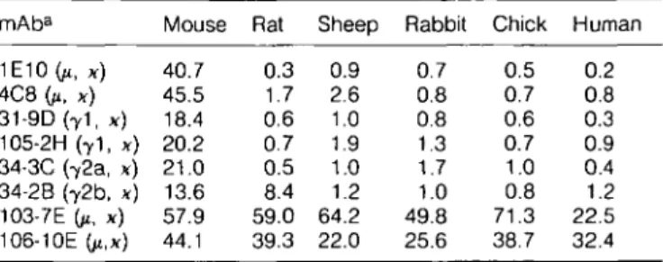
Eight hybridomas secreting IgM (1E10, 4C8, 103-7E, and 106-10E), lgG1 (31-9D and 105-2H), lgG2b (34-2B), or lgG2a (34-3C) anti-MRBC mAb were prepared from four separate fusions with spleen cells of anemic NZB mice. All eight mAb exhibited significantly increased binding activities to intact MRBC at 4°C compared with control mAb and polyclonal mouse IgG (Table 1). Essentially identical activities were observed at room temperature and at 37°C except for the 4C8 and 103-7E mAb, which showed slightly higher binding activity at 4°C (35.2 and 23.6%) than at room temperature (21.6 and 13.4%) or 37°C (21.2 and 12.4o/o).
When the binding of these anti-MRBC mAb with RBC from five different species (rat, sheep, rabbit, chick, and human) was examined, three different groups of anti-MRBC mAb were
Table 1 . In vitro binding of anti-MRBC mAb to different species
of RBC mAba 1E10 (ji, x) 4C8 Gt, x) 31-9D(71, x) 105-2H (71 , x) 34-3C (72a, x) 34-2B (72b, x) 103-7E (11, x) 106-10E (/t,x) Mouse 40.7 45.5 18.4 20.2 21.0 13.6 57.9 44.1 Rat 0.3 1.7 0.6 0.7 0.5 8.4 59.0 39.3 Sheep 0.9 2.6 1.0 1.9 1.0 1.2 64.2 22.0 Rabbit 0.7 0.8 0.8 1.3 1.7 1.0 49.8 25.6 Chick 0.5 0.7 0.6 0.7 1.0 0.8 71.3 38.7 Human 0.2 0.8 0.3 0.9 0.4 1.2 22.5 32.4 a2 fig of each mAb were incubated with RBC from different species overnight at 4°C. Results are expressed as percentage of 125l-labeled anti-mouse Ig bound to RBC. Background binding activities obtained by control IgM and IgG mAb or by polyclonal mouse IgG from a pooled normal mouse serum were <2°/o.
identified (Table 1). The first group, consisting of 1E10, 4C8, 31-9D, 105-2H, and 34-3C mAb, reacted with only MRBC. The second group (34-2B mAb) exhibited binding activity with both mouse and rat RBC. The third group (103-7E and 106-10E) reacted with the RBC of all species tested.
To further define the specificity of the first group of anti-MRBC mAb, 34-3C, 1E10, or 4C8 mAb was iodinated and the competitive inhibition assay was performed in the presence of excess amounts of different mAb. As shown in Fig. 1, not only the 34-3C mAb but also the 105-2H mAb were able to inhibit almost completely the MRBC-binding by the [125I]34-3C mAb. However, the inhibitory activity of 34-3C mAb was ~25-fold stronger than that of 105-2H mAb. A slight inhibition (15%) by 200-fold excess of 1E10 or 4C8 mAb was observed, and no inhibition by other anti-MRBC mAb (31-9D, 34-2B, 103-7E, and 106-10E). When the competition assay was performed with [125I]1E1O mAb, 34-3C mAb exhibited the highest inhibitory activity (Fig. 1). In fact, 80 ng of 34-3C mAb completely inhibited the binding of 50 ng of [12s|]1E10 mAb, while only 50% inhibi-tion was achieved by the same amount of 1E10 mAb. The 4C8 and 105-2H mAb exhibited similar inhibitory activity to that of the 1E10 mAb: all three mAb at a dose of 2 pg completely inhibited the MRBC-binding of [l25|]1E10 mAb. Partial inhibition was observed by 103-7E and 31-9D mAb at 40- to 200-fold excess, and no inhibition by 34-2B and 106-10E mAb. When [125|]4C8 mAb was used for the competition assay, results were essentially identical to those obtained with [125I]1E1O mAb (data not shown). These results suggested that 1E10, 4C8, 105-2H, and 34-3C mAb may recognize the same antigen—different from that recognized by the 31-9D mAb—and, if so, the affinity of the 34-3C mAb is far higher than that of the others.
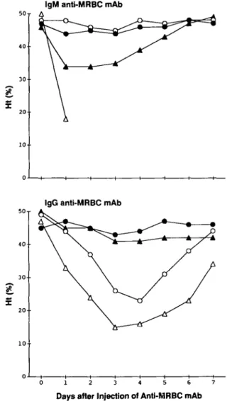
Induction of anemia by the injection of anti-MRBC mAb
Since eight anti-MRBC mAb exhibited substantial binding activity to intact MRBC in vitro at 37°C as well as at 4°C, the pathogenicity of anti-MRBC mAb was investigated by a single i.p. injection of purified mAb in BALB/c mice. The injection of 100 HQ of purified mAb of two IgM (1E10 and 4C8), lgG1 31-9D, or lgG2a 34-3C mAb caused a rapid decrease in Ht values, and 1E10 mAb even killed all animals within 1 or 2 days after its injection (Fig. 2). In contrast, lgG1 105-2H, lgG2b 34-2B, and
125l-34-3C mAb 125!-1E10mAb iooT C 60 2 0.08 0.4 2 Inhibitors (^g) 10
Fig. 1 . Inhibitory activities of various amounts of unlabeled anti-MRBC mAb(O,34-3C; • , 105-2H; A, 1E10; A , 31-9D; O, 4C8; • , 103-7E; o , 34-2B; • , 106-10E) on the MRBC-binding by 50 ng of p25|]34-3C or [125I]1E1O anti-MRBC mAb. Results are expressed as percentage inhibition of [125I]34-3C or p25|]iE10 mAb-binding to MRBC.
two IgM (103-7E and 106-10E) mAb were not able to significantly decrease Ht values.
To compare the pathogenic activity of the anti-MRBC mAb quantitatively, various amounts of mAb were injected and the quantities of mAb required to induce anemia (decreasing Ht values to <4O°/o), to cause a 50% decrease in Ht values (decreasing Ht values to <24%), and to kill animals were estimated. The 31-9D mAb exhibited the strongest activity for inducing anemia (Table 2). In fact, only 5/tg of mAb were sufficient to induce anemia; Ht values dropped from 50 to 34% 4 days after the injection. This dose was 5 - 1 0 times less than that of the other three highly pathogenic mAb (34-3C, 1E10, and 4C8). Similar differences were observed when compared with the amount of mAb required for a 50% decrease in Ht values. However, the 1E10 mAb was most toxic, because only 100 ng were sufficient to kill mice, while 2 . 5 - 5 times more mAb was required for 31-9D, 4C8, and 34-3C mAb. Although 100 ^g of 105-2H mAb failed to induce anemia, anemia was observed at
IgM anti-MRBC mAb
*
IgG anti-MRBC mAb
0 1 2 3 4 5 6 7
Days after Injection of Anti-MRBC mAb
Fig. 2. Development of anemia in BALB/c mice after a single i.p. injection of 100 pg of purified IgM anti-MRBC mAb (upper panel: A, 1E10; A.4C8; O, 103-7E; • , 106-10E) or IgG anti-MRBC mAb (lower panel: A, 31-9D; A , 105-2H; O, 34-3C; • , 34-2B). Results are mean Ht values of five mice. Note that all the mice injected with 1E10 mAb died within 2 days after injection.
Table 2. Estimation of quantities of mAb required for inducing
anemia and killing mice
mAb 1E10 4C8 31-9D 105-2H 34-2C
Dose required for inducing anemiaa kg) 25 50 5 250 50
Dose required for 50% decrease in Htb kg) 75 200 15 1200 100 Lethal dose kg) 100 250 250 NDC 500
aThe quantity of mAb required for inducing anemia. Ht values < 4 0 % ( = < 3 SD from mean values of 3- to 4-month-old BALB/c mice) were considered as anemic.
bThe quantity of mAb required for causing a 50% decrease in Ht values (Ht values decreased to <24%).
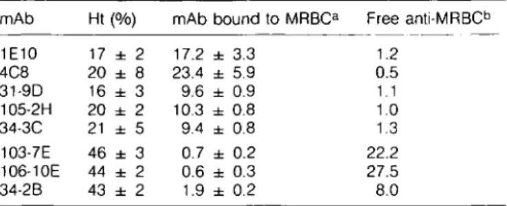
Table 3. In vivo MRBC binding by pathogenic but not
non-pathogenic anti-MRBC mAb
mAb 1E10 4C8 31-9D 105-2H 34-3C 103-7E 106-10E 34-2B Ht 17 20 16 20 21 46 44 43 (% ± ± ± ± ± ± ± ± ) 2 8 3 2 5 3 2 2 mAb 17.2 23.4 9.6 10.3 9.4 0.7 0.6 1.9 bound to MRBCa ± ± ± ± ± ± ± ± 3.3 5.9 0.9 0.8 0.8 0.2 0.3 0.2 Free anti-MRBCb 1.2 0.5 1.1 1.0 1.3 22.2 27.5 8.0
a5 - 1 0 days after injection of 5 x 10⁶ hybridoma cells, the amount of mAb bound to MRBC in vivo was determined by direct RIA. Results are expressed as percentage binding of |125l]anti-mouse Ig on MRBC (mean of 4 - 8 mice ± 1 SD). Background binding activities obtained in mice injected with control hybridomas were < 2 % .
bFree anti-MRBC activities in 1/1000 diluted sera pooled from 4 - 8 mice were determined by indirect RIA. Results are expressed as percentage binding of [125l]anti-mouse Ig bound to MRBC. Background binding activities in sera from mice injected with control hybridomas were
a dose of 250 ^g, which was 50 times higher than that of 31-9D mAb of the same Ig subclass, and 1.2 mg was required to cause a 50% decrease in Ht values. Notably, no anemia developed following the injection of ascites containing 10 mg of lgG2b 34-2B, IgM (103-7E and 106-10E) anti-MRBC mAb, or control mAb of any IgG subclass or IgM, including anti-bromelinized MRBC mAb.
Lack of in vivo MRBC binding by non-pathogenic anti-MRBC mAb
Since experiments of anti-MRBC mAb injection have shown that three of eight anti-MRBC mAb failed to induce anemia, we examined whether these mAb were able to bind MRBC in vivo. In mice developing anemia 5 - 1 0 days after the injection of five different hybridoma cells, significant amounts of antibodies bound to MRBC were detectable in all cases, while there was no demonstrable activity of free anti-MRBC antibodies in their sera (Table 3). In contrast, none of the mice transplanted with three non-pathogenic anti-MRBC hybridomas had MRBC bound with anti-MRBC antibodies at a significant level, although substantial activity of unbound anti-MRBC antibodies was found in their sera.
Histopathological manifestations induced by the injection of anti-MRBC mAb
Histological examinations revealed two different pathological changes, depending on the mAb, in spleens and livers of the mice which had either died of acute anemia or developed a severe anemia after the injection of mAb. The first group of mAb, i.e. two IgM (1E10 and 4C8) and lgG1 (31-9D) mAb at a dose of 100 - 250 IIQ, induced a marked splenomegaly associated with an enormous accumulation of agglutinated RBC in the spleen, which prevented recognition of splenic architecture (Fig. 3A). In the liver agglutinated RBC were accumulated in sinusoids, causing marked necrosis of hepatic parenchymal cells (Fig. 3B). However, erythrophagocytosis by Kupffer cells or by splenic macrophages was barely detectable. In contrast, in mice administered by the second group of mAb, 500 ng of lgG2a
(34-3C) or 2 mg of IgGi (105-2H), erythrophagocytosis by Kupffer cells was the most remarkable pathological change (Fig. 3C and D). Notably, no significant histological alterations were observed in mice injected with ascites containing 10 mg of three other anti-MRBC mAb (34-2B, 103-7E, and 106-10E) or control mAb (Fig. 3E and F).
Induction of anemia in C5-deficient or C3-depleted mice by anti-MRBC mAb
To determine the role of complement-mediated lysis in anemia induced by MRBC mAb, 100 ng of 4C8 (IgM), 31-9D (lgG1), or 34-3C (lgG2a) were injected i.p. into C5-deficient DBA/2 mice (17), and CVF-treated or control BALB/c mice. The extent and kinetics of anemia induced by these mAb in DBA/2 mice were essentially identical to those in BALB/c mice (Table 4). CVF-treated BALB/c mice similarly developed anemia, although their anemia was more severe than that of the control group of mice. This was probably caused by the effect of CVF alone, since CVF-treated mice receiving control non-anti-MRBC mAb exhibited a slight decrease in Ht values. It should be noted that, when MRBC opsonized with these mAb were subjected to complement-mediated lysis in vitro in the presence of guinea pig, rabbit, or mouse serum, no macroscopically visible hemolysis was observed (data not shown).
Fc receptor-mediated phagocytosis and hemagglutination of MRBC opsonized with anti-MRBC mAb
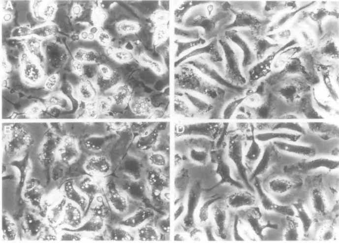
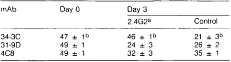
Because none of the mAb capable of inducing anemia caused complement-mediated hemolysis in vitro, and because both C5-deficient and C3-depleted mice developed anemia, the observed difference in pathological manifestations may be related to a difference in the ability of the anti-MRBC mAb to bind the FC7 receptors of macrophages and/or to induce hemag-glutination. As shown in Fig. 4, MRBC opsonized with 31-9D, 4C8, or 1E10 mAb were not significantly phagocytosed by peritoneal macrophages. In contrast, remarkably high and moderate levels of phagocytosis were observed when MRBC were opsonized with lgG2a 34-3C and lgG1 105-2H mAb, respectively. Notably, this phagocytosis was completely inhibited in the presence of a rat anti-murine Fc> receptor mAb (2.4G2) (data not shown). Finally, it was found that an i.v. administration of 1 mg of 2.4G2 mAb 2 h before the anti-MRBC mAb injection completely inhibited the development of anemia induced by lgG2a 34-3C mAb, but did not inhibit at all that induced by lgG1 31-9D or IgM 4C8 mAb (Table 5).
When the hemagglutination activity was determined in vitro, both 1E10 and 4C8 IgM mAb markedly agglutinated MRBC (hemagglutination titers of culture supernatants: 1E10,1024; 4C8, 4096). However, none of the other anti-MRBC mAb, including two non-pathogenic IgM anti-MRBC (103-7E and 106-10E) mAb, agglutinated the MRBC, although they reacted well with MRBC under this condition, as determined by RIA.
Discussion
The present study represents an extensive analysis of the pathological manifestations of eight monoclonal anti-MRBC autoantibodies obtained from unimmunized NZB mice. Although all eight anti-MRBC mAb reacted well with MRBC in vitro at 37°C
Fig. 3. (A and 3) Representative histological appearance of spleen and liver trom bALb/c mice wnicn aiea or anemia z aays arter tne injection of 100 ^g of 1E10 anti-MRBC mAb. Note an enormous accumulation of agglutinated RBC in the entire spleen, rendering the splenic architecture hardly recognizable (A; HE, x 24), and marked necrosis of hepatic parenchymal cells secondary to the accumulation of agglutinated RBC in sinusoids of liver (B; HE, x24). Essentially identical lesions were observed in mice injected with 31-9D or 4C8 mAb. (C and D) Representative histological appearance of spleen and liver from BALB/c mice which died of anemia 4 days after the injection of 500 ^g of 34-3C anti-MRBC mAb. Note the absence of accumulation of agglutinated RBC in spleen (C; HE, x 24) and liver (D; HE, x 240) but remarkable erythrophagocytosis by Kupffer cells in liver (D). Similar histological changes were observed in mice developing anemia after the injection of 2 mg of 105-2H mAb. (E and F) Represen-tative histological appearance of spleen and liver in mice injected with 10 mg of 34-2B anti-MRBC mAb. Note that there are no significant histological alterations in spleen (E; HE, x24) and liver (F; HE, x24). Mice injected with either 103-7E or 106-10E mAb exhibited no appreciable changes in spleens and livers (not shown).
as well as at 4°C, only five of them were able to bind MRBC
in vivo, causing anemia either by Fc receptor-mediated
erythrophagocytosis or by marked sequestration of agglutinated MRBC in spleens and livers, but not by complement-mediated hemolysis. The distinct difference in the specificity between pathogenic and non-pathogenic anti-MRBC mAb strongly
Table 4. Development of anemia in C5-deficient DBA/2 and
CVF-treated BALB/c mice after injection of anti-MRBC mAb
mAb 4C8 31-9D 34-3C 41-5C" DBA/2 36 ± 3a 20 ± 1 26 ± 2 47 ± 2 BALB/c CVF 28 ± 3a ND 19 ± 2 41 ± 1 Control 35 ± 3a 17 ± 1 24 ± 1 47 ± 1 aHt values (mean of 5 mice ± 1 SD) were determined 3 days after i.p. injection of 100 HQ of anti-MRBC mAb. CVF were injected i.v. 8 h before and 1 and 2 days after the anti-MRBC mAb injection. Note that serum levels of C3 at the time of anti-MRBC mAb injection were - 5 % of control values, which remained until day 3. ND, not done.
blgM non-anti-MRBC mAb.
suggests the importance of autoantibody specificity for the pathogenesis of autoimmune hemolytic anemia.
It is significant that all five pathogenic autoantibodies recognize only species-specific antigens (at least two distinct epitopes) on MRBC, while non-pathogenic antibodies recognize cross-reactive determinants present only on rat RBC or on RBC from many species such as human, sheep, rabbit, and chick. In this regard, it should be mentioned that one IgM anti-MRBC mAb (G-8), established by Caulfield etal. (8) and whose pathogenic activity was demonstrated by the transplantation of hybridoma cells, also reacts specifically with MRBC. These results are consistent with earlier findings that autoantibodies eluted from RBC of Coombs-positive NZB mice react only with MRBC but not with human, rat, sheep, guinea pig, or rabbit RBC (2,18). Thus, at least two species-specific epitopes, as suggested by the present competition experiment, are likely to be involved in the pathogenesis of autoimmune hemolytic anemia. Clearly, the identification of the biochemical nature of pathogenic epitopes is of importance to understand the immunopathogenesis of autoimmune hemolytic anemia.
The importance of the specificity of anti-MRBC mAb in the pathogenesis of autoimmune hemolytic anemia is best documented by the finding that among four IgM anti-MRBC mAb
Fig. 4. In vitro phagocytosis by peritoneal macrophages of MRBC opsonized with either 34-3C (A), 105-2H (B), 31-9D (C), or 4C8 (D) anti-MRBC mAb. Note the marked and moderate phagocytosis of MRBC opsonized with 34-3C and 105-2H mAb, respectively, but the absence of significant phagocytosis of MRBC opsonized with either 31-9D or 4C8 mAb. No phagocytosis was observed with MRBC coated with 1E10 anti-MRBC mAb (not shown).
Table 5. Effect of anti-Fc7 receptor mAb (2.4G2) on the
development of anti-MRBC mAb-induced anemia
mAb 34-3C 31-9D 4C8 Day 0 47 ± 49 ± 49 ± 1b 1 1 Day 3 2.4G2a 46 ± 1b 24 ± 3 32 ± 3 Control 21 ± 3b 26 ± 2 35 ± 1 a1 mg of anti-Fc7 receptor mAb (2.4G2) was given i.v. 2 h before the injection of anti-MRBC mAb.
bHt values (mean of 5 mice ± 1 SD) were determined 3 days after i.p. injection of 100 /ig of anti-MRBC mAb.
only two (1E10 and 4C8) are able to induce anemia. The marked
in vitro and in vivo hemagglutination by pathogenic 1E10 and
4C8 mAb but not by non-pathogenic 103-7E and 106-10E mAb can be explained by the difference in specificity of these two groups of mAb. It may be that the density of antigenic determinant recognized by 1E10 and 4C8 mAb is much higher than that recognized by 103-7E and 106-10E mAb. Alternatively, the antigen recognized by 1E10 and 4C8 mAb may be distributed in small clusters on MRBC, as recently demonstrated for the complement receptor type 1 on human RBC (19). Such differences in the distribution of target antigens could also account for the difference in in vivo binding to MRBC by these two groups of anti-MRBC mAb.
The present finding that none of the anti-MRBC mAb, even IgM mAb, are able to lyse MRBC in vitro in the presence of complement from mice, rabbits or guinea pigs may not be totally surprising. In fact, non-complement-fixing IgM antibodies with diverse specificities have been described in various species, including mouse (20-23). Since the structure of the antigen appears to influence the capacity of IgM antibodies to fix and/or activate complement (24,25), the failure of hemolysis by our autoantibodies may be related to the particular structure of the target RBC antigen. This notion was further supported by a recent observation that some lymphocyte surface antigens are better targets for complement-mediated lysis than others, independent of their densities (26). Thus, it may be possible that the antigens involved in anti-MRBC autoimmune responses and bound by anti-MRBC mAb could not efficiently activate C1, probably because of their low mobility in the RBC membrane. Whatever the reason, the development of anemia in C5-deficient DBA/2 mice as well as normal BALB/c mice clearly indicates a minor role, if any, of complement-mediated hemolysis in the pathogenesis of anemia induced by our anti-MRBC mAb. In this regard, it should be noted that NZB mice are congenially deficient in hemolytic complement activity (27) yet develop severe autoimmune hemolytic anemia. Finally, the complete prevention of anemia induced by the lgG2a 34-3C mAb by the injection of anti-Fc? receptor mAb and the similar development of anemia in both C3-depleted and control mice argue against the possible involvement of C3 receptor-mediated erythrophagocytosis in the development of anemia induced by our anti-MRBC mAb. Notably, our results do not exclude the possibility that combinations of antibodies against different epitopes might lead to complement-mediated hemolysis.
Our in vitro and in vivo results combined with histological
examinations have clearly shown that two different mechanisms are responsible for the development of anemia induced by our anti-MRBC mAb. In the case of lgG2a 34-3C and lgG1 105-2H anti-MRBC mAb, which bind efficiently to Fc receptors on macrophages, the major cause of anemia is apparently a rapid Fc receptor-mediated phagocytosis of opsonized MRBC, most remarkably by Kupffer cells. In contrast, with the two IgM (1E10 and 4C8) and lgG1 31-9D anti-MRBC mAb which fail to mediate Fc receptor-dependent phagocytosis, anemia is most likely caused by the marked sequestration of agglutinated RBC in spleen and liver. Subsequent liver damage and hemodynamic failure are likely to be additional contributing factors to the animals' death, explaining why this group of mAb was more toxic than the first group of pathogenic mAb. It should be mentioned that both 31-9D and 105-2H mAb are of the lgG1 class, yet a substantial difference exists in their ability to mediate Fc receptor-dependent phagocytosis. Since both mAb recognize different epitopes, limited phagocytosis of MRBC opsonized with the 31-9D mAb may be due to the difference in the distribution and nature of antigenic determinants recognized by each mAb. Although the specificity of autoantibodies is important, it is apparent that other factors may determine the pathogenic activity of anti-MRBC mAb. The best example is the difference in the pathogenic potential between lgG2a 34-3C and lgG1 105-2H mAb. Although both mAb appear to recognize the same epitope, the competition experiment strongly suggested that the affinity of 34-3C mAb to MRBC is far higher than that of 105-2H mAb. In addition, in vitro experiments on Fc receptor-mediated phagocytosis have shown that lgG2a 34-3C mAb exhibits greater binding to Fc receptor than does lgG1 105-2H mAb, owing to the difference in the affinity of the Fc receptor for lgG2a and of the Fc receptor for lgG1/lgG2b (28,29). Both differences in the affinities of mAb for the target antigen and for the Fc receptor may well explain the fact that the pathogenic activity of lgG2a 34-3C mAb is ~ 10 times stronger than that of lgG1 105-2H mAb. Our present studies have demonstrated several important points: (i) not all the autoantibodies reactive with intact MRBC
in vitro are able to induce anemia; (ii) all five pathogenic
autoantibodies recognize antigenic determinants expressed only on MRBC but not on other species of RBC, suggesting the importance of the specificity of anti-MRBC autoantibodies; and (iii) the affinities of autoantibodies for the target antigens and for the Fey receptors of macrophages, and the ability to cause hemagglutination are additional factors determining the pathogenic activity of anti-MRBC autoantibodies. Although it is obvious that the anti-MRBC mAb studied here may not represent the full range of pathogenic anti-MRBC autoantibodies occurring in the NZB mouse, the substantial variation of pathogenic activities observed among anti-MRBC mAb could explain the remarkable individual variability in the progression of autoimmune hemolytic anemia in the NZB mouse. Since recent structural analysis of autoantibodies has revealed the oligoclonal origin of autoimmune responses (30), the selection of different autoreactive clones during the early course of autoimmune responses may markedly influence the onset and progression of autoimmune hemolytic anemia in the NZB mouse. Finally, the development of the experimental model of autoimmune hemolytic anemia described here certainly represents a good opportunity to evaluate various therapeutic approaches based on interference at the level of the essential pathogenic pathways.
Acknowledgements
We thank Dr Alex Lussow for critically reviewing the manuscript, Ms Genevieve Leyvraz for histological preparations, and Ms Claire Desjeux for the preparation of this manuscript. This work was supported by grant no. 31-28782.90 from the Swiss National Foundation for Scientific Research, by a grant from the Ministry of Education, Science and Culture, Japan, by the Swiss Confederation acting on the proposal of the 'Commissions Federates des Maladies Rhumatismales', and by the Roche Research Foundation. Abbreviations CVF DMEM ELISA HE Ht mAb MRBC RIA
cobra venom factor
Dulbecco's modified Eagle's medium enyzme-linked immunosorbent assay hematoxylin and eosin
hematocrit
monoclonal antibody mouse red blood cells radioimmunoasay
References
1 Helyer, B. J. and Howie, J. B. 1963. Spontaneous auto-immune disease in NZB/BI mice. Br. J. Haematol. 9:119.
2 Linder, E. and Edgington, T. S. 1972. Antigen specificity of anti-erythrocyte autoantibody responses to NZB mice: identification and partial characterization of two erythrocyte surface autoantigens. J.
Immunol. 108:1615.
3 Deheer, D. H., Linder E. J., and Edgington, T. S. 1978. Delineation of spontaneous erythrocyte autoantibody responses of NZB and other strains of mice. J. Immunol. 120:825.
4 Miescher P. A. and Dayer, J. M. 1976. Autoimmune hemolytic anemia. In Miescher, P. A. and Muller-Eberhard, H. J., eds, Textbook of
Immunopathology, p. 649. Grune & Stratton, New York.
5 Lewis, D. E., Griswold, S., and Warner, N. L. 1981. Monoclonal anti-erythrocyte auto-antibodies from unimmunized NZB mice.
Hybridoma 1:71.
6 Cooke, L. A., Staines, N. A., Morgan, A., Moorhouse, C, and Harris, G. 1982. Haemolytic disease in mice induced by transplanta-tion of hybridoma cells secreting monoclonal anti-erythrocyte auto-antibodies. Immunology 47:569.
7 Ozaki, S., Nagasawa, R., Sato, H., and Shirai, T. 1984. Hybridoma autoantibodies to erythrocytes from NZB mice and the induction of hemolytic anemia. Immunol. Lett. 8:115.
8 Caulfield, M. J., Stanko, D., and Calkins, C. 1989. Characterization of the spontaneous autoimmune (anti-erythrocyte) response in NZB mice using a pathogenic monoclonal autoantibody and its anti-idiotype. Immunology 66:233.
9 Cerottini, J. C, Engers, H. D., MacDonald, H. R., and Brunner, K. T. 1974. Generation of cytotoxic T lymphocytes in vitro. I. Response of normal and immune spleen cells in mixed leukocyte cultures. J. Exp.
Med. 140:703.
10 Luzuy, S., Merino, J., Engers, H., Izui, S., and Lambert, P. H. 1986. Autoimmunity after induction of neonatal tolerance to alloantigens:
role of B cell chimerism and F, donor B cell activation. J. Immunol. 136:4420.
11 Poncet, P., Kocher, H. P., Pages, J., Jaton, J. C , and Bussard, A. E. 1985. Monoclonal autoantibodies against mouse red blood cells: a family of structurally restricted molecules. Mol. Immunol. 22:541. 12 Spertini, F., Coulie, P. G., Van Snick, J., Davidson, E., Lambert, P. H.,
and Izui, S. 1989. Inhibition of cryoprecipitation of murine lgG3 anti-dinitrophenyl (DNP) monoclonal antibodies by anionic DNP-amino acid conjugates. Eur. J. Immunol. 19:273.
13 Unkeless, J. C. 1979. Characterization of a monoclonal antibody directed against mouse macrophages and lymphocyte Fc receptors.
J. Exp. Med. 150:580.
14 Garci'a-Gonzalez, M., Bettinger, S., CM, S., Olivier, P., Kadouche, J., and Pouletty, P. 1988. Purification of murine lgG3 and IgM monoclonal antibodies by euglobulin precipitation. J. Immunol. Methods 111:17. 15 McConahey, P. J. and Dixon, F. J. 1980. Radioiodination of proteins by the use of the chloramine-T method. Methods Enzymol. 70:210. 16 Bazin, H., Cormont, F., and de Clercq, L. 1986. Purification of rat
monoclonal antibodies. Methods Enzymol. 121:638.
17 Cinader, B., Dubiski, S., and Wardlaw, A. C. 1964. Distribution, inheritance, and properties of an antigen, MuB1, and its relation to hemolytic complement. J. Exp. Med. 120:897.
18 Long, G., Holmes, M. C, and Burnet, F. M. 1963. Autoantibodies produced against mouse erythrocytes in NZB mice. Aust. J. Exp. Biol.
Med. Sci. 41:315.
19 Paccaud, J. P., Carpentier, J. L, and Schifferli, J. A. 1988. Direct evidence for the clustered nature of complement receptors type 1 on the erythrocyte membrane. J. Immunol. 141:3889.
20 Plotz, P. H., Colten, H., and Talal, N. 1968. Mouse macroglobulin antibody to sheep erythrocytes: a non-complement-fixing type. J.
Immunol. 100:752.
21 Spiegelberg, H. 1974. Biological activities of immunoglobulins of different classes and subclasses. Adv. Immunol. 19:259. 22 Van Snick, J. L. and Masson, P. 1978. The effects of complement
on the ingestion of soluble antigen - antibody complexes and IgM aggregates by mouse peritoneal macrophages. J. Exp. Med. 148:903. 23 Klaus, G. G. B., Pepys, M. B., Kitajima, K., and Askonas, B. A. 1979.
Activation of mouse complement by different classes of mouse antibody. Immunology 38:687.
24 Ishizaka, T., Tada, T., and Ishizaka, K. 1968. Fixation of complement and C'1a by rabbit IgG and IgM antibodies with paniculate and soluble antigens. J. Immunol. 100:1148.
25 Cunniff, R. V. and Stollar, B. D. 1968. Properties of 19S antibodies in complement fixation. I. Temperature dependence and role of antigen structure. J. Immunol. 100:7.
26 Bindon, C. I., Hale, G., and Waldmann, H. 1988. Importance of antigen specificity for complement-mediated lysis by monoclonal antibodies.
Eur. J. Immunol. 18:1507.
27 Andrews, B. S. and Theofilopoulos, A. N. 1978. A microassay for the determination of hemolytic complement activity in mouse serum. J.
Immunol. Methods 22:273.
28 Unkeless, J. C, Scigliano, E., and Freedman, V. H. 1988. Structure and function of human and murine receptors for IgG. Annu. Rev.
Immunol. 6:251.
29 Yamada, A. and Suzuki, T. 1989. Fc72b receptor-mediated phagocytosis by a murine macrophage-like cell line (P388D,) and peritoneal resident macrophages. J. Immunol. 142:2457. 30 Shlomchik, M. J., Marshak-Rothstein, A., Wolfowicz, C. B., Rothstein,
T. L., and Weigert, M. G. 1987. The role of clonal selection and somatic mutation in autoimmunity. Nature 328:805.