ORIGINAL PAPER
Patient characteristics differently affect early cup
and stem loosening in THA: a case-control study
on 7,535 patients
C. Röder&S. Eggli&P. Münger&M. Melloh&A. Busato
Received: 3 October 2006 / Revised: 11 October 2006 / Accepted: 11 October 2006 / Published online: 15 February 2007
# Springer-Verlag 2007
Abstract We postulated that certain patient characteristics have different effects on early THA component loosening. With two matched case-control studies we assessed 3,028 cups and 5,224 stems. Loosening was defined using signs of mechanical component failure on routine follow-up radio-graphs or revision for aseptic loosening. Women and men had similar cup-loosening odds, but women had lower odds for stem loosening (p<0.0001). Odds for cup loosening de-creased by 2.1% per additional year of age (p=0.0004), those for stem loosening by 2.4% (p<0.0001). Each additional kilogram of weight decreased cup loosening odds by 1.3% (p=0.0051). Each additional unit of BMI increased stem loosening odds (p=0.0109). Charnley classes B and C
were protective factors against loosening of both compo-nents. There were no risk differences for the various main diagnoses. Certain patient characteristics differently affected early cup and stem loosening, although some characteristics had the same protective or harmful effect on component survival.
Résumé IO-10-06-555.R1. A partir de l’hypothèse que certaines caractéristiques des patients avaient un effet sur les descellements précoces des prothèses de la hanche, nous avons étudié 3028 cupules et 5224 tiges fémorales dans le cadre d’une étude controlée. Le descellement a été défini par des anomalies radiographiques ou l’existence d’une reprise pour descellement aseptique. Les hommes et les femmes avaient un taux similaire de descellement cotyloidien mais les femmes avaient un taux de descellement de tige plus faible (p<0,0001). La fréquence des descellements cotyloi-diens diminuait de 2,1% par année d’âge (p=0,0004), et celle des descellements de tige de 2,4% (p<0,0001) . Chaque kilog de poids supplémentaire diminuait de 1,3% les descellements cotyloidiens (p=0,0051). Chaque unité sup-plémentaire de BMI augmentait le descellement de la tige (p=0,0109).Les classes B et C de Charnley étaient des facteurs protecteurs du descellements des deux composants.. Il n’y avait pas de différence de risque entre les principaux diagnostics. Les caractéristiques des patients affectent de façon différente les descellements précoces et certaines caractéristiques sont protectrices ou nuisibles vis à vis de la survie des composants.
Introduction
Although modern component designs and fixation tech-niques have decreased the rate of total hip arthroplasty
C. Röder (*)
:
P. Münger:
M. Melloh:
A. Busato MEM Research Center in Orthopaedic Surgery, Institute for Evaluative Research in Orthopaedic Surgery, University of Bern, Stauffacherstrasse 78, 3014 Bern, Switzerland e-mail: [email protected] M. Melloh e-mail: [email protected] S. EggliDepartment of Orthopaedic Surgery, Inselspital, University of Bern,
3001 Bern Switzerland e-mail: [email protected] A. Busato
Health Technology Assessment and Clinical Epidemiology, MEM Research Center in Orthopaedic Surgery, Institute for Evaluative Research in Orthopaedic Surgery, University of Bern
Stauffacherstrasse 78, 3014 Bern, Switzerland
(THA) failures, the increasing number of primary proce-dures, particularly in younger patients, results in a new epidemic of patients requiring joint revisions [14]. Three main factors influence the outcome and survival time of primary THA: (1) the surgeon’s skills and experience [10,
17], (2) the implant design and method of fixation [2,17], and (3) patient characteristics like sex, age, weight, underlying disease, and activity level [2,13,15].
Because of the reduction in the infection rate by improved operative techniques, laminar flow operative theatres, and intravenous antibiotic prophylaxis [5], me-chanical failure remains the most common complication requiring revision. To maximise prosthetic survival time, and to educate patients about the long-term results of operative treatment, the influence of patient characteristics on prosthetic survival must be considered. The literature provides a multitude of articles about this issue [2–5,8,13,
15, 17, 22–24]. Most authors use revision for cup and/or stem loosening [2] as endpoint for their analyses, but others also include symptomatic patients with radiographic loos-ening [13]. Whilst some authors agree that demographic factors affect loosening of sockets and stems to a different extent [23], others report virtually identical rates of survivorship for the acetabular and femoral components, which may merely be a result of different follow-up times in the studies [2]. In addition, many studies represent single-surgeon or single-department reports, with only one type of prosthesis. This makes generalisation of findings difficult.
In order to further elucidate the influence of patient characteristics on mechanical component failure in THA, and to overcome some of the aforementioned methodolog-ical problems, we have conducted a two-arm matched case-control study based on data from a multitude of centres and surgeons from various European countries that also includ-ed different types of sockets and stems. We postulatinclud-ed that patient demographics like age, sex, BMI and main diagnosis—but also activity levels—affect the risks for cup and stem loosening to different extents. The study design was selected to allow more general conclusions about the principal research question, and not to have to restrict conclusions to one specific prosthesis type or surgical technique.
Materials and methods
The current study used the database of the Institute for Evaluative Research in Orthopaedic Surgery at the Univer-sity of Bern, Switzerland, formerly Department of Educa-tion and DocumentaEduca-tion of the Maurice E. Müller Foundation. Preoperative and postoperative clinical and radiological data were prospectively documented using
optically readable code sheets from consecutive primary total hip replacements according to the standards of the International Documentation and Evaluation System (IDES) [20].
The inclusion criteria were: (1) an underlying diagnosis of osteoarthritis, developmental dysplasia, inflammatory arthritis, fracture or osteonecrosis, primary THA, (2) age more than 20 years at the time of surgery, and (3) at least one follow-up examination with a complete set of the AP pelvic view and lateral radiographs of the treated side (preoperative, immediately postoperative, follow-up), or a documented first revision of one or both components for mechanical failure.
The IDES follow-up form provides a section for the radiographic evaluation of the prosthetic components, and for changes in bone or tissue structures (e.g. ossifications, change in cortex density, cavitations). Radiographic assess-ment was performed based on standardised AP pelvic and lateral view radiographs with the MEM-template for evaluation of THA as a standardised measurement tool. Component loosening was defined by comparing the postoperative and follow-up radiographs and measuring the superior and medial cup migration, tilt, radiolucencies, broken socket, or broken cement. The stem was assessed for subsidence, radiolucencies at the bone–stem or bone– cement interface, a progressive tilt, cavitations, and stem fracture.
The following IDES variables were used: (1) continuous radiolucencies around the socket in Zones 1–3 [10], (2) a superior migration ≥5 mm with a severe protrusion or a progressive tilt of the socket, (3) a fracture of the socket or the cement mantle after cemented fixation, (4) stem subsidence ≥3 mm, (5) radiolucencies >2 mm at the bone–cement or bone–stem interface, (6) continuous radio-lucencies at the bone–cement or bone–stem interface, and (7) multiple small cavitations or large defects; and 8) stem or cement fractures.
The revision diagnosis and exchanged components were recorded on the IDES revision forms. Depending on cup or stem revision, or exchange of both components, the patient was referred to one (cup or stem study) or both (cup and stem study) study groups. The percentage of cases derived from revisions was 26.5% for the cup study and 17.9% for the stem study respectively.
Controls were derived from IDES follow-up forms. They were defined as patients without radiographic signs of cup and stem loosening. Patients defined as their own control in case of bilateral THA, and clinical examinations after revision surgery were excluded as controls.
Patients and controls were matched on the following criteria: (1) THA performed in the same hospital, (2) date of surgery within ± 2.5 years, (3) follow-up examination performed within less than six months, (4) stem type (stem
study), (5) head size (stem study), and (6) cup design, size, and material (cup study).
This procedure resulted in two sets of matched patients and controls to analyse risk factors of cup and stem loosening derived from 2,796 patients for the cup study and 4,739 patients for the stem study. There were 3,028 (549 cases, 2,489 controls) and 5,224 (840 cases, 4,384 controls) primary hip replacements respectively, and data for 1,639 joints were used in both studies. We used data collected between 1981 and 2003 in 30 hospitals in seven European countries (Austria, Belgium, Switzerland, Ger-many, France, The Netherlands, and Italy). An average of 4.6 and 5.2 controls per case were included. There were 48.6% male patients in the cup study and 48.1% in the stem study. Of these patients, 8.3% and 10.24 % had both hips treated. The mean follow-up was 4.8 years for sockets (range, 0.11–20.1 years) and 4.15 years for stems (range, 0.2–21.7 years). Twenty-three cup types and 64 stem types were included in the respective studies.
The relationships between component loosening/revision and patient characteristics were analysed as a 1:M matched pairs case-control study (multiple controls matched to each case) using multiple conditional logistic models. The first model was fitted to the data to investigate the overall effects of age and BMI adjusted for sex, main diagnosis, and Charnley class. Age and BMI were included as continuous explanatory variables. Sex, main diagnosis (five diagnoses), and Charnley class (A, B, C) were included as categorised variables. Similar models were used to estimate effects of patient weight.
Cup and stem type were included in the matching criteria. Because this procedure does not control for effects of component fixation (cemented, uncemented) an addi-tional explanatory variable taking into account the type of component fixation was included in the models. 28.6% of cups and 83.8% of stems were cemented in the two studies. The results of continuous variables were interpreted as an estimate of the change in risk per unit increase of each variable. Additional models were evaluated with continu-ous explanatory variables categorised into groups. Body mass index was classified as normal with BMI< 25 kg/m2, overweight with BMI between 25 and 30 kg/m2, and obese with BMI> 30 kg/m2. Age was classified into four groups: <60 years, 60–70 years, 70–80 years, and >80 years. Weight was categorised into quartiles of the observed data. Results were interpreted as risk difference to a reference level. The respective reference levels were normal weight (BMI < 25 kg/m2), age <60 years, and the lowest weight class. A model was used to estimate the significance of all first-order interaction terms of explanatory variables. None of the interaction terms appeared to be significant, using a backward elimination process. Adjusted population risks or aetiological fractions for modifiable preoperative risks were
calculated. Power calculations were performed for sex, age, diagnosis, BMI, Charnley class, and the corresponding ranges of exposure levels. Power levels of significant outcome variables were all above 0.8. For insignificant results, the sample sizes of the studies would allow the detection of odds ratios of less than three at a power of 0.8. All calculations were done using SAS 9.1 (SAS Institute Inc, Cary, NC) andp<0.05 was considered significant. Results
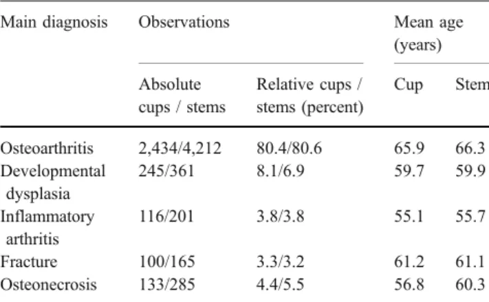
Main diagnoses were almost equally distributed in the cup and stem study, with about 80% of patients having had osteoarthritis, 8% developmental dysplasia, 4% inflamma-tory arthritis, 3% having had a fracture and 5% osteone-crosis (Table1). The mean age ranged from 55.1 years to 60.3 years in the groups with inflammatory arthritis, developmental dysplasia and osteonecrosis, and from 61.1 to 66.3 years in the groups with osteoarthritis and fracture (Table 1). Overall, the mean age at primary surgery was 64.4 years in the cup study and 65.1 years in the stem study. The average bodyweight was 73.4 kg and 73.5 kg in the cup study and stem study respectively The BMI groups were equally distributed for the cup study and the stem study, with about 38% having a BMI below 25, 44% between 25 and 30, and 18% over 30. The mean BMI at surgery was 26.9 kg/m2in the cup study and 27 kg/m2in the stem study. The distributions of patients across Charnley classes in the cup and stem studies were: Charnley Class A, 66.5%/66.7%; Charnley Class B, 31.2%/31.2%; and Charnley Class C, 2.3%/2.1%.
Women and men had similar odds for cup loosening, but women had lower odds for stem loosening than men (p<0.001: Table2).
Age-related reduction of odds for cup and stem loosen-ing decreased in a linear fashion across the age groups: <60 years (reference level), 60–70 years (cup p=0.111,
Table 1 Basic demographic and diagnostic data
Main diagnosis Observations Mean age (years) Absolute cups / stems Relative cups / stems (percent) Cup Stem Osteoarthritis 2,434/4,212 80.4/80.6 65.9 66.3 Developmental dysplasia 245/361 8.1/6.9 59.7 59.9 Inflammatory arthritis 116/201 3.8/3.8 55.1 55.7 Fracture 100/165 3.3/3.2 61.2 61.1 Osteonecrosis 133/285 4.4/5.5 56.8 60.3
stem p=0.029) , 70–80 years (cup p=0.002, stem p< 0.001) and >80 years (cup p=0.038, stem p=0.0005). When age was analysed as a continuous variable, risks for cup loosening decreased (p=0.0004) by 2.1% per addi-tional year of age. Similarly, the odds for stem loosening decreased (p<0.0001) by 2.4% per additional year of age (Table2).
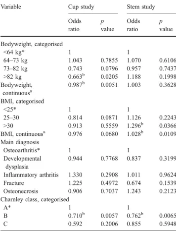
Compared to the reference group (<64 kg), weight-related cup-loosening odds were increased (p=0.786) for the weight group 64–73 kg and decreased for weight groups 73–82 kg (p=0.08) and >82 kg (p=0.021). There were slight and inconsistent risk changes of higher body weight for stem loosening (p=0.2–0.611). When weight was analysed as a continuous variable, the odds for cup loosening decreased (p=0.005) by 1.3% per additional kilogram of body weight. The odds for stem loosening were only minimally increased (p=0.363) (Table 3).
Compared to the reference level (BMI<25) odds for cup loosening were decreased for overweight (BMI=25–30, p=0.087) and obese (BMI>30, p=0.556) patients. In contrast, odds for stem loosening increased in a linear fashion for overweight (p=0.224) and obese (p=0.037) patients. Similarly, when BMI was analysed as a continuous variable, the odds for cup loosening decreased (p=0.068) per additional unit of BMI, whilst those for the stem increased (p=0.011) (Table 3). The aetiological fraction of BMI on stem loosening was 0.099, i.e., a BMI of greater than 25 was accompanied by a 9.9% increase in cases of radiographic or actual stem loosening.
Patients with developmental dysplasia had decreased loosening odds for both components (cupp=0.777, stem p=0.32) compared to the reference group with osteoar-thritis. For patients with inflammatory arthritis, the odds for component loosening were increased (cup p=0.291, stem p=0.962). For the patient groups with a fracture or osteonecrosis cup and stem loosening, odds did not
change in the same direction. Whilst the fracture group displayed increased (p=0.497) stem-loosening and de-creased (p=0.154) cup-loosening odds, the osteonecrosis group showed risk changes in the opposite way with decreased (p = 0.704) cup-loosening and increased (p=0.212) stem-loosening odds (Table3).
Patients in Charnley Class A were defined as the reference group. Component loosening odds were de-creased for both, cups and stems, in Charnley class B (cupp=0.006, stem p=0.007) and in Charnley class C (cup p=0.201, stem p=0.598) (Table3).
Discussion
Our findings indicate older age protects against mechanical component failure. In the model with continuous explana-tory variables, the reduction in risk for each additional year of intervention postponement was 2% for cup loosening and 2.4% for stem loosening. The model with age groups confirmed these findings. The protective effect is in accordance with the opinion that younger patients have higher loosening risks because of higher levels of physical
Table 3 Effects of demographics and diagnosis on evidence of component loosening
Variable Cup study Stem study
Odds ratio p value Odds ratio p value Bodyweight, categorised <64 kg* 1 1 64–73 kg 1.043 0.7855 1.070 0.6106 73–82 kg 0.743 0.0796 0.957 0.7437 >82 kg 0.663b 0.0205 1.188 0.1998 Bodyweight, continuousa 0.987b 0.0051 1.003 0.3628 BMI, categorised <25* 1 1 25–30 0.814 0.0871 1.126 0.2243 >30 0.913 0.5559 1.296b 0.0366 BMI, continuousa 0.976 0.0680 1.028b 0.0109 Main diagnosis Osteoarthritis* 1 1 Developmental dysplasia 0.944 0.7768 0.837 0.3199 Inflammatory arthritis 1.330 0.2908 1.011 0.9624 Fracture 1.225 0.4972 0.674 0.1539 Osteonecrosis 0.906 0.7037 1.243 0.2123 Charnley class, categorised
A* 1 1
B 0.710b 0.0057 0.762b 0.0065
C 0.592 0.2006 0.855 0.5948
*reference level;aanalysed using a model with continuous
explana-tory variables;bp≤0.05 is significant; BMI=body mass index
Table 2 Effects of sex and age on component loosening
Variable Cup study Stem study
Odds ratio p value Odds ratio p value Sex Male* 1 1 Female 1.129 0.3462 0.623b <0.0001 Age (years) <60* 1 1 60–70 0.814 0.1111 0.797b 0.0286 70–80 0.623b 0.0019 0.543b <0.0001 >80 0.486b 0.0380 0.364b 0.0005 Age, continuousa 0.979b 0.0004 0.976b <0.0001 *reference level;aanalysed using a model with continuous
activity [2,24,25]. Only one article reports similar revision rates in patients less than 50 years compared to older patients [3].
We analysed the absolute and relative dimensions of weight on component loosening risks. Our findings showed a protective effect of heavier absolute bodyweight on cup-loosening risks in both models, whilst the influence on stem-loosening risks was small and nonlinear. Body mass index revealed changing effects on cup-loosening risks, yet resulted in increased risks for stem loosening. High weight as an absolute index, and overweight as expressed by a high BMI, should not be considered detrimental for cup survival, but for stem fixation. Calculation of population-attributable risks showed that a BMI of greater than 25 was accompa-nied by a 9.9% increase in cases of radiographic or actual stem loosening.
Many studies have not differentiated between weight and obesity as expressed by BMI [25]. Focusing on the functional outcome of THA, obesity may play a more important role than patient weight, since a higher BMI corresponds with decreased walking distances after THA [21]. One would also expect obesity to be the more influential factor in the extent of radiographic stability. The extent to which the reduced activity levels offsets the increased biomechanical stresses on the THA cannot be concluded from our study. Nevertheless, our data indicate that a strong and linear relation between weight or BMI and socket-loosening risk can be denied, whilst the stem fixation is more directly affected. The literature similarly reports that the time to implant failure decreases in a linear fashion with increased patient weight [7]. In contrast to our findings, the cup was included in these conclusions, which may primarily result from the underlying diagnosis. All patients had osteonecrosis which more often affects young and active persons [11].
While being of female sex was protective for stem loosening, there were no differences in cup-loosening risks between males and females. These results are in accordance to the findings of other authors with similar case definitions (aseptic loosening). They found age-adjusted stem-loosen-ing risks to be significantly higher in the male population, and concluded that lighter bodyweight was responsible for a lower loosening risk in female patients, because after adjusting for weight the risk differences became insignifi-cant [22, 24]. Another study confirmed the increased femoral loosening risks in men, but did not detect differ-ences in weight between males with and without radio-graphic loosening, hypothesising that the greater muscular force was an aetiological factor [6]. Based on the protective effects of female sex and advanced age, and our earlier findings about decreased walking capacities in these patient groups [21], the activity level seems to have a more detrimental effect on stem fixation than on cup fixation.
Accordingly, one study reporting more than a two-fold increase of femoral failure in male patients also reported that patients with the highest activity levels had the highest incidence of femoral loosening [23]. Stating one main aetiology for femoral loosening differences is difficult to appoint, and other authors have suggested additional explanations like difference in size of the femoral canals [15, 18]. We consider the different risks for femoral loosening between male and female patients to be dynamic and multidimensional. Factors such as weight, BMI, muscular forces, and anatomical differences are compo-nents of a complex equation.
Compared to osteoarthritis, which served as reference pathology, odds ratios for component loosening for the other main diagnoses were changing and inconsistent. While patients with developmental dysplasia and osteonec-rosis showed decreased cup-loosening risks, those with inflammatory arthritis and a hip fracture had increased risks. On the femoral side, all patients except those with osteonecrosis had lower loosening risks than the reference group. There is disagreement about increased [1, 16] or decreased [4] component loosening risks for patients with rheumatoid arthritis. The bone stock of patients with rheumatoid arthritis is poorer because of disease and steroid medication. A periarticular osteopenia and generalised osteoporosis can also be found in these patients. However, the deficient bone stock does not necessarily lead to a compromised cup fixation with increased risks for aseptic cup loosening, most probably due to low activity levels.
Osteonecrosis is also considered as a risk-increasing diagnosis regarding component loosening, which may be a result of increased activity levels in these patients, as mentioned before [9, 11]. Our study merely showed tendencies for increased stem-loosening risks.
Charnley classes B and C revealed lower component loosening risks than Charnley class A. Differences were, however, only significant between classes A and B. The Charnley classification represents the patients’ overall mobility status. Differences in the percentage of patients ambulating longer than 60 minutes without walking aids are significant between all the three Charnley groups [21]. Therefore, we consider the Charnley classification as an indirect, but good proxy measure of activity level. Accordingly, the decreased overall mobility as expressed by Charnley Classes B and C also resulted in decreased risks for component loosening. Findings in the literature are still inconclusive. One study found heavy physical labour (agricultural work) to have a significant relationship with component loosening [12], whilst another one reported an increased component failure rate in Charnley Class B patients, hypothesising that there was added stress imposed on the operated hip while the contralateral hip was symptomatic [7].
References
1. Ahnfelt L, Herberts P, Malchau H, Andersson GB (1990) Prognosis of total hip replacement: a Swedish multicenter study of 4,664 revisions. Acta Orthop Scand Suppl 238:1–26
2. Berry DJ, Harmsen WS, Cabanela ME, Morrey BF (2002) Twenty-five-year survivorship of two thousand consecutive primary Charnley total hip replacements: factors affecting survi-vorship of acetabular and femoral components. J Bone Joint Surg Am 84:171–177
3. Boeree NR, Bannister GC (1993) Cemented total hip arthroplasty in patients younger than 50 years of age: ten- to 18-year results. Clin Orthop 287:153–159
4. Bulstrode C (1987) Keeping up with orthopaedic epidemics. Br Med J (Clin Res Ed) 295:514
5. Carlsson AS, Gentz CF, Lindberg HO (1983) Thirty-two noninfected total hip arthroplasties revised due to stem loosening. Clin Orthop 181:196–203
6. Carlsson AS, Gentz CF (1980) Mechanical loosening of the femoral head prosthesis in the Charnley total hip arthroplasty. Clin Orthop 147:262–270
7. Cornell CN, Salvati EA, Pellicci PM (1985) Long-term follow-up of total hip replacement in patients with osteonecrosis. Orthop Clin North Am 16:757–769
8. DeLee JG, Charnley J (1976) Radiological demarcation of cemented sockets in total hip replacement. Clin Orthop 121:20–32
9. Dutton RO, Amstutz HC, Thomas BJ, Hedley AK (1982) Tharies surface replacement for osteonecrosis of the femoral head. J Bone Joint Surg Am 64:1225–1237
10. Fender D, van der Meulen JH, Gregg PJ (2003) Relationship between outcome and annual surgical experience for the charnley total hip replacement. Results from a regional hip register. J Bone Joint Surg Br 85:187–190
11. Hedley AK, Kim W (1983) Prosthetic replacement in osteone-crosis of the hip. Instr Course Lect 32:265–271
12. Inoue K, Ushiyama T, Tani Y, Hukuda S (1999) Sociodemographic factors and failure of hip arthroplasty. Int Orthop 23:330–333 13. Kavanagh BF, Wallrichs S, Dewitz M, Berry D, Currier B, Ilstrup
D, Coventry MB (1994) Charnley low-friction arthroplasty of the hip. Twenty-year results with cement. J Arthroplast 9:229–234
14. Kavanagh BF, Ilstrup DM, Fitzgerald RH Jr (1985) Revision total hip arthroplasty. J Bone Joint Surg Am 67: 517–526
15. Kobayashi S, Eftekhar NS, Terayama K (1994) Predisposing factors in fixation failure of femoral prostheses following primary Charnley low friction arthroplasty: a 10- to 20-year follow-up study. Clin Orthop 306:73–83
16. Lachiewicz PF, McCaskill B, Inglis A, Ranawat CS, Rosenstein BD (1986) Total hip arthroplasty in juvenile rheumatoid arthritis: two to eleven- year results. J Bone Joint Surg Am 68:502–508
17. Malchau H, Herberts P, Ahnfelt L (1993) Prognosis of total hip replacement in Sweden. Follow-up of 92,675 operations per-formed 1978–1990. Acta Orthop Scand 64:497–506
18. Noble PC, Box GG, Kamaric E, Salama JK, Ochi T, Tullos HS (1995) The effect of aging on the shape of the proximal femur. Clin Orthop 316:31–44
19. Paterson M, Fulford P, Denham R (1986) Loosening of the femoral component after total hip replacement: the thin black line and the sinking hip. J Bone Joint Surg Br 68:392– 397
20. Röder C, Eggli S, EL-Kerdi A, Müller U, Ambrose T, Röösli E, Busato A, Aebi M (2003) The international documentation and evaluation system (IDES) - ten years experience. Int Orthop 27:259–261
21. Röder C, Parvizi J, Eggli S, Berry DJ, Müller ME, Busato A (2003) Demographic factors affecting long-term outcome of total hip arthroplasty. Clin Orthop 417:62–73
22. Schurman DJ, Bloch DA, Segal MR, Tanner CM (1989) Conventional cemented total hip arthroplasty: assessment of clinical factors associated with revision for mechanical failure. Clin Orthop 240:173–180
23. Sutherland CJ, Wilde AH, Borden LS, Marks KE (1982) A ten-year follow-up of one hundred consecutive muller curved-stem total hip-replacement arthroplasties. J Bone Joint Surg Am 64:970–982
24. Surin VV, Sundholm K (1983) Survival of patients and prostheses after total hip arthroplasty. Clin Orthop 177:148–153
25. Young NL, Cheah D, Waddell JP, Wright JG (1998) Patient characteristics that affect the outcome of total hip arthroplasty: a review. Can J Surg 41:188–195