2009/163
Metzincin’s canonical methionine is responsible
for the structural integrity of the zinc-binding site
Anselm E. Oberholzer
a, Mario Bumann
a,b,
Thomas Hege, Santina Russo
cand
Ulrich Baumann*
Department of Chemistry and Biochemistry, University
of Bern, CH-3012 Bern, Switzerland
* Corresponding author
e-mail: [email protected]
Abstract
The metzincins constitute a subclan of metalloproteases
possessing a HEXXHXXGXXH/D zinc-binding consensus
sequence where the three histidines are zinc ligands
and the glutamic acid is the catalytic base. A completely
conserved methionine is located downstream of this
motif. Families of the metzincin clan comprise, besides
others, astacins, adamalysins proteases, matrix
metallo-proteases, and serralysins. The latter are extracellular
50 kDa proteases secreted by Gram-negative bacteria
via a type I secretion system. While there is a large
body of structural and biochemical information available,
the function of the conserved methionine has not been
convincingly clarified yet. Here, we present the crystal
structures of a number of mutants of the serralysin
member protease C with the conserved methionine being
replaced by Ile, Ala, and His. Together with our former
report on the leucine and cysteine mutants, we
demon-strate here that replacement of the methionine side chain
results in an increasing distortion of the zinc-binding
geometry, especially pronounced in the x
2angles of the
first and third histidine of the consensus sequence. This
is correlated with an increasing loss of proteolytic activity
and a sharp increase of flexibility of large segments of
the polypeptide chain.
Keywords: crystal structure; directed mutagenesis;
metalloprotease; metzincin; serralysin.
Introduction
Zinc-dependent endo-metalloproteases occur in all
king-doms of life and are involved in manifold biological
func-tions. Many of them are characterized by a consensus
sequence HEXXH, where the histidines are zinc ligands
and the glutamic acid functions as catalytic base. A third
zinc ligand is provided by an amino acid side-chain (His,
Glu, Asp) downstream of this motif and the coordination
These authors contributed equally to this work.
a
Present address: MRC France, CRG BM14 ESRF, B.P. 220,
b
F-38043 Grenoble Cedex, France.
Present address: Swiss Light Source, Paul Scherrer Institute,
c
CH-5232 Villigen, Switzerland.
sphere is completed by the activated water molecule that
attacks the peptide bond in the course of the reaction
(Jiang and Bond, 1992; Tronrud et al., 1992). These
enzymes are grouped in clan MA of the MEROPS
pro-tease database (Rawlings et al., 2006). The metzincins
are a subclan, possessing an elongated
HEXXH-XXGXX(H/D) zinc-binding motif with the last amino acid
(mostly His) being the third zinc ligand (Bode et al., 1993;
Stocker et al., 1995; Gomis-Ruth, 2003, 2009). The name
of this subclan originates from a conserved methionine
located in a 1,4-turn (so-called ‘Met-turn’), which is
sub-jacent to the base of the pyramidal arrangement of the
three zinc ligands (Bode et al., 1993). Protease families
belonging to the metzincins are, besides others, the
asta-cins (Bond and Beynon, 1995), matrix metalloproteases
(MMPs; Nagase et al., 2006), adamalysins (Blobel, 2005),
and serralysins (Maeda and Morihara, 1995). The latter
are proteases secreted by Gram-negative bacteria
util-izing a type I secretion system. Typical members are
ser-ralysin from Serratia marcescens, alkaline protease from
Pseudomonas aeruginosa and protease C (PrtC) from
Erwinia chrysanthemi. These 50 kDa proteins possess,
besides an N-terminal approximately 220 amino acid
long protease domain, a C-terminal b-roll moiety which
is formed by tandem repeats of the glycine-rich
nona-peptide GGXGXDXLX. This domain binds calcium and
probably functions as an intramolecular chaperone
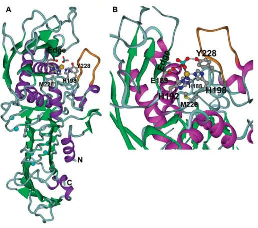
(Baumann et al., 1993; Meier et al., 2007) (Figure 1A).
Serralysins are classical metzincins where His-188,
His-192, and His-198 are the zinc ligands and the
Met-turn possesses the sequence 224-SIMSY-228. The tyrosine
(Tyr-228) in this element stabilizes the tetrahedral
transi-tion state of the hydrolysis reactransi-tion (Hege and Baumann,
2001b) and is not present in all metzincin families (Figure
1B). Another unique feature of the serralysins is the loop
comprising residues 201–210 in PrtC (drawn in orange in
Figure 1), which is quite flexible in the apo-state and
undergoes an induced-fit closure upon substrate binding.
The various metzincin families are rather distant to
each other in evolution, exhibiting sequence identities of
typically much less than 20%, and sequence alignments
frequently pick up only the zinc-binding consensus motif.
Between these families, the Met-turn is located in a
non-conserved sequence stretch and the spacing between
zinc-binding consensus motif and Met-turn varies.
Nev-ertheless, as identified by structural alignment, the
methi-onine always occupies the same spatial position and is
in the same conformation (Figure 1). It lies below the
base of the pyramid formed by the three histidines which
are the amino acid zinc ligands. The sulfur atom is
approximately 6 A˚ away from the metal ion and its lone
pairs point in the opposite direction. The carbonyl oxygen
accepts a hydrogen bond from the Nd1 of His-188, which
is the first zinc ligand.
Figure 1 Overall structure and active center of wild-type PrtC.
(A) Ribbon diagram. The proteolytic domain is at the top, the b-roll domain at the bottom. Zinc is shown as golden sphere, calcium ions in light blue. A phosphate ion from the crystallization buffer coordinating to the zinc is shown as cyan and red sticks. The flexible loop (residues 200:206) is shown in orange. (B) Close-up of the active site. The substrate-binding edge strand is labeled as ‘edge’. The sulfur atom of the methionine is depicted in yellow.
Methionine is not a conservative amino acid and ranks
among the top 25% most rapidly mutating amino acids,
being replaced most frequently by leucine, isoleucine, or
valine (Jones et al., 1992; Tourasse and Li, 2000). Hence,
owing to the apparently perfect conservation in a large
set of very distantly related proteins, one should expect
that the Met-turn methionine is very important for
pro-teolytic activity and/or structural integrity. However,
ex-perimental studies replacing this residue by other amino
acids in different metzincins did not deliver unequivocal
results. In the case of MMPs, substitution of Met by
sele-nomethionine (Qoronfleh et al., 1995; Pieper et al., 1997)
or Leu or Ser (Butler et al., 2004) showed none or only
minor changes in catalytic activity. Similar results were
obtained by introduction of difluoromethionine in the
serralysin alkaline protease (Walasek and Honek, 2005).
On the other hand, mutation to Leu, Ile, or Ser in tumor
necrosis factor a-converting enzyme led to a loss of
ectodomain shedding (Perez et al., 2007). Our previous
mutagenesis study of PrtC showed decreased proteolytic
activity of mutants bearing Leu, Ile, and Ala at the
posi-tion of Met-226. Crystal structures of the leucine mutant
M226L and a double mutant EM189,226KC revealed
subtle changes in the active site (Hege and Baumann,
2001a). The latter construct had an unwanted additional
mutation of the active site glutamic acid, thus inactivating
the enzyme completely. This allowed for more convenient
purification and crystallization procedures.
In order to put the effects of methionine mutations on
a more extended structural basis, we prepared more
mutants and characterized their proteolytic activity.
Fur-thermore, we determined the crystal structures of double
mutants carrying Ile, Ala, and His at the methionine
posi-tion and report the results here.
Results
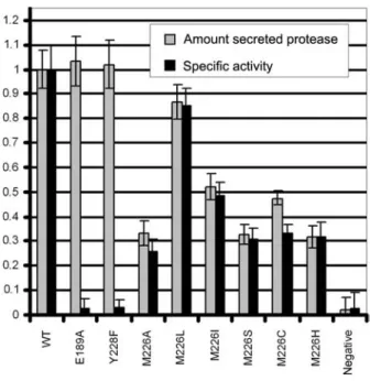
Proteolytic activity
In the past, we have reported Michaelis-Menten
para-meters for the M226L, M226I, and M226A mutants (Hege
and Baumann, 2001a). Other mutants, such as M226H
and M226N, could not be purified. Since the proteins
were secreted into the extracellular medium and no other
proteases were present there, we assayed proteolytic
activities directly from the supernatant employing a
reso-rufin-casein assay, normalized for protein concentrations
determined by a BioRad assay as reported previously
(Hege and Baumann, 2001b; see Figure 2). The results
are in qualitative agreement with our previous data. Not
surprisingly, the specific activity decreases
Met)Leu-)Ile)His)Ala, with the conservative mutations Leu and
Ile having some 85% and 50% of the wild-type activity,
respectively. The levels of secreted protein decrease in
the same order, indicating some defect in synthesis and
secretion or stability of the mutants.
Crystal structure analysis
Since yield and purity of some of the single methionine
mutants was not sufficient for structural studies, we
Figure 2 Activity assays.
Activity was assayed using resorufin-casein and media super-natant after spinning down the cells as described in the mate-rials and methods section. Shown is the average of three independent measurements. Light bars indicate the ratio of secreted protein to cell density, and the dark bars indicate the specific activity. Values for the wild-type protein were normalized to 1.0. Cells carrying only the transport machinery plasmid were used as negative control (‘negative’).
Table 1 Data collection and refinement.
Protein M226I-E189A M226A- M226A- M226A-
M226H-E189A_inh E189A_SLS E189A_DESY E189A
X-ray source DESY X11 Ru300 SLS PXI DESY X11 DESY X11
Unit cell a (A˚)a 102.41 101.96 102.26 101.89 102.11
Unit cell c (A˚) 121.03 123.22 123.40 122.52 122.86
Wavelength (A˚) 0.812 1.542 1.281 0.812 0.812 Resolution (A˚) 50.0–1.75 50.0–1.95 26.6–2.05 50.0–2.13 50.0–1.77 (outer shell) (1.78–1.75) (1.98–1.95) (2.16–2.05) (2.17–2.13) (1.80–1.77) Rmerge(%) 4.8 (32.9) 3.4 (18.4) 11.7 (19.1) 8.4 (39.7) 4.0 (17.4) Completeness (%) 99.9 (98.5) 99.6 (100.0) 99.9 (99.8) 99.9 (100.0) 100.0 (100.) Redundancy 5.6 (3.8) 6.0 (5.1) 7.5 (5.8) 6.7 (3.8) 7.4 (5.7) I/sigma (I) 23.0 (5.0) 28.3 (7.5) 15.5 (6.1) 12.9 (3.3) 45.1 (10.2) refinement No. of reflections 74 320 (2183) 54 271 (1631) 47 147 (1412) 41 498 (1260) 72 453 (2138) work (test) set
No. atoms protein 3525 2937 2961 2947 3554
Ca2q/Zn2q 7/1 7/1 7/1 7/0 7/2c Water 548 512 481 227 486 R/Rfree(%) 15.5/17.9 15.3/16.6 14.8/17.0 18.2/20.7 16.6/18.7 RMS bonds (A˚) 0.017 0.007 0.007 0.005 0.009 RMS angles (deg.) 1.56 1.0 0.97 0.88 1.63 Ramachandran 0 0 0 0 0 outliers (%)b
aCrystals belong to space group P3 121.
bAccording to Lovell et al. (2003) and Richardson (2003).
cThe two Zn2qions occupy alternating sites with approximately 50% occupancy.
The numbers in parentheses refer to the outer resolution shell.
introduced an additional mutation E189A, knocking
out the catalytic base and thus inactivating the mutants
completely. This greatly helped in purification and
crys-tallization. Relevant statistics are given in Table 1. No
significant changes in the active site were introduced by
this mutation (data not shown), besides the
disappear-ance of a zinc-bound phosphate ion originating from the
crystallization buffer and a slight movement of His-198.
Therefore, we conclude that the differences we observe
for the Met-226 mutants originate largely from the
replaced methionine (Qoronfleh et al., 1995; Pieper et al.,
1997; Walasek and Honek, 2005).
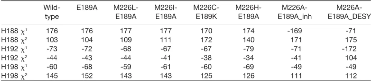
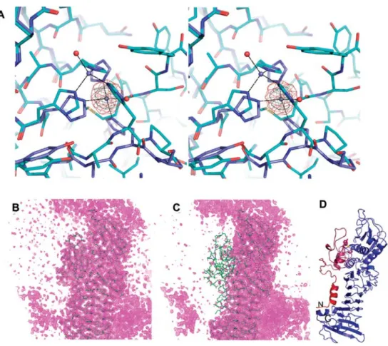
An overlay of the active sites of the more conservative
mutants (M226L, M226I, M226C) shows mainly a change
in the geometry of the imidazole side chains of the
coor-dinating histidine residues (Figure 3). In particular, the x
1angle of His-188 increases with decreasing activity (Table
2) and with decreasing size of the side-chain at position
226. This change in side-chain conformation causes a
movement of the imidazole ring towards the cavity
cre-ated by the mutation. The effect is most dramatic by
cut-ting down the methionine side-chain to the C
batom, i.e.,
in the M226A mutant (Figure 4). As can be seen in Figure
4A, the zinc, which is clearly present as documented by
a large peak in the anomalous difference Fourier map,
moves downwards by approximately 3 A˚, while the
imidazole side-chain of His-188 is nearly perpendicular
to the orientation found in the wild-type enzyme. In order
to keep the coordination sphere, His-198 changes
posi-tion as well. This latter change is possible due to a
dra-matic order-disorder transition: amino acids 18–61 and
199–238, all very well defined in all the other mutants
with the exception of M226H, become completely
dis-ordered. The second segment comprises the Met-turn
and would clash with the new side-chain conformation
of His-198. In other words, the polypeptide chain
cov-Table 2 Side-chain dihedral angles of active-site histidines.
Wild- E189A M226L- M226I- M226C- M226H- M226A-
M226A-type E189A E189A E189K E189A E189A_inh E189A_DESY
H188 x1 176 176 177 177 170 174 -169 -71 H188 x2 103 104 109 111 172 140 171 175 H192 x1 -73 -72 -68 -67 -67 -79 -71 -172 H192 x2 -44 -43 -44 -41 -38 -34 -41 104 H198 x1 -60 -68 -59 -61 -60 -69 -49 -49 H198 x2 145 152 143 143 125 126 111 112
Figure 3 Active centers of M226 mutants revealing a distortion of the Zn2q-binding site and a shift in the coordinating histidines.
Stereo view of mutants E189A (cyan carbon atoms), M226L (magenta), EM189,226AI (gray), and EM189,226KC (wheat). The phos-phate ion still present in the M226L structure has been omitted as well as the zinc-coordinating water molecules. The systematic change in the side-chain dihedrals, especially x2of His-188, with decreasing size of the side-chain at position 226 is clearly visible
(compare to Table 2).
ering the left side of the active center in Figure 4B–D
(right side in Figure 1) becomes completely disordered.
A patch of peptide-shaped electron density is clearly
vis-ible in the active site, forming an antiparallel b-strand
with the edge of the sheet in the protease domain. It is
unlikely that this peptide is one of the proteolysis
prod-ucts sometimes observed in the active centers of
serra-lysins (Baumann et al., 1993): firstly, the E189A mutation
inactivates PrtC completely, and secondly the putative
peptide extends over the zinc-binding position, which
means it resembles more the educt than the products of
the hydrolytic reaction.
However, the situation in the EM189,226AA mutant is
even more complicated, owing to the additionally
occur-ring complete loss of the zinc ion, as revealed by analysis
of other crystals. Two out of four datasets show the
con-dition depicted in Figure 4, while the remaining two
crys-tals exhibit an entire loss of the zinc ion concomitant with
a complete rearrangement of the active-site histidines
(Figure 5, Table 2).
Another variation of the distortion of the active site
could be observed in the EM189,226AH mutant. Here,
an alternative zinc-binding site with approximately 50%
occupancy, as derived by comparison of B-factors from
neighboring atoms and the situation in other mutants,
is created by the presence of the additional imidazole
group, as is clearly revealed by the imaginary electron
density component (Figure 6). His-188 occurs in two
alternative conformations coordinating to both zinc
sites. Polypeptide segments 18–61 and 199–238 exhibit
slightly higher average B-factors than in the E189A
mutant, but are clearly visible with the exception of
res-idues 203–210 belonging to the active center loop.
Discussion
Currently, some 800 members of the metzincin clan are
known (Gomis-Ruth, 2003, 2009). All of them appear to
possess the conserved methionine, although this residue
is sometimes only tentatively identified by sequence
alignments. Mutagenesis of such a conserved amino
acid would be expected to exhibit large effects on activity
and stability. However, conservative substitution by Leu
or Ile, the naturally most frequently occurring mutations,
displays only moderate effects. However, the previously
reported distortions of the zinc environment have been
confirmed, and they become increasingly larger with
decreasing side-chain size at position 226. In order to
assess the fit of the methionine side-chain compared to
other amino acids, we calculated the shape
complemen-tarity value (SC value) (Lawrence and Colman, 1993) from
our X-ray structures. A perfect fit would be indicated by
a value of 1.0. The values for wild-type, M226L, M226I,
and M226C mutants are 0.81, 0.73, 0.56, and 0.42,
respectively. This clearly shows that in the jigsaw
pack-ing of hydrophobic amino acids at the base of the
zinc-binding pyramid, methionine is best fitting. Thus, even
replacement by leucine leads to observable distortions
and a reduction in activity by approximately 15%. While
this effect is small, it might nevertheless contribute to the
selection pressure on the methionine. It should be noted
Figure 4 The EM189,226AA mutant as seen with the in-house and SLS data.
(A) Stereo view of an overlay E189A (cyan carbons, zinc shown as blue sphere) and EM189,226AA (blue carbon atoms, zinc depicted as gray sphere). Coordinating water molecules are shown as red spheres. An imaginary Fourier map is contoured at 10 and 15 standard deviations above the mean (magenta and gray mesh, respectively; data collected at SLS). (B) 2Fo-Fcelectron density (contour
level 1 sigma) of E189A. (C) 2Fo-Fcdensity map contoured at 1.0 sigma for the EM189,226AA double mutant. The orientation is
similar as in panel (B). (D) Ribbon diagram of EM189,226AA colored according to B-factors from blue to red (-25 A˚2: blue; )220 A˚2:
red). The orientation is approximately 180 degrees around the vertical from Figure 1 and similar as in panels (B) and (C). M226 and Y228 are shown as sticks as well as H188 and H192. The purple and red segments are completely invisible and have been omitted in panel (A).
Figure 6 The EM189, 226AH mutant possesses two alternative Zn2qsites.
The imaginary component of the electron density is shown in pink and contoured at 8 standard deviations above the mean (data collected at DESY). Zinc ions are shown as yellow spheres.
Figure 5 Active site of EM189,226AA as seen in data from DESY.
The 2Fo-Fcmap (magenta) is contoured at 1.0 sigma above the
mean. Anomalous difference Fourier maps show no signal for a Zn2qion anywhere close to the active site. This is in contrast to
Figure 4A where the Zn2qsite is shifted.
that most other studies have replaced the methionine
with selenomethionine or difluoromethionine, i.e., amino
acids with different electronic properties at the d-position
of the side-chain but with a very similar size and shape
(Qoronfleh et al., 1995; Pieper et al., 1997; Walasek and
Honek, 2005). Only the substitution of methionine by
serine in MMP-2 (Butler et al., 2004) does not fit into
our observations on PrtC. While in MMP2 no difference
in activity and stability has been detected, in PrtC the
M226S mutant exhibits approximately 30% of the
wild-type activity, very similar to the cysteine mutant. Further
studies will be needed to resolve these conflicting results
and to attain a deeper understanding of the conservation
of the methionine.
Materials and methods
Activity assay
LB medium (5 ml: 50
m
g/ml kanamycin, 50m
g/ml chloramphen-icol) was inoculated with a single colony carrying two plasmids pRUW520 (PrtC) and pRUW4inh1 (transport machinery) and incubated under shaking at 378C overnight. The cell density was measured at 500 nm. Cells were removed by centrifugation and the protein concentration in the supernatant was measured by a Bradford assay. An aliquot was precipitated by trichloroacetic acid and analyzed on sodium dodecyl sulfate polyacrylamide gel electrophoresis. Proteolytic activity was assayed employing resorufin-casein (Roche Diagnostics, Basel, Switzerland) accor-ding to the manufacturer’s instructions. Cells transformed only with pRUW4inh1 and grown in LB medium without kanamycin served as negative control.Mutagenesis, protein expression, and purification
Site-directed mutagenesis was performed as described previ-ously (Jones and Winistorfer, 1992; Hege and Baumann, 2001b) using the E189A DNA as template. Oligonucleotides were ordered from MWG Biotec (Ebersberg, Germany). The helper pri-mer had the sequence 59-CGC GGC CTC GAG CAA GAC GTT TCC CG-39. The following mutagenic primers were employed (base changes are underlined):M226I sense: 59-CAA TTC AGT ATC ATAAGC TAC TGG GG-39; M226A sense: 59-CAA TTC AGT ATCGCGAGC TAC TGG GG-39; and
M226H sense: 59-CAA TTC AGT ATC CAT AGC TAC TGG GG-39.
Expression was performed in Escherichia coli XL1 BLUE cells according to Delepelaire and Wandersman (1989). Briefly, cells were grown in LB medium containing 50
m
g/ml kanamycin and 50m
g/ml chloramphenicol to an OD600 of 0.6 at 378C, theninduced with 1 mM isopropyl-beta-D-thiogalactopyrano-side (IPTG) and grown overnight at 378C. Secreted proteins were purified by anion exchange chromatography as described pre-viously (Hege and Baumann, 2001a,b). Trigonal-bipyramidal crystals grew in hanging drops at 208C from a solution of 10 mg/ ml protein and a reservoir containing 2MNaCl, 0.1MNaH2PO4,
0.1MKH2PO4, 0.1M2-(N-morpholino)ethanesulfonic acid (MES),
pH 6.5.
Data collection and structure refinement
Crystals were cryo-protected by the addition of 25% (v/v) gly-cerol and flash-cooled in a 110 K nitrogen stream. Data collec-tion in-house employed a Rigaku (Tokyo, Japan) RU300 rotating anode and an RaxisIV image plate detector (Rigaku, Tokyo, Japan). Synchrotron data were collected at the EMBL outstation at Deutsches Elektronensynchrotron (DESY) in Hamburg (Ger-many) utilizing an MAR165 CCD detector. Data were processed
using the HKL2000 package (Otwinowski and Minor, 1997). Interactive model rebuilding was effected with O (Jones et al., 1991) and COOT (Emsley and Cowtan, 2004). Refinement was initially carried out using CNS (Brunger et al., 1998) and com-pleted by PHENIX using TLS refinement (Adams et al., 2002). Figures were prepared with PYMOL (http://www.pymol.org). Coordinates of the various structures have been deposited in the Protein Data Bank (PDB) under the entry codes 3HB2, 3HBU, 3HBV, and 3HDA.
Acknowledgments
This work has been supported by the Swiss National Science Foundation and by the University of Bern and its supporting foundation. We are grateful to Philippe Delepelaire and Cecile Wandersman, Institute Pasteur, Paris (France) for the plasmids encoding PrtC and the transport machinery. We thank Drs. Man-fred S. Weiss and Andrea Schmidt for their support during data collection at the EMBL outstation at DESY, Hamburg (Germany).
References
Adams, P.D., Grosse-Kunstleve, R.W., Hung, L.W., Ioerger, T.R., McCoy, A.J., Moriarty, N.W., Read, R.J., Sacchettini, J.C., Sauter, N.K., and Terwilliger, T.C. (2002). PHENIX: building new software for automated crystallographic structure determination. Acta Crystallogr. D Biol. Crystallogr. 58, 1948– 1954.
Baumann, U., Wu, S., Flaherty, K.M., and McKay, D.B. (1993). Three-dimensional structure of the alkaline protease of
Pseu-domonas aeruginosa: a two-domain protein with a calcium
binding parallel b-roll motif. EMBO J. 12, 3357–3364. Blobel, C.P. (2005). ADAMs: key components in EGFR signalling
and development. Nat. Rev. Mol. Cell Biol. 6, 32–43. Bode, W., Gomis-Ruth, F.X., and Stockler, W. (1993). Astacins,
serralysins, snake venom and matrix metalloproteinases exhibit identical zinc-binding environments (HEXXHXXGXXH and Met-turn) and topologies and should be grouped into a common family, the ‘metzincins’. FEBS Lett. 331, 134–140. Bond, J.S. and Beynon, R.J. (1995). The astacin family of
metal-loendopeptidases. Protein Sci. 4, 1247–1261.
Brunger, A.T., Adams, P.D., Clore, G.M., DeLano, W.L., Gros, P., Grosse-Kunstleve, R.W., Jiang, J.S., Kuszewski, J., Nilges, M., Pannu, N.S., et al. (1998). Crystallography and NMR system: a new software suite for macromolecular structure determination. Acta Crystallogr. D Biol. Crystallogr. 54, 905– 921.
Butler, G.S., Tam, E.M., and Overall, C.M. (2004). The canonical methionine 392 of matrix metalloproteinase 2 (gelatinase A) is not required for catalytic efficiency or structural integrity: probing the role of the methionine-turn in the metzincin metalloprotease superfamily. J. Biol. Chem. 279, 15615– 15620.
Delepelaire, P. and Wandersman, C. (1989). Protease secretion by Erwinia chrysanthemi. Proteases B and C are synthesized and secreted as zymogens without a signal peptide. J. Biol. Chem. 264, 9083–9089.
Emsley, P. and Cowtan, K. (2004). Coot: model-building tools for molecular graphics. Acta Crystallogr. D Biol. Crystallogr. 60, 2126–2132.
Gomis-Ruth, F.X. (2003). Structural aspects of the metzincin clan of metalloendopeptidases. Mol. Biotechnol. 24, 157–202. Gomis-Ruth, F.X. (2009). Catalytic domain architecture of
met-zincin metalloproteases. J. Biol. Chem. 284, 15353–15357. Hege, T. and Baumann, U. (2001a). The conserved methionine
residue of the metzincins: a site-directed mutagenesis study. J. Mol. Biol. 314, 181–186.
Hege, T. and Baumann, U. (2001b). Protease C of Erwinia
chry-santhemi: the crystal structure and role of amino acids Y228
and E189. J. Mol. Biol. 314, 187–193.
Jiang, W. and Bond, J.S. (1992). Families of metalloendopepti-dases and their relationships. FEBS Lett. 312, 110–114. Jones, D.H. and Winistorfer, S.C. (1992). Recombinant circle
PCR and recombination PCR for site-specific mutagenesis without PCR product purification. Biotechniques 12, 528– 535.
Jones, T.A., Zou, J.Y., Cowan, S.W., and Kjeldgaard, M. (1991). Improved methods for building protein models in electron density maps and the location of errors in these models. Acta Crystallogr. A 47, 110–119.
Jones, D.T., Taylor, W.R., and Thornton, J.M. (1992). The rapid generation of mutation data matrices from protein sequen-ces. Comput. Appl. Biosci. 8, 275–282.
Lawrence, M.C. and Colman, P.M. (1993). Shape complemen-tarity at protein/protein interfaces. J. Mol. Biol. 234, 946–950. Lovell, S.C., Davis, I.W., Arendall, W.B. III, de Bakker, P.I., Word, J.M., Prisant, M.G., Richardson, J.S., and Richardson, D.C. (2003). Structure validation by Ca geometry: w, c and Cb deviation. Proteins 50, 437–450.
Maeda, H. and Morihara, K. (1995). Serralysin and related bac-terial proteinases. Methods Enzymol. 248, 395–413. Meier, R., Drepper, T., Svensson, V., Jaeger, K.E., and Baumann,
U. (2007). A calcium-gated lid and a large b-roll sandwich are revealed by the crystal structure of extracellular lipase from Serratia marcescens. J. Biol. Chem. 282, 31477–31483. Nagase, H., Visse, R., and Murphy, G. (2006). Structure and function of matrix metalloproteinases and TIMPs. Cardio-vasc. Res. 69, 562–573.
Otwinowski, Z. and Minor, W. (1997). Processing of X-ray dif-fraction data collected in oscillation mode. Methods Enzy-mol. 276, 307–326.
Perez, L., Kerrigan, J.E., Li, X., and Fan, H. (2007). Substitution of methionine 435 with leucine, isoleucine, and serine in tumor necrosis factor a converting enzyme inactivates ecto-domain shedding activity. Biochem. Cell Biol. 85, 141–149. Pieper, M., Betz, M., Budisa, N., Gomis-Ruth, F.X., Bode, W.,
and Tschesche, H. (1997). Expression, purification, charac-terization, and X-ray analysis of selenomethionine 215 variant of leukocyte collagenase. J. Protein Chem. 16, 637–650. Qoronfleh, M.W., Ho, T.F., Brake, P.G., Banks, T.M., Pulvino, T.A.,
Wahl, R.C., Eshraghi, J., Chowdhury, S.K., Ciccarelli, R.B., and Jones, B.N. (1995). Production of selenomethionine-labeled recombinant human neutrophil collagenase in
Esche-richia coli. J. Biotechnol. 39, 119–128.
Rawlings, N.D., Morton, F.R., and Barrett, A.J. (2006). MEROPS: the peptidase database. Nucleic Acids Res. 34, D270–D272. Richardson, J.S. (2003). All-atom contacts: a new approach to structure validation. Methods Biochem. Anal. 44, 305–320. Stocker, W., Grams, F., Baumann, U., Reinemer, P., Gomis-Ruth,
F.X., McKay, D.B., and Bode, W. (1995). The metzincins – topological and sequential relations between the astacins, adamalysins, serralysins, and matrixins (collagenases) define a superfamily of zinc-peptidases. Protein Sci. 4, 823–840. Tourasse, N.J. and Li, W.H. (2000). Selective constraints, amino
acid composition, and the rate of protein evolution. Mol. Biol. Evol. 17, 656–664.
Tronrud, D.E., Roderick, S.L., and Matthews, B.W. (1992). Struc-tural basis for the action of thermolysin. Matrix (Suppl) 1, 107–111.
Walasek, P. and Honek, J.F. (2005). Nonnatural amino acid incor-poration into the methionine 214 position of the metzincin
Pseudomonas aeruginosa alkaline protease. BMC Biochem. 6, 21.