9 2001 Society for In Vitro Biology 1071-2690/01 $10.00+0.00
P H E N O T Y P I C CHARACTERIZATION OF H U M A N UMBILICAL VEIN E N D O T H E L I A L
( E C V 3 0 4 ) A N D U R I N A R Y CARCINOMA ( T 2 4 ) CELLS: E N D O T H E L I A L VERSUS
EPITHELIAL FEATURES
KAYOSHI SUDA, BARBARA ROTHEN-RUTISHAUSER, MAJA GUNTHERT, AND HEIDI WUNDERLI-ALLENSPACH t Biopharmacy, Department of Applied BioSciences, ETH Zurich, CH-8057 Zurich, Switzerland
(Received 13 March 2001; accepted 8 June 2001)
SUMMARY
ECV304 cells reported as originating from human umbilical vein endothelial cells by spontaneous transformation have been used as a model cell line for endothelia over the last decade. Recently, deoxyribonucleic acid fingerprinting revealed an identical genotype for ECV304 and T24 cells (urinary bladder carcinoma cell line). In order to resolve the apparent discrepancy between the identical genotype and the fact that ECV304 cells phenotypically show important endothelial characteristics, a comparative study was performed. Immortalized porcine brain mierovascular endothelial cells/C1-2, and Madin Darby canine kidney cells were included as typical endothelial and epithelial cells, respectively. Various methods, such as confocal laser scanning microscopy, Western blot, and protein activity tests, were used to study the cell lines. ECV304 and T24 cells differ in criteria, such as growth behavim; cytoarchitecture, tight junction arrangement, transmembrane electrical resistance, and activity of ~/-glutamyltransferase. Several endothelial markers (yon Willebrand factor, uptake of low-density lipoprotein, vimentin) could clearly be identified in ECV304, but not in T24 ceils. Des- moglein and cytokeratin, both known as epithelial markers, were found in ECV304 as well as in T24 cells. However, differences were found for the two cell lines with respect to the type of cytokeratin: in ECV304 cells mainly cytokeratin 18 (45 kDa) is found, whereas in T24 cells eytokeratin 8 (52 kDa) is predominant. As we eould demonstrate, the ECV304 cell line exposes many endothelial features which, in view of the scarcity of suitable endothelial cell lines, still make it an attractive in vitro model for endothelia.
Key words: endothelia; epithelia; tight junction; cytoarchitecture; cell markers. INTRODUCTION
Endothelial cells are involved in a wide range of pathological processes including inflammation (Abbot et al., 1992), tumor in- vasion (Folkman, 1992), and atheroselerosis (Massy and Keane, 1996). This has led to considerable efforts to isolate and culture endothelial cells from both human and animal sources in order to further investigate their role. One major problem of primary cell cultures is their heterogeneity and the loss of specific markers dur- ing cultivation (Scott and Bicknell, 1993; De Boer et al., 1999) which makes comparison of the results between different studies difficult. For reproducibility- and easy handling, stable cell lines are a valid alternative under the assumption that standard culture con- ditions are established, and the resulting phenotype is characterized with regard to specific markers. Only a few endothelial cell lines, particularly of human origin, are available today. This is the reason why ECV304 cells, a spontaneously transformed human umbilical vein endothelial cell line (Takahashi et al., 1990), became very popular for studies on angiogenesis (Hughes, 1996), cell migration (Kikkawa et al., 1996), glucose transport studies (Vinals et al., 1999), or signal transduetion of the vascular growth factor (Abedi
1 To whom correspondence should be addressed at Biopharmacy, Depart- ment of Appliect BioSeiences, ETH Zurich, Winterthurerstrasse 190, CH- 8057 Zurich, Switzerland. E-mail: [email protected]
and Zachary, 1997). In addition, the ECV304 cells were often used for investigation on the blood-brain barrier (BBB) as they express many characteristic BBB features, especially under the influence of glial ceils (Hurst and Fritz, 1996; Dobbie et al., 1999).
The origin of ECV304 cells has recently been questioned by the finding that the deoxyribonucleic acid (DNA) fingerprints of ECV304 are identical with those of the T24 cell line (Dirks et al., 1999). As T24, a urinaly bladder cancer cell line, was established before the ECV304 cell line (Bubenik et al., 1973), ECV304 has to be regarded as a T24 variant clone that arose by cross-contam- ination of the endothelial culture. DNA fingerprint screening was performed independently at the DSMZ (German Collection of Mi- croorganisms and Cell Cultures), ATCC (American Type Culture Collection), ECACC (European Collection of Animal Cell Cultures), and JCRB (Japanese Collection of Research Bioresourees) with the same outcome: i.e., an identical handing pattern of the two cell lines.
Although an identical genotype has been established for ECV304 and T24 cells, it is clear from the published data that ECV304 cells express some endothelial characteristics (Takahashi et al., 1990; Takahashi and Sawasaki, 1991; Lechardeur et al., 1995; Hughes, 1996; Dobbie et al., 1999; Kiessling et al., 1999). Recognizing the importance of ECV304 for endothelial studies in the present situ- ation, a systematic investigation on the phenotype of the two cells
5 0 6 SUDA ET AL.
was u n d e r t a k e n . For c o m p a r i s o n , M a d i n Darby c a n i n e k i d n e y (MDCK) cells (McRoberts et al., 1981), a typical epithelial cell line, a n d porcine b r a i n m i e r o v a s c u l a r e n d o t h e l i a l cells ( P B M E C ) / C 1 - 2 cells (Teifel a n d Friedl, 1996), w h i c h are of endothelial origin, were included. E C V 3 0 4 a n d T 2 4 cells were first investigated with regard to general features, s u c h as growth behavior, cytoarehitecture, ex- p r e s s i o n of P-glycoprotein (P-gp) a n d ~/-glutamyltransferase (~-GT activity). T h e a r r a n g e m e n t of tight j u n c t i o n s (TJ) (Bowman et al., 1992; D e j a n a et al., 1995; D u n i n a - B a r k o v s k a y a , 1998) was ex- plored by m e a n s of confocal l a s e r s c a n n i n g microscopy (CLSM) a n d t r a n s m e m b r a n e electrical r e s i s t a n c e (TEER) m e a s u r e m e n t s . T h e cells were t h e n investigated with r e s p e c t to specific endothelial characteristics. A s a typical endothelial feature, the u p t a k e of acet- ylated low-density lipoprotein (LDL) was m e a s u r e d as well as the e x p r e s s i o n of yon W i l l e b r a n d factor (vWF), platelet endothelial cell a d h e s i o n m o l e c u l e - 1 (PECAM-1) (Hewett a n d Murray, 1993), VE- c a d h e r i n , a n d v i m e n t i n . For c o m p a r i s o n , s o m e epithelial m a r k e r s were also studied, s u c h as E - c a d h e r i n , cytokeratin, a n d desmoglein.
MATERIAI,S AND METHODS
Cell culture. The following cells were used: ECV304 (ATCC CRL-1998; passage #133-150), T24 (ATCC HTB-4; passage #42-50), MDCK (Rothen- Rutishauser et ah, 1998; passage #216-270), and PBMEC/C1-2 (kindly pro- vided by Prof. Friedl; Teifel and Friedl, 1996; passage #91-198). No differ- ence was found in any cell line between the early and late passages. Cell monolayers were propagated on plastic TPP * flasks (Winiger AG, Wohlen, Switzerland). The media were M199 (Sigma Chemical Co., Buehs, Switzer- land) with 10 mM N-2-hydroxyethylpiperazine-N'-2-ethane-sultonic acid (HEPES) (Sigma) fi)r ECV304, PBMEC/C1-2, and T24 ('.ells, and Eagle's minimum essential medium with Earl's salts supplemented with 2 mM t,- glutamine (GIBCO BRL Life Technologies, Basel, Switzerland) and 0.225% NaCO:~ ibr the MDCK ceils. In addition to M199, McCoy's 5A medium (Sig- ma) with 2 mM L-glutamine (GIBCO BRL) was used to test the diflerences in growth and electrical resistance in T24 cells. All media contained 1()% tetal calf serum (PAA Laboratories GmbH, Linz, Austria), 100 units penicil- lin/ml, and 100 ~zg streptomycin/ml (Penicillin-Streptomycin, GIBCO BRL #1.5140-114). Incubations were in a 5% CO2 atmosphere at 37 ~ C. [n the case of the PBMEC/C1-2 ('ells, TPW flasks were coated with l% porcine skin gelatine (Sigma). In order to establish the growth curves, cells were propagated in TPP ~ 24-well plates. Cells were trypsinized and counted with a Neubauer counting chamber. Experimental cultures were, grown on Falcon ~ cell culture inserts with a PET" membrane (0.4 p~(n, 4.3 c,n =, Bectnn Dick- inson, Basel, Switzerland, #3090) with 2.5 nil medium above the insert and 3.0 ml below the insert. Confluence was reached at the same time as with cuhures grown on TPP * plastic. For experimental cultures, the medium was changed twice weekly. Elec, trieal resistance was measured with the Millicell- ERS system (MERS 000 01, Millipore, Volketswil, Switzerland) at 37 ~ C about 6 h after medium change.
Confi~cal laser scanning microscopy. A Zeiss LSM 410 inverted microscope was used with the following lasers: HeNe 633 nm, HeNe 543 nm, Ar 488/ 514 nm, and Ar UV 364 nm. Optical sections at intervals of 0.3 txm were taken with a • Plan-Apochromat objective. Image processing was (lone on a Silicon Graphics workstation using IMARIS, a three-dimensional (3D) muhichannel image processing software tbr confocal microscopic images (Bit- plane AG, Zurich, Switzerland).
Antibodies and fluorescent reagents. For CLSM, the tbllowing antibodies were used. An anti-ZO-1 polyclonal antibody (Zymed, South San Francisco, USA), an anti-E-cadherin monoclonal antibody (Sigma), and affinity purified secondary antibodies (IgG) conjugated with cyanine 5 (Chemicon, Temecula, CA, USA) or cyanine 3 (Chemicon) were used. F-actin was labeled with phtdloidin Oregon Green (Molecula r Probes, Eugene, OR, USA). Cell nuclei were visualized with 4,6-diamidino-2-phenylindole (DAPI, Hoechst AG, Frankturt, Germany). Uptake studies were performed with l,l'dioctadecyl- 3,3,3',3'-tetramethyl-indocarbocyanine perchlorate acetylated LDL (DiI-Ac- LDL) (Molecular Probes). Antibodies used in Western blots were as follows: anti-P-gp monoclonal antibody (C219) and anti-human vWF polyclonal an- tibody (DAKO), and antivimentin monoclonal antibody, anti-E-cadherin
monoclonal antibody, and anti-Pan-cytokeratin monoclonal antibody (Sigma). The antidesmogleinl~ monoclonal antibody was from Progen (PROGEN Bio- technik GmbH, Heidelberg, Germany). For VE-cadherin, an anti-VE-cad- herin monoclonal antibody (1104.1; BD PharMingen, Basel, Switzerland) m; alternatively, an anti-VE-cadherin monoclonal antibody (Chemicon) was used. PECAM-1 was stained with an anti-human monoclonal antibody (WM- 59, Sigma) or a monoclonal anti-mouse antibody (MEC 13.3, BD Phar- Mingen). For detection on Western blots, primary antibodies were reacted with the secondary goat anti-mouse IgG, goat anti-rabbit IgG, or goat anti- rat IgG, all coupled to alkaline phosphatase (Pierce, Rockford, USA).
Immunofluorescent labeling. Inserts with cell layers were prepared for CLSM as described (Rothen-Rutishauser et al., 1998). Briefly, cell layers were fixed tbr 15 min at room temperature in 3% paraformaldehyde (PFA) in phosphate-buft~red saline (PBS) pH 7.4 (10 mM Na~HPO4/KHzPO4, 130 mM NaC1). They were treated with 0.1 M glycine in PBS for 5 min and permeabilized in 0.2% Triton X-100 in PBS for 15 min. Samples were incubated at 37 ~ C for 60 min with the primary antibody, and for 90 rain with the secondary antibody. Antibodies were diluted in PBS containing 3% bovine serum albumin as follows: anti-ZO-1, 1:100; anti- E-cadherin, 1:100; anti-rabbit cyanine 5 and anti-rat cyanine 3, each 1:50. The dilution for phalloidin Oregon Green was 1:10, and the con- centration for DAPI was 1 txg/ml. Preparations were mounted in 0.1 M Tris-HC1 (pH 9.5)/glycerol (3:7) containing 50 mg n-propyl-gallate per milliliter (Sigma).
DiI-Ac-LDL uptake. The cellular uptake of DiI-Ae-LDL was assessed by the method of Voyta et al. (1984). In briet~ cells were incubated with DiI- Ac-LDL (10 txg/ml) in growth medium for 6 h at 37 ~ C, then washed in PBS pH 7.4, and fixed in 3% PFA fi)r 15 rain. At}er washing with PBS, cells were labeled with DAPI for 90 min, and nmunted as described above.
hnmunoblots. For vWF detection, cells were extracted in gel electropho- resis sample buffer (50 nrM HEPES, 150 mM NaC1, 10% glycerol, 1% Triton X-100, 1.5 mM MgC12-6H20, 1 mM ethylene glycol-bis(aminoethylether)-te- traacetic acid [EGTA], I mM phenylmethylsulfonyl fluoride [PMSF], 1% aprotinin, and 100 txM benzamidine). Samples were denatured by heating for 5 min at 95 ~ C. For the detection of all other antibodies, crude membranes were prepared as described (Hrycyna et al., 1998). They were not denatured betore loading on the gel. Briefly, cells were harvested by scraping in ice- cold PBS containing ]% aprotinin (Sigma). At)er centrifngation tor 5 rain at 13,000 X g and 4. ~ C, the cells were resuspended in hypotonic. ]ysis buffer (10 mM Tris pH 7.5, 10 mM NaC], 1 mM MgCI~, and 1% aprotinin) and fi'ozen at - 8 0 ~ C. Subsequently, the cells were thawed and incubated on ice tbr 30 rain. They were then disrupted using 50 strokes with a POTTER| homogenizer (B.Braun Biotech ]nternatiomd, Melsungen, Germany). After dif- t~erential centrifugation, the membranes were resuspended in buffer contain- ing 10% glycerol and stored at - 8 0 ~ C until use. The protein concentration ot" each sample was measured with the BioRad assay (BioRad, Glattbrugg, Switzerland). Sodium dodecy] snfft~te-polyacrylamide-ge[ e[ectrophoresis was carried out on either 7.5 or 12% mini-gels (BioRad) with around 30 t~g ptvtein per slot. C, els were blolted on to hybond-C nitrocellulose sheets (Anmrsham, Dtibmulm'f, Switzerland), and the primary antibodies were re- acted with the secm,la,~y antibodies coupled to alkaline phosphatase (Pierce). Detection of phosphatase activity was with a chemiluminescence systmn (BioRad) on preflashed X-ray film (Fujifilm).
For VE-cadherin and PECAM-l, an additional method was used tor the collection of the samples (Tang et al., 1993). Cells were washed twice in washing solution (10 mM PBS pH 7.4, 5 mM PMSE and 1% aprotinin) and then harvested by scraping in TNC lysis butter (0.01 M Tris-acetate pH 8.0, 0.5% Nonidet P40, 1 mM CaCl~, l mM MgC12, 5 mM PMSK 1% aprotinin, 1 mM benzamidine, and 0.1% trasylo]). They were lysed tot 45 rain at 4 ~ C and centrifuged for 30 min at 4 ~ C, 14,000 X g, and stored at 80 ~ C until use. This procedure was only applied tor samples that were stained with the mouse anti-VE-cadherin fi'om Chemicon and the mouse anti-PECAM-1 fi'mn Sigma.
T-GT assay. The 3'-GT activity was measured by a kinetic colorimetric measurement (Naftalin et al., 1969). Cells were scraped in lysis buffer pH 7.4 containing 50 mM HEPES, 150 mM NaCI, 20% glycerol, 1% Triton-X- 100, 1.5 mY/MgC12, 1 n~]//EGTA, in the presence of the following protease inhibitors: PMSF 1 mM (Sigma), benzamidine 0.1 mM (Sigma), and trasylol 0.1% (Bayer, Leverkusen, Germany). Samples were then lysed for 30 min at 4 ~ C and stored at - 8 0 ~ C until the ~/-GT activity was quantified with L- glutamyl-p-nitlvanilide as substrate (Sigma kit). The protein concentration of each sample was deten~ained by the Bradtbrd method (BioRad).
A
o 6 O ,r .o 4 E c-2
D i ~ - - D I'~ i I I I 0 10 20 i 84 30B
I ' I I I 35O 250 n," uJ uJ 150 i I i I i I i I 0 10 20 30 40 time[d]
FIG. ]. Growth characteristics of ECV304 and T24 cells. Cell numbers (A) and TEER values (B) were determined as described ("Materials and Meth- ods" section). (e) ECV304 cells~ (o) T24 cells. One representative experiment for each cell line is shown with the mean 4- SD determined from three culture wells.
RESULTS
Growth characteristics of ECV304 and T24 cells.
Growth curves for ECV304 and T24 cells (Fig. 1A) were determined as de- scribed(see
"Materials and Methods" section). Both were seeded at a density of 3 - 4 X 10⁴ cells/cm 2 in M199 medium. ECV304 cells were confluent after 1 d in culture. The cell number reached a plateau of about 8 • 105 cells/cm 2 between days 16 and 21. T24 cells were confluent within 3 d, and reached a pla- teau of about 2.5 • !05 cells/cm 2 between days ! 5 and 20, The same was found when T24 cells were cultured in McCoy's 5A medium. For both cell lines, the cell numbers remained constant at least up to 28 d. T E E R values were measured at various times after seeding (Fig. 1B). With ECV304 cells, values of about 2 5 0 - 350 tlcnF were reached between days 10 and 15. They remained within this range for up to at least 28 d, whereas i n T24 cells T E E R values stayed at a lower level, i.e., around 50-75 ~-~cnl 2 between days 5 and 28. Both cell lines could be maintained in culture for up to 45 d without major changes in the eytoarchi- tecture and TEER values (data not shown).Characterization of cytoarchitecture by CLSM.
In order to study the cytoarchitectare, cells were prepared for CLSM as described(see
"Materials and Methods" section). The cell lines were investi- gated in the stationary phase of the growth curve, with the exception of PBMEC/C1-2 cells which detached from the insert after around 10 d in cultme before reaching a plateau in the growth curve. The PBMEC/C1-2 cel!s showed a multilayer formation (Fig. 2A). This is clearly visible in the x,z- and y,z-projection, where the cell nuclei are arranged in different layers (Fig. 2A',arrows).
Strong F-actin bundles were expressed mainly along the cell borders (Fig. 2A), The TJ protein ZO-1 was localized at the cell borders (Fig. 2B), but did not form a complete network. ECV304 cells formed a mono- layer (Fig. 2D) with TJ localized near the apical surface of the cells (Fig. 2D",arrow).
The 3D reconstruction showed that ZO-1 was expressed at the cell-cell contacts and formed a nearly complete TJ network (Fig. 2E). The morphology of T24 cells in turn was significantly different from that of ECV304 cells, with respect to the cytoarchitecture and localization of the ZO-1 protein. Although the T24 ceils formed flat monolayers, the F-actin bundles did not line the cell borders as in ECV304 ceils, but were expressed in a parallel ordered fashion instead (Fig. 2G). The ZO-1 protein was expressed throughout the cells (Fig. 2H) and in part seemed to colocalize with F-actin (Fig.2G, arrows).
The irregular arrangement throughout the cytoplasm was particularly striking in the 3D recon- struction (Fig. 2H). A typical flat monolayer was also found for MDCK ceils (Fig. 2K), as seen from the arrangement of the cell nuclei (Fig. 2K' and K"). The TJs, visualized with the anti ZO-1 antibody, were localized in the upper part of the cells at the cell- cell contacts (Fig. 2K",arrow),
A regular, complete TJ network was formed (Fig. 2L). For occludin, another TJ-related protein, the same staining patterns as with ZO-! were observed in all the cell lines (data not shown).Comparative studies of DiI-Ac-LDL uptake.
DiI-Ac-LDL uptake, which is a characteristic of endothelial cells, was tested in the four cell types. Highest uptake was found for PBMEC/C!-2 cells (Fig. 2C) followed by ECV304 cells (Fig. 2F). Minute traces were found in T24 cells (Fig. 2/), and no DiI-Ac-LDL uptake was observed in MDCK cells (Fig. 2M).T-GT activity.
~-GT activity was determined in the four cell types (Fig. 3). The activity was high in ECV304 (25.6 nmol/min/mg protein) and MDCK (28.1 nmol/min/mg protein) cells, but hardly detectable in T24 (0.7 nmol/min/mg protein) and PBMEC/C1-2 (0.8 nmol/min/mg protein) cells. Fibroblasts (BHK21), which were used as a negative control, did not show any activity (data not shown).P-gp expression.
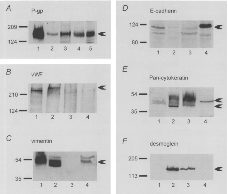
P-gp expression in the four cell lines was com- pared by Western blots (Fig. 4A), P-gp expression was highest in PBMEC/C1-2 cells (7 d) in culture, but significant amounts were also found in MDCK (11 d) and in T24 (21 d) cells. Only traces of P-gp were present in ECV304 cells up to day 21, whereas at a later time between days 31 and 50, P-gp expression increased to a com- parable level as in T24 cells.Expression of vWF, vimentin, VE-cadherin, and PECAM-1.
Ex- pression of vWF, vimentin, VE-cadherin, and PECAM-1 was inves- tigated by Western blots. In ECV304 cells (Fig. 4B), vWF could be detected already at day 4 as a clear band of around 240 kDa. This could be confirmed by the positive control, the PBMEC/C1-2 cells, which showed a strong band at a slightly lower molecular mass. In T24 cells, however, no vWF could be detected for up to 21 d in culture, the same result as was found for MDCK cells used as a negative control.FIG. 3.
PBMEC ECV T24 MDCK
3,-GT activity in PBMEC/CI-2, ECV304, T24, and MDCK cells. t -
3o
g
.~ 20 E "5 E ,[,.- >,, 10 > 0 (9 ~- 0Cells were grown for the times indicated, and the enzyme activity measured
(see "Materials and Methods" section): PBMEC/C1-2 (PB) cells, day 7;
ECV304 and T24 cells, day 21; MDCK cells, day 11. Each column shows one representative experiment with the mean + SD of triplicate measure- m e n t s .
Similar results were found for vimentin (Fig. 4C). Strong expres- sion was detected in PBMEC/C1-2 and ECV304 cells, even though with a slight difference in the molecular weight. In PBMEC/C1-2 cells, vimentin is localized slightly above its predicted molecular weight of 52 kDa, which can be seen in ECV304 cells. T24 cells were completely negative for vimentin, whereas in MDCK cells, some vimentin expression could be detected. Two bands were vis- ible, from which the lower band could be assigned to vimentin according to its described molecular weight of 52 kDa. The upper band, seen also in ECV304 cells, could be either unspecific staining or a phosphorylated form of vimentin.
VE-cadherin as well as PECAM-1 was not found in any of the four cell lines investigated, although various antibodies and differ- ent methods for the collection of samples were used (data not shown).
Expression of E-cadherin, Pan-cytokeratin, and desmoglein.
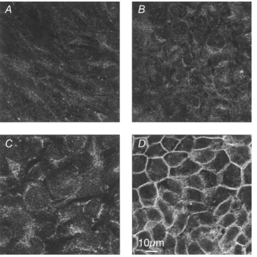
MDCK, T24, ECV304, and PBMEC/C1-2 cells were compared with respect to E-cadherin expression by Western blots (Fig. 4D) and by CLSM (Fig. 5). In Western blots, a strong band of 120 kDa for E- cadherin was detected in MDCK cells; the expression was low in T24 and PBMEC/C1-2 cells, and lowest in ECV304 cells. Similar results were found by CLSM (Fig. 5). Significant staining was pre- sent in the MDCK cells (Fig. 5D), particularly at the cell-cell con- tacts. Faint cytoplasmic staining was found in the T24 cell prepa- ration (Fig. 5C). Both ECV304 (Fig. 5B) and PBMEC/C1-2 (Fig. 5A) cells showed no fluorescence at all.
Cytokeratin was stained with a Pan-cytokeratin antibody (Fig. 4E) which is able to detect cytokeratin 4, 5, 6, 8, 10, 13, and 18. In
PBMEC/C1-2 cells, faint staining was detected for cytokeratins 8. In ECV304 and T24 cells, some of the cytokeratins were stained. Interestingly, the two cell lines did not stain the same type of cy- tokeratin. In ECV304 cells, a band at 45 kDa (cytokeratin 18) and a very weak band at 52 kDa (cytokeratin 8) appeared; T24 cells instead showed strong staining at 52 kDa and little staining at 45 kDa. In MDCK cells, only cytokeratin 8 was detected. The inves- tigations with desmoglein are shown in Fig. 4E As expected, des- moglein was not found in PBMEC/C1-2 cells. ECV304 and T24 cells were positive for this protein, whereas in MDCK cells, no staining could be detected.
DISCUSSION
Comparative studies on the phenotypes of the ECV304 and the T24 cell line showed significant differences (summarized in Table 1), although both have been demonstrated to have the same geno- type (Dirks et al., 1999). The cell density in the stationary phase is threefold higher for ECV304 than for T24 cells. The same was found also if T24 cells were grown in McCoy's 5A medium (unpub- lished data). Slight differences in cell density have already been described earlier for two subclones of the T24 cell line. However, they only differed by a factor of 1.5 (Flatow et al., 1987). In ac- cordance with published data, ECV304 as well as T24 cells form monolayers (Flatow et al., 1987; Hughes, 1996), which are, however, not as homogeneous and compact as those formed by MDCK epi- thelial ceils (Rothen-Rutishauser et al., 1998). PBMEC/C1-2, the typical endothelial ceils included in the study were not contact inhibited and grew in ilTegular inuhilayers. This behavior was also found in other transfected cells, e.g., the MDR1-MDCK cells (H/im- merle et al., 2000).
Another important difference between ECV304 and T24 cells is found in the formation and maintenance of TJ. The arrangement of TJ, as visualized in the CLSM by labeling of the TJ-related protein ZO-1, is strikingly different between the two cell types. A distinct network is present in ECV304 cells, which is in accor- dance with data of Kiessling et al. (1999), whereas TJ formation is rudimentary- in T24 ceils, although the ZO-1 protein is highly expressed throughout the cells. The same pattern was obtained for occludin, another TJ protein (unpublished data). This situation is also reflected in the TEER values: around 300 l')cnl 2 for ECV304 but only about 50 l ) e m 2 in the case of T24 cells. It has to be noted, howevel, that TEER values do not depend only on the TJ arrangement. For instance, ECV304 and MDCK (type II) cells exhibit similar TEER values, although the MDCK cells form a more regular network of TJ than the ECV304 cells. Also subtypes of MDCK cells (types I and II), which differ in TEER values by a factor of about 60, do not show morphological differences in TJ arrangement in the CLSM (Richardson et al., 1981; Wong, 1997). The functional expression of -/-GT is an important feature of the
zr
FIG. 2. Characterization of cell cultures by CLSM. Cells were grown on filter inserts for the times indicated, and prepared for CLSM
(see "Materials and Methods" section). (A-C) PBMEC/C1-2 cells, day 7; (D-F) ECV304 cells, day 21; (G-l) T24 cells, day 21; (K-M)
MDCK cells, day 14. (A, D, G, K) Preparations were triple-stained for F-actin (green), cell nuclei (blue), and ZO-1 (red); arrows point to TJ (D", K"), or to cell nuclei (A'). Colocalization of F-actin and ZO-1 results in yellow staining. (B, E, H, L) 3D reconstructions of the same areas as in (A, D, G, K), but only ZO-1 (red) and cell nuclei (blue) are shown. (C, F, I, M) DiI-Ac-LDL uptake (see "Materials and Methods" section), 3D reconstructions with DiI-Ac-LDL (red) and cell nuclei (blue). (A, D, G, K) single optical sections; (A', D', G', K') x,z-projections; (A", D", G", K") y,z-projections.
5 1 0 SUDA ET AL.
FiG. 4. Expression of P-gp, vW1 ~, vimentin, E-cadherin, Pan-c.ytokeratin, and desmoglein. The expression of the indicated proteins was determined in PBMEC/CI-2, ECV304, T24, and MDCK cells by Western blots at the times indicated: (A) PBMEC/C1-2 cells cultured for 7 d (lane 1); ECV304 }:ells for 21 d (lane 2); and 50 d (lane 3); '1"24 ('ells for 21 d (lane 4) and MDCK {:ells tor J 1 d (lane 5). (B- F) PBMEC/C]-2 (day 7, lane 1); ECV304 (day 21, lane 2); T24 (day 211, lane 3); and MDCK ((lay l I, lane 4) exc.ept in the blot of vWF (B), in which the sample of ECV304 cells was already taken on (lay 4.
BBB because this enzyme is, among others, responsible for the deg- radation of xenobiotics (Frey, 1993; C o m m a n d e u r et al., 1995). Even though ~/-GT is generally absent in urinary bladder ceils (Van- derlaan and Phares, 1981), some enzyme activity was found in T24, a carcinoma cell line. This finding is compatible with the data re- ported by Vanderlaan and P h a r e s (1981), who found m e a s u r a b l e ~- GT activity in some cultured cell lines of b l a d d e r tumors, and thus proposed the use of this activity as a m a r k e r for tuinor ceils. In comparison, the E C V 3 0 4 ceils show very high enzyme activity. It was five times the activity rep6rted by L e c h a r d e u r et al. (1995) using the same detection kit. Unfortunately, there is no indication about the source of the E C V 3 0 4 cell line in this study. The ~/-GT activity found in P B M E C / C 1 - 2 cells is significantly lower, i.e., in the same range as that reported by "feifel and Friedl (1996). The
high enzyme activity measured in the E C V 3 0 4 cells is in a range similar to the activity in the MDCK cells, in accordance with pre- viously p u b l i s h e d values (Verkoelen et al., 1995).
P-gp is highly expressed in tumor ('.ells. It is also found in the b r u s h b o r d e r of proximal t u b u l e s of the kidney, in the bile cana- licular m e m b r a n e of hepatocytes, in the apical m e m b r a n e of mu- cosal cells in the intestine, a n d in the l u m i n a l m e m b r a n e of en- dothelial cells at b l o o d - t i s s u e b a r r i e r sites (Thiebaut et al., 1987; Relling, 1996). P-gp was found in all the four cell lines tested, although to a variable extent. The highest amount was found in P B M E C / C 1 - 2 cells c u l t u r e d for 7 d, whereas for MDCK (day 11), T24, and E C V 3 0 4 cells, similar amounts were found. T24 cells exhibited P-gp expression w h e n cultured for 21 d. At that time, little P-gp was detected in E C V 3 0 4 cells. Only after prolonged
FIG. 5. Expression of E - c a d h e r i n as vi- sualized by CLSM. Cells were cultured for the times indicated and p r e p a r e d for CLSM (see
" M a t e r i a l s a n d M e t h o d s " section). (A) P B M E C / C 1 - 2 , day 7; (B) ECV304, day 21; (C) T24, day 21; (D) MDCK, day 11. All irrtages
are single optical sections taken in the middle
of the cell layers.
TABLE 1
CHARACTERISTICS OF P B M E C / C I = 2 , ECV304, T24, AND MDCK CELLS IN STATIONARY P H A S E (SUMMARYy
PBMEC/C1-2 ECV304 T24 MDCK
G e n e r a l features
cell n u m b e r [cells/cm 2] no stationary p h a s e 8 x 10⁵ 2 . 5 x 10⁵ 5 • l 0 s T E E R [llcm 2] 150 -+ 3 0 3 0 0 + 5 0 5 0 +- 2 0 2 5 0 + 30 TJ network (ZO1) + + + - - + + + P - g p + + + + + ( 2 1 d) + + ( 2 1 d) + + + + + (5O d) 3'-GT [nmol/min/mg protein] 0.76 -+ 0.31 2 5 . 6 3 + 2 . 7 8 0 . 7 + 0 . 3 7 28.13 -+ 2.26 Endothelial markers A c - L D L uptake + + + + + + - - v W F + + + + + + ---: - - vimentin + + + + + + - - + V E - c a d h e r i n . . . . PECAM-1 . . . . Epithelial markers E - c a d h e r i n + + + + + + cytokeratin 8 + + + + + + cytokeratin 18 - - + + + - - desmoglein - - + + + - -
5 1 2 SUDA ET AL.
culturing (30-50 d), comparable amounts of P-gp protein were present. This difference could be explained by the different growth behavior of the two cell lines. In T24 cells, the stationary phase of the growth curve is reached between days 10 and 12 after seed- ing (seeding density: 5 • 104 cells/cm2), whereas with ECV304 cells, this plateau is only attained after about 21 d in culture (seeding density: 5 • 104 cells/cm2). Therefore, the differentiation of ECV304 cells, e.g., with regard to transporters, is delayed when compared with T24 cells. A similar situation can be found in Caco-2 cells which need at least 3 wk to build up their specific transporter systems (Hosoya et al., 1996).
The uptake of DiI-Ac-LDL is regarded as a characteristic feature of endothelial cells (Hewett and Murray, 1993), and therefore high uptake of DiI-Ac-LDL was expected for the PBMEC/C1-2 cells. This was indeed the case. Uptake was also found in ECV304 cells, although to a lesser extent than in PBMEC/C1-2 cells. No signifi- cant uptake was registered in the T24 cell line and in MDCK ceils. DiI-Ac-LDL uptake in ECV304 cells has already been described though to a lesser extent than in freshly isolated human umbilical vein endothelial cells (Hughes, 1996). Furthermore, the vWF is a widely used endothelial marker. It was shown to be expressed in endothelial cells, megakaryocytes, and platelets, where it is con- centrated in the Weibel-Palade bodies, presumable storage and/or processing vesicles for this protein (Wagner et al., 1982). We could identify this marker in the ECV304 and PBMEC/C1-2 cells. The finding for ECV304 cells is in agreement with Hughes (1996). Both, DiI-Ac-LDL uptake and the vWF have been reported to be present in mesothelial cells (Chung-Welch et al., 1997). Mesothelial cells are similar to endothelial cells, with which they share many mor- phological and functional properties. However, vWF expression and DiI-Ac-LDL uptake are not found in epithelial (,'ells and never have been described in bladder cells. An additional hint for an endothe- lial phenotype in ECV304 cells is the finding that they organize into an extensive network of capillal"y tube-like structures within 1 d when cultured on Matrigel, a laminin-based support (Hughes, 1996). No comparable studies have been performed with T24 cells. Two other endothelial markers, VE-cadherin and PECAM-1, could not be detected in any of the studied cell lines, although various antibodies were used (two for each protein). The fact that in PBMEC/C 1-2 ceils too no expression of VE-cadherin or PECAM- 1 could be detected may be interpreted to indicate that the anti- bodies, which were raised against humans or mice, are species- specific and do not bind to the porcine forms. This argument does not, however, hold for ECV304, a human cell line. These cells have already been described to be negative for PECAM-1 and VE-cad- herin (Hughes, 1996; Kim et al., 1998; Kiessling et al., 1999).
Vimentin is mainly found in cells of mesenchymal origin, such as fibroblasts, some leukocytes, and endothelial cells, but it was also found in epithelial cells, such as mesothelial cells and the human kidney (Hohhofer et al., 1984; Chung-Welch et al., 1997). Our data perfectly agree with these findings. PBMEC/C1-2 cells strongly expressed vimentin. Strong expression was also found in ECV304 cells, whereas T24 cells did not express it at all. The weak expression found in MDCK ceils is in accordance with the pub- lished data (Stuart et al., 1994). Staining for Pan-cytokeratin re- vealed cytokeratin expression in all the four cell lines. Even in PBMEC/C1-2 cells, faint staining was observed. Cytokeratin as well is not restricted to epithelial tissue. Cytokeratins of types 8, 18, and 19 are usually expressed in simple epithelia. They have also
been found in lymph nodes, peripheral blood cells, skin fibroblasts, freshly isolated endothelial cells (Traweek et al., 1993), human aor- tic smooth muscle cells (Glukhova et al., 1991), mesothelial cells (Connell and Rheinwald, 1983), and pulmonary microvessel endo- thelial cells (Alexander et al., 1991) as well as subsynovial capillary blood vessels (Stosiek et al., 1990). The coexpression of vimentin and cytokeratin documented in this study has been described before (Schmid et al., 1983; Hohhofer et al., 1984; Czernobilsky et al., 1985).
E-cadherin was used as a typical epithelial cell marker. Sig- nificant amounts of E-cadherin were detected in the MDCK cells by CLSM as well as by Western blots. The ECV304, T24, and PBMEC/C1-2 cells showed a faint staining in Western blots, but only background staining in the CLSM. The results for ECV304 are in contrast with Kiessling et al. (1999) who clearly showed E-cadherin staining for ECV304 ceils in Western blots, and also reported focal areas of lateral membrane staining by immunoflu- orescence in the CLSM. It cannot be excluded that this discrep- ancy could be caused by the different antibodies used. However, the findings of strong staining in MDCK cells, as well as the positive staining in the other cell lines, make this explanation unlikely. Another explanation could be the use of different sub- clones of the same cell line. Kiessling et al. (1999) worked with ECV304 cells of the ECACC culture collection, whereas the ECV304 cells in this work stem from the ATCC cell bank. Scism et al. (1999) reported signifieant differences between the ECV304 cells of the ECACC and ATCC collections, respectively; in particular, differences in T E E R and sucrose permeability were observed. A very important difference is that ECV304 cells from the ATCC are not inducible for BBB characteristics by astrocyte or glioma cocuhure, whereas the ceils from ECACC can be in- dueed. In view of these findings, differences in the protein pat- tern of the two subclones could be expected as well. Systematic studies have yet to be performed, but comparing the markers used in this study and in that of Hughes (1996), some indications can already be found regarding the expression of vWF and of cytokeratin types 6 and 10.
The weak signal for E-cadherin in T24 ceils is consistent with the previously published loss of E-cadherin expression in T24 cells as compared with normal urinary bladder cells (Wakatsuki et al., 1996; Imao et al., 1999). The authors suggested an inverse con:e- lation between E-cadherin expression and clinical aggressiveness of tumors. It is interesting to note that E-cadherin has also been found in primary brain endothelial cells (Rubin et al., 1991; Pal et al., 1997). High E-cadherin expression, which is typical for an ep- ithelial phenotype, such as MDCK cells, is neither found in ECV304 nor in T24 cells.
Desmosomes are structures typically found in epithelial cells, whereas in endothelial cells, syndesmos, desmosomal-like struc- tures, were described. Desmoglein and desmoplakin expression was demonstrated in ECV304 cells (Kiessling et al., 1999). Our results confirmed this finding for desmoglein which was found in ECV304 and T24 ceils. No staining has been performed with desmoplakin because this protein is a member of syndesmos as well and therefore cannot be used to distinguish between an epithelial or an endothe- lial feature.
To sum up, although the ECV304 and T24 cell lines have an identical genotype, they exhibit clear phenotypic differences. They differ not only in their TEER, growth behavior, and cytoarchitecture,
but also in the expression pattern of endothelial and epithelial markers, respectively. Neither the ECV304 cell line nor the T24 cell line showed a complete endothelial phenotype. However, T24 cells were lacking in important endothelial features, such as sig- nificant LDL uptake, expression of v W F and vimentin, or the for- mation of a complete TJ network, whereas ECV304 cells clearly exhibited these endothelial traits. Regarding all available data, we conclude that at the present moment, with a paucity of endothelial cell lines available, the E C V 3 0 4 cell line is still a suitable endo- thelial model. ECV304 cells from ATCC exhibit endothelial char- acteristics and a nearly complete TJ network without coculture with astrocytes or astroeyte supernatants.
ACKNOWLEDGMENTS
We thank the ATCC culture collection for the kind gift of the T24 cell line. We are also grateful to Stefanie Kramer for inspiring suggestions during the preparation of the manuscript.
REFERENCES
Abbot, S. E.; Kaul, A.; Stevens, C. R.; Btake, D. tl. Isolation and culture of synovial microvaseular endothelial ceils. Characterization and as- sessment of adhesion molecule expression. Arthritis Rheum. 35:401M,06; 1992.
Abedi, H.; Zachary, I. Vascular endothelial growth factor stimulates tyrosine phosphorylation and recruitment to new focal adhesions of focal ad- hesion kinase and paxillin in endothelial cells. J. Biol. Chem. 272:15,442-15,451; 1997.
Alexander, J. S.; Patton, W. F.; Yoon, M. U.; Shepro, D. Cytokeratin filament modulation in pulmonary nfierovessel endothelial cells by vasoaetive agents and culture confluency. Tissue Cell 23:141-150; 1991. Bowman, P. D.; Du Bois, M.; Shivers, R. R.; Dorovini-Zis, K. Endothelial
tight junctions. In: Cereijido, M., ed. Tight junctions. Boca Raton, FL: CRC Press; 1992:305-320.
Bubenik, J.; Baresova, M.; Viklicky, V\; Jakoubkova, J.; Sainernva, H.; Don- ner, J. Established cell line of urinary bladder carcinoma (T24) con- taining turnout-specific antigen. Int. J. Cancer 11:765-773; 1973. Chung-Welch, N.; Patton, W. E; Shepro, D.; Cambria, R. P. Two-stage iso-
lation procedure for obtaining homogenous populations of microvas- cular endothelial and mesothelial cells from human omentum. Micro- vasc. Res. 54:121-134; 1997.
Commandeur, J. N. M.; Stijntjes, G. J.; Vermeulen, N. P. E. Enzymes and transport systems involved in the formation and disposition of gluta- thione S-conjugates: role in bioactivation and detoxication mecha- nisms of xenobiotics. Pharumcoh Rev. 47:271-330; 1995.
Connell, N. D.; Rheinwald, J. G. Regulation of the cytoskeleton in mesothelial cells: reversible loss of keratin and increase in vinmntin during rapid growth in culture. Cell 34:245-253; 1983.
Czeruobilsky, B.; Moll, R.; Levy, R.; Franke, W. W. Co-expression of cyto- keratin and vimentin filaments in mesothelial, granulosa and rete ova- rii cells of the human ovmy. Eur. J. Cell Biol. 37:175-190; 1985. De Boel; A. G.; Galliard, R J.; Bi~inmr, D. D. The transterenee of results
between blood-brain barrier cell culture systems. Eur. J. Phaim. Sci. 8:1-4; 1999.
Dejana, E.; Corada, M.; Lampugnani, M. G. Endothelial cell-to-cell junctions. FASEB J. 9:910-918; 1995.
Dirks, W. G.; Macleod, R. A. E; Drexler, H. G. ECV304 (endothelial) is really T24 (bladder carcinoma): cell line m'oss-eontamination at source. In Vitro Cell. Dev. Biol. 35A:558-559; 1999.
Dobbie, M. S.; Hurst, R. D.; Klein, N. J.; Surtees, R. A. H. Upregulation of intercellular adhesion molecule-1 expression on human endothelial cells by tumour necrosis factor-e~ in an in vitro model of the blood- brain barrier. Brain Res. 830:330-336; 1999.
Dunina-Barkovskaya, A. Tight junctions: facts and models. Membr. Cell Biol. 11:555-589; 1998.
Flatow, U.; Rabson, A. B.; Rabson, A. S. Tumorigenicity of T24 urinary bladder carcinoma cell sublines. Int. J. Cancer 40:240-245; 1987.
Folkman, J. The role of angiogenesis in tmnor growth. Semin. Cancer Biol. 3:65-71; 1992.
Frey, A. Gamma-glutamyl transpeptidase: molecular cloning and structural and functional features of a blood-brain barrier marker protein. In: Pardridge, W. M., ed. The blood-brain ban'ier: cellular and molecular biology. New York: Raven Press; 1993:339-368.
Glukhova, M. A.; Shekhonin, B. V.; Kruth, H.; Koteliansky, V. E. Expression of cytokeratin 8 in human aortic smooth muscle cells. Am. J. Physiol. 261:72-77; 1991.
H~mmerle, S. P.; Rothen-Rutishausm; B.; Kr~mer, S. D.; Gtinthert, M.; Wun- derli-Allenspach, H. P-gp in celt cultures: a combined approach to study expression, localisation, and functionality in the confocal mi- croscope. Eur. J. Pharm. Sei. 12:69-77; 2000.
Hewett, R W.; Murray, J. C. Human microvessel endothelial cells: isolation, culture and characterization. In Vitro Cell. Dev. Biol. 29A:823-830; 1993.
Holthofer, H.; Miettinen, A.; Lehto, V. R; Lehtonen, E.; Virtanen, I. Expres- sion of vimentin and cytokeratin types of intermediate filament pro- teins in developing and adult human kidneys. Lab. Invest. 50:552-559; 1984.
Hosoya, K. I.; Kim, K. J.; Lee, V. H. Age-dependent expression of P-glyeo- protein gpl70 in Caeo-2 cell monolayers. Phann. Res. 13:885-890; 1996.
Hrycyna, C. A.; Airan, L. E.; Germann, U. A.; Ambudkal; S. V.; Pastan, I.; Gottesman, M. M. Structural flexibility of the linker region of human P-glyeoprotein permits ATP hydrolysis and drug transport. Biochem- istry 37:13,660-13,673; 1998.
Hughes, S. E. Functional characterization of the spontaneously transformed human umbilical vein endothelial cell line ECV304: use in an in vitro model of angiogenesis. Exp. Cell Res. 225:171-185; 1996. Hurst, R. D.; Fritz, I. B. Properties of an immortalised vascular endothelial/
glioma cell co-culture model of the blood-brain barrier. J. Cell. Phy- siol. 167:81-88; 1996.
hnao, T.; Koshida, K.; Endo, Y.; Uchibayashi, T.; Sasaki, T.; Namiki, M. Dominant role of E-cadherin in the progression of bladder cancer. J. Urol. 161:692-698; 1999.
Kiessling, E; Kartenbeck, J.; Haller, C. Cell-cell contacts in the human cell line ECV304 exhibit both endothelial and epithelial characteristics. Cell Tissue Res. 297:131-140; 1999.
Kikkawa, Y.; Akaogi, K.; Mizushima, H.; Yamanaka, N.; Umeda, M.; Miya- zaki, K. Stimulation of endothelial cell migration in culture by ladsin, a laminin-5-1ike cell adhesion protein. In Vitro Cell. Dev. Biol. 32A:46-52; 1996.
Kim, C. S.; Wang, T.; Madri, J. A. Platelet endothelial cell adhesion molecule- 1 expression modulates endothelial cell migration in vitro. Lab. In- vest. 78:583-590; 1998.
Leehardeur, D.; Schwartz, B.; Paulin, D.; Seherman, D. Induction of blood- brain barrier differentiation in a rat brain-derived endothelial cell line. Exp. Cell Res. 220:161-170; 1995.
Massy, Z. A.; Keane, W. K Pathogenesis of atherosclerosis. Semin. Nephroh 16:12-20; 1996.
McRoberts, J. A.; Taub, M.; Saier, M. H. The Madin Darby canine kidney (MDCK) cell link. In: Sato, G. H., ed. Functionally differentiated cell lines. New York: Alan R. Lisa; 1981:117-139.
Naftalin, L.; Sexton, M.; Whitaker, J. E; Traeey, D. A routine procedure for estimating serum ~-glutamyltranspeptidase activity. Ctin. Chim. Acta 26:293-296; 1969.
Pal, D.; Audus, K. L.; Siahaan, T. J. Modulation of cellular adhesion in bovine brain microvessel endothelial cells by a decapeptide. Brain Res. 747:103-113; 1997.
Relling, M. V. Are the major effects of P-glycoprotein modulators due to altered pharmaeokinetics of antieaneer dnigs? Ther. Drug Moult. 18:350-356; 1996.
Richardson, J. C. W.; Sealera, V.; Simmons, N. L. Identification of two strains of MDCK cells which resemble separate nephron tubule segments. Biochim. Biophys. Acta 673:26-36; 1981.
Rothen-Rutishauser, B. M.; Kramer, S. D.; Braun, A.; Gtinthert, M.; Wun- derli-Allenspach, H. MDCK cell cultures as an epithelial in vitro model: cytoskeleton and tight junctions as indicators for the definition of age-related stages by confocal microscopy. Pharln. Res. 15:964-971; 1998.
5 1 4 SUDA ET AL.
Rubin, L. L.; Hall, D. E.; Porter, S.; Barbu, K.; Cannon, C.; Homer, H. C.; Janatpour, M.; Liaw, C. W.; Manning, K.; Morales, J. A cell culture model of the blood-brain balTier. J. Cell Biol. 115:1725-1735; 1991. Schmid, E.; Schiller; D. L.; Grand, C.; Stadler, J.; Franke, W. W. Tissue type- specific expression of intermediate filament proteins in a cultured epithelial cell line from bovine mannnary gland. J. Cell Biol. 96:37-50; 1983.
Scism, J. L.; Laska, D. A.; Horn, J. W.; Gimple, J. L.; Pratt, S. E.; Shepard, R. L.; Dantzig, A. H.; Wrighton, S. A. Evaluation of an in vitro co- culture model for the blood-brain barrier: cmuparison of hmnan um- bilical vein endothelial cells (ECV304) and rat glioma cells (C6) from two commercial sources. In Vitro Cell. Dev. Biol. 35A:580-592; 1999. Scott, R A.; Bieknell, R. The isolation and culture of microvaseular endo-
thelium. J. Cell Sei. 105:269-273; ]993.
Stosiek, R; Kasper, M.; Conrad, K. Immunhistoehemisehe Untersuchungen zur Cytokeratin-Expression in menschlichen Gefgssendothelien unter besonderer Be~qieksiehtigung des Gelenkbindegewebes. Acta Histo- chem. 89:61~56; 1990.
Stuart, R. O.; Sun, A.; Paniehas, M.; Hebert, S. C.; Brenner, B. M.; Nigam, S. K. Critical role for intracellular calcium in tight junction biogen- esis. J. Cell. Physiol. 159:423M,33; 1994.
Takahashi, K.; Sawasaki, Y. Hmnan endothelial cell line, ECV304, produces pro-urokinase [letter]. In Vitro Cell. Dev. Biol. 27A:766-768; 1991. Takahashi, K.; Sawasaki, Y.; Hata, J.; Mukai, K.; Goto, 3: Spontaneous trans-
formation and imraoitalization of human endothelial cells. In Vitro Cell. Dev. Biol. 26:265-274; 1990.
Tang, D. G.; Chen, Y. Q.; Newman, P. J.; Shi, L.; Gao, X.; Diglio, C. A.; Honn, K. V. Idehtification of PECAM-1 in solid tumor ceils and its potential involvement in tumor cell adhesion to endothelium. J. Biol. Chem. 268:22,883-22,894; 1993.
Teifel, M.; Friedl, R Establishment of the permanent microvascular endothe- lial cell line PBMEC/CI-2 fi~om porcine brains. Exp. Cell Res. 228:50-57; 1996.
Thiehaut, IF.; Tsuro, T.; Hamada, H.; Gottesman, M. M.; Pastan, I.; Willingh- am, M. C. Cellular localization of the nmltidrug-resistanee gene prod- uet P-glycoprotein in normal hmnan tissues. Proe. Natl. Acad. Sci. USA 84:7735-7738; 1987.
Traweek, S. T.; Liu, J.; Battifora, H. Keratin gene expression in non-epithelial tissues. Detection with polymerase chain reaction. Am. J. Pathol. 142:1111-1118; 1993.
Vanderlaan, M.; Phares, W. ~-Glutamyltranspeptidase: a tumour cell marker with a pharmacological function. Histochem. J. 13:865-877; 1981. Verkoelen, C. F.; Romijn, J. C.; De Bruijn, W. C.; Boeve, E. R.; Cao, L. C.;
Schroder, F. H. Association of calcium oxalate monohydrate crystals with MDCK cells. Kidney Int. 48:129-138; 1995.
Vinals, F.; Gross, A.; Testar, X.; Palaein, M.; F'osen, R; Zorzano, A. High glucose concentrations inhibit glucose phosphorylation, hut not glu- cose transport, in human endothelial cells. Bioehim. Biophys. Acta 1450:119-129; 1999.
Voyta, J. C.; Via, D. R; Butterfield, C. E.; Zetter, B. R. Identification and isolation of endothelial cells based on their increased uptake of aeetylated-low density lipoprotein. J. Cell Biol. 99:2034-2040; 1984. Wagner, D. D.; Olmsted, J. B.; Marder, V. J. Innnunolocalization of yon Wil- lebrand protein in Weibel-Palade bodies of human endothelial cells. J. Cell Biol. 95:355-360; 1982.
Wakatsuki, S.; Watanabe, R.; Saito, K.; Saito, T.; Katagiri, A.; Sato, S.; Tomita, Y. Loss of human E-cadherin (ECD) correlated with invasiveness of transitional cell cancer in the renal pelvis, ureter and urinary bladden Cancer Lett. 103:11-17; 1996.
Wong, V. Phosphorylation ~ff occludin cotTelates with occludin localization and function at the tight junction. Am. J. PhysM. 273:C1859-C1867; 1997.