Controversial Issues Concerning Norepinephrine
and Intensive Care Following Severe Traumatic
Brain Injury
John F. Stover, Peter Steiger, Reto Stocker
1Ab stract
Norepinephrine and corresponding intra- and interorgan pathways are of clinical pathophysiologic and pharma-cologic importance as exaggerated activation needs to be reduced and insufficient activation must be support-ed to prevent further deterioration and therapy-induced organ damage. This is of high relevance in critically ill patients in whom various norepinephrine-influenced organ systems are simultaneousy affected with varying degrees of tolerability and resistance to norepinephrine-induced cell damage and finds its maximal challenge in patients suffering from severe traumatic brain injury (TBI). This comprehensive review describes complex pathophysiologic interactions, including hemodynamic, microcirculatory, hormonal, metabolic, inflammatory, and thrombocytic alterations overshadowed by differential consequences of com-monly applied pharmacological interventions following TBI. Overall, investigations published to date suggest that receptor-dependent effects of norepinephrine might predispose to complex evolving deterioration especially during intensive care which is characterized by differentiated complication-driven changes and specific complication-dependent needs. In this context, thrombocytes and leukocytes with their adrenergic receptors and differential norepinephric functional regulation are ideal candidates to influence all organs at once. Despite its secure integration of norepineph-rine in clinical routine, future emphasis must be direct-ed at unmasking, monitoring, and controlling possible receptor-mediated detrimental influences which could offset anticipated organ protection.
1 Department of Surgery, Division of Surgical Intensive Care Medicine, University Hospital Zürich, Zürich, Switzerland. Received: January 12, 2006; accepted: January 19, 2006
Key Words
Catecholamines · Secondary injury · Monitoring · Critical care
Eur J Trau ma 2006;32:10–27
DOI 10.1007/s00068-006-0004-4
Introduction
An integral part of modern therapy aimed at preventing secondary injury following traumatic brain injury (TBI) is to maintain adequate cerebral perfusion. Elevating mean arterial blood pressure(MABP) and cerebral per-fusion pressure (CPP) is achieved pharmacologically by continuously infusing vasopressors, e.g., norepinephrine. Experimental conditions following TBI mainly focus on early cerebral changes during short continuous norepi-nephrine infusion which does not necessarily reflect criti-cally ill pati-ents. As outlined in the schematic drawing (Figure 1) challenge to improve our understanding and thus ameliorate modern treatment modalities following TBI is to simultaneously consider the temporal profile of local and evolving systemic alterations with potential re-ciprocal influences which are simultaneously influenced by current therapeutic interventions. Although catechol-amines are readily used in critically ill patients, differen-tial organ-specific changes induced by catecholamines need to be considered and should be monitored to prevent affecting the anticipated neuroprotection. This comprehensive review focuses on pathophysio-logically relevant inter- and intraorgan norepinephric pathways and characterizes various potentially harmful
pharmaco-dynamic effects of continuous norepinephrine infusion within the routine intensive care treatment of patients suffering from severe TBI.
Endogenous Release and Exogenous Norepinephrine Administration
Within minutes following a stressful event, activation of the hypothalamic–pituitary–adrenal axis amplifies re-lease of norepinephrine, epinephrine, and cortisol from the adrenal gland [1]. These exhaustive alterations maintain hemodynamic stability, mobilize energetic reserves, influence the immune system, and adapt neu-roendocrinological and hormonal alterations [1, 2]. Ac-tivation of the noradrenergic locus coeruleus stimulates various neuronal functional networks responsible for the increased level of alertness and sustained analgesia, and inhibits secretion of various hypothalamic and pitu-itary hormones, thereby suppressing reproductive, growth and thyroid functions [2]. The magnitude of this response reflects the extent of underlying injury and contributes to subsequent worsening if not controlled [3]. Under clinical conditions, norepinephrine is infused continuously together with volume resuscitation and hemorrhage/coagulation control to maintain defined blood pressure or CPP levels (Figure 2). In parallel, the sustained endogenous sympatho-autonomic activity is reduced by sedatives and analgetics.
Organ-Specific Effects of Norepinephrine
Extensive investigations of acute and chronic norepi-nephrine infusion have revealed important influences
on various organs which, among others, depend on administered dose, organ-specific receptor distribution, and binding availability of these receptors. Overall,
cerebral
changes
time
systemic
changes
treatment
norepinephrine fentanyl midazolam norepinephrine norepinephrine leukocytes thrombocytes glucoseFigure 1. Schematic drawing depicting the
prin-cipal pillars of simultaneous time-dependent pathophysiologic and pharmacologic changes which balance beneficial and disadvantageous norepinephrine-mediated actions. In critically ill patients, specific needs are dictated by char-acteristic changes over time, possibly requiring individual adjustment of interventions, making detailed monitoring indispensable.
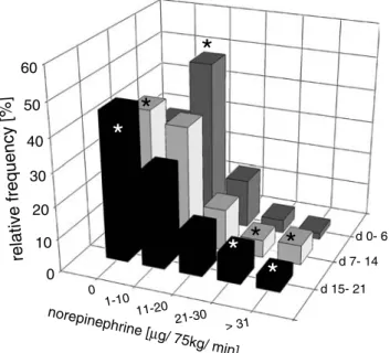
0 10 20 30 40 50 60 0 1-10 11-20 21-30 > 31 d 15- 21 d 7- 14 d 0- 6 relative frequency [%] norepinephrine [ µg/ 75kg/ min]
*
*
*
*
*
* *
Figure 2. Relative frequency distribution of norepinephrine dose in
50 patients suffering from severe TBI up to 3 weeks following injury. Norepinephrine dose was adjusted to maintain CPP between 70 and 110 mmHg. During the first week predominant norepinephrine dose ranged from 1–10 µg/min. While the majority of patients stabilized, reflected by the sustained frequency without norepinephrine and the decreasing frequency within the norepinephrine dose ranging from 0.013–0.133 µg/kg/min, patients with a more difficult clinical course showed an increased frequency in high norepinephrine dose
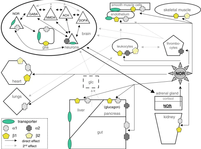
beneficial effects on individual organs may be offset by simultaneous alterations of other organ systems which demand following a ‘brain-oriented’ and avoiding a ‘brain-centered’ therapy. In this context, norepinephrine-driven improvement of cerebral perfusion and metabo-lism due to increased CPP occurs in face of reduced kidney, liver, and testis perfusion and metabolism [4]. The general concept of modern intensive care treatment is to guarantee adequate volume replacement and catecholamine administration within acceptable, i.e., organ-protecting limits. While increased volume admin-istration allows to significantly reduce norepinephrine dosage [5], prevention of organ-endangering volume overload must be considered. Careful judgement of the individual situation is required to guide sequential or parallel administration of norepinephrine and fluids. In principal, single administration of high-dose norepi-nephrine in a patient in whom intravascular volume is depleted or reduced should be avoided as norepineph-rine-mediated vasoconstriction will induce organ dam-age. A careful review of the literature produced only few clinical studies investigating the pharmacodynamic and pharmacokinetic profile of continuous norepineph-rine infusion in healthy volunteers, nonseptic, and TBI patients. The majority of findings are derived from hemodynamically instable patients suffering from sep-sis and corresponding experimental sepsep-sis models in various species. Given the facts that sepsis fulfills the same criteria of the systemic inflammatory response syndrome (SIRS) expanded by a bacterial infection and that SIRS can even develop in patients with isolated severe TBI [6], certain changes observed during these conditions might also be of relevance for the treatment of TBI patients, especially if sepsis develops. The com-plexity and diversity of posttraumatic intensive care involving various norepinephrine-influenced organs is depicted in the schematic drawing (Figure 3).
Heart, Circulation, and Macrohemodynamics
According to its characteristic receptor distribution, norepinephrine increases cardiac contractility (b1) and peripheral resistance (a1 receptors), thereby elevating systolic and diastolic blood pressure, increasing MABP, cardiac index, and total peripheral resistance (TPR) [7–11]. Pressure-dependently, coronary perfusion is improved in healthy animals [7]. However, norepineph-rine might endanger cardiac viability as chronic admin-istration of high-dose norepinephrine induces left ventricular hypertrophy [12–14] and may activate car-diotoxic cascades via stimulation of b1 -adrenergic-driv-en apoptotic changes due to intracellular activation of Ca2+-activated calmodulin kinase and release of free
oxygen radicals which is inhibited by b2 stimulation [15]. It is important to keep in mind that improving MABP does not necessarily reflect ameliorated organ perfu-sion especially under pathological conditions [8, 10]. Despite normalized MABP, failure of improving im-paired renal and mesenteric perfusion [8, 10] could re-flect maintained autoregulation or insufficient increase in MABP due to massively disturbed autoregulation. This is also suggested by recent findings in septic pa-tients in whom a further increase in MABP from 65 to 85 mmHg did not improve renal function [16].
Lungs
Apart from pressure- and volume-passive influences, norepinephrine interferes with pulmonary function by activating adrenergic receptors and stimulating the in-flammatory response. Under experimental conditions, norepinephrine dose-dependently induces a1A- and a1B -adrenergic-mediated pulmonary vasoconstriction [17] which can be inhibited by fentanyl in vitro [18]. High-dose norepinephrine (0.1 mg/kg/h 1.7 µg/kg/min, 128 µg/75 kg/ min) continuously infused up to 72 h in healthy rats results in reversible pleural effusion and pulmonary venous congestion related to increased hydrostatic pressure [13]. The reversible left ventricular hypertro-phy appears necessary to compensate and clear pleural effusion upon termination of norepinephrine infusion [13]. In addition, adrenergic-mediated inflammatory response with alveolar and interstitial edema formation contributes to functional and structural lung injury [14]. Under pathological conditions, the lungs are primed for sustained accumulation, activation, and sequestration of leukocytes [19] which could be aggravated by infused norepinephrine. Vasoconstriction in combination with increased leakage could impair preexisting regional perfusion/ventilation mismatch in intubated and venti-lated ICU patients.
Intestines
a-adrenergic and b-adrenergic activation influences gut motility and intestinal functions. Splanchnic vasocon-striction shunts blood to heart, lungs, brain, and muscles. While epinephrine reduces intestinal and splanchnic perfusion leading to mucosa damage [20, 21], norepinephrine at 0.05 µg/kg/min (3.75 µg/75 kg/min) is not associated with negative effects in animals and pa-tients [8, 9, 22] as it does not impair intestinal perfusion and mucosal integrity despite a dose-dependent increase in splanchnic oxygen extraction [9]. Even under adverse conditions, as e.g. sepsis with or without ensuing shock high-dose norepinephrine at 0.18 or 0.45 µg/kg/min is not harmful [20, 21]. Norepinephrine is superior to
phenylephrine, dopamine, vasopressin, and epineph-rine by improving splanchnic perfusion, oxygen deliv-ery, and lactate uptake [23–26].
Kidneys
The strong oxygen dependency and low critical thresh-old for oxygen consumption in combination with the required high renal perfusion pressure (kidney: 80–180 mmHg vs. liver: 50–150 mmHg) make the kid-neys highly vulnerable to impaired perfusion and oxygen supply [27, 28]. Apart from volume administra-tion, norepinephrine is beneficial, especially in face of nitric oxide (NO)-mediated vasodilation and disease-related vasoparalysis which disturbs various modulators (catecholamines, NO, angiotensin II, vasopressin, and endothelin-1) and intracellular pathways [29]. Despite
norepinephrine-induced reduction in renal perfusion observed in healthy volunteers at 0.118 ± 0.03 µg/kg/min [30], norepinephrine pressure-dependently elevates renal perfusion, increases urine output and creatinine clearance in healthy [7, 31] and septic animals [31]. Adverse effects were ruled out in a retrospective study including 200 cardiac surgery patients in whom norepi-nephrine infusion did not increase serum creatinine levels [32]. Under experimental and clinical septic con-ditions norepinephrine required to elevate MABP to 70 mmHg needs to be increased severalfold [31, 11], reach-ing values as high as 1.3 ± 0.3 µg/kg/min (97.5 ± 22.5 µg/75 kg/min) in humans [11] or 3.1 ± 0.3 versus 0.2–1 µg/kg/min (232.5 ± 22.5 µg/75 kg/min vs. 15–75 µg/75 kg/min) in septic versus control rats [8]. While increas-ing MABP in nonseptic patients does not alter renal brain heart liver pancreas gut kidney thrombo-cytes leukocytes glia neurons
smooth muscle cells
glc cortisol NOR transporter α1 β1 α2 GABA NMDA ACH DOPA skeletal muscle lungs (glucagon) endothelium NOR β2 direct effect 2ndeffect NOR adrenal gland
Figure 3. Schematic drawing of different organ systems with their complex intra- and interorgan influences involved in
norepinephrine-medi-ated functional circuits. An unspecific stressful event stimulates adrenal release of norepinephrine which then receptor-dependently stimulates, inhibits, or disinhibits subsequent pathways with their own secondary cascades (specific details are given in the main text). Intravenously infused norepinephrine targets the same adrenergic receptors. The intact lines depict direct or primary norepinephrine-mediated effects; the broken lines show secondary effects involving leukocytes, thrombocytes, and elevated glucose levels, possibly inducing or aggravating cell damage (details are described in the main text).
function [11, 32], suggesting intact autoregulation, elevating MABP from 51 ± 3 to 79 ± 7 mmHg in septic patients significantly increases urine flow and creatinine clearance [11]. This, however, is contrasted by the recently published findings that an increase in MABP from 65 to 85 mmHg does not improve renal function in septic patients [16]. Filtration and resorption processes are under adrenergic influence: renal vasoconstriction in conjunction with stimulated b1 secretion of renin with subsequent activation of the vasoconstrictor angiotensin II and release of aldosterone results in decreased glo-merular filtration and increased retention of sodium, re-ducing loss of fluid via urine. In addition, norepinephrine decreases tubular sodium secretion.
Metabolism
Complex Regulation of Lipolysis, Proteolysis, Glycolysis, and Glycogenolysis
Catecholamines differentially influence actions of insulin and glucagon, hormones which control fat, pro-tein, and glucose metabolism. In critically ill patients, sustained release of proinflammatory and catabolism-aggravating cytokines [33] occurs in face of disturbed hormonal regulation, impaired nutrient uptake, and sustained metabolism. Lipolysis mediated by activation of a2 and b1-adrenergic receptors and regulated at the level of cAMP production by different intracellular cas-cades releases free fatty acids and glycerol and produces free oxygen radicals [34, 35]. Proteolysis is not only restricted to injured muscle due to activation of the ubiquitin/proteasome system, calcium- and calpain-dependent release of myofilaments from the sarcomere and upregulation of macrophage-associated lysosomal proteolysis [36, 37], but involves all muscles as observed clinically by the generalized muscle loss in critically ill patients without any obvious muscle trauma. Thus, nor-epinephrine by itself or in conjunction with glucocorti-coids, cytokines, and altered insulin responsiveness with inadequate amino acid supply can induce myofibrillary breakdown and insufficient synthesis [38, 39]. Sustained ATP and oxygen consumption of b-adrenergic-stimu-lated muscular Na+–K+–ATPase could contribute to
muscle degradation. Norepinephrine-induced proteoly-sis [40] has been challenged by recent reports suggesting anabolic effects via stimulation of b2- and b3-adrenergic receptors in rats [41]. For the complex regulation of glu-cose metabolism, activation of a2-adrenergic receptors inhibits insulin secretion, thus elevating blood glucose levels due to attenuated uptake in myocytes and lipo-cytes while stimulation of a1- and b-adrenergic recep-tors increases pancreatic release of insulin which decreases blood glucose due to increased cellular
uptake and intracellular degradation [42]. During critical care with disturbed peripheral glucose uptake and metabolism [43], the predominant a1-adrenergic stimulation with sustained hepatic gluconeogenesis and glycogenolysis will increase blood glucose levels during high-dose norepinephrine infusion [21, 44]. In addition, pancreatic glucagon release stimulated by a1, a2, and b receptors increases blood glucose [45, 46]. In healthy volunteers, norepinephrine at 0.1 µg/kg/min significant-ly increases glucose production and uptake [47]. In patients, norepinephrine at 0.18 or 0.45 µg/kg/min [21] elevates blood glucose ³10 mM (180 mg/dl) which aggravates underlying brain damage and impairs sur-vival [48] due to local acidosis and sustained cerebral inflammatory response. Thus, increased insulin admin-istration might become inevitable whenever high-dose norepinephrine is required to maintain certain MABP and CPP levels.
Oxidative Metabolism and Organ Energetics
As observed in healthy volunteers, norepinephrine dose-dependently increases whole body oxygen con-sumption between 0.06 and 0.2 µg/kg/min which could contribute to adverse effects in critically ill patients [49] despite increasing oxygen delivery, especially with un-derlying disturbed cell function. Under clinical condi-tions, the use of more invasive procedures, including pulmonary artery catheter, transjugular cannulation of the hepatic vein, assessment of hepatic indocyanine-green clearance, endoluminal positioning of a tonomet-ric gasttonomet-ric tube and laser Doppler catheters allows to determine cardiac index, hepatosplanchnic oxygen extraction, lactate production, alanine uptake, and blood flow, gastric mucosal pCO2 production, and jeju-nal mucosal perfusion, respectively [9, 21]. In septic pa-tients, norepinephrine significantly increases splanchnic oxygen and lactate extraction. Elevated lactate predom-inantly results from b-adrenergic-stimulated muscular Na+-K+-ATPase which is then oxidized by hepatic
glu-coneogenesis (Cori cycle). These effects are mainly me-diated by epinephrine [50] or high-dose norepinephrine. The dose-dependent increase in splanchnic oxygen ex-traction especially in patients with low baseline cardiac index values < 2.4 l/min/m2 suggests that intravascular
volume depletion had not been restored [9]. Under ex-perimental conditions, parameters of organ energetics as e.g., ATP, phosphocreatinine, and lactate/pyruvate ratio determined in the muscle, liver, gut, kidney, and heart, as well as humoral arterial parameters (glucose, lactate, lactate/pyruvate ratio, ketone body ratio) are not altered by norepinephrine at 0.2 µg/kg/min in other-wise healthy rats [8].
Inflammatory response
Complex Alterations Contributing to Cellular Dysfunction
The inflammatory response comprises a plethora of complex cellular and humoral alterations which support local inflammation aimed at confining existing tissue damage by concomitantly inhibiting systemic inflamma-tion to prevent uncontrollable damage of other primar-ily uninjured organs. However, this fine-tuning is disturbed in critically ill patients, resulting in SIRS [6]. TBI induces local and systemic inflammation as evi-denced by an upregulation of intestinal NF-kB, ICAM-1, TNF-a, and IL-6 [5ICAM-1, 52].
Differential Influence of Norepinephrine
Initially, b2-adrenergic activation increases circulating lymphocytes derived from the marginal pool and the spleen, while a-adrenergic activation subsequently ele-vates circulating neutrophil granulocytes released from the marginal pool and the lungs [53] due to reduced adhesion to vascular endothelium [54]. Subsequently,
lymphopenia with a mismatch between T helper and T cytotoxic lymphocytes with sustained neutrophil activi-ty [55] develops. Released proinflammatory cytokines, in turn, can influence central noradrenergic pathways [56]. Overall, norepinephrine interferes with immuno-competence [57] which could contribute to evolving multiorgan failure [58] (Figures 4a, 4b, and 4d). Norepi-nephrine induces apoptosis, impairs mitochondrial membrane potential in lymphocytes and natural killer (NK) cells [59], inhibits cytokine secretion, target bind-ing, and programming for cytotoxicity in NK cells [60] and suppresses phagocytosis, generation of oxygen rad-icals, and neutrophilic and lymphocytic chemotaxis dur-ing prolonged adrenergic stimulation [61, 63]. In addition, norepinephrine dose-dependently inhibits oxygen consumption in nonstimulated human periph-eral blood mononuclear cells, while in activated cells b-adrenergic receptors are desensitized and a-adrener-gic receptors are sensitized, resulting in sustained norepinephrine-mediated stimulation of oxygen con-sumption [62]. Dendritic cells important in fine-tuning
neutrophil granulocytes [x10 3/ µ l] 0 2 4 6 8 10 12 14 lymphocytes [x10 3/ µ l] 0.0 0.5 1.0 1.5 2.0
days after TBI
0- 3 4- 7 8- 14 15- 21
days after TBI
0- 3 4- 7 8- 14 15- 21 IL-6 [ng/ ml] 0 40 80 120 thrombocytes [x 10 3/ µ l] 0 100 200 300 400 500 A B C D
Figures 4a to 4d. Temporal profile of changes in neutrophils (a), lymphocytes (b) thrombocytes (c) and IL-6 (d) determined in 20 patients with
severe TBI up to 3 weeks following injury. The dashed lines reflect upper (neutrophils, IL-6) or lower normal limits (lymphocytes, thrombocytes). Color-coded bars reflect the degree of pathological deviation from normal values (black: strong; dark grey: moderate; light grey: mild; white: normal).
the appropriate immune response to invading patho-gens and tolerance to self-antipatho-gens are under differen-tial b-adrenergic control [64]. In addition, b-adrenergic activation controls release of pro- and antiinflammatory cytokines [44, 65, 66] and contributes to depressed cell-mediated inflammation by stimulating the peroxisome proliferator-activated receptor gamma (PPARgamma), a nuclear hormone receptor that mediates antiinflam-matory actions [67], as well as inhibiting NF-kB and activating I-kBa [68, 69]. At low norepinephrine con-centrations (»20 nM), a2 receptor activation stimulates TNF-a and IL-1b production in hepatic Kupffer cells which is inhibited by high concentrations via b2 -adren-ergic receptors [56]. b2-adrenergic stimulation also induces cellular immunosuppression by downregulating various receptors on stimulated human peripheral blood mononuclear cells [70]. A loss in endogenous norepi-nephrine appears crucial in inducing, maintaining, and impairing resolution of brain inflammation [71].
Thrombocytes
Physiological Control of Organ Function
Thrombocytes are crucial in functionally interlocking coagulation with the innate immune system [72]. Apart from stopping hemorrhage by receptor-mediated (P-Selectin) adherence to endothelial cells, leukocytes, and other thrombocytes [73], thrombocytes activate the coagulation cascade and release a multitude of different mediators which also control vascular tone, e.g., sero-tonin, norepinephrine, thrombin, prostacyclin, hista-mine, and bradykinin. Thrombocytes restrict local tissue injury, recruit and activate neutrophils through the release of IL-8 [61], enable leukocyte tissue penetra-tion and further thrombocyte aggregapenetra-tion via release of matrix-degrading metalloproteinases [74]. The func-tional complexity is reflected by the plethora of intracel-lular pathways [75], and the involvement of cytokines (TNF-a) and endothelial cells (NO) within the regula-tion of thrombus formaregula-tion [76].
Pathological Response
Exaggerated local thrombocyte–leukocyte activation can impair microvascular blood flow [77, 78] and com-promise thrombocyte-mediated stabilization of endo-thelial cells and protection against oxidative tissue injury [79] as increased neutrophil activation and in-duced endothelial damage result in a burst of free radi-cals and release of digestive enzymes also observed in thrombocytopenic patients suffering from multiorgan disease [80]. P-Selectin-activated pathways promoting leukocyte and thrombocyte adhesion contribute to post-traumatic brain edema formation in knock-out mice
[81]. Released thrombin exerts neurotoxic effects, impairs memory functions, and decreases cerebral per-fusion under experimental conditions [82] which is in-hibited pharmacologically [83]. Following severe injury, elective orthopedic surgery or vascular graft insertion, thrombocytes are in a state of increased activation as judged by expression of surface proteins, release of sol-uble adhesion molecules [84–89], hyperaggregation, and sustained adhesiveness [90–92].
Noradrenergic Influence
Formation of thrombocyte–neutrophil aggregates as well as receptor expression on thrombocytes and neu-trophils are increased through a-adrenergic stimulation [93], possibly aggravating disease-related changes. a2-adrenergic stimulation activates intracellular cas-cades and dose-dependently promotes thrombocyte activation [94, 95] which is inhibited pharmacologically [94, 96, 97]. Sustained norepinephrine-stimulated acti-vation with subsequent consumption and peripheral sequestration of thrombocyte-bound leukocytes can decrease circulating thrombocytes and contribute to multiorgan failure [85, 98, 99] (Figure 4c).
Brain
Transmitter and Local Functional Circuits
The excitatory neurotransmitter norepinephrine origi-nates in the locus coeruleus and lateral tegmental nuclei of the brain stem from where it activates different dien-cephalic and telendien-cephalic regions, modulates cortical neuronal acitivity, induces arousal and alertness, en-ables memory formation, consolidation, reinforcement, and information retrieval [100–109] by influencing hip-pocampal input [110]. Norepinephrine also modulates hormone release from pineal gland [111], pituitary [112–114], and hypothalamus [115], influences process-ing of arterial chemoreceptor afferent inputs [116], co-ordinates respiratory pacemaker and nonpacemaker neurons [117], and controls the esophageal–gastric relaxation reflex [118] by a-adrenergic receptors. Age-related reduction in cortical noradrenergic neurotrans-mission affects spatial learning and memory performance [119]. Norepinephrine exerts anti- and prooxidative functions on various isolated neurons [120–122]. As all transmitters, norepinephrine not only influences neuro-nal and glial function but is also subject to site-depen-dent regulatory influences by other transmitters: norepinephrine stimulates glial release of ATP which regulates postsynaptic efficacy of glutamatergic neurons [123]; activation of presynaptic cholinergic receptors facilitates noradrenergic transmission [124]; stimulation of presynaptic GABAA receptors on glutamatergic
neurons within the locus coeruleus contributes to the excitability and activity of noradrenergic neurons due to functional disinhibition [125]; noradrenergic stimula-tion of basal ganglia and cortical glutamatergic neurons can be inhibitory (a2) [126, 127] or excitatory (b1) [126]; activation of a1 receptors inhibits dopamine release in midbrain neurons [128] but induces dopamine release in the medial prefrontal cortex [129]; hippo-campal and cortical norepinephrine release are under glutamatergic and dopaminergic influence [129, 130]; a2-adrenergic presynaptic activation diminishes norepinephrine release and reduces the inhibitory action of GABA-ergic inputs in brainstem neurons, thereby disinhibiting histaminergic neurons [131]; glial glutamate uptake is mediated by a1-adrenergic stimulation and inhibited by b-adrenergic activation [132].
Vasoregulation
Apart from static, myogenic, and metabolic influences, including various circulating and local endothelial mediators, norepinephrine modulates proximal, large diameter segments of cerebral arteries and arterioles (10–20 µm). The resulting local vasodilation and vaso-constriction assures constancy of cerebral perfusion with MABP values ranging from 50–170 mmHg. En-dogenous norepinephrine released from adrenergic neurons in close apposition to vessels and glia [133] stimulates Ca2+-mediated astrocytic-driven
vasocon-striction [134], and activates b2 receptors on nitrigeric nerve terminals, thereby releasing vasodilating NO while co-localized a2 receptors inhibit NO release and mediate vasoconstriction [135]. Exogenous norepineph-rine primarily targets endothelial a- and b-adrenergic receptors as the BBB with its enzymes [136] and specific transporter localization inhibits free norepinephrine penetration [137]. However, a-adrenergic-induced en-dothelial permeability enables uncontrolled passage with subsequent neuronal and glial activation [138].
Metabolism
In addition to its effect on glial glycogenolysis and gly-colysis [139], glycogen synthesis [140, 141], and gluta-mine uptake [142], norepinephrine increases lactate uptake in cultured mouse cortical neurons [143] to assure sufficient energy transfer from astrocytes to neu-rons under conditions of increased energetic demand.
Glucose-Dependent Changes
Hypoglycemia activates central counter-regulatory pro-cesses to correct low blood-glucose levels and avoid brain damage. In this context, glutamatergic stimulation of the sympathoadrenal and hypothalamic-pituitary
adrenal axis [144], and release of norepinephrine within the ventromedial hypothalamus result in central a2- and b-adrenergic activation [145] and adrenal secretion of counter-regulatory hormones [146].
Plasticity and regeneration
Within the functional and structural complexity of the brain, various transmitters including norepinephrine receptor-dependently modulate excitability and modify neuronal threshold for activity-dependent synaptic changes which influence cortical plasticity [147], pro-long survival of cultured human neuroblastoma cells, induce neuronal differentiation, and influence synaptic connectivity [148]. Further evidence supporting norepi-nephrine-mediated regeneration is found in the facts that noradrenergic depletion increases cerebral inflam-mation [149] and that administration of clonidine, which selectively reduces a2-mediated synaptic norepineph-rine release and reduces plasma catecholamine levels [150], impairs posttraumatic functional recovery and even reinstates neurological deficits [151, 103].
Norepinephrine and Traumatic Brain Injury
Following TBI, norepinephrine is of clinical interest for several reasons: (1) disturbed cerebral noradrenergic circuits contribute to evolving brain damage; (2) these changes give rise to potential pharmacological targets ameliorating neuropsychological and cognitive distur-bances; and (3) infused norepinephrine is used to im-prove reduced cerebral perfusion following TBI.
Posttraumatic Changes in Brain Norepinephrine and Potential for Pharmacologic Regeneration Cerebral Functional and Structural Disturbances Following an initial transient increase, norepinephrine turnover is depressed in TBI rats [152, 153] which together with reduced axonal transport and decreased brain norepinephrine amount induces behavioral and psychological abnormalities [154]. Furthermore, dis-turbed noradrenergic circuits upregulate potentially harmful excitatory pathways [152] and constrict isolated rat middle cerebral artery [155] and posttraumatic pial arterioles [156], inducing injury-aggravating cerebral ischemia.
Differential Pharmacological Targets
The initial sustained clearance of norepinephrine from the extracellular space is thought to be autoprotective and should not be influenced pharmacologically as this promotes edema formation [153]. The subsequently de-pressed norepinephrine turnover, however, should be targeted to support noradrenergic influence on
regen-eration, plasticity, behavioral, and cognitive improve-ment [157–159]. In this context, a1 and b1-adrenergic antagonists (prazosin; propranolol) and a2-adrenergic ag-onists (clonidine) should not be given, as these drugs im-pair cognitive functions [160] and reinstate neurological deficits [151, 161] without worsening or inducing histological damage. This is in sharp contrast to the differential pharmacologic interventions used within the LUND concept, an ICP-oriented, low CPP-con-trolled and volume-guided treatment paradigm, where clonidine and metoprolol together with low-dose thio-pental and continuous fentanyl and midazolam infusion are used [162]. Posttraumatic disturbance of the norad-renergic system shares certain similarities with patho-physiological alterations involved in depression and Parkinson’s disease. Thus, norepinephrine, mixed sero-tonin/norepinephrine, and dopamine/norepinephrine reuptake inhibitors, tricyclic antidepressants, mono-amine oxidase inhibitors, amphetmono-amines (norepineph-rine release and inhibited uptake), amantadine and memantine (NMDA receptor antagonists with dopa-mine release), L-DOPA (norepinephrine precursor), and bromocriptine (dopamine agonist) used to treat these chonic neurodegenerative diseases have been in focus to ameliorate posttraumatic psychomnestic defi-cits [163]. First clinical trials with small patient numbers showed promising results in treating posttraumatic de-pression and improving cognitive functions following administration of milnacipran, desipramine, or amanta-dine [164–166]. Based on experimental data in non-TBI rats, additional a2-adrenergic inhibition to increase ex-tracellular norepinephrine [167] as well as repetitive ad-ministration are required to induce beneficial effects, since antidepressants usually need 2–3 weeks of chronic administration before cellular and clinical alterations are detected [168, 169]. Unfortunately, psychostimula-tive antidepressants carry side effects [163] and may also impair memory consolidation [170]. Modulating a-ad-renergic changes may be age-or model-dependent [171] which makes a simple transfer from bench-to-bed difficult. Prospective controlled studies are required to evaluate the beneficial effects of adjuvant neuropsycho-pharmacotherapy started early after TBI, i.e., before patients are transferred to neurorehabilitation centers.
Posttraumatic Changes in Cerebral Perfusion and Metabolism
Secondary Damage
In principal, severe TBI is characterized by a primary lesion which can be worsened during its clinical course owing to secondary injuries [172] – e.g., insufficient cerebral perfusion which is considered a treatable and avoidable event.
Regional and Temporal Heterogeneity
Observational studies reveal regional and temporal het-erogenous changes in perfusion (hypoperfusion, vaso-spasm, and hyperemia) [173], metabolism (hypo- and hypermetabolism [174] with enzymatic disturbances [175]), and vascular reactivity [176]. Norepinephrine can influence these alterations. This regional and tem-poral heterogeneity conveys to pharmacologically targeted perfusion deficits, which, in turn, requires intensified monitoring to avoid exaggerated and insuf-ficient treatment. In this context, experimentally elevat-ing MABP and CPP at 24 h after TBI, when pericon-tusional perfusion normalizes, induces hyperemia [177]. Hyperemia, a sign of impaired cerebral autoregulation [178], elevates ICP and could aggravate brain damage via norepinephrine-induced increase in hydrostatic pressure or receptor-mediated activation of detrimental cellular changes.
Norepinephrine-Induced Increase in MABP, Cerebral Perfusion and Metabolism
Apart from the endogenous increase in metabolism-driven cerebral perfusion [179], norepinephrine dose-dependently increases MABP which – depending on the investigated species and the induced level of arterial hypertension [155] – increases cerebral blood flow (CBF) and metabolism [180, 181], has no effect [182], increases CBF without influencing glucose metabolism [155] or even decreases CBF [183, 184]. With a structur-ally injured or functionstructur-ally impaired BBB encountered following TBI [185] and induced by norepinephrine [184], respectively, infused norepinephrine can pene-trate the brain [186] and increase CBF via b-adrenergic activation of glial and neuronal activity [181, 183]. Nor-epinephrine-induced increase in cerebral perfusion also improves cerebral oxygenation in rats [177, 187] and patients [188–190]. The pressure-dependent increase in cerebral perfusion is also related to widening of spastic cortical arterioles and flushing of vessels with micro-thrombosis as revealed by in vivo intravital microscopy in TBI rats [177]. Contrary to experimental conditions, the norepinephrine-ameliorated cerebral perfusion and reduction in ischemic brain volume in patients was not associated with increased cerebral metabolism, pos-sibly related to the concomitant administration of seda-tives and analgetics. In fact, cerebral oxygen consump-tion was significantly reduced, possibly related to increased inflow of sedatives and analgetics or reversal of ischemic changes due to improved perfusion [191]. As observed under experimental conditions, norepi-nephrine-induced regional alterations might contribute toprolonged increase in CBF [187], related to locally re-leased vasoactive mediators – e.g., NO and augmented
cellular activity. Sustained NO production due to increased glutamate-mediated neuronal activity induces cGMP-dependent smooth muscle relaxation resulting in vasodilation and increased perfusion to meet meta-bolic demands. In addition, catecholamines could con-tribute to vasodilation by scavenging free radicals [119] which have been shown to inactivate NO [192]. The sig-nificant increase in extracellular pericontusional gluta-mate concentrations related to b-mediated reduced glial glutamate uptake [131], sustained neuronal release, and facilitated penetration via a damaged BBB could explain the increased cortical EEG activity [187]. Alter-natively, elevated EEG power could reflect preserved neuronal integrity due to improved tissue perfusion and oxygenation.
Increased Posttraumatic Brain Damage
To avoid additional posttraumatic ischemic damage, MABP and the calculated CPP are increased and maintained ³70 mmHg which prevents an increase in cortical contusion volume in TBI rats [193]. However, experimental and clinical studies clearly show that CPP values ³ 90 mmHg are indispensable to increase and normalize local cerebral perfusion [177, 187, 191]. Con-sequently, higher norepinephrine amounts are required. This, in turn, could increase the risk for additional nor-epinephrine-dependent alterations – e.g., sustained pericontusional hermorrhage [194]. While low-dose norepinephrine (0.15 µg/kg/min) significantly reduced cortical contusion volume, higher dose (0.3 and 1.0 µg/ kg/min) did not influence contusion compared to control rats. Pericontusional hemorrhage was signifi-cantly increased at all doses, being mostly pronounced at 0.3 and 1.0 µg/kg/min. To limit potential detrimental side effects, CPP should not exceed 120 mmHg which significantly increased cortical contusion volume in rats [193]. In TBI patients, CPP values between 100 and 120 mmHg appeared safe as they did not induce intracranial hypertension in patients with or without vasopressors [195]. It remains to be determined if these adverse ef-fects are caused by elevated hydrostatic pressure due to increased TPR or related to direct, possibly additive norepinephrine-induced pharmacodynamic influences. In cases of intracranial hypertension as investigated experimentally by increasing intracranial volume inflat-ing a balloon [196] or infusinflat-ing fluid into the cisterna magna [197] cerebral perfusion is impaired and the up-per limit of CBF autoregulation is reduced, respectively. Thus, the ICP-dependent narrowing of the cerebral autoregulation interval might increase the risk for norepinephrine-mediated brain injury, as higher nor-epinephrine dose is required to elevate CPP. Then
again, impaired cerebral perfusion might prevent its penetration, thereby reducing the risk of norepineph-rine-mediated cell damage.
Traumatic Brain Injury, Norepinephrine, and Inter-Organ Changes
Pharmacokinetics
Plasma norepinephrine is influenced by organ dysfunc-tion. While continuous norepinephrine infusion dose-dependently increases plasma levels [198] in nonseptic TBI patients, septic patients show a significant decrease in norepinephrine clearance resulting in prolonged half-life [199]. This, in turn, could aggravate adrenergic organ damage. To properly control administration of drugs in the critically ill, changes in volume of distribu-tion, elimination half-life, protein binding, clearance, and active metabolites need to be considered on an indi-vidual and daily basis to determine the appropriate dose and possibly attenuate developing tolerance [200] and also improve treatment of withdrawal symptoms [201].
Inflammation- and sepsis-mediated encephalopathy This area comprises a plethora of complex pathophysi-ological alterations related to microorganisms and their toxins, inflammatory mediators, metabolic disturbanc-es, changes in cerebral perfusion, alterations in amino acid and neurotransmitter homeostasis, and aggravated energy expenditure [202]. In otherwise healthy rats, systemic endotoxemia induces cerebral inflammation [203, 204] but fails to influence cerebral perfusion [205]. In brain-injured rats, sustained systemic inflammation significantly impairs cerebral vascular and metabolic response [206] and aggravates TBI-induced local inflammation [207]. Under these conditions, norepi-nephrine is of importance as the increased cerebral oxygen consumption and cerebral perfusion are medi-ated by b-adrenergic activation [208], cerebral norepi-nephrine uptake and synthesis is impaired [209, 210], and central (brain) as well as peripheral (thrombocytes) a2-adrenergic transmission is disturbed [211]. While norepinephrine infusion does not adversely affect cere-bral perfusion in endotoxemic sheep [212], similar investigations have not yet been performed following TBI with severe inflammation.
Receptor Regulation
Chronic receptor stimulation or inhibition alters receptor affinity and activity due to phosphorylation, posttranscriptional, and posttranslational changes. In this context, prolonged endogenous as well as exoge-nous catecholamine administration reduces a2 receptor affinity in human thrombocytes [95] and rat brain [213],
and decreases b2 receptors in human mononuclear leu-kocytes [214] which might be influenced by certain genetic predisposition to differential b2 adrenergic re-ceptor regulation as seen in human lymphocytes [215] and human neutrophils [216]. In critically ill patients, b-adrenergic receptors of circulating lymphocytes are reduced [217] and inflammatory cytokines might impair b-adrenergic receptor-dependent production of the reg-ulatory cAMP [217]. Adrenergic receptors are also influenced by steroids, retinoids, and thyroid hormones at the level of transcription, resulting in a decreased ex-pression of adrenergic receptors in critically ill patients with disturbed hormonal influence. Ensuing arterial hypotension requires steroid substitution to increase sensitivity to a1 receptor stimulation [218].
Influence of Opioids and Benzodiazepines
Basic treatment of patients suffering from severe TBI includes continuous intravenous infusion of opioids (e.g., fentanyl) and benzodiazepines (e.g., midazolam). Apart from sedation and analgesia, opioids and benzo-diazepines can induce tolerance, predispose to with-drawal symptoms, influence thrombocyte and leukocyte functions, and modulate adrenergic responsiveness of smooth muscle cells. Midazolam inhibits norepineph-rine release from sympathetic synapses [219] and allo-sterically modulates a-adrenergic receptors of smooth muscle cells [219, 220]. Midazolam dose-dependently inhibits activation of human thrombocytes [221, 222], reduces thrombocyte–leukocyte interactions [221], inhibits neutrophil apoptosis and monocyte chemotaxis [223, 224], thereby influencing the inflammatory response. Chronic administration of fentanyl inhibits dobutamine-related hemodynamic changes by modu-lating b-adrenergic receptors [225] and reduces a1 pulmonary vasoconstriction [18]. In addition, chronic opioids promote astrogliosis which is reduced by a2 inhibition [226]. Immunosuppressive properties of opioids [227] and a2-mediated thrombocyte activation are discussed controversially [228–230].
Withdrawal Symptoms
Chronic opioid and benzodiazepine infusion changes function of opioid and adrenergic receptors, thereby promoting drug dependence and disturbed arousal. Ensuing withdrawal symptoms can be modulated phar-macologically by a2-adrenergic agonists and a1 and a2 antagonists to suppress excessive norepinephrine release [231] and activation of the hypothalamus–pitu-itary–adrenocortical axis [232]. Pharmacological control of withdrawal symptoms, however, is complex as a2 inhibition (yohimbine) preceeding a2 stimulation
(clonidine) is superior to pretreatment using yohimbine or clonidine alone [233, 234]. Under clinical conditions, opioids and benzodiazepines should be reduced slowly [201]. Arising “sympathetic storm”, characterized by hypertension, tachycardia, tachypnea, arousal without adequate responsiveness, sweating, and increased energy expenditure [235] usually requires further seda-tion. While clonidine is commonly used, newer data suggest its avoidance. An internationally valid concept of which agents to use and how to proceed is still lacking and is strongly needed to avoid interfering with antici-pated neuroprotection.
Open Questions for Future Clinical and Experimental Research
Despite its daily use, relatively few data is available related to time-dependent differential influences of norepinephrine-induced and receptor-mediated organ-specific alterations in critically ill patients suffering from severe TBI with and without additional organ dysfunc-tion. To improve current treatment modalities, future research is warranted to address specific questions. 1. Pharmacokinetics and pharmacodynamics
Is there a characteristic temporal profile for criti-cally ill patients?
Are there differences in complicated (SIRS/ sepsis) vs. noncomplicated cases?
Do these changes correlate with systemic and local monitoring parameters?
Can changes within the injured brain be assessed by calculating arterio-jugularvenous differences? 2. Systemic and local monitoring
Which parameters should be integrated in daily clinical routine?
How many measurements are required?
Will changes reflect evolving impairment or com-pleted perturbation?
3. Detrimental effects of infused norepinephrine Are potential adverse effects dependent on dose or
length of administration?
Is activation of thrombocytes and modulation of leukocytes really induced by infused norepineph-rine or a mere in vitro effect?
Does infused norepinephrine promote brain contu-sion growth and hemorrhage?
4. Disturbed vascular reactivity and autoregulation Does norepinephrine infusion increase the risk of
cell damage in case of disturbed vascular reactivity and autoregulation?
Should other vasoconstricting agents be used when testing autoregulation?
Is pretreatment with b-blockers essential to prevent impairment of cerebral metabolism upon increas-ing norepinephrine dose?
5. Induced dependence, tolerance, and withdrawal How should drug dosage be reduced to avoid a
surge in norepinephrine release?
Is this sustained noradrenergic response detrimental? Does clonidine administration impair anticipated neuroprotection and affect neurorehabilitation processes?
Which pharmacological paradigm should be fol-lowed to replace clonidine?
6. Pharmacological promotion of norepinephrine-de-pendent regeneration
Can plasticity, regeneration, and neuropsychom-nestic deficits be influenced in patients with severe TBI?
Which pharmacological compounds should be used?
When should administration of these drugs start? 7. Change in therapeutic strategy
Will an increase in cerebral metabolism depressing drugs reduce the required norepinephrine dose and thus decrease potential adverse side effects? Will this relate to an improved clinical course and subsequent neurorehabilitation?
References
1. Carrasco GA, Van de Kar LD. Neuroendocrine pharmacology of
stress. Eur J Pharmacol 2003;463:235–72.
2. Tsigos C, Chrousos GP. Hypothalamic-pituitary-adrenal axis, neuro-endocrine factors and stress. J Psychosom Res 2002;53:865–71. 3. Koiv L, Merisalu E, Zilmer K, et al. Changes of sympatho-adrenal
and hypothalamo-pituitary-adrenocortical system in patients with head injury. Acta Neurol Scand 1997;96:52–8.
4. Kraut A, Barbiro-Michaely E, Mayevsky A. Differential effects of norepinephrine on brain and other less vital organs detected by a multisite multiparametric monitoring system. Med Sci Monit 2004;10:BR215–20 (Epub 2004 Jun 29).
5. Sakka SG, Meier-Hellmann A, Reinhart K. Do fluid administration and reduction in norepinephrine dose improve global and splanchnic haemodynamics? Br J Anaesth 2000;84:758–62. 6. Keel M, Trentz O. Pathophysiology of polytrauma. Injury
2005;36:691–709.
7. Di Giantomasso D, May CN, Bellomo R. Norepinephrine and vital
organ blood flow. Intensive Care Med 2002;28:1804–9. 8. Levy B, Bollaert PE, Charpentier C, Nace L, et al. Comparison of
norepinephrine and dobutamine to epinephrine for hemody-namics, lactate metabolism, and gastric tonometric variables in septic shock: a prospective, randomized study. Intensive Care Med 1997;23:282–7.
9. Nygren A, Thoren A, Ricksten SE. Effects of norepinephrine alone and norepinephrine plus dopamine on human intestinal mucosal perfusion. Intensive Care Med 2003;29:1322–8.
10. Krouzecky A, Matejovic M, Radej J, et al. Perfusion pressure ma-nipulation in porcine sepsis: effects on intestinal hemodynamics. Physiol Res 2005; (Epub ahead of print).
11. Albanese J, Leone M, Garnier F, et al. Renal effects of norepineph-rine in septic and nonseptic patients. Chest 2004;126:534–9. 12. Leon-Velarde F, Bourin MC, Germack R, et al. Differential
altera-tions in cardiac adrenergic signaling in chronic hypoxia or nor-epinephrine infusion. Am J Physiol Regul Integr Comp Physiol 2001;280:R274–81.
13. Rassler B, Barth W, Zimmer HG. Transient pleural effusion in nor-epinephrine-stimulated rats. Basic Res Cardiol 2001;96:471–7. 14. Rassler B, Reissig C, Briest W, et al. Catecholamine-induced
pul-monary edema and pleural effusion in rats – alpha- and beta-adrenergic effects. Respir Physiol Neurobiol 2003;135:25–37. 15. Communal C, Colucci WS. The control of cardiomyocyte apoptosis
via the beta-adrenergic signaling pathways. Arch Mal Coeur Vaiss 2005;98:236–41.
16. Bourgoin A, Leone M, Delmas A, et al. Increasing mean arterial pressure in patients with septic shock: effects on oxygen vari-ables and renal function. Crit Care Med 2005;33:780–6. 17. Kaye AD, Hoover JM, Baber SR, et al. Effects of norepinephrine on
alpha-subtype receptors in the feline pulmonary vascular bed. Crit Care Med 2004 ;32:2300–3.
18. Sohn JT, Ding X, McCune DF, et al. Fentanyl attenuates alpha1B-adrenoceptor-mediated pulmonary artery contraction. Anesthe-siology 2005;103:327–34.
19. Abraham E. Neutrophils and acute lung injury. Crit Care Med 2003;31:Suppl 4:S195–9.
20. Sautner T, Wessely C, Riegler M, et al. Early effects of catechol-amine therapy on mucosal integrity, intestinal blood flow, and oxygen metabolism in porcine endotoxin shock. Ann Surg 1998;228:239–48.
21. De Backer D, Creteur J, Silva E, et al. Effects of dopamine, norepi-nephrine, and epinephrine on the splanchnic circulation in septic shock: which is best? Crit Care Med 2003;31:1659–67.
22. Schwarz B, Hofstotter H, Salak N, et al. Effects of norepinephrine and phenylephrine on intestinal oxygen supply and mucosal tis-sue oxygen tension. Intensive Care Med 2001;27:593–601. 23. Reinelt H, Radermacher P, Kiefer P, et al. Impact of exogenous
be-ta-adrenergic receptor stimulation on hepatosplanchnic oxygen kinetics and metabolic activity in septic shock. Crit Care Med 1999;27:325–31.
24. Guerin JP, Levraut J, Samat-Long C, et al. Effects of dopamine and norepinephrine on systemic and hepatosplanchnic hemodynam-ics, oxygen exchange, and energy balance in vasoplegic septic patients. Shock 2005;23:18–24.
25. Martikainen TJ, Tenhunen JJ, Uusaro A, et al. The effects of vaso-pressin on systemic and splanchnic hemodynamics and metabo-lism in endotoxin shock. Anesth Analg 2003;97:1756–63. 26. Martikainen TJ, Tenhunen JJ, Giovannini I, et al. Epinephrine
induc-es tissue perfusion deficit in porcine endotoxin shock: evaluation
by regional CO2 content gradients and lactate-to-pyruvate ratios.
Am J Physiol Gastrointest Liver Physiol 2005;288:G586–92.
27. Schlichtig R, Kramer DJ, Boston JR, et al: Renal O2 consumption
during progressive hemorrhage. J Appl Physiol 1991;70:1957–62. 28. Ba ZF, Wang P, Koo DJ, et al. Alterations in tissue oxygen
consump-tion and extracconsump-tion after trauma and hemorrhagic shock. Crit Care Med 2000;28:2837–42.
29. Rajapakse NW, Oliver JJ, Evans RG. Nitric oxide in responses of re-gional kidney blood flow to vasoactive agents in anesthetized rabbits. J Cardiovasc Pharmacol 2002;40:210–9.
30. Richer M, Robert S, Lebel M. Renal hemodynamics during norepi-nephrine and low-dose dopamine infusions in man. Crit Care Med 1996;24:1150–6.
31. Booke M, Hinder F, McGuire R, et al. Noradrenaline and nomega-monomethyl-L-arginine (L-NMMA): effects on haemodynamics and regional blood flow in healthy and septic sheep. Clin Sci (Lond) 2000;98:193–200.
32. Morimatsu H, Uchino S, Chung J, et al. Norepinephrine for hypo-tensive vasodilatation after cardiac surgery: impact on renal function. Intensive Care Med 2003;29:1106–12.
33. Van Hall G, Steensberg A, Sacchetti M, et al. Interleukin-6 stimu-lates lipolysis and fat oxidation in humans. J Clin Endocrinol Metab 2003;88:3005–10.
34. Lafontan M, Barbe P, Galitzky J, et al. Adrenergic regulation of adi-pocyte metabolism. Hum Reprod 1997;12:Suppl 1:6–20.
35. Qvisth V, Hagstrom-Toft E, Enoksson S, et al. Human skeletal mus-cle lipolysis is more responsive to epinephrine than to norepi-nephrine stimulation in vivo. J Clin Endocrinol Metab 2005; (Epub ahead of print).
36. Mitch WE, Price SR. Mechanisms activating proteolysis to cause muscle atrophy in catabolic conditions. J Ren Nutr 2003;13:149–52. 37. Watford M. Not all injury-induced muscle proteolysis is due to
in-creased activity of the ubiquitin/proteasome system: evidence for up-regulation of macrophage-associated lysosomal proteoly-sis in a model of local trauma. Nutr Rev 2003;61:34–8.
38. Hasselgren PO, Fischer JE. Counter-regulatory hormones and mechanisms in amino acid metabolism with special reference to the catabolic response in skeletal muscle. Curr Opin Clin Nutr Metab Care 1999;2:9–14.
39. Hasselgren PO, Wray C, Mammen J. Molecular regulation of mus-cle cachexia: it may be more than the proteasome. Biochem Bio-phys Res Commun 2002;290:1–10.
40. Rooyackers OE, Nair KS. Hormonal regulation of human muscle protein metabolism. Annu Rev Nutr 1997;17:457–85.
41. Navegantes LC, Resano NM, Baviera AM, et al. CL 316,243, a selective beta(3)-adrenergic agonist, inhibits protein breakdown in rat skeletal muscle. Pflugers Arch 2005; (Epub ahead of print).
42. Garcia-Barrado MJ, Sancho C, Palomero J, et al. Role of alpha2-ad-renoceptors on the hyperglycaemic and insulin secretory effects derived from alpha1- and beta-adrenoceptor stimulation in the rabbit. J Auton Pharmacol 1998;18:287–95.
43. Van den Berghe G. How does blood glucose control with insulin save lives in intensive care? J Clin Invest 2004;114:1187–95. 44. Stover JF, Sakowitz OW, Schoning B, et al. Norepinephrine
infu-sion increases interleukin-6 in plasma and cerebrospinal fluid of brain-injured rats. Med Sci Monit 2003;9:382–8.
45. Filipponi P, Marcelli M, Nicoletti I, et al. Characterization of adren-ergic control of glucagon secretion from isolated perfused rat pancreas. Diabetes Metab 1982;8:313–8
46. Hirose H, Maruyama H, Itoh K, et al. Alpha-2 adrenergic agonism stimulates islet glucagon release from perfused rat pancreas: possible involvement of alpha-2A adrenergic receptor subtype. Acta Endocrinol (Copenh) 1992;127:279–83.
47. Kreisman SH, Ah Mew N, Halter JB, et al. Norepinephrine infusion during moderate-intensity exercise increases glucose production and uptake. J Clin Endocrinol Metab 2001;86:2118–24.
48. Jeremitsky E, Omert LA, Dunham CM, et al. The impact of hyper-glycemia on patients with severe brain injury. J Trauma 2005; 58:47–50.
49. Ensinger H, Weichel T, Lindner KH, et al. Effects of norepinephrine, epinephrine, and dopamine infusions on oxygen consumption in volunteers. Crit Care Med 1993;21:1502–8.
50. McCarter FD, James JH, Luchette FA, et al. Adrenergic blockade re-duces skeletal muscle glycolysis and Na(+), K(+)-ATPase activity during hemorrhage. J Surg Res 2001;99:235–44.
51. Hang CH, Shi JX, Li JS, et al. Up-regulation of intestinal nuclear factor kappa B and intercellular adhesion molecule-1 following traumatic brain injury in rats. World J Gastroenterol 2005 28;11:1149–54.
52. Hang CH, Shi JX, Li JS, et al. Expressions of intestinal NF-kappaB, TNF-alpha, and IL-6 following traumatic brain injury in rats. J Surg Res 2005;123:188–93.
53. Benschop RJ, Rodriguez-Feuerhahn M, Schedlowski M. Catechol-amine-induced leukocytosis: early observations, current research, and future directions. Brain Behav Immun 1996;10:77–91. 54. Stevenson JR, Westermann J, Liebmann PM, et al. Prolonged
al-pha-adrenergic stimulation causes changes in leukocyte distribu-tion and lymphocyte apoptosis in the rat. J Neuroimmunol 2001;120:50–57.
55. Chao HJ, Hsu YC, Yuan HP, et al. The conditioned enhancement of neutrophil activity is catecholamine dependent. J Neuroimmunol 2005;158:159–69.
56. Miksa M, Wu R, Zhou M, et al. Sympathetic excitotoxicity in sep-sis: pro-inflammatory priming of macrophages by norepineph-rine. Front Biosci 20051;10:2217–29.
57. Kohm AP, Sanders VM. Norepinephrine and b2-adrenergic recep-tor stimulation regulate CD4+ T and B lymphocyte function in vi-tro and in vivo. Pharmacol Rev 2001;53:487–525.
58. Menges T, Engel J, Welters I, et al. Changes in blood lymphocyte populations after multiple trauma: association with posttrau-matic complications. Crit Care Med 1999;27:733–40.
59. Takabayashi A, Kanai M, Kawai Y, et al. Change in mitochondrial membrane potential in peripheral blood lymphocytes, especially in natural killer cells, is a possible marker for surgical stress on the immune system. World J Surg 2003;27:659–65.
60. Gan X, Zhang L, Solomon GF, et al. Mechanism of norepinephrine-mediated inhibition of human NK cytotoxic functions: inhibition of cytokine secretion, target binding, and programming for cyto-toxicity. Brain Behav Immun 2002;16:227–46.
61. Elenkov IJ, Wilder RL, Chrousos GP, et al. The sympathetic nerve – an integrative interface between two supersystems: the brain and the immune system. Pharmacol Rev 2000;52:595–638. 62. Lünemann JD, et al. Effects of norepinephrine on oxygen
con-sumption of quiescent and activated human peripheral blood mononuclear cells. Ann NY Acad Sci 2002;966:365–8.
63. Garcia JJ, del Carmen Saez M, De la Fuente M, et al. Noradrenaline and its end metabolite 3-methoxy-4-hydroxyphenylglycol inhibit lymphocyte chemotaxis: role of alpha- and beta-adrenorecep-tors. Mol Cell Biochem 2003;254:305–9.
64. Maestroni GJ. Adrenergic modulation of dendritic cells function: relevance for the immune homeostasis. Curr Neurovasc Res 2005;2:169–73.
65. Uusaro A, Russell JA. Could anti-inflammatory actions of cate-cholamines explain the possible beneficial effects of supranor-mal oxygen delivery in critically ill surgical patients? Intensive Care Med 2000;26:299–304.
66. Madrigal JL, Feinstein DL, Dello Russo C. Norepinephrine protects cortical neurons against microglial-induced cell death. J Neurosci Res 2005;81:390–6.
67. Klotz L, Sastre M, Kreutz A, et al. Noradrenaline induces expres-sion of peroxisome proliferator activated receptor gamma (PPAR-gamma) in murine primary astrocytes and neurons. J Neurochem 2003;86:907–16.
68. Bergmann M, Sautner T. Immunomodulatory effects of vasoac-tive catecholamines. Wien Klin Wochenschr 2002;114:752–61
69. Gavrilyuk V, Dello Russo C, Heneka MT, et al. Norepinephrine in-creases I kappa B alpha expression in astrocytes. J Biol Chem 2002;277:29662–8.
70. Kuroki K, Takahashi HK, Iwagaki H, et al. Beta2-adrenergic recep-tor stimulation-induced immunosuppressive effects possibly through down-regulation of co-stimulatory molecules, ICAM-1, CD40 and CD14 on monocytes. J Int Med Res 2004;32:465–83. 71. Galea E, Heneka MT, Dello Russo C, et al. Intrinsic regulation of
brain inflammatory responses. Cell Mol Neurobiol. 2003 Oct;23:625-35.
72. Esmon CT. Interactions between the innate immune and blood coagulation systems. TRENDS in Immunology 2004;25. 73. Weyrich AS, et al. The evolving role of platelets in inflammation.
J Thrombo Haemost 2003;1:1897–905.
74. Falcinelli E, Guglielmini G, Torti M, et al. Intraplatelet signaling mechanisms of the priming effect of matrix metalloproteinase-2 on platelet aggregation. J Thromb Haemost. 2005;3:2526–35.
75. Gibbins JM. Platelet adhesion signalling and the regulation of thrombus formation. J cell Sci 2004;117:3415—25.
76. Cambien B, et al. Antithrombotic activity of TNF-a. J Clin Invest 2003;112:1589–96.
77. Kirschenbaum LA, Aziz M, Astiz ME, et al. Influence of rheologic changes and platelet-neutrophil interactions on cell filtration in sepsis. Am J Respir Crit Care Med 2000;161:1602–7.
78. Esposito CJ, Popescu WM, Rinder HM, et al. Increased leukocyte-platelet adhesion in patients with graft occlusion after peripheral vascular surgery. Thromb Haemost 2003;90:1128–34.
79. Strange C, Gottehrer A, Birmingham K, et al. Platelets attenuate oxidant-induced permeability in endothelial monolayers: gluta-thione-dependent mechanisms. J Appl Physiol 1996;81:1701–6. 80. Ueno H, Hirasawa H, Oda S, et al. Coagulation/fibrinolysis
abnor-mality and vascular endothelial damage in the pathogenesis of thrombocytopenic multiple organ failure. Crit Care Med 2002;30:2242–8.
81. Whalen MJ, Carlos TM, Dixon CE, et al. Reduced brain edema after traumatic brain injury in mice deficient in P-selectin and intercel-lular adhesion molecule-1. J Leukoc Biol 2000;67:160–8. 82. Mhatre M, Nguyen A, Kashani S, et al. Thrombin, a mediator of
neurotoxicity and memory impairment. Neurobiol Aging 2004;25:783–93.
83. Lu D, Mahmood A, Goussev A, et al. Atorvastatin reduction of in-travascular thrombosis, increase in cerebral microvascular paten-cy and integrity, and enhancement of spatial learning in rats sub-jected to traumatic brain injury. J Neurosurg 2004;101:813–21. 84. Endo S, Inada K, Kasai T, et al. Levels of soluble adhesion
mole-cules and cytokines in patients with septic multiple organ failure. J Inflamm 1995–1996;46:212–9.
85. Gawaz M, Fateh-Moghadam S, Pilz G, et al. Platelet activation and interaction with leucocytes in patients with sepsis or multiple or-gan failure. Eur J Clin Invest 1995;25:843–51.
86. Boldt J, Muller M, Kuhn D, et al. Circulating adhesion molecules in the critically ill: a comparison between trauma and sepsis pa-tients. Intensive Care Med 1996;22:122–8.
87. Jacoby RC, Owings JT, Holmes J, et al. Platelet activation and func-tion after trauma. J Trauma 2001;51:639–47.
88. Russwurm S, Vickers J, Meier-Hellmann A, et al. Platelet and leu-kocyte activation correlate with the severity of septic organ dys-function. Shock 2002;17:263–8.
89. Bunescu A, Widman J, Lenkei R, et al. Increases in circulating levels of monocyte-platelet and neutrophil-platelet complexes follow-ing hip arthroplasty. Clin Sci (Lond) 2002;102:279–86.
90. Gawaz M, Dickfeld T, Bogner C, et al. Platelet function in septic multiple organ dysfunction syndrome. Intensive Care Med 1997;23:379–85.
91. Kirschenbaum LA, Adler D, Astiz ME, et al. Mechanisms of plate-let-neutrophil interactions and effects on cell filtration in septic shock. Shock 2002;17:508–12.
92. Kirschenbaum LA, McKevitt D, Rullan M, et al. Importance of platelets and fibrinogen in neutrophil-endothelial cell interac-tions in septic shock. Crit Care Med 2004;32:1904–9.
93. Horn et al. Epinephrine enhances platelet-neutrophil adhesion in whole blood in vitro. Anesth Analg 2005;100:520– 6. 94. Hikasa Y, et al. Effects of imidazoline and non-imidazoline
a-adrenergic agents on canine platelet aggregation. Pharmacology 1999;58:171–82.
95. Ikarugi H, Taka T, Nakajama S, et al. Norepinephrine, but not epi-nephrine, enhances platelet reactivity and coagulation after exercise in humans. J Appl Physiol 1999;86:133–8.
96. Hollister AS, Fitzgerald GA, Nadeau JH, et al. Acute reduction in hu-man platelet a2-adrenoreceptor affinity for agonist by endogenous and exogenous catecholamines. J Clin Invest 1983;72:1498–505. 97. Pinthing D, Songsermsakul, Rattanachamnong P, et al. The
ef-fects of imidazoline agents on the aggregation of human plate-lets. JPP 2004;56:213–20.
98. Alt E, Amann-Vesti BR, Madl C, et al. Platelet aggregation and blood rheology in severe sepsis/septic shock: relation to the Sepsis-related Organ Failure Assessment (SOFA) score. Clin Hemorheol Microcirc 2004;30:107–15.
99. Vanderschueren S, et al. Thrombocytopenia and prognosis in in-tensive care. Crit Care Med 2000;28:1871–6.
100. Berridge CW, Waterhouse BD. The locus coeruleus-noradrenergic system: modulation of behavioral state and state-dependent cognitive processes. Brain Res Brain Res Rev 2003;42:33–84. 101. Gibbs ME, Summers RJ. Alpha 2-adrenoceptors in the basal
gan-glia have a role in memory consolidation and reinforcement. Neuropharmacology 2003;45:355–67.
102. Knox D, Sarter M, Berntson GG. Visceral afferent bias on cortical processing: role of adrenergic afferents to the basal forebrain cholinergic system. Behav Neurosci 2004;118:1455–9. 103. Murchison CF, Zhang XY, Zhang WP, et al. A distinct role for
nor-epinephrine in memory retrieval. Cell. 2004;117:131–43. 104. Arnsten AF, Li BM. Neurobiology of executive functions:
catecholamine influences on prefrontal cortical functions. Biol Psychiatry 2005;57:1377–84.
105. Devoto P, Flore G, Saba P, et al. Stimulation of the locus coeruleus elicits noradrenaline and dopamine release in the medial prefrontal and parietal cortex. J Neurochem 2005;92:368–74.
106. Jurgens CW, Rau KE, Knudson CA, et al. Beta1 adrenergic receptor-mediated enhancement of hippocampal CA3 network activity. J Pharmacol Exp Ther 2005;314:552–60 (Epub 2005 May 20).
107. Riba J, Rodriguez-Fornells A, Morte A, et al. Noradrenergic stimulation enhances human action monitoring. J Neurosci 2005;25:4370–4.
108. Yavich L, Jakala P, Tanila H. Noradrenaline overflow in mouse dentate gyrus following locus coeruleus and natural stimula-tion: real-time monitoring by in vivo voltammetry. J Neurochem 2005;95:641–50.
109. Nieuwenhuis S, Aston-Jones G, Cohen JD. Decision making, the P3, and the locus coeruleus-norepinephrine system. Psychol Bull 2005;131:510–32.
110. Otmakhova NA, Lewey J, Asrican B, et al. Inhibition of perforant path input to the CA1 region by serotonin and noradrenaline. J Neurophysiol 2005;94:1413–22 (Epub 2005 May 11). 111. Gupta BB, Spessert R, Vollrath L. Molecular components and
mechanism of adrenergic signal transduction in mammalian pi-neal gland: regulation of melatonin synthesis. Indian J Exp Biol 2005;43:115–49.