EVALUATION OF COMMERCIALLY AVAILABLE TESTS FOR
CHLAMYDIA NUCLEIC ACID DETECTION IN SYNOVIAL
FLUID OF PATIENTS
S. BAS, B. NINET,* O. DELASPRE* and T. L. VISCHER
Division of Rheumatology and *Division of Infectious Diseases, Department of Medicine, University Hospital, Geneva, Switzerland
SUMMARY
Since the presence of Chlamydia nucleic acids has been shown in synovial fluid (SF) from some patients with Chlamydia reactive arthritis, we investigated whether commercially available tests, developed to detect Chlamydia nucleic acids in urogenital samples, could also be used for their detection in SF samples. We therefore tested SF samples, found positive with at least two different systems of DNA amplification in a previous study, with three commercially available kits. No positive results were obtained. It is concluded that the commercially available tests Gen-Probe PACE 2, Amplicor (developed by Roche Molecular Systems) and LCx (developed by Abbott Laboratories) do not have sufficient sensitivity to detect reliably Chlamydia RNA or DNA in SF.
K : Chlamydia, Synovial fluid, rRNA, DNA.
S it has been shown that chlamydial rRNA and DNA are present in synovial fluid (SF) and tissue [1–4] of some patients with reactive arthritis and undifferen-tiated seronegative oligoarthropathies, it could be tempting to use the different commercially available kits, developed to detect Chlamydia nucleic acids in urogenital samples, to look for Chlamydia nucleic acids in SF samples. These tests are much easier to perform than nested polymerase chain reaction (PCR) followed by gel electrophoresis, blotting and hybridization.
The first kit developed was a DNA–RNA hybridiz-ation system with a chemiluminescent-labelled, single-stranded DNA probe complementary to the Chlamydia trachomatisrRNA (PACE 2 developed by Gen-Probe). Later, a PCR test (Amplicor, developed by Roche Molecular Systems) became available, using a single set of biotinylated oligonucleotide primers directed against a 207 bp fragment of the C. trachomatis plasmid. Biotinylated amplicons are detected colorimetrically after specific hybridization with an immobilized oligonucleotide capture probe bound to the bottom of wells of a 96 microwell plate [5]. The last one is a ligase chain reaction (LCR)-based assay (LCx, developed by Abbott Laboratories). This uses two complementary pairs of probes targeted at a 48 bp fragment of the plasmid, which, when the correct template is available, hybridize next to each other. Polymerase acts to fill in the gap and then ligase can covalently join the pair of probes. Amplified products are detected with a microparticle sandwich immunoassay based on co-valent labelling of the four probes with two different immunoreactive chemical groups [6]. These tests are aimed at use with urogenital samples, and the PCR-and LCR-based assays with urine samples as well. The
last two give excellent specificity and sensitivity when compared to culture and antigen detection [6, 7].
PATIENTS AND METHODS
Patients
Thirteen to 16 SF samples found positive with at least two different systems of DNA amplification and 5–23 samples found negative in a previous study [1] were used to evaluate the commercially available tests.
Quantitation of Chlamydia trachomatis elementary bodies(EB)
A dilution panel of C. trachomatis serovar D (American Type Culture Collection, Rockville, MD, USA) was prepared from stock grown in McCoy cells. A fluorescein isothiocyanate (FITC)-conjugated mono-clonal antibody (MicroTrak, Syva, Palo Alto, CA, USA) was added as specified in the manufacturer’s instructions. The FITC–antibody-stained EB were counted by epifluorescent microscopy.
Treatment of samples
SF pellets were prepared from aliquots of 1 ml as previously described [1]. Aliquots of 100ml were also prepared for each sample at the time of collection of the SF and stored at −80°C until used.
Detection of Chlamydia trachomatis rRNA by Gen -Probe PACE2system(Gen-Probe Inc., San Diego, CA, USA)
SF pellets and 100ml aliquots were thawed, set into Gen-Probe transport tubes and vortexed to ensure suspension in the transport medium. They were otherwise processed according to the instructions of the kit manufacturer.
Submitted 20 June 1996; revised version accepted 1 August 1996. Correspondence to: S. Bas, Research Laboratory, Division of Rheumatology, University Hospital, 1211 Geneva 14, Switzerland.
= 1997 British Society for Rheumatology 198
F. 1.—Gen-Probe PACE 2 system: response for 10 000 Chlamydia trachomatis elementary bodies; influence of the SF cell number on the relative light unit (RLU) values.
Detection of Chlamydia trachomatis plasmid DNA by Amplicor (Roche Molecular Systems, Basel, Switzer-land)
Different experimental conditions were tested. SF pellets and 100ml aliquots were either treated with hyaluronidase and proteinase K as previously de-scribed [1], or processed as urine samples according to the manufacturer’s instructions.
Detection of Chlamydia trachomatis plasmid DNA by LCx (Abbott Laboratories, North Chicago, IL, USA)
Different experimental conditions were tested. Hundred microlitre aliquots of SF were either diluted 1/2 or 1/10 (with or without proteinase K treatment [1] for dilution 1/2). After heating to 97°C for 15 min,
100ml of the mixture were used for the amplification procedure.
Statistics
Differences between groups were estimated using the two-tail t-test (Student).
RESULTS
Sensitivity of the Gen-Probe PACE 2 system
A positive result was always obtained when 10 000 EB were added to four different pellets prepared from 1 ml of control SF (2.4 × 10⁵–6.2 × 10⁷ cells) (Fig. 1). However, as mentioned previously, the dissolution of the SF pellets was not always complete. With 100ml aliquots, a positive result (close to the
TABLE I
Results of testing SF for the presence of Chlamydia trachomatis (CT) rRNA by the Gen-Probe PACE 2 system Sensitivity
Number of CT elementary Number of SF cells Relative light units RLU sample/
bodies present in the test present in the test (RLU) Cut-off RLU cut-off Results SF pellet 2000 12 × 106 1655 403 4.1 + 200 12 × 106 174 403 0.43 − 20 12 × 106 144 403 0.36 − 100ml aliquot 2000 1.2 × 10⁶ 444 403 1.1 + 200 1.2 × 10⁶ 133 403 0.33 − 20 1.2 × 10⁶ 60 403 0.15 −
Results obtained with samples tested
Number of SF cells Relative light units RLU sample/ present in the test (RLU) RLU cut-off
Type of samples (minimum–maximum) (minimum–maximum) Cut-off (mean2 ..) Results SF pellets previously 0.05 × 10⁶–44 × 106 33–239 403 or 455 0.242 0.12 − found positive n= 15 SF pellets previously 1 × 10⁶–18 × 106 27–191 403 or 455 0.182 0.1 − found negative n= 23 t-test ns
TABLE II
Results of testing SF for the presence of Chlamydia trachomatis (CT) plasmid DNA by Amplicor (Roche Molecular Systems) Sensitivity
Number of CT
elementary bodies Number of SF cells OD
present in the test present in the test (cut-off: 0.25) Results 1000 0.008 × 10⁵ 0.058 −
1000 0.03 × 10⁵ 1.364 +
1000 0.8 × 10⁵ 0.054 −
1000 3.5 × 10⁵ 0.062 −
1000 7.3 × 10⁵ 0.041 −
Results obtained with samples tested
Number of SF cells OD present in the test (cut-off: 0.25) Type of samples (minimum–maximum) (mean2 ..) Results SF previously found positive n= 16 0.2 × 10⁵–4.5 × 10⁵ 0.0882 0.040 − SF previously found negative n= 5 0.008 × 10⁵–7.3 × 10⁵ 0.0762 0.01 − t-test ns TABLE III
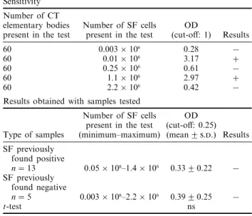
Results of testing SF for the presence of Chlamydia trachomatis (CT) plasmid DNA by LCx (Abbott Laboratories)
Sensitivity Number of CT
elementary bodies Number of SF cells OD
present in the test present in the test (cut-off: 1) Results 60 0.003 × 10⁶ 0.28 −
60 0.01 × 10⁶ 3.17 +
60 0.25 × 10⁶ 0.61 −
60 1.1 × 10⁶ 2.97 +
60 2.2 × 10⁶ 0.42 −
Results obtained with samples tested
Number of SF cells OD present in the test (cut-off: 0.25) Type of samples (minimum–maximum) (mean2 ..) Results SF previously found positive n= 13 0.05 × 10⁶–1.4 × 10⁶ 0.332 0.22 − SF previously found negative n= 5 0.003 × 10⁶–2.2 × 10⁶ 0.392 0.25 − t-test ns
detection limit) was also obtained when 10 000 EB were added (2000 EB present in the test) (Table I).
Sensitivity of the Amplicor system
When SF pellets were mixed with 1.25 ml of urine resuspension buffer and 1.25 ml of urine diluent, no dissolution was observed, except for one of the 34 pellets tested. This pellet contained the lowest number of SF cells (5 × 10⁴) and a positive result was obtained when 20 EB were added.
However, since these experimental conditions did not allow the majority of the pellets to be dissolved, other experimental conditions were tested. With samples previously found positive [1], the highest optical density values were obtained when 100ml aliquots of SF were mixed with 100ml of urine resuspension buffer and 100ml of urine diluent. In these conditions, 1000 EB were added to five different SF preparations and one positive result was obtained for one sample containing 0.03 × 10⁵ cells (in the test) (Table II).
Sensitivity of the LCx system
With samples previously found positive [1], the highest rates of fluorescence were obtained when aliquots of SF were mixed with an equal volume of urine specimen resuspension buffer. In these con-ditions, 60 EB were added to five different SF preparations and two positive results were obtained for the samples containing 0.01 × 10⁶ and 1.1 × 10⁶ cells (in the test) (Table III).
Comparison of the sensitivities of the different systems to detect Chlamydia trachomatis nucleic acids in SF
The sensitivities varied from 20 EB/ml with nested PCR targeting plasmid sequences [1] to 100 000 EB/ml of SF with the Gen-Probe PACE 2 system. The best sensitivity of the commercially available tests was
obtained with the LCx system (1200 EB/ml of SF) (Table IV).
Detection of Chlamydia trachomatis rRNA and plasmid DNA by commercially available tests in SF samples
SF samples previously found positive with at least two different systems of DNA amplification [1] and SF samples previously found negative were tested. No positive results were obtained according to the instructions of the kit manufacturers and the values obtained for samples previously found positive were not significantly different from those obtained for samples previously found negative (Tables I–III).
With the Gen-Probe PACE 2 system, three SF aliquots of 100ml previously found positive and 12 previously found negative were also tested and found negative.
TABLE IV
Sensitivities of the different systems tested to detect Chlamydia
trachomatisnucleic acids in SF
Minimal number of Chlamydia
trachomatiselementary bodies
to obtain positivity Present in Present in 1 ml
the test of SF Nested PCR targeting plasmid
sequences [1] 1–10 20–200 PACE 2 (Gen-Probe) 2000 10 000†–100 000‡ Amplicor (Roche Molecular
Systems) (20)*–1000 (1000)*–60 000 LCx (Abbott Laboratories) 60 1200
*Result obtained only with the SF pellet containing the lowest number of cells (5 × 10⁴).
†Result obtained with SF pellets (not always completely dissolved).
For the commercially available PCR system (Am-plicor), processing the specimens after heat treatment to 95°C and centrifugation decreased the values for both groups of samples.
DISCUSSION
Three commercially available tests, developed to detect Chlamydia nucleic acids in urogenital samples, were tested with SF samples. We performed the assays either directly on SF pellets (with Gen-Probe PACE 2 and Amplicor) or on 100ml aliquots, without nucleic acid extraction, in order to minimize sample handling and to see whether these kits could be used in a very simple way to detect Chlamydia nucleic acids in SF specimens. However, it is important to be aware that the sensitivity of these tests depends largely on the specimen processing. To be used with good chances of success, it is necessary to test a large amount of a sample, but this requirement is contraindicated with the presence of Taq polymerase in two of these kits. Indeed, this enzyme is sensitive to inhibitors present in SF samples and will be less inhibited if the specimens are more diluted. The ‘ideal’ experi-mental conditions are a balance between these two requirements.
The use of SF pellets could be an advantage since it allows a larger number of cells to be tested without inhibitor products susceptible to being present in SF. The comparison of the sensitivities obtained for the SF pellets and the 100ml aliquots was performed with the Gen-Probe PACE 2 system and with Amplicor. For both systems, the best sensitivity is obtained with SF pellets. However, their use is limited by the difficulty of dissolving them and, if the pellets are not completely dissolved, it is difficult to assume an efficient detection of nucleic acids.
The sensitivity of the kits was tested by adding increasing amounts of C. trachomatis EB to SF samples before processing. With Amplicor, a sensitivity of 20 EB was obtained when a non-inflammatory SF containing 5 × 10⁴ cells/ml was used. However, it was not possible to test the other samples in the same experimental conditions since they were not dissolved. In conditions allowing the processing of all the samples, the best sensitivity of the three commercially available tests varied from 2000 (Gen-Probe PACE 2) to 60 EB (LCx), corresponding to 100 000 (Gen-Probe PACE 2) to 1200 EB (LCx) per millilitre of SF.
For the Gen-Probe PACE 2 system, we obtained a higher sensitivity than Kuipers et al. [8] who reported a sensitivity of 1 × 10⁶EB/ml of SF. This discrepancy could be explained by differences in the specimen processing or by the number of cells present in the samples. Indeed, our results show that the higher the number of cells, the lower the sensitivity of this system. For the PCR and LCR systems, the sensitivities also depended on the samples and not only on the cell number. The presence of more amplification inhibitors in some particular SF could explain the variable sensitivities observed.
Samples previously found positive with at least two different systems of DNA amplification [1] were tested with these commercially available tests and no positive results were obtained. This could be explained by the insensitivity of these tests. Indeed, the DNA amplifica-tion systems developed by Bas et al. [1] had a sensitivity between 1 and 50 EB (present in the test). However, the LCx system had a sensitivity close to 50 EB for 2/5 samples. It could be possible to find positive results in samples without a strong inhibition effect or in synovial tissue samples since Branigan et al. [9] found that chlamydial DNA is more reliably identified in tissues than in SF. It is also important to be aware of the difficulty of determining the sensitivity of these assays, the DNA template coming from added EB is probably not comparable in quality and quantity to the DNA coming from SF samples.
For each test, we compared the means obtained for samples previously found positive with the means obtained for samples previously found nega-tive in order to see whether a difference could be observed in spite of the fact that the values were lower than the cut-off. No significant differences were observed.
In conclusion, in our experimental conditions, the commercially available kits Gen-Probe PACE 2, Amplicor and LCx cannot be reliably used to detect ChlamydiaRNA or DNA in SF.
R
1. Bas S, Griffais R, Kvien TK, Glenna˚s A, Melby K, Vischer TL. Amplification of plasmid and chromosome Chlamydia DNA in synovial fluid of patients with reactive arthritis and undifferentiated seronegative oligoarthropathies. Arthritis Rheum 1995;38:1005–13. 2. Taylor-Robinson D, Gilroy CB, Thomas BJ, Keat ACS.
Detection of Chlamydia trachomatis DNA in joints of reactive arthritis patients by polymerase chain reaction. Lancet 1992;340:81–2.
3. Rahman MU, Hudson AP, Schumacher HR. Chlamydia and Reiter’s syndrome (reactive arthritis). Rheum Dis Clin North Am 1992;18:67–79.
4. Rahman MU, Cheema MA, Schumacher HR, Hudson AP. Molecular evidence for the presence of Chlamydia in the synovium of patients with Reiter’s syndrome. Arthritis Rheum 1992;35:521–9.
5. Loeffelholz MJ, Lewinski CA, Silver SR, Purohit AP, Herman SA, Buonagurio DA et al. Detection of Chlamydia trachomatis in endocervical specimens by polymerase chain reaction. J Clin Microbiol 1992;30:2847–51.
6. Chernesky MA, Jang D, Lee H, Burczak JD, Hu H, Sellors J et al. Diagnosis of Chlamydia trachomatis infections in men and women by testing first-void urine ligase chain reaction. J Clin Microbiol 1994;32: 2682–5.
7. Skulnick M, Chua R, Simor AE, Low DE, Khosid HE, Fraser S et al. Use of the polymerase chain reaction for the detection of Chlamydia trachomatis from endocervi-cal and urine specimens in an asymptomatic low-preva-lence population of women. Diagn Microbiol Infect Dis 1994;20:195–201.
8. Kuipers JG, Scharmann K, Wollenhaupt J, Netteln-breker E, Hopf S, Zeidler H. Sensitivities of PCR,
MicroTrak, Chlamydia EIA, IDEIA, and PACE 2 for purified Chlamydia trachomatis elementary bodies in urine, peripheral blood, peripheral blood leukocytes, and synovial fluid. J Clin Microbiol 1995;33:3186–90. 9. Branigan PJ, Gerard HC, Saaibi D, Hudson AP,
Schumacher HR. Screening of synovial tissue vs. fluid from patients with Reiter’s syndrome (RS), other spondyloarthropathies, or reactive arthritis (ReA) for Chlamydia via polymerase chain reaction (PCR). Arthritis Rheum 1995;38:R20.