The use of two density gradient centrifugation techniques and the swim-up method to separate spermatozoa with chromatin and nuclear DNA anomalies
5
0
0
Texte intégral
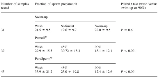
(2) Separation of spermatozoa with nuclear anomalies. Table I. The mean (⫾SD) percentage of spermatozoa positive to chromomycin A3 in the initial sample (wash) and various fractions after isolation using swim-up, Percoll® and PureSperm® techniques. All samples tested were from different men Number of samples tested. Fraction of sperm preparation. Paired t-test (wash versus swim-up or 90%). Swim-up. 31. Wash 21.5 ⫾ 9.5. Sediment 19.6 ⫾ 9.7. Swim-up 22.0 ⫾ 9.5. P ⫽ 0.6. 45% 30.72 ⫾ 18.3. 90% 18.1 ⫾ 12.1. P ⬍ 0.001. 45% 25.0 ⫹ 19.8. 90% 12.4 ⫾ 12.6. P ⬍ 0.001. Percoll®. 39. Wash 29.9 ⫾ 15.5 PureSperm®. 45. Wash 33.9 ⫾ 21.2. i.e. the way the DNA is packaged, while nick translation represents damage in the actual DNA itself. The presence of these abnormal spermatozoa in our insemination medium may have consequences during the fertilization process and for the developing embryo (Sakkas et al., 1996; Sun et al., 1997; Lopes et al., 1998). Therefore, the ability of sperm preparation techniques to remove spermatozoa with such anomalies has important implications for assisted reproduction techniques outcome. This study aims to determine the efficacy of the swim-up and density gradient centrifugation techniques in the removal of spermatozoa with nuclear aberrations. Materials and methods Preparation of semen samples and slides Semen samples were obtained from men attending the Clinic of Sterility, University Hospital of Geneva, Geneva, Switzerland and the Assisted Conception Unit, Birmingham Women’s Hospital, Birmingham, UK. All men attending the clinics were used. The only limiting parameter was whether we could isolate over 10 000 spermatozoa to prepare the slides. Patient semen characteristics ranged from a concentration of 1⫻106 spermatozoa per ml and 10% progressive motility to 289⫻106 spermatozoa per ml and 58% progressive motility (WHO, 1999). All samples were prepared using the same techniques and products in both laboratories and once slides were prepared they were sent to the same reader using a code, so that the reader did not know the type of preparation. The semen sample from the same man was not used to test all parameters. Samples were prepared according to the WHO (WHO, 1999) manual using 1 ml starting volumes. Density gradient centrifugation was carried out using 0.5 ml volumes of either Percoll® (Sigma Pharmaceutical, Buchs, Switzerland) or PureSperm® (Nidacon International, Gothenburg, Sweden). Briefly, 0.5 ml of a 45% suspension was layered over 0.5 ml of 90% and centrifuged for 20 min (300 g). Spermatozoa in the upper (0.5 ml) layer of the swim-up and both PureSperm® and Percoll® fractions (45 and 90%) were fixed in 3.5% paraformaldehyde. Three smears of each were then prepared on slides and left to air dry. It should be noted that the manufacturers have now withdrawn Percoll® from use in human assisted reproduction techniques.. In-situ nick translation assay and CMA3 staining In-situ nick translation was performed as previously described (Manicardi et al., 1995) by omitting the endonuclease treatments since, in the presence of pre-existing DNA endogenous nicks, the DNA polymerase I, by virtue of its 5⬘-3⬘ exonucleotic activity, can catalyse movement of the nicks along the double helix. The only difference with the previously described method was that Digoxigenin11-dUTP (Boehringer Mannheim, Rotkrenz, Switzerland) was used. For CMA3 staining slides were treated for 20 min with 100 µl of CMA3 solution (0.25 mg/ml McIlvaine buffer, pH 7.0, containing 10 mmol/l MgCl2) (Manicardi et al., 1995). They were then rinsed in buffer, air-dried and mounted with a 1:1 mixture of phosphate buffered saline (PBS) and glycerol. In nearly all cases an operator, working blind, examined at least 500 spermatozoa on each coded slide. The nick translation and CMA3 staining were predominantly of all-ornothing type and the rare cells showing ambiguous fluorescence were not considered. We have previously shown that a strong correlation exists between chromatin packaging, as revealed by CMA3 positivity, and the presence of nicks in sperm DNA (Manicardi et al., 1995). Fluorescence analysis was performed using a Zeiss Photomikroskop® III (Zeiss, Oberkochen, Germany). Statistical analysis was performed using SPSS 9.0 for Windows and the mean values of each sample compared using the paired t-test.. Results When examining the different fractions prepared after using the swim-up technique, no significant difference was found between the percentage of spermatozoa recovered from the upper swim-up layer that were positive to the CMA3 fluorochrome when compared to those spermatozoa in the initial wash sample and the sediment (Table I). When prepared using Percoll®, the mean percentage (⫾SD) of spermatozoa positive to the CMA3 fluorochrome was significantly lower (paired t-test, P ⬍ 0.001) in the 90% fraction compared to those remaining in the 45% fraction and in the initial washed sample (Table I). Similarly, using PureSperm®, the percentage of spermatozoa positive to the CMA3 fluorochrome was again significantly lower (paired t-test, P ⬍ 0.001) in the 90% fraction compared to those 1113.
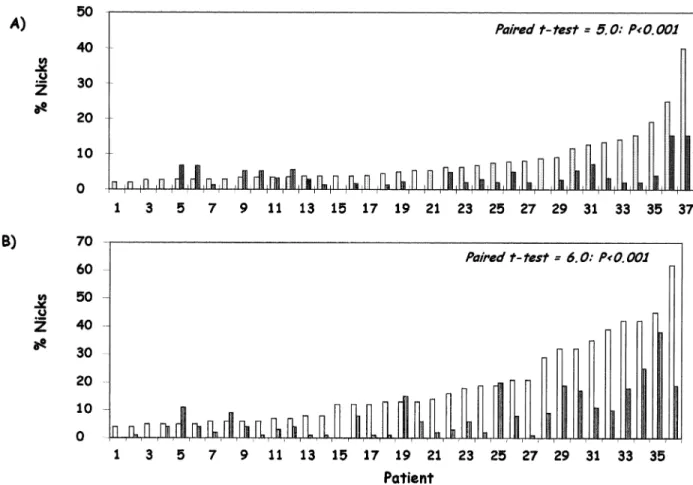
(3) D.Sakkas et al.. Figure 1. The percentage of human spermatozoa exhibiting endogenous DNA nicks in the 45% (white columns) and 90% (dark columns) fractions after preparation using (A) Percoll® and (B) PureSperm®. The patients used in A and B are not the same. Statistical comparisons using the paired t-test are included in the figure.. remaining in the 45% fraction and in the initial washed sample (Table I). When examining for the percentage of spermatozoa with DNA damage we found a similar pattern as for the CMA3 fluorochrome. Spermatozoa isolated in the 90% fractions of both Percoll® and PureSperm® possessed a significantly lower percentage of DNA damage when compared to the 45% fraction (Figure 1). Discussion In this study we have shown that the different standard sperm preparation techniques used in the routine assisted reproduction techniques laboratory vary in their ability to separate spermatozoa possessing nuclear anomalies from those which are normal. Sperm preparation using the swim-up technique does not appear to be as efficient at isolating a population of spermatozoa with a low percentage of nuclear anomalies. This is in contrast to previous reports (Angelopoulos et al., 1998; Spano et al., 1999) where post-rise spermatozoa prepared using the swimup technique represented a subpopulation characterized by a general increase of the green head sperm percentage when using acridine orange. Spano and co-workers (Spano et al., 1999) also reported that this subpopulation exhibited improved chromatin structure properties as assessed using the sperm chromatin structure assay (SCSA). The SCSA utilizes the metachromatic properties of acridine orange to distinguish 1114. between low-pH or heat-denatured (red fluorescence ⫽ singlestranded) and native (green fluorescence ⫽ double stranded) DNA in sperm chromatin. We have found however, using the CMA3 fluorochrome, that although the swim-up technique is adequate for the isolation of a highly motile sperm population, overall sperm quality may be compromised, with a similar percentage of CMA3 positive spermatozoa found in the swimup to those found in the sediment. Interestingly, Colleu and co-workers (Colleu et al., 1996) found that Percoll® gradients appeared to enrich for spermatozoa with less intermediate proteins, and more mature nucleoproteins of the protamine 2 family, a feature not observed with the swim-up spermatozoa. In contrast, Larson et al. (1999) reported that glass wool filtration produced sperm suspensions with improved chromatin integrity (Larson et al., 1999), as measured with the SCSA, compared to density gradient centrifugation using Enhance-S plus™ (Conception Technologies, San Diego, CA, USA). To clarify the differences between the results observed using acridine orange and CMA3 a direct comparison of the two stains using the different preparation techniques is needed. When examining the ability of Percoll® to isolate spermatozoa of normal chromatin structure both Angelopoulos et al. (1998) and Golan et al. (1997) again found that the percentage of green fluorescent spermatozoa improved in the 90% fraction (Golan et al., 1997; Angelopoulos et al., 1998). In the current study, sperm preparation using density centrifugation techniques with either the now largely obsolete Percoll® or.
(4) Separation of spermatozoa with nuclear anomalies. one of its licensed replacements, PureSperm®, is able to reduce significantly the percentage of spermatozoa with nuclear abnormalities. We have assessed two forms of nuclear anomalies: firstly, using CMA3 we have shown that spermatozoa with a more compacted chromatin are more likely to be present in the 90% fraction. We have previously reported that CMA3 can be used to distinguish populations of spermatozoa with differing levels of protamination (Bizzaro et al., 1998) and that there is a strong relationship between CMA3 accessibility and the presence of endogenous DNA nicks in human spermatozoa (Manicardi et al., 1995). Our current findings with both Percoll® and PureSperm® therefore substantiate the work of Colleu (Colleu et al., 1996). More importantly, both Percoll® and PureSperm® significantly reduced the percentage of spermatozoa with nuclear DNA damage. This was clearly evident in the majority of patients who had high levels of DNA damage. The patients who exhibit high levels of CMA3 positivity and nuclear DNA damage are likely to have poor semen parameters (Manicardi et al., 1995; Bianchi et al., 1996a,b; Sun et al., 1997; Esterhuizen et al., 2000). This brings to light the question of what the benefits are of isolating a fraction of spermatozoa with lower levels of nuclear anomalies? A number of studies now clearly indicate that an abnormal sperm nucleus may have a detrimental effect on fertilization, embryo development and pregnancy outcome. We have previously shown that spermatozoa from men with high levels of DNA damaged spermatozoa are more likely to exhibit anomalies in sperm decondensation after ICSI (Sakkas et al., 1996). In addition, Lopes et al. (1998) showed that DNA damage in spermatozoa may contribute to fertilization failure after ICSI. More importantly, the group of Robaire has shown that damage to rat sperm DNA may be linked to an increase in early embryo death (Qiu et al., 1995a,b). Recently, Evenson showed that the SCSA could be used as a prognostic factor for human fertility (Evenson et al., 1999), stating that men who have an SCSA value of greater than 30% would have difficulties in achieving pregnancy. Clearly, isolating spermatozoa of a better nuclear consistency will increase the likelihood of achieving pregnancies with normal embryos. Sperm preparation for assisted reproduction should aim to minimize the risk of abnormal spermatozoa being used for fertilization. By performing techniques such as ICSI, we remove many of the barriers set in place to select the best spermatozoa for fertilization. Unique checkpoints have already been observed after ICSI in the rhesus monkey (Hewitson et al., 1999) showing that both gametes can attempt to maintain reproductive capabilities even under abnormal conditions. It has also been shown that human spermatozoa possessing DNA damage have the same potential as control spermatozoa to decondense and form pronuclei when micro-injected into hamster oocytes (Twigg et al., 1998). The possible detrimental effects that an abnormal paternal genome may have on the fate of the human embryo after ICSI have been highlighted by a number of authors including ourselves (Sakkas et al., 1998; Sakkas, 1999). Sperm preparation for assisted reproduction techniques should therefore aim to minimize the risk that abnormal spermatozoa can have on outcome. A reduction in the number. of spermatozoa possessing nuclear anomalies in the final insemination preparation should be a priority, in particular as little is known about the relative influences spermatozoa with abnormal nuclei can have on the embryo and ensuing offspring. Therefore, density gradient centrifugation techniques using approved separating agents should be the preferred method.. Acknowledgements We thank the staff of the Laboratoire des Gametes, University Hospital of Geneva and the IVF and Andrology Laboratories, Assisted Conception Unit, Birmingham Women’s Hospital for their assistance, in particular Nicole Jaquenoud, Ingrid Wagner, Jo Turner and Yvonne D’Arcy. Part of this study was supported by a grant from the Fonds National Suisse.. References Angelopoulos, T., Moshel, Y.A., Lu, L. et al. (1998) Simultaneous assessment of sperm chromatin condensation and morphology before and after separation procedures: effect on the clinical outcome after in vitro fertilization. Fertil. Steril., 69, 740–747. Ballachey, B.E., Hohenboken, W.D. and Evenson, D.P. (1986) Sperm head morphology and nuclear chromatin structure evaluated by flow cytometry in a diallel cross in mice. Can. J. Genet. Cytol., 28, 954–966. Bianchi, P.G., Manicardi, G.C., Bizzaro, D. et al. (1993) Effect of deoxyribonucleic acid protamination on fluorochrome staining and in situ nick-translation of murine and human mature spermatozoa. Biol. Reprod., 49, 1083–1088. Bianchi, P.G., Manicardi, G., Bizzaro, D. et al. (1996a) Use of the guaninecytosine (GC) specific fluorochrome, chromomycin A3, as an indicator of poor sperm morphology. J. Assist. Reprod. Genet., 13, 246–250. Bianchi, P.G., Manicardi, G.C., Urner, F. et al. (1996b) Chromatin packaging and morphology in ejaculated human spermatozoa: evidence of hidden anomalies in normal spermatozoa. Mol. Hum. Reprod., 2, 139–144. Bizzaro, D., Manicardi, G.C., Bianchi, P.G. et al. (1998) In-situ competition between protamine and fluorochromes for sperm DNA. Mol. Hum. Reprod., 4, 127–132. Colleu, D., Lescoat, D. and Gouranton, J. (1996). Nuclear maturity of human spermatozoa selected by swim-up or by Percoll gradient centrifugation procedures. Fertil. Steril., 65, 160–164. Esterhuizen, A.D., Franken, D.R., Lourens, J.G.H. et al. (2000) Sperm chromatin packaging as an indicator of in vitro fertilisation rates. Hum. Reprod., 15, 657–661. Evenson, D.P. (1986) Flow cytometry of acridine orange stained sperm is a rapid and practical method for monitoring occupational exposure to genotoxicants. Prog. Clin. Biol. Res., 207, 121–132. Evenson, D.P. (1990) Flow cytometric analysis of male germ cell quality. Methods Cell. Biol., 33, 401–410. Evenson, D.P., Darzynkiewicz, Z. and Melamed, M.R. (1980) Comparison of human and mouse sperm chromatin structure by flow cytometry. Chromosoma, 78, 225–238. Evenson, D.P., Jost, L.K., Marshall, D. et al. (1999) Utility of the sperm chromatin structure assay as a diagnostic and prognostic tool in the human fertility clinic. Hum. Reprod., 14, 1039–1049. Golan, R., Shochat, L., Weissenberg, R., et al. (1997) Evaluation of chromatin condensation in human spermatozoa: a flow cytometric assay using acridine orange staining. Mol. Hum. Reprod., 3, 47–54. Gorczyca, W., Traganos, F., Jesionowska, H. and Darzynkiewicz, Z. (1993) Presence of DNA strand breaks and increased sensitivity of DNA in situ to denaturation in abnormal human sperm cells: analogy to apoptosis of somatic cells. Exp. Cell Res., 207, 202–205. Hewitson, L., Dominko, T., Takahashi, D. et al. (1999) Unique checkpoints during the first cell cycle of fertilization after intracytoplasmic sperm injection in rhesus monkeys. Nature Med., 5, 431–433. Larson, K.L., Brannian, J.D., Timm, B.K. et al. (1999) Density gradient centrifugation and glass wool filtration of semen remove spermatozoa with damaged chromatin structure. Hum. Reprod., 14, 2015–2019. Lopes, S., Sun, J.G., Juriscova, A. et al. (1998) Sperm deoxyribonucleic acid fragmentation is increased in poor quality sperm samples and correlates. 1115.
(5) D.Sakkas et al. with failed fertilization in intracytoplasmic sperm injection. Fertil. Steril., 69, 528–532. Manicardi, G.C., Bianchi, P.G., Pantano, S. et al. (1995) Presence of endogenous nicks in DNA of ejaculated human spermatozoa and its relationship to chromomycin A3 accessibility. Biol. Reprod., 52, 864–867. Mortimer, D. (1999) Structured management as a basis for cost effective infertility care. In Gagnon, C. (ed.), The Male Gamete: From Basic Science to Clinical Applications. Cache River Press, Illinois, USA, pp. 363–370. Qiu, J., Hales, B.F. and Robaire, B. (1995a) Damage to rat spermatozoal DNA after chronic cyclophosphamide exposure. Biol. Reprod., 53, 1465–1473. Qiu, J., Hales, B.F. and Robaire, B. (1995b) Effects of chronic low-dose cyclophosphamide exposure on the nuclei of rat spermatozoa. Biol. Reprod., 52, 33–40. Sakkas, D. (1999) The use of blastocyst culture to avoid inheritance of an abnormal paternal genome after ICSI. Hum. Reprod., 14, 4–5. Sakkas, D., Urner, F., Bianchi, P.G. et al. (1996) Sperm chromatin anomalies can influence decondensation after intracytoplasmic sperm injection. Hum. Reprod., 11, 837–843. Sakkas, D., Urner, F., Bizzaro, D. et al. (1998) Sperm nuclear DNA damage and altered chromatin structure: effect on fertilization and embryo development. Hum. Reprod. 4 (Suppl.), 11–19. Spano, M., Cordelli, E., Leter, G. et al. (1999) Nuclear chromatin variations in human spermatozoa undergoing swim-up and cryopreservation evaluated by the flow cytometric sperm chromatin structure assay. Mol. Hum. Reprod., 5, 29–37. Sun, J.G., Jurisicova, A. and Casper, R.F. (1997) Detection of deoxyribonucleic acid fragmentation in human sperm: correlation with fertilization in vitro. Biol. Reprod., 56, 602–607. Trasler, J.M., Hales, B.F. and Robaire, B. (1985) Paternal cyclophosphamide treatment of rats causes fetal loss and malformations without affecting male fertility. Nature, 316, 144–146. Twigg, J.P., Irvine, D.S. and Aitken, R.J. (1998) Oxidative damage to DNA in human spermatozoa does not preclude pronucleus formation at intracytoplasmic sperm injection. Hum. Reprod., 13, 1864–1871. World Health Organization (1999) WHO Laboratory Manual for Examination of Human Semen. Cambridge University Press, Cambridge. Received on October 20, 1999; accepted on February 7, 2000. 1116.
(6)
Figure
Documents relatifs
On the other hand, there are cases where compatible maps are not sufficient: we prove in [13] the cor- rectness of a distributed abstract machine, where mechanisms introduced by
The design of surveillance systems for detection and applicable testing of lyssaviruses should be based in part upon the primary intent of each programme, the available resources
Next to techniques aimed at the detection of lyssavirus antigens, such as the direct fluorescent antibody test (DFAT; see Chapter 11), the direct rapid immuno- histochemistry test
Due to time restriction we could only submit one cross-language run with English as query language using the phrase search component of Mpro-IR which is restricted to search only
tive chromatin fraction from round spermatids after 1.5 rain of digestion, lane C same as lane B but after 3-min digestion, lane D MNase-sensitive
crystal as a function of the direction-cosines of the direction of strain measurement and the direc- tion of magnetization, we have obtained for the single.
The calculated SEDM lineshape using the superoperator formalism and assuming this spin flip relaxation mechanism shows a peak at this position for a rela- xation time of te =
Different techniques (SEM, optical microscopy, image analysis, XPS, XRD, solvent extraction, wettability measurements, etc.) can be successfully used to
