Images in cardio-thoracic surgery
Pseudoaneurysm of the left ventricle near the non-coronary sinus
valsalvae after aortic valve replacement
J.P. Muller
a,*, O. Reuthebuch
a, R. Jenni
b, M.I. Turina
aa
Department of Cardio-Vascular Surgery, University Hospital Zurich, Raemistrasse 100, CH-8008 Zurich ZH, Switzerland
bDepartment of Cardiology, University Hospital Zurich, Zurich ZH, Switzerland
Received 18 September 2003; accepted 20 October 2003
Keywords: Aortic valve replacement; Pseudoaneurysm; Transesophagal echocardiography
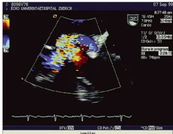
Fourteen days after aortic valve replacement a 60-year-old
male patient presented with sternal infection. Subsequent
transesophagal echocardiography revealed a huge and
pulsatile pseudoaneurysm near the non-coronary sinus of
Valsalva. To prevent potential rupture pseudoaneurysm was
resected and aortic root replaced with a homograft. Under
antibiotic therapy postoperative course was uneventful
(
Figs. 1 and 2
).
1010-7940/$ - see front matter q 2003 Elsevier B.V. All rights reserved. doi:10.1016/j.ejcts.2003.10.020
European Journal of Cardio-thoracic Surgery 25 (2004) 283
www.elsevier.com/locate/ejcts
Fig. 1. TEE: CM 23, artificial aortic valve (Carbomedicsw
); L, left atrium; LVOT, left ventricular outlet tract; PA, pulmonal artery; ! , in-flow from
the LVOT to the pseudoaneurysm. Fig. 2. Doppler-TEE of the pseudoaneurysm.
* Corresponding author. Tel.: þ41-1-255-1111; fax: þ41-1-255-8920. E-mail address: [email protected] (J.P. Muller).