The role of the yeast cleavage and polyadenylation
factor subunit Ydh1p/Cft2p in pre-mRNA 3¢-end
formation
Andrea Kyburz, Martin Sadowski, Bernhard Dichtl and Walter Keller*
Department of Cell Biology, Biozentrum, University of Basel, Klingelbergstrasse 70, CH-4056 Basel, Switzerland
Received April 17, 2003; Revised and Accepted May 23, 2003
ABSTRACT
Cleavage and polyadenylation factor (CPF) is a multi-protein complex that functions in pre-mRNA 3¢-end formation and in the RNA polymerase II (RNAP II) transcription cycle. Ydh1p/Cft2p is an essential component of CPF but its precise role in 3¢-end processing remained unclear. We found that mutations in YDH1 inhibited both the cleavage and the polyadenylation steps of the 3¢-end formation reaction in vitro. Recently, we demonstrated that an important function of CPF lies in the recognition of poly(A) site sequences and RNA binding analyses suggesting that Ydh1p/Cft2p interacts with the poly(A) site region. Here we show that mutant ydh1 strains are de®cient in the recognition of the ACT1 cleavage site in vivo. The C-terminal domain (CTD) of RNAP II plays a major role in coupling 3¢-end pro-cessing and transcription. We provide evidence that Ydh1p/Cft2p interacts with the CTD of RNAP II, several other subunits of CPF and with Pcf11p, a component of CF IA. We propose that Ydh1p/Cft2p contributes to the formation of important interaction surfaces that mediate the dynamic association of CPF with RNAP II, the recognition of poly(A) site sequences and the assembly of the polyadenylation machinery on the RNA substrate.
INTRODUCTION
All eukaryotic mRNA precursors (pre-mRNA) are extensively modi®ed before they can serve as templates for protein synthesis. Pre-mRNA 3¢-end processing is initiated by endonucleolytic cleavage at the poly(A) site. Subsequently, the upstream cleavage product is polyadenylated whereas the downstream fragment is rapidly degraded (for review see 1). The yeast 3¢-end processing reaction can be reconstituted in vitro with the cleavage and polyadenylation factor IA (CF IA), cleavage and polyadenylation factor IB (CF IB), cleavage and polyadenylation factor (CPF) and the poly(A) binding protein (Pab1p) (2,3). Interactions between their subunits
allow bridging of the different factors and ensure their coordinated action on the substrate. So far the CPF components Fip1p and Pfs2p have been shown to bridge CPF with CF IA by interacting with Rna14p, a subunit of CF IA (3,4). A table of the factors involved in 3¢-end processing and their subunits is provided as supplementary material.
The polyadenylation signals that guide the processing machinery in yeast are redundant and more degenerate compared to the well-conserved sequences in higher eukary-otes. Still, conserved elements can be found, which are the ef®ciency element (EE), the positioning element (PE), the poly(A) site and U-rich sequences. The EE is located at variable distances upstream of the cleavage site (5). The PE is often found ~20 nt upstream of the cleavage site and consists of an A-rich sequence (6). U-rich regions, located directly upstream and downstream of the cleavage site, and the poly(A) site itself act in concert to produce multiple recognition sites (5,7±9).
RNase H protection mapping experiments with a CYC1 pre-mRNA suggested that CPF is involved in the recognition of the poly(A) site by speci®c interactions of its subunits Yhh1p/Cft1p, Ydh1p/Cft2p and Yth1p with sequences sur-rounding the poly(A) site (9±11). Nab4p/Hrp1p (CF IB) binds to the EE (12,13) and, through interaction with Rna14p, possibly enables CF IA to bind to the PE (14). However, it was also reported that Nab4p/Hrp1p (15) as well as the PE and EE (9) are not essential for cleavage in vitro, underscoring the importance of the poly(A) site region itself.
The yeast and mammalian 3¢-end processing factors are highly homologous (reviewed in 16). In mammals cleavage and polyadenylation speci®city factor (CPSF) contributes to poly(A) site selection by binding to the well conserved AAUAAA element upstream of the cleavage site (17); CPSF-160 and possibly additional CPSF subunits are thought to mediate speci®c interactions to the RNA (18). The binding of puri®ed CPSF is weak, but is strongly enhanced by a cooperative interaction with cleavage stimulation factor (CstF) bound to the downstream elements (19±21).
Transcription by RNA polymerase II (RNAP II) and pre-mRNA processing reactions are coupled events (reviewed in 22±25). The C-terminal domain (CTD) of RNAP II plays a central role in linking the processing reactions to transcription. Current models suggest that the CTD is hypo-phosphorylated
*To whom correspondence should be addressed. Tel: +41 61 267 2060; Fax: +41 61 267 2079; Email: [email protected] Present address:
Martin Sadowski, Cancer Research Program, Garvan Institute of Medical Research, Sydney, NSW 2010, Australia
during transcription initiation and that escape of RNAP II into the elongation phase is accompanied by phosphorylation of the CTD. It has been proposed that the change in charge upon phosphorylation enables proteins involved in pre-mRNA processing to bind to the CTD (26±28). This includes proteins involved in capping (29±32), splicing (33) and 3¢-end formation (10,28,34±37). It is assumed that the assembled proteins subsequently travel with the elongating RNAP II during transcription and act on the nascent RNA transcript.
In mammals the CTD was suggested to play a direct role in 3¢-end cleavage in vivo (34) and in vitro (38,39). Experiments in yeast provided evidence that transcription in the absence of the CTD was accompanied by a reduction of cleavage ef®ciency and the resulting mRNAs had shorter poly(A) tails (28,40). Correct transcription termination requires a functional poly(A) signal in all organisms (41,42, reviewed in 43), and yeast strains carrying mutations in CPF and CF IA were shown to be de®cient in correct transcription termin-ation, indicating the coupling between 3¢-end processing and transcription termination (10,35,44±47). Furthermore, correct transcriptional termination was found to require the transcrip-tion factors Sub1p (48) and Res2p (49) and chromatin remodeling factors (50). The Nrd1p complex was shown to be required for correct termination at snoRNA genes (51).
Ydh1p/Cft2p (which will be referred to as Ydh1p in the remainder of this paper) is the 105 kDa subunit of CPF. It shares 24.4% identity and 43% similarity with the mammalian CPSF-100 protein. It is also signi®cantly related to Ysh1p and to CPSF-73 (52). Ydh1p is essential for cell viability (53) and was shown to bind RNA (54). RNase H protection experi-ments suggested that the protein interacts with the poly(A) site region (9). Here, we show that Ydh1p is required for cleavage and polyadenylation in vitro and for poly(A) site recognition in vivo. Furthermore, we provide evidence that Ydh1p interacts with several subunits of CPF, with Pcf11p, a subunit of CF IA, and with the CTD of RNAP II. The results suggest that Ydh1p is part of an interaction surface that mediates important contacts with CF IA, the CTD of RNAP II and the pre-mRNA substrate.
MATERIALS AND METHODS Yeast strains
For random mutagenesis of YDH1, mutagenic PCR was carried out with a low concentration of ATP (3). The primers used for PCR were: Ydh1-N (5¢-CCCTTACGGATTGAAGT-CATT-3¢) and Ydh1-C (5¢-TTGAACCTTTTATTTGTGCTG-3¢). Plasmid pBD63 (YDH1±LEU2±CEN) was obtained by subcloning of the YDH1 gene from pIA115 (YDH1±URA3± CEN) (53) into the BamHI and SacI restriction sites of pRS415. The plasmid was digested with the restriction enzymes NsiI and SpeI. The fragment containing plasmid sequences and sequences ¯anking the YDH1 gene was co-transformed together with the mutagenized PCR product into the yeast strain YPP106 (53). Transformants were selected on minimal medium lacking leucine and replica-plated onto 5-FOA plates in order to shuf¯e out the pIA115 plasmid. The colonies were then replica-plated onto YPD-plates and incubated at 25, 33 or 37°C, respectively. Plasmids of candidate colonies that showed a ts phenotype were isolated
by standard procedures. The conditional growth phenotype was then veri®ed by retransformation of the isolated plasmids into YPP106, 5-FOA treatment and growth tests at elevated temperatures.
Genotypes of yeast strains used in this study were: YPP106: MATa; ura3-1; ade2-1; leu2-3,112; his3-11,15; trp1D; ydh1::TRP1 [pIA115; CEN4±URA3±YDH1] (53); YAK1: MATa; ura3-1; ade2-1; leu2-3,112; his3-11,15; trp1D; ydh1::TRP1 [pAK21 CEN±LEU2±ydh1-1]; YAK2: MATa; ura3-1; ade2-1; leu2-3,112; his3-11,15; trp1D; ydh1::TRP1 [pAK22 CEN±LEU2±ydh1-2]; YAK3: MATa; ura3-1; ade2-1; leu2-3,112; his3-11,15; trp1D; ydh1::TRP1 [pAK23 CEN± LEU2±ydh1-3]; Y190: ura3-52; trp1-901; ade2-101; leu2-3; 112 his3-200r; gal4D; gal 80D; URA3:GAL1-lacZ; LYS2::GAL1-HIS3; cyhr; Clontech.
Plasmids and primers
The plasmids encoding the C-terminally truncated Ydh1p fragments were obtained by digestion of plasmid pBD71 (9) with restriction enzymes AccI (pBD91; encoding the recom-binant protein DC338), BamHI/XbaI (pBD92; encoding DC182), BamHI/SpeI (pAK5; encoding DC137), BamHI/ A¯II (pAK6; encoding DC555), BamHI/AgeI (pAK7; encod-ing DC613). GST-Ysh1p was encoded by pBD38, which was obtained by subcloning of Ysh1p into p20 (GST expression vector) using the restriction enzymes NdeI and BamHI. GST-Tev-Pta1p was encoded by pBD51, which was obtained by PCR ampli®cation of Pta1p followed by digestion with NdeI and BamHI and ligation into p26 (GST-Tev expression vector). GST-Yhh1p-H6was encoded by pBD75 (10).
The plasmid encoding the protein used to produce antibody directed against Ydh1p was constructed by digestion of pQE-9 (His6 expression vector Qiagen) with BamHI/HindIII. The insert was constructed by PCR, amplifying the sequence between primer Ydh5¢ (5¢-ATCGCGGATCCATGACTTAT-AAATACAATTG-3¢) and Ydh3¢ (5¢-AGCCCAAGCTTATT-TACTCAATTCGTTTGGT-3¢) of YDH1. For details about pBD-CTD, pACT2-YHH1, pACT2-YSH1 and pACT2-PCF11 see (10).
Expression of recombinant proteins in Escherichia coli BL21 E.coli cells carrying the respective plasmid were grown at 25°C in 23 YT until they reached an OD600 of ~1.
Following induction by 0.5 mM IPTG, incubation was continued for 6 h. The proteins were puri®ed at 4°C on glutathione±Sepharose 4B as recommended (Pharmacia) and the protein was eluted with GST-elution buffer [75 mM KCl, 50 mM Tris±HCl pH 7.9, 10% glycerol, 10 mM glutathione (reduced), 0.01% NP-40, 1 mM DTT].
Protein±protein interactions
In vitro translations were performed with the TNT-coupled transcription±translation system (Promega). GST fusion protein (100 ng) was incubated with in vitro translated [35S]methionine-labeled proteins for 1 h. The mixture was
bound in a total volume of 860 ml to 20 ml glutathione sepharose (Pharmacia), which was previously equilibrated in 1 ml PBS, 0.01% NP-40 and 100 mg BSA. The matrix was washed three times with IPP150 (150 mM KCl, 20 mM Tris± HCl pH 8.0, 0.01% NP-40) and the proteins were eluted by addition of protein loading buffer and incubation at 95°C.
Bound proteins were separated by SDS±PAGE and visualized by autoradiography. Phosphorylation of GST±CTD was performed as described previously (38) and the assay of the CTD interaction was carried out as described (10). The two-hybrid tests were carried out as described (55).
Extract preparation and in vitro cleavage and polyadenylation assays
Extracts competent for in vitro processing were prepared following the procedure previously described (3). The cleav-age and polyadenylation assays were carried out as described (56). To restrict the assay to cleavage only, EDTA replaced MgAc and CTP replaced ATP. For each reaction 30±40 mg total protein was used and, in the case of the reactions carried out at 34°C, pre-incubated at this temperature for 5 min. The RNA substrates were prepared by run-off transcription following the previously described procedure (9).
RNA analyses
Northern analyses and RNase H experiments were carried out as described (10). In addition we employed the oligonucle-otides anti-U24 (5¢-TCAGAGATCTTGGTGATAAT-3¢) and anti±snR13 (5¢-GGCAAAAGCCAAACAGCAACTCGA-GCCAAATGCACTCATATTCATCATAT-3¢), which were labeled with [g-32P]ATP by T4 polynucleotide kinase.
The reverse transcription analysis was performed as described (45) with primers downstream of snoRNA genes as previously described (51).
Protein extraction for western blotting
The cells were grown at 25°C and shifted to 37°C; during incubation their growth was kept in the log phase. At each time point, 40 ml of the culture (OD600= 0.4) was harvested.
The following procedure was carried out on ice. The cells were resuspended in IPP150 and an equal volume of glass beads was added. The cells were opened by rigorous vortexing. Five millilitres of IPP150 and protease inhibitors were added and the mixture was centrifuged for 1 h at 8000 g r.p.m. Four millilitres of the supernatant was thereafter centrifuged for 2 h at 200 000 g and the protein was concentrated by centrifugation in a centricon YM10 (Millipore). The protein concentration was determined by Bradford analysis and equal amounts of total proteins were separated by SDS±PAGE.
RESULTS
Ydh1p is required for cleavage and polyadenylation in vitro
In order to investigate the role of Ydh1p in 3¢-end processing we generated temperature sensitive ydh1 alleles (see Materials and Methods). Figure 1A shows growth curves of ydh1 mutant and isogenic wild-type cells following shift from 25 to 37°C. The ydh1-1 strain displayed growth arrest at 37°C after 3 h, whereas the strains ydh1-2 and ydh1-3 ceased growth after ~5 h. A drop-test revealed that the mutant strains did not form colonies at the restrictive temperature (Fig. 1B).
Next, we analysed whether mutations in YDH1 affect cleavage and polyadenylation in vitro and tested extracts from wild-type, ydh1-1, ydh1-2 and ydh1-3 cells for their ability to
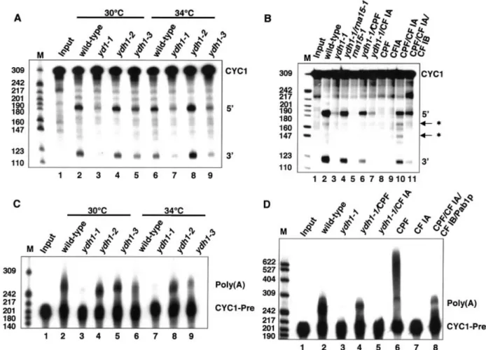
cleave a synthetic CYC1 pre-mRNA (Fig. 2A). Extracts from wild-type (lanes 2 and 6), ydh1-2 (lanes 4 and 8) and ydh1-3 cells (lanes 5 and 9) accurately cleaved the substrate RNA at 30 and 34°C. Notably, ydh1-3 extract (lane 9) showed a reduced ef®ciency of cleavage at 34°C compared to wild-type (lane 6). In contrast, extract of ydh1-1 cells was de®cient in cleavage at both temperatures (lanes 3 and 7).
To show that the de®ciency in cleaving the RNA substrate is due to inactive Ydh1p or CPF, respectively, we carried out reconstitution assays either with extract which is mutant in a CF IA subunit (rna15-1) or with puri®ed factors at 30°C (Fig. 2B). The cleavage activity of ydh1-1 extract was restored upon addition of rna15-1 extract (lane 4) or puri®ed CPF (lane 6), but not by addition of puri®ed CF IA (lane 7). As expected, the rna15-1 extract on its own lacked cleavage activity (lane 5) (56) and puri®ed CPF and CF IA alone were not able to cleave the substrate (lanes 8 and 9); cleavage occurred at speci®c and at cryptic sites when both factors were added to the reaction (lane 10); site-speci®c cleavage was obtained by including CF IB in the reaction (lane 11) (15).
Next we analysed whether mutations in YDH1 affect the polyadenylation reaction of pre-cleaved CYC1 substrate (CYC1-Pre) in vitro (Fig. 2C). No polyadenylation activity could be observed in ydh1-1 extract at 30 and 34°C (lanes 3 and 7). In contrast, the ydh1-2 and ydh1-3 extracts speci®cally and ef®ciently polyadenylated the substrate at both tempera-tures (lanes 4, 5, 8 and 9), comparable to wild-type (lanes 2 and 6). Speci®c polyadenylation activity was restored in ydh1-1 extract by addition of puri®ed CPF (Fig. 2D, lane 4) but not by addition of puri®ed CF IA (lane 5). As shown before, CPF by itself unspeci®cally polyadenylated the substrate (lane 6) (3), whereas CF IA alone displayed no polyadenylation activity (lane 7). Speci®c polyadenylation activity was restored upon combination of CPF, CF IA, CF IB and Pab1p (lane 8).
Notably, addition of 100±250 ng recombinantly expressed Ydh1p failed to reconstitute the cleavage and polyadenylation activities in ydh1-1 extract, possibly because the recombinant
Figure 1. YDH1 mutant strains display a temperature sensitive phenotype. (A) Growth curves of wild-type and mutant ydh1 strains at 37°C. (B) 10-fold serial dilution of cultures spotted on YPD plates followed by incubation at the indicated temperature for 2 days.
protein was not able to replace its mutant counterpart in the CPF complex (results not shown).
The above results showed that the ydh1-1 extract was strongly reduced in cleavage and polyadenylation activities in vitro and that both activities could be reconstituted by addition of puri®ed CPF. Furthermore, the cleavage activity of the ydh1-3 mutant was lower at 34°C compared to 30°C. The results indicate that Ydh1p is involved in both steps of the 3¢-end processing reaction.
mRNAs are unstable in ydh1 mutant strains at restrictive temperature
mRNAs without poly(A) tails are rapidly degraded in living cells. Yeast strains with a 3¢-end processing de®ciency are therefore expected to under-accumulate mRNAs after shift to the restrictive temperature. For this reason, we performed northern blot analyses on total RNA extracted from strains grown at 25°C and after shift to 37°C (Fig. 3A). The amount of 18S rRNA served as control for the loading of the RNA (panel
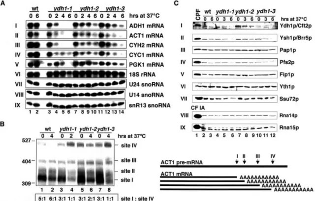
VI). ADH1, ACT1, CYH2 and CYC1 mRNA levels diminished in the ydh1-1 mutant cells after 2 h shift to 37°C (panels I±IV, lanes 3±6). The level of the stable PGK1 mRNA (t1/2= 45 min)
dropped signi®cantly only after the cells were shifted to 37°C for 6 h (panel V, lanes 3±6). In comparison the ADH1, ACT1, CYH2 and CYC1 mRNA levels of the ydh1-2 mutant strain only dropped slightly after 4 h, and the PGK1 mRNA level remained stable even after shift to 37°C for 6 h (panels I±V, lanes 7±10). The mutant strain ydh1-3 under-accumulated ACT1 mRNA after only 2 h at 37°C; ADH1, CYH2 and CYC1 mRNA levels were reduced after 4 h (panels I±IV, lanes 11± 14). The PGK1 mRNA level showed a slight decrease after 4 h and more so after 6 h (panel V, lanes 11±14). The RNA polymerase II transcribed U24, U14 and snR13 snoRNAs were stable in all mutants tested, even after shift to 37°C for 6 h (panels VII±IX), indicating that snoRNA processing was not affected in ydh1 mutant strains. These results showed that ydh1 mutant strains under-accumulated mRNAs at restrictive temperature. The ydh1 mutant phenotypes are possibly due to
Figure 2. Ydh1p is required for cleavage and polyadenylation in vitro. In vitro analysis of cleavage (A and B) and polyadenylation activities (C and D) of ydh1 mutant extracts. Input lanes (1) represent mock-treated reactions. The migration positions of the CYC1 and CYC1-precleaved (CYC1-Pre) RNA sub-strate, speci®c (5¢ and 3¢) and cryptic (asterisks) cleavage products and the polyadenylation products [Poly(A)] are indicated on the right of each panel. The position and size (in number of nucleotides) of the marker bands are indicated on the left. (A) Extracts prepared from yeast strains (as indicated above the lanes) were monitored for their ability to cleave internally32P-labelled CYC1 RNA substrate at 30 and 34°C. (B) Reconstitution of cleavage activity in ydh1-1
extract at 30°C. As indicated above the lanes, ydh1-1 extract was combined with equal amounts of rna15-1 extract (lane 4), puri®ed CPF (lane 6) or CF IA (lane 7). (C) Speci®c polyadenylation was analysed at 30 and 34°C with internally32P-labelled precleaved CYC1 RNA substrate that ends at the natural
poly(A) site. (D) Reconstitution of speci®c polyadenylation activity in ydh1-1 extract at 30°C. ydh1-1 extract was combined with puri®ed CPF (lane 4) or CF IA (lane 5).
a de®ciency of the strains in poly(A) site recognition (see below) which might impair the ef®ciency in 3¢-end processing. Ydh1p is required for poly(A) site selection of ACT1 pre-mRNA
The previous observation that Ydh1p binds RNA around the poly(A) site (9), raised the possibility that the protein is required for poly(A) site recognition. To test this, we analysed poly(A) site usage of ACT1 pre-mRNA in wild-type and mutant cells (57). The ACT1 3¢ untranslated region (UTR) contains at least four polyadenylation sites, of which the most proximal one (site I) is used with highest frequency in wild-type cells (Fig. 3B; lanes 1 and 2). All tested ydh1 mutants used site I three times more often than site IV at 25°C (lanes 3, 5 and 7). At restrictive temperature, however, processing shifted from site I towards site IV in a ratio of 1:1 in ydh1-1 and ydh1-3 cells (lanes 4 and 8); the ydh1-2 mutant showed a ratio of 2:1 (lane 6). These results indicated that mutations in ydh1-1, ydh1-2 and ydh1-3 impair poly(A) site recognition in vivo. To verify that the ACT1 mRNAs were polyadenylated we incubated the reactions with RNase H and the oligos ACT1-RnaseH and dT to digest the poly(A) tails. This resulted
in a slight downward shift and sharpening of the RNA bands. This showed that the ACT1 mRNAs were polyadenylated (results not shown).
Western blot analyses were carried out to test whether the observed phenotypes were the result of an under-accumulation of Ydh1p or other subunits of the 3¢-end processing machinery (Fig. 3C). As control, puri®ed CPF or CF IA was analysed in parallel (lane 1). In wild-type and ydh1-2 cells Ydh1p was stable even after 6 h at 37°C (panel I, lanes 2, 3 and 7±9), whereas it was reduced in ydh1-1 and ydh1-3 cells after 3 h at 37°C (lanes 5, 6, 11 and 12). Ysh1p/Brr5p, Pfs2p and Ssu72p (panels II, IV and VII) under-accumulated in ydh1-1 and ydh1-3 cells after 6 h at 37°C (lanes 6 and 12). The levels of Pap1p, Fip1p and Yth1p were constant even after 6 h at 37°C (panels III, V and VI). In addition, mutations in YDH1 did not affect the levels of the CF IA subunits Rna14p and Rna15p (panels VIII and IX).
These results indicate that the defects observed in the ydh1 mutant strains might be caused by a destabilization of the Ydh1p protein. Since the destabilization at restrictive tem-perature of Ysh1p/Brr5p, Pfs2p and Ssu72p was detectable at later time-points compared to the appearance of phenotypes
Figure 3. mRNAs are unstable in ydh1 mutant strains at restrictive temperature and the mutant strains are de®cient in the recognition of ACT1 poly(A) site in vivo. (A) Northern analysis of total RNA extracted from wild-type and mutant ydh1 cells grown at 23°C, or after shift to 37°C for 2, 4 and 6 h as indicated above each lane. The RNAs were separated on formaldehyde/1.2% agarose gels (panels I±VI) or 8.3 M urea/8% polyacrylamide gels (panels VII±IX). The ®lters were developed with random-primed labeled probe (panels I±V) or end-labeled oligonucleotides (panels VI±IX) directed against the RNA species indi-cated at the right of each panel. (B) Analysis of ACT1 poly(A) site usage in wild-type and mutant ydh1 cells. The schematic on the right shows the relative positions of the different poly(A) sites. Total RNA extracted from wild-type and mutant ydh1 cells after growth at 23°C, or following shift to 37°C as indi-cated above each lane, was treated with an oligonucleotide complementary to the 3¢ region of the ACT1±mRNA (ACT1±RnaseH) and RNaseH. The RNAs were separated on an 8 M urea/6% polyacrylamide gel and the ®lters were incubated with random primed labeled probe directed against the 3¢-end of the ACT1 mRNA. RNA levels were quanti®ed by PhosphorImager scanning (Molecular Dynamics); the ratios of poly(A) site I:site IV usage for each lane are indicated at the bottom. (C) Western analysis of wild-type and ydh1 mutant extracts prepared from cells grown at 23°C, or after shift to 37°C for 3 and 6 h. Equal amounts of total protein were loaded in each lane. Lane 1 shows puri®ed CPF (panels I±VII) and CF IA (panels VIII and IX). The ®lters were treated with antibodies directed against the proteins indicated at the right.
[see northern blot and ACT1 poly(A) site selection], we consider it unlikely that lower levels of factors other than Ydh1p are responsible for the de®ciencies of the ydh1 mutant cells.
Ydh1p interacts with the C-terminal domain of RNA polymerase II
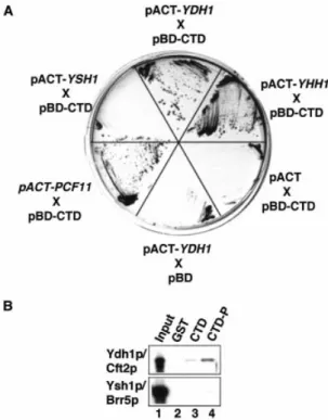
The CTD of RNAP II plays an important role in coupling transcription and 3¢-end formation (reviewed in 22,23). We were interested to know whether Ydh1p contributes to this process by physically interacting with the CTD. To test this, we carried out GAL4-based yeast two-hybrid tests and assayed for the activation of expression of the HIS3 and lacZ reporter genes. Strain Y190 was co-transformed with plasmids carry-ing the GAL4 DNA bindcarry-ing domain fused to the CTD (pBD-CTD) and the GAL4 activation domain fused to test genes. As positive controls we analysed the known CTD interactors YHH1/BRR5 (pACT2-YHH1) and PCF11 (pACT2-PCF11) (10,28,47) and pACT2-YSH1 as negative control. As shown in Figure 4A the plasmids carrying YDH1, YHH1 and PCF11 in combination with the plasmid pBD-CTD enabled cells to grow on medium lacking histidine under stringent con-ditions (in the presence of 35 mM 3¢-aminotriazole). In contrast, no growth was observed with pACT2-YSH1 or when empty pBD and pACT2 plasmids were tested. These results were con®rmed by an X-Gal ®lter lift assay, which monitors b-galactosidase expression (results not shown). Thus, YDH1 showed a two-hybrid interaction with the CTD of RNAP II.
To con®rm that Ydh1p interacts with the CTD, and to test whether this interaction is in¯uenced by the phosphorylation state of the CTD we performed GST pull-down experiments with in vitro translated radioactively labeled proteins and recombinant GST±CTD fusion protein. Ydh1p interacted with the CTD (Fig. 4B, lane 3) and this interaction was enhanced upon phosphorylation of the CTD (lane 4); no signal was observed with GST alone (lane 2). Ysh1p/Cft1p interacted with neither form of the GST±CTD protein. These results suggested that Ydh1p binds the CTD and that the interaction is enhanced upon phosphorylation of the CTD.
Correct transcription termination by RNAP II requires a functional poly(A) site on the nascent RNA and the CTD plays an important role in coupling transcription and pre-mRNA processing (reviewed in 43). The observations that Ydh1p is involved in poly(A) site recognition and that it interacted with the CTD raised the possibility that Ydh1p might also be involved in transcriptional termination. Therefore, we carried out transcriptional run-on analysis with the well-characterized GAL1/10 controlled CYC1 gene in vivo (44). We did not observe a signi®cantly increased RNAP II density downstream of the CYC1 terminator in ydh1-1 and ydh1-3 cells grown at 25°C or following shift to 37°C compared to wild-type (results not shown). Similarly, we did not detect the accumulation of read-through products in ydh1 strains in reverse transcription experiments with primers annealing downstream of the cleavage site of different snoRNA species (snR39b, snR45, snR3, snR50, snR71) (51). This suggested that Ydh1p is not generally required for termination of RNAP II during transcription of pre-mRNAs and snoRNAs.
Ydh1p interacts with other subunits of CPF and with Pcf11p, a subunit of CF IA
As Ydh1p is part of a multi-protein complex we were interested to examine with which of the other CPF subunits the protein interacts. Therefore, we expressed a N-terminally GST-tagged and C-terminally His-tagged version of the Ydh1 protein (G-Ydh1-H) in E.coli and carried out GST pull-down experiments with radioactively labeled in vitro translated proteins. As shown in Figure 5A, G-Ydh1-H interacted with itself and with the CPF subunits Yhh1p/Cft1, Ysh1p/Brr5p, Pta1p, Pfs2p, Ssu72p, YDL094cp (panels I±VII) and the CF IA subunit Pcf11p (panel X). G-Ydh1-H did not pull-down the CPF subunits Fip1p and Yth1p (panels VII and IX), nor the CF IA subunits Rna14p, Clp1p and Rna15p (panels XI±XIII), Nab4p/Hrp1 (CF IB) or Pab1p (panels XIV and XV). Since we identi®ed a large number of potential Ydh1p interaction partners, we decided to investigate whether these interactions were speci®c and therefore assignable to distinct regions of Ydh1p. For this purpose the pull-downs were repeated with C-terminally truncated Ydh1p proteins (Fig. 5B). Figure 5A shows that the very C-terminus was required for interaction of Ydh1p with itself (panel II) as most of the interaction was lost upon deletion of the C-terminal 137 amino acids. The
Figure 4. Ydh1p interacts with the CTD of RNAP II. (A) GAL4-based two-hybrid analysis. Y190 cells were co-transformed with a plasmid encoding the GAL4 DNA binding domain fused to the CTD (pBD-CTD) and plasmids (pACT2) encoding the GAL4 activation domain fused to the genes indi-cated. Transformants were tested for expression of the HIS3 reporter gene on medium lacking histidine, and containing 35 mM 3-amino-1,2,4-triazole. As control, pBD CTD was co-transformed with the empty pACT2 plasmid and pACT2-YDH1 with the empty pBD plasmid. (B) Pull-down experiments with 1 mg GST (lane 2), GST±CTD (lane 3), phosphorylated GST±CTD (lane 4) and in vitro translated, [35S]methionine-labeled proteins
(indicated on the left). Lane 1 shows 10% of the in vitro translated reactions used in the assay. Bound proteins were separated by SDS±PAGE and visualized by autoradiography.
N-terminal 246 amino acids were suf®cient for interaction with Pfs2p (panel V) and central protein sequences most likely mediated interactions with Yhh1p/Cft1p, Ysh1p/Brr5p, Pta1p, Ssu72, pYDL094c and Pcf11p (panels I, III, IV, VI, VII and X). These results suggested that the Ydh1p subunit might contribute to the stability and structural order of CPF and bridge the two factors CPF and CF IA.
To obtain a better understanding of how CPF interacts with CF IA we decided to assay for further interactions between the two factors. Figure 5C shows a GST pull-down with in vitro translated protein and GST-tagged recombinant proteins. Of the analysed CPF subunits we found that only Yhh1p/Cft1p interacted strongly with Rna14p. Yhh1p/Cft1p also bound to Pcf11p, and more weakly to Clp1p (lane 3). Ydh1p pulled-down only Pcf11p (lane 4). Ysh1p/Brr5p interacted strongly with Clp1p and Pcf11p (lane 5). A weak signal was observed for the interaction between Pta1p and Pcf11p (lane 6). These results suggested that Ydh1p, Yhh1p/Cft1p, Ysh1p/Brr5p and possibly also Pta1p are involved in forming a protein±protein interaction surface between CPF and CF IA. Our combined results indicate that Ydh1p plays an important role in determining the RNA binding speci®city of CPF and in the assembly of the 3¢-end formation machinery and its tethering to RNAP II.
DISCUSSION
The yeast and mammalian 3¢-end formation machineries display a surprisingly complex subunit composition. At present, up to 20 polypeptides are known that constitute the factors that catalyse the yeast 3¢-end formation reaction in vitro (45,58). The ongoing characterization of the components suggests distinct and specialized tasks of the individual polypeptides in both steps of the 3¢-end processing reaction as well as in the coupling of 3¢-end formation to transcription. In this work we characterized the role of the yeast CPF subunit Ydh1p in these processes.
To analyse if Ydh1p is necessary for cleavage and polyadenylation we produced conditional ydh1 mutant strains and carried out in vitro assays. The experiments revealed a de®ciency of the mutant extracts in both steps of the 3¢-end processing reaction. The activity could be rescued upon addition of puri®ed CPF. mRNAs without a poly(A) tail are prone to rapid degradation in vivo. Northern analysis revealed that the levels of a number of different mRNAs were reduced at restrictive temperature in Ydh1p mutants. The results support a requirement for Ydh1p in both steps of 3¢-end processing. However, we do not expect Ydh1p to be directly involved in the catalysis of the 3¢-end processing reaction,
Figure 5. Ydh1p interacts with other subunits of CPF and with Pcf11p, a subunit of CF IA. (A) GST pull-down experiments with 0.5 mg GST, 100 ng G-Ydh1-H (Ydh1p) or 100 ng of the different C-terminally truncated, GST-tagged Ydh1 fragments (as indicated above each lane) and in vitro translated [35S]methionine-labeled proteins (indicated on the right). The ®rst lane shows 10% of the in vitro translated protein used in the binding reactions.
(B) Schematic drawing of the full length and C-terminal truncations of Ydh1p. The numbers indicate the length of the constructs in amino acids. (C) GST pull-down experiment with GST-tagged recombinant proteins (indicated above each lane) and in vitro translated, [35S]methionine-labeled proteins (indicated
because it took several hours at the restrictive temperature before the mRNA levels were reduced. Furthermore, the ACT1 poly(A) site selection assay revealed that a substantial amount of pre-mRNA appeared to be cleaved and polyadenylated in vivo at the restrictive temperature in all mutants.
We reported previously that Ydh1p binds to a CYC1 pre-mRNA around the poly(A) site; this suggested a role for the protein in poly(A) site selection (9). Here we show that Ydh1p is involved in poly(A) site selection of ACT1 pre-mRNA in vivo, as all analysed ydh1 mutant strains displayed more frequent use of alternative poly(A) sites compared to the wild-type strain. These results underscore our previously postulated model that Ydh1p contributes to poly(A) site selection (9). In contrast to mammalian polyadenylation signals, the cis-acting sequences in yeast are highly degenerate (reviewed in 1,8). We proposed that the RNA binding subunits of CPF act in concert to achieve speci®c recognition of the correct poly(A) site (9). The preferential use of cleavage sites located downstream of the major site in Ydh1 mutants may be caused by the coupling of transcription elongation and 3¢-end processing. Reduced RNA binding ef®ciency of CPF contain-ing mutant Ydh1p could lead to skippcontain-ing of the ®rst cleavage site of the nascent pre-mRNA emerging from the elongating RNAP II. Ydh1p (9), Yhh1p/Cft1p (10) and Yth1p (11) were shown to bind preferentially to U-rich elements localized directly upsteam and downstream of the poly(A) site, thus colocalizing with the region to which the complete CPF factor binds (9). These interactions with the RNA substrate are thought to be crucial for poly(A) site selection and for cleavage activity. Thus, the observed in vitro cleavage de®ciency of the ydh1 mutant cells might result from insuf®cient poly(A) site selection. It remains to be determined how Ydh1p interacts with RNA. The primary sequence of the protein does not display any clear similarities to known RNA binding domains. Preliminary RNA binding experiments with portions of Ydh1p indicated that the RNA binding activity is distributed throughout the entire length of the protein (results not shown). However, detailed analyses have not been done yet. Interestingly, RNA binding activity has not been demon-strated for the mammalian homolog of Ydh1p, CPSF 100 kDa. Considering the highly conserved cis-acting elements in mammalian pre-mRNAs (reviewed in 1), it seems possible that a smaller set of proteins is suf®cient for speci®c poly(A) site recognition, whereas in yeast a cooperative interplay of more RNA binding proteins is required to recognize the more degenerate sequence elements in a speci®c fashion.
We have shown that Ydh1p interacted with the CTD of RNAP II both in a two-hybrid test and in a GST pull-down assay. This interaction was enhanced upon phosphorylation of the CTD. The only other CPF subunit found to interact with the phosphorylated CTD is Yhh1p/Cft1p (10). We suggest that Ydh1p might be involved in tethering CPF to transcribing RNAP II and thereby contributes to the coupling of 3¢-end processing and transcription. However, the ydh1 mutants did not reveal a defect in transcriptional termination at the CYC1 terminator. As this was the only pre-mRNA that was tested by transcriptional run-on analysis we cannot exclude that Ydh1p might be involved in transcriptional termination of other genes. Moreover, reverse transcription analysis on snoRNAs in ydh1 mutant strains did not reveal 3¢-extended transcripts, indicating that Ydh1p was not required for snoRNA
termination either. We propose that Ydh1p helps to tether CPF to elongating RNAP II but that it is not needed for general transcription termination. Ydh1p and Yhh1p/Cft1p show a phenotypic similarity in that both proteins bind RNA, are involved in poly(A) site recognition and bind to the CTD (10). However, the observation that mutants in yhh1 are severely impaired in transcription termination at the CYC1 gene (10) whereas ydh1 mutants have no general termination defect, indicates that the functional roles of the individual 3¢-end formation factor subunits can be substantially different.
It is not well understood how the subunits of the CPF and CF IA factors assemble to form functional 3¢-end processing complexes on the RNA substrate. CPF consists of up to 15 polypeptides, which interact with each other and form a stable factor. We found that Ydh1p interacted with the CPF subunits Yhh1p/Cft1p, Ysh1p/Brr5p, Pta1p, Pfs2p, Ssu72p and pYDL094c. These many interactions indicate that the protein contributes substantially to the assembly and structural order of the CPF factor. Strikingly, Ydh1p also bound strongly to itself in the GST pull-down assays. This may indicate that there is more than one Ydh1p molecule present per 3¢-end processing unit. However, silver stain analysis of puri®ed CPF factor suggested an apparent stoichiometric presence of Ydh1p compared to other CPF subunits (45). Interestingly, we found that the CF IA subunit Pcf11p interacted with the CPF components Ydh1p, Yhh1p/Cft1p, Ysh1p/Brr5p, and weakly with Pta1p. In addition, we showed that the CPF subunits Yhh1p/Cft1p and Ysh1p/Brr5p interacted with the CF IA subunits Rna14p and Clp1p, respectively. The latter interactions were also observed between the homologous subunits of the mammalian 3¢-end formation machinery. CPSF 160 kDa interacts with the CstF 77 kDa protein (18) and CPSF 73 kDa interacts with hClp1p (59). So far, only Pfs2p and Fip1p were shown to bridge CPF and CF IA (3,4). We propose that Ydh1p, Yhh1p/Cft1p, Ysh1p/Brr5p, Pfs2p, Fip1p, and possibly Pta1p, contribute to a protein±protein interaction surface which acts in the assembly of the 3¢-end formation machinery and which appears to be conserved in evolution (Fig. 5D).
Our analysis of Ydh1p suggests that the protein is an important constituent of protein interaction surfaces which act in the association of 3¢-end processing factors with RNAP II, the recognition of poly(A) signal sequences and the assembly of an active 3¢-end formation complex on the RNA substrate.
SUPPLEMENTARY MATERIAL
Supplementary Material is available at NAR Online.
ACKNOWLEDGEMENTS
We thank Isabelle Kaufmann, Myriam Schaub and Jeannette Wolf for comments on the manuscript. This work was supported by the University of Basel, the Swiss National Science Fund, the European Community (www.euronomics. org) via the Bundesamt fuÈr Bildung und Wissenschaft, Bern (grant 01.0123) and the Louis-Jeantet-Foundation for Medicine.
REFERENCES
1. Zhao,J., Hyman,L. and Moore,C. (1999) Formation of mRNA 3¢ ends in eukaryotes: mechanism, regulation, and interrelationships with other steps in mRNA synthesis. Microbiol. Mol. Biol. Rev., 63, 405±445. 2. Chen,J. and Moore,C. (1992) Separation of factors required for cleavage
and polyadenylation of yeast pre-mRNA. Mol. Cell. Biol., 12, 3470± 3481.
3. Ohnacker,M., Barabino,S.M., Preker,P.J. and Keller,W. (2000) The WD-repeat protein Pfs2p bridges two essential factors within the yeast pre-mRNA 3¢-end-processing complex. EMBO J., 19, 37±47. 4. Preker,P.J., Lingner,J., Minvielle-Sebastia,L. and Keller,W. (1995) The
FIP1 gene encodes a component of a yeast pre-mRNA polyadenylation factor that directly interacts with poly(A) polymerase. Cell, 81, 379±389. 5. Graber,J.H., Cantor,C.R., Mohr,S.C. and Smith,T.F. (1999) Genomic
detection of new yeast pre-mRNA 3¢-end-processing signals. Nucleic Acids Res., 27, 888±894.
6. Russo,P., Li,W.Z., Hampsey,D.M., Zaret,K.S. and Sherman,F. (1991) Distinct cis-acting signals enhance 3¢ endpoint formation of CYC1 mRNA in the yeast Saccharomyces cerevisiae. EMBO J., 10, 563±571. 7. van Helden,J., del Olmo,M. and Perez-Ortin,J.E. (2000) Statistical
analysis of yeast genomic downstream sequences reveals putative polyadenylation signals. Nucleic Acids Res., 28, 1000±1010.
8. Guo,Z. and Sherman,F. (1996) 3¢-end-forming signals of yeast mRNA. Trends Biochem. Sci., 21, 477±481.
9. Dichtl,B. and Keller,W. (2001) Recognition of polyadenylation sites in yeast pre-mRNAs by cleavage and polyadenylation factor. EMBO J., 20, 3197±3209.
10. Dichtl,B., Blank,D., Sadowski,M., HuÈbner,W., Weiser,S. and Keller,W. (2002) Yhh1p/Cft1p directly links poly(A) site selection and RNA polymerase II transcription termination. EMBO J., 21, 4125±4135. 11. Barabino,S.L.M., Ohnacker,M. and Keller,W. (2000) Distinct roles of
two Yth1p domains in 3¢-end cleavage and polyadenylation of yeast pre-mRNAs. EMBO J., 19, 3778±3787.
12. Chen,S. and Hyman,L.E. (1998) A speci®c RNA±protein interaction at yeast polyadenylation ef®ciency elements. Nucleic Acids Res., 26, 4965± 4974.
13. Kessler,M.M., Henry,M.F., Shen,E., Zhao,J., Gross,S., Silver,P.A. and Moore,C.L. (1997) Hrp1, a sequence-speci®c RNA-binding protein that shuttles between the nucleus and the cytoplasm, is required for mRNA 3¢-end formation in yeast. Genes Dev., 11, 2545±2556.
14. Gross,S. and Moore,C.L. (2001) Rna15 interaction with the A-rich yeast polyadenylation signal is an essential step in mRNA 3¢-end formation. Mol. Cell. Biol., 21, 8045±8055.
15. Minvielle-Sebastia,L., Beyer,K., Krecic,A.M., Hector,R.E.,
Swanson,M.S. and Keller,W. (1998) Control of cleavage site selection during mRNA 3¢-end formation by a yeast hnRNP. EMBO J., 17, 7454± 7468.
16. Shatkin,A.J. and Manley,J.L. (2000) The ends of the affair: capping and polyadenylation. Nature Struct. Biol., 7, 838±842.
17. Keller,W., Bienroth,S., Lang,K.M. and Christofori,G. (1991) Cleavage and polyadenylation factor CPF speci®cally interacts with the pre-mRNA 3¢ processing signal AAUAAA. EMBO J., 10, 4241±4249.
18. Murthy,K.G.K. and Manley,J.L. (1995) The 160-kD subunit of human cleavage-polyadenylation speci®city factor coordinates pre-mRNA 3¢-end formation. Genes Dev., 9, 2672±2683.
19. MacDonald,C.C., Wilusz,J. and Shenk,T. (1994) The 64-kilodalton subunit of the CstF polyadenylation factor binds to pre-mRNAs downstream of the cleavage site and in¯uences cleavage site location. Mol. Cell. Biol., 14, 6647±6654.
20. Wilusz,J., Shenk,T., Takagaki,Y. and Manley,J.L. (1990) A multicomponent complex is required for the AAUAAA-dependent cross-linking of a 64-kilodalton protein to polyadenylation substrates. Mol. Cell. Biol., 10, 1244±1248.
21. Gilmartin,G.M. and Nevins,J.R. (1991) Molecular analyses of two poly(A) site-processing factors that determine the recognition and ef®ciency of cleavage of the pre-mRNA. Mol. Cell. Biol., 11, 2432±2438. 22. Bentley,D. (2002) The mRNA assembly line: transcription and
processing machines in the same factory. Curr. Opin. Cell Biol., 14, 336± 342.
23. Proudfoot,N.J., Furger,A. and Dye,M.J. (2002) Integrating mRNA processing with transcription. Cell, 108, 502±512.
24. Howe,K. (2002) RNA polymerase II conducts a symphony of pre-mRNA processing activities. Biochim. Biophys. Acta, 1577, 308.
25. Proudfoot,N. and O'Sullivan,J. (2002) Polyadenylation: a tail of two complexes. Curr. Biol., 12, R855±R857.
26. Cho,E.J., Kobor,M.S., Kim,M., Greenblatt,J. and Buratowski,S. (2001) Opposing effects of Ctk1 kinase and Fcp1 phosphatase at Ser 2 of the RNA polymerase II C-terminal domain. Genes Dev., 15, 3319±3329. 27. Komarnitsky,P., Cho,E.J. and Buratowski,S. (2000) Different
phosphorylated forms of RNA polymerase II and associated mRNA processing factors during transcription. Genes Dev., 14, 2452± 2460.
28. Licatalosi,D.D., Geiger,G., Minet,M., Schroeder,S., Cilli,K., McNeil,J.B. and Bentley,D.L. (2002) Functional interaction of yeast pre-mRNA 3¢ end processing factors with RNA polymerase II. Mol. Cell, 9, 1101± 1111.
29. McCracken,S., Fong,N., Rosonina,E., Yankulov,K., Brothers,G., Siderovski,D., Hessel,A., Foster,S., Shuman,S. and Bentley,D.L. (1997) 5¢-Capping enzymes are targeted to pre-mRNA by binding to the phosphorylated carboxy-terminal domain of RNA polymerase II. Genes Dev., 11, 3306±3318.
30. Cho,E.J., Takagi,T., Moore,C.R. and Buratowski,S. (1997) mRNA capping enzyme is recruited to the transcription complex by phosphorylation of the RNA polymerase II carboxy-terminal domain. Genes Dev., 11, 3319±3326.
31. Ho,C.K., Sriskanda,V., McCracken,S., Bentley,D., Schwer,B. and Shuman,S. (1998) The guanylyltransferase domain of mammalian mRNA capping enzyme binds to the phosphorylated carboxyl-terminal domain of RNA polymerase II. J. Biol. Chem., 273, 9577±9585. 32. Cho,E.J., Rodriguez,C.R., Takagi,T. and Buratowski,S. (1998) Allosteric
interactions between capping enzyme subunits and the RNA polymerase II carboxy-terminal domain. Genes Dev., 12, 3482±3487.
33. Yuryev,A., Patturajan,M., Litingtung,Y., Joshi,R.V., Gentile,C., Gebara,M. and Corden,J.L. (1996) The C-terminal domain of the largest subunit of RNA polymerase II interacts with a novel set of serine/ arginine-rich proteins. Proc. Natl Acad. Sci. USA, 93, 6975±6980. 34. McCracken,S., Fong,N., Yankulov,K., Ballantyne,S., Pan,G.,
Greenblatt,J., Patterson,S.D., Wickens,M. and Bentley,D.L. (1997) The C-terminal domain of RNA polymerase II couples mRNA processing to transcription. Nature, 385, 357±361.
35. Barilla,D., Lee,B.A. and Proudfoot,N.J. (2001) Cleavage/
polyadenylation factor IA associates with the carboxyl-terminal domain of RNA polymerase II in Saccharomyces cerevisiae. Proc. Natl Acad. Sci. USA, 98, 445±450.
36. Rodriguez,C.R., Cho,E.J., Keogh,M.C., Moore,C.L., Greenleaf,A.L. and Buratowski,S. (2000) Kin28, the TFIIH-associated carboxy-terminal domain kinase, facilitates the recruitment of mRNA processing machinery to RNA polymerase II. Mol. Cell. Biol., 20, 104±112. 37. Fong,N. and Bentley,D.L. (2001) Capping, splicing, and 3¢ processing
are independently stimulated by RNA polymerase II: different functions for different segments of the CTD. Genes Dev., 15, 1783±1795. 38. Hirose,Y. and Manley,J.L. (1998) RNA polymerase II is an essential
mRNA polyadenylation factor. Nature, 395, 93±96.
39. Ryan,K., Murthy,K.G., Kaneko,S. and Manley,J.L. (2002) Requirements of the RNA polymerase II C-terminal domain for reconstituting pre-mRNA 3¢ cleavage. Mol. Cell. Biol., 22, 1684±1692.
40. McNeil,J.B., Agah,H. and Bentley,D. (1998) Activated transcription independent of the RNA polymerase II holoenzyme in budding yeast. Genes Dev., 12, 2510±2521.
41. Orozco,I.J., Kim,S.J. and Martinson,H.G. (2002) The poly(A) signal, without the assistance of any downstream element, directs RNA polymerase II to pause in vivo and then to release stochastically from the template. J. Biol. Chem., 277, 42899±42911.
42. Tran,D.P., Kim,S.J., Park,N.J., Jew,T.M. and Martinson,H.G. (2001) Mechanism of poly(A) signal transduction to RNA polymerase II in vitro. Mol. Cell. Biol., 21, 7495±7508.
43. Proudfoot,N.J. (1989) How RNA polymerase II terminates transcription in higher eukaryotes. Trends Biochem. Sci., 14, 105±110.
44. Birse,C.E., Minvielle-Sebastia,L., Lee,B.A., Keller,W. and
Proudfoot,N.J. (1998) Cleavage of the primary transcript couples 3¢-end RNA-processing with termination of pol II transcription. Science, 280, 298±301.
45. Dichtl,B., Blank,D., Ohnacker,M., Friedlein,A., Roeder,D., Langen,H. and Keller,W. (2002) A role for SSU72 in balancing RNA polymerase II transcription elongation and termination. Mol. Cell, 10, 1139±1150.
46. Hammell,C.M., Gross,S., Zenklusen,D., Heath,C.V., Stutz,F., Moore,C. and Cole,C.N. (2002) Coupling of termination, 3¢ processing, and mRNA export. Mol. Cell. Biol., 22, 6441±6457.
47. Sadowski,M., Dichtl,B., HuÈbner,W. and Keller,W. (2003) Independent functions of yeast Pcf11p in pre-mRNA 3¢ end processing and in transcription termination. EMBO J., 22, 2167±2177.
48. Calvo,O. and Manley,J.L. (2001) Evolutionarily conserved interaction between CstF-64 and PC4 links transcription, polyadenylation, and termination. Mol. Cell, 7, 1013±1023.
49. Aranda,A. and Proudfoot,N. (2001) Transcriptional termination factors for RNA polymerase II in yeast. Mol. Cell, 7, 1003±1011.
50. Alen,C., Kent,N.A., Jones,H.S., O'Sullivan,J., Aranda,A. and
Proudfoot,N.J. (2002) A role for chromatin remodeling in transcriptional termination by RNA polymerase II. Mol. Cell, 10, 1441±1452. 51. Steinmetz,E.J., Conrad,N.K., Brow,D.A. and Corden,J.L. (2001)
RNA-binding protein Nrd1 directs poly(A)-independent 3¢-end formation of RNA polymerase II transcripts. Nature, 413, 327±331.
52. Jenny,A., Minvielle-Sebastia,L., Preker,P.J. and Keller,W. (1996) Sequence similarity between the 73-kilodalton protein of mammalian CPSF and a subunit of yeast polyadenylation factor I. Science, 274, 1514±1517.
53. Preker,P.J., Ohnacker,M., Minvielle-Sebastia,L. and Keller,W. (1997) A multisubunit 3¢-end processing factor from yeast containing poly(A)
polymerase and homologues of the subunits of mammalian cleavage and polyadenylation speci®city factor. EMBO J., 16, 4727±4737.
54. Zhao,J., Kessler,M.M. and Moore,C.L. (1997) Cleavage factor II of Saccharomyces cerevisiae contains homologues to subunits of the mammalian cleavage/polyadenylation speci®city factor and exhibits sequence-speci®c, ATP-dependent interaction with precursor RNA. J. Biol. Chem., 272, 10831±10838.
55. Fromont-Racine,M., Rain,J.C. and Legrain,P. (1997) Toward a functional analysis of the yeast genome through exhaustive two-hybrid screens. Nature Genet., 16, 277±282.
56. Minvielle-Sebastia,L., Preker,P.J. and Keller,W. (1994) RNA14 and RNA15 proteins as components of a yeast pre-mRNA 3¢-end processing factor. Science, 266, 1702±1705.
57. Mandart,E. and Parker,R. (1995) Effects of mutations in the Saccharomyces cerevisiae RNA14, RNA15, and PAP1 genes on polyadenylation in vivo. Mol. Cell. Biol., 15, 6979±6986.
58. Gavin,A.C., Bosche,M., Krause,R., Grandi,P., Marzioch,M., Bauer,A., Schultz,J., Rick,J.M., Michon,A.M., Cruciat,C.M. et al. (2002) Functional organization of the yeast proteome by systematic analysis of protein complexes. Nature, 415, 141±147.
59. deVries,H., RuÈegsegger,U., HuÈbner,W., Friedlein,A., Langen,H. and Keller,W. (2000) Human pre-mRNA cleavage factor II(m) contains homologs of yeast proteins and bridges two other cleavage factors. EMBO J., 19, 5895±5904.