Third structure determination by powder diffractometry round robin
„SDPDRR-3…
A. Le Baila兲Laboratoire des Oxydes et Fluorures, Université du Maine, CNRS UMR 6010, Av. O. Messiaen, 72085 Le Mans, France
L. M. D. Cranswick
Chalk River Laboratories, Canadian Neutron Beam Centre, National Research Council Canada, Building 459, Chalk River, Ontario K0J 1J0, Canada
K. Adil
Laboratoire des Oxydes et Fluorures, Université du Maine, CNRS UMR 6010, Av. O. Messiaen, 72085 Le Mans, France
A. Altomare
Istituto di Cristallografia, CNR, Via G. Amendola, 122/o, 70126 Bari, Italy M. Avdeev
Bragg Institute, Australian Nuclear Science and Technology Organisation, Building 87, PMB 1, Menai, New South Wales 2234, Australia
R. Cerny
Laboratoire de Cristallographie, 24, quai Ernest-Ansermet, CH-1211 Geneva 4, Switzerland C. Cuocci and C. Giacovazzo
Istituto di Cristallografia, CNR, Via G. Amendola, 122/o, 70126 Bari, Italy I. Halasz
Department of Chemistry, Faculty of Science, University of Zagreb, Horvatovac 102a, 10000 Zagreb, Croatia
S. H. Lapidus
Department of Physics and Astronomy, Stony Brook University, Stony Brook, New York 11794-3800, USA J. N. Louwen
Research Center Catalysts, Albemarle Corporation, P.O. Box 37650, 1030 BE Amsterdam, The Netherlands A. Moliterni
Istituto di Cristallografia, CNR, Via G. Amendola, 122/o, 70126 Bari, Italy L. Palatinus
Laboratoire de Cristallographie, Le Cubotron, Ecole Polytechnique Fédérale de Lausanne, 1015 Lausanne, Switzerland
R. Rizzi
Istituto di Cristallografia, CNR, Via G. Amendola, 122/o, 70126 Bari, Italy E. C. Schilder
Research Center Catalysts, Albemarle Corporation, P.O. Box 37650, 1030 BE Amsterdam, The Netherlands P. W. Stephens and K. H. Stone
Department of Physics and Astronomy, Stony Brook University, Stony Brook, New York 11794-3800, USA J. van Mechelen
Laboratory of Crystallography, University of Amsterdam, Valckenierstraat 65, 1018XE Amsterdam, The Netherlands
共Received 15 April 2009; accepted 28 May 2009兲
The results from a third structure determination by powder diffractometry共SDPD兲 round robin are discussed. From the 175 potential participants having downloaded the powder data, nine sent a total of 12 solutions 共8 and 4 for samples 1 and 2, respectively, a tetrahydrated calcium tartrate and a lanthanum tungstate兲. Participants used seven different computer programs for structure solution 共ESPOIR, EXPO, FOX, PSSP, SHELXS, SUPERFLIP, and TOPAS兲, applying Patterson, direct methods,
direct space methods, and charge flipping approach. It is concluded that solving a structure from powder data remains a challenge, at least one order of magnitude more difficult than solving a problem with similar complexity from single-crystal data. Nevertheless, a few more steps in the a兲Author to whom correspondence should be addressed. Electronic mail: [email protected]
direction of increasing the SDPD rate of success were accomplished since the two previous round robins: this time, not only the computer program developers were successful but also some users. No result was obtained from crystal structure prediction experts. © 2009 International Centre for
Diffraction Data. 关DOI: 10.1154/1.3200881兴
Key words: powder diffraction, crystal structure determination, round robin, blind test
I. INTRODUCTION
Two structure determinations by powder diffractometry 共SDPD兲 round robins 共RRs兲 were organized in 1998 and 2002 共Le Bail and Cranswick, 2001, 2003兲. In both cases, materials共raw powder diffraction patterns兲 were distributed worldwide on the internet and the competition was opened to all. The number of potential participants共counting the data downloads兲 was 70 for the SDPDRR-1, only two of them sent a solution to the second of two problems共one inorganic and one pharmaceutical兲. Both participants were the conceivers/developers of the computer programs they ap-plied共DASHandCSD兲. The SDPDRR-2 was proposed in two
steps, indexing and then structure solution. Again, only two participants completed the second step, sending the solutions for the first two of three samples共one inorganic, one orga-nometallic, and a fullerene compound兲. And again, these two participants were the conceivers/developers of the computer programs共FOX andTOPAS兲. Because no external user of the
structure solution computer programs could provide any so-lution, it was concluded that the SDPD of these compounds was not attainable in a routine manner. As a consequence of the one-dimensional character of powder diffraction data, in-ducing considerable overlap of diffraction peaks, the com-plexity of a problem is mainly dependent on two causes, instrumental 共or instrumental and sample dependent if the crystallinity is low via contribution to the broadening兲 and structural. High instrumental resolution can reduce peak overlap problems, favouring the use of synchrotron radiation since more complex problems can in principle be solved. The number of degrees of freedom共DOFs兲 at the solution stage is the second parameter that determines the complexity of a SDPD problem. This DOF number will depend on the num-ber of atoms in the asymmetric unit or on the numnum-ber of independent molecules and torsion angles if the molecular geometry is known 共one molecule corresponding to six DOFs, three positional and three orientational; one atom in general position corresponding to three DOFs兲. The effective number of DOF at the solving 共sDOF兲 stage is the central value that we will consider here, being the smallest number of unknown parameters necessary to estimate the initial structural model that will allow for the completion and 共Ri-etveld兲 refinement of the final crystal structure. At the refine-ment stage, rDOF can be defined, represented by the number of atomic coordinates which should be refined. The sDOF can be considerably smaller than the rDOF depending on the level of chemical knowledge about the sample which will determine the choice of the structure solution method 共clas-sical methods such as Patterson or direct methods or the direct space approach兲. We are now close to 1500 published structures determined from powder diffraction data, 200/ year. Since the previous SDPDRR-2 in 2002, many new computer programs appeared for the purpose of SDPD, and the access to them共academic or commercial兲 is also much
broader. Recently, the charge flipping approach 共Oszlányi and Sütő, 2004兲 was adapted to powder data 共Baerlocher
et al., 2007兲. Moreover, structure prediction has progressed
after a series of blind tests 共Day et al., 2005兲. It was thus considered timely to try to verify by a SDPDRR-3 if we are now closer to the expected “routine SDPD.” Moreover it was explicitly requested by the Chairman of the IUCr Commis-sion on Powder Diffraction: “It is a decade since Armel Le
Bail and Lachlan Cranswick issued a challenge to the pow-der diffraction community to solve two crystal structures from powder diffraction data alone. Despite a generous time scale of around 6 weeks, there were less than a handful of correct solutions. Would the situation be different today? I think so—but I do have a number of caveats. 共…兲 With all the diversity of methodologies presented in this newsletter, are we nearer to saying that structure determination from powder diffraction data is routine. My personal view is that for the general user, it is not.共…兲 Perhaps then, Armel and Lachlan, we will all be ready for another SDPD round robin.”共David, 2007兲.
II. SDPD
The possibilities to completely solve complex structures from powder diffraction data alone are described in many review articles and a few recent books 共David et al., 2002; Pecharsky and Zavalij, 2003; Clearfield et al., 2008; Dinne-bier and Billinge, 2008兲. The topic is young since less than 300 published SDPDs were realized ten years ago and less than 2000 today共now running at 200/year兲. It is thus a quite small niche when compared to structure determination from single-crystal data 共⬃40 000/year兲, mainly gathered in the repositories of organic crystal structures, the Cambridge structural database 共CSD兲 共Allen, 2002兲, and of inorganics 共ICSD兲 共Belsky et al., 2002兲. However, during the last 20 years from several research teams, considerable efforts have been made in order to improve our abilities in that domain. For such a young topic, it is desirable from time to time to offer the community some problems to solve in order to compare the efficiency of various approaches and to provide guidelines to the users, as was frequently the case for the powder diffraction community in the past 共surveys of crys-tallographic program packages, interlaboratory intercompari-sons of procedures兲 or more recently to give a few refer-ences: the Rietveld RR 共Hill and Cranswick, 1994兲, the Rietveld refinement guidelines共McCusker et al., 1999兲, the quantitative phase analysis RR 共Madsen et al., 2001兲, the indexing 共Bergmann et al., 2004兲, and the size strain RR 共Balzar et al., 2004兲. Because SDPD attempts are either suc-ceeding or failing, we do not expect to be able to provide many new and meaningful recommendations but at least the target is to show the state of the art, how experts may ap-proach and eventually solve differently some typical and
relatively difficult problems, using the same data. Forming an opinion by only reading the computer program manuals gives a relatively false impression that SDPD can be easily performed in a routine manner.
III. ROUND-ROBIN ORGANIZATION, SAMPLES, AND TIMETABLE
The SDPDRR-3 was proposed for two indexed powder patterns共similarly to the RR-1兲, a new tetrahydrated calcium tartrate polymorph and a lanthanum tungstate. Because so few solutions were obtained during the first two RRs, more time was allocated, exactly 3 months from February 1st to April 30, 2008共Figure 1兲. The information was distributed worldwide through the SDPD and Rietveld mailing lists 共having respectively more than 700 and 1000 subscribers兲, the sci.techniques.xtallography newsgroup, and personal electronic mails were sent to the conceivers/developers of SDPD computer programs and to the previous participants of the crystal structure prediction blind tests. A probably non-exhaustive list of published dedicated SDPD software from which structure solution could be expected for one or both samples is as follow共alphabetical order兲:DASH共David et al.,
2006兲,EAGER共Harris et al., 1998兲,ENDEAVOUR共Putz et al.,
1999兲,ESPOIR共Le Bail, 2001兲,EXPO共Altomare et al., 2004兲, FOX共Favre-Nicolin and Cerny, 2002兲,GEST共Feng and Dong,
2007兲, OCTOPUS 共Harris et al., 1994兲, ORGANA 共Brodski et
al., 2005兲, POWDERSOLVE 共Engel et al., 1999兲, PSSP
共Stephens and Huq, 2002兲, SAFE 共Brenner et al., 2002兲, SA
共Andreev et al., 1997兲,SIMPEL共Jansen et al., 1993兲, SUPER-FLIP 共Palatinus and Chapuis, 2007兲,TOPAS 共Coelho, 2000兲,
andXLENS共Rius, 2004兲. A list of computer programs for the
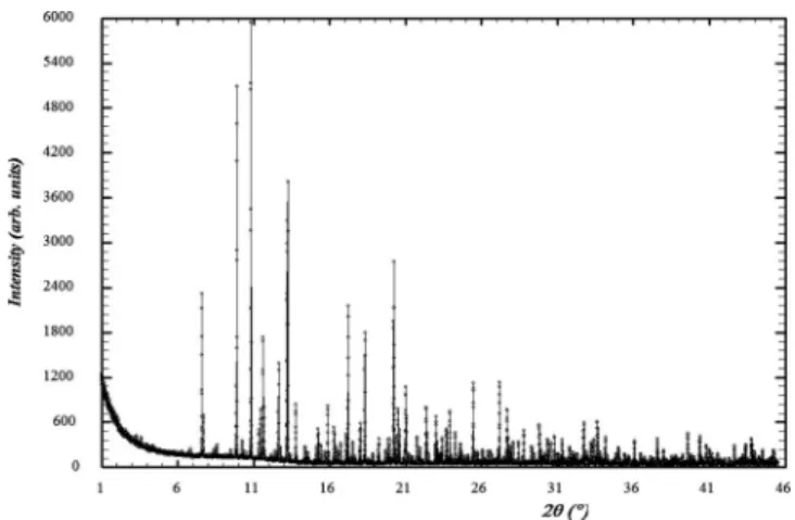
prediction of the packing of molecular structure, which could have been used for the structure solution of sample 1, can be found in Day et al., 2005. The powder patterns for the two samples were experimental, supplied in various standard for-mats共Figures 2–4兲. The participants were warned about the possibility of impurity presence as well as systematic zero-point error or even some preferred orientation. The addi-tional details provided are gathered in Table I.
For the new calcium tartrate tetrahydrate form, a CIF provided the tartrate molecular formula as established from two previous single-crystal structure determinations 共Haw-thorne et al., 1982; Boese and Heinemann, 1993兲. For the lanthanum tungstate, the formula and cell were provided ac-cording to the published literature mentioning composition variations. If the cell parameters given for sample 1 could allow directly for a Pawley共1981兲 or Le Bail 共2005兲 fit, this was not the case for sample 2. Moreover, the space groups were not defined, either P1 or P-1 for sample 1, whereas the list of possibilities was longer for sample 2. Those looking
Figure 2. X-ray powder pattern for sample 1共calcium tartrate tetrahydrate兲. The second reflection at low angle culminates at⬃116 000 counts.
Figure 3. Synchrotron powder pattern for sample 2共lanthanum tungstate兲. Figure 1.共Color online兲 Data downloads and solutions received along the
12-week duration共2008兲 of the round robin.
accurately at the publications mentioned in the ICDD-JCPDS cards could have found more details, but they were contra-dictory: “possible space groups P63/mmc, P63nmc, and
P-62c,⬙ favouring the centrosymmetrical group 共negative
re-sult at the generation of second harmonic兲” 共Yanovskii and Voronkova, 1975兲; “hexagonal with R-62c space group” 共Kovalevsky et al., 1999兲. Table II provides the sDOF and rDOF corresponding to the various samples of the SDPDRR-1, 2, and 3. However, they are estimated as if the final structure and space group were known by the partici-pants, which was not always the case.
IV. CRYSTAL STRUCTURES AS SOLVED BY THE ORGANIZERS
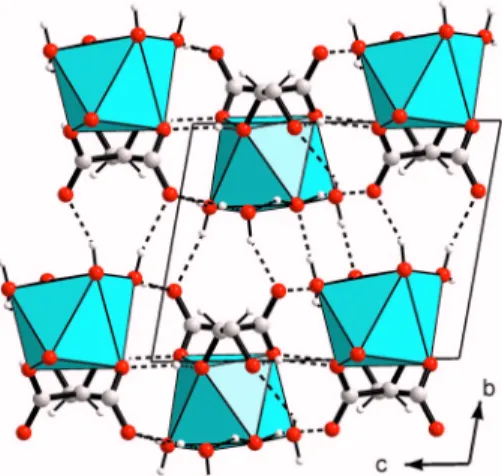
A. Sample 1: Calcium tartrate tetrahydrate
Large crystals共100 to 200 m兲 were extracted from rat kidney. The chemical analysis suggested a hydrated calcium tartrate. The crystal structure determination revealed a new tetrahydrated form. Crystals were found in massive inter-growths and first resisted to the characterization attempts due to the difficulties to separate a real single crystal. Indexing in a triclinic cell was realized from powder diffraction data by
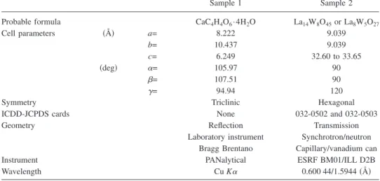
TABLE I. Data provided for samples 1 and 2.
Sample 1 Sample 2
Probable formula CaC4H4O6· 4H2O La14W8O45or La8W5O27
Cell parameters 共Å兲 a= 8.222 9.039 b= 10.437 9.039 c= 6.249 32.60 to 33.65 共deg兲 ␣= 105.97 90 = 107.51 90 ␥= 94.94 120
Symmetry Triclinic Hexagonal
ICDD-JCPDS cards None 032-0502 and 032-0503
Geometry Reflection Transmission
Laboratory instrument Synchrotron/neutron Bragg Brentano Capillary/vanadium can
Instrument PANalytical ESRF BM01/ILL D2B
Wavelength Cu K␣ 0.600 44/1.5944共Å兲
TABLE II. Supposing the space group and structure known, minimal number of degrees of freedom at the structure solution共sDOF兲 stage from X-ray data for various methods, and total number of refinable 共rDOF兲 atomic coordinates共non-H atoms兲. DM: direct methods, Patt: Patterson method, DS: direct space method, References: SDPDRR-1, samples 1共Zhu et al., 1999兲 and 2 共Clegg and Teat, 2000兲; SDPDRR-2 samples 1 共Adil et al., 2007兲 and 2 共Le Bail, 2003兲; and SDPDRR-3 samples 1 共Le Bail et al., 2009兲 and 2 共Chambrier et
al., 2009兲.
SDPDRR-1 SDPDRR-2 SDPDRR-3
Sample 1 Sample 1 Sample 1
Sample 2 Sample 2 Sample 2
Chemical formula 关Co共NH3兲5CO3兴NO3· H2O Al2F10关C6N4H20兴 CaC4H4O6· 4H2O
C22H24N2O8HCl Sr5V3共F/O/H2O兲22 La18W10O57 Space group P21 P2/c P-1 P212121 P21/c P-62c Z 2 2 2 4 4 2 Total independent 15 12 15 non-H 33 30 20 Sites to find by DM/Patt ⬃10 ⬃10 ⬃10 to 15 ⬃20 ⬃15 6W + 4La sDOF DS: 6⫻3+3=21 6⫻3=18 6 + 5⫻3=21 mol+ atoms 6 + 3 = 9 6⫻8=48 10⫻3=30 rDOF 44 30 45 99 90 43
Data quality Low Low Low
using theMCMAILLEsoftware共Le Bail, 2004兲. The structure was solved from the powder data by direct space method using theESPOIR software共Le Bail, 2001兲 applied to inten-sities extracted by the Le Bail共2005兲 method in space group
P-1. The tartrate molecule was rotated and translated
to-gether with the calcium and remaining water oxygen atoms up to find an optimum by a Monte Carlo process. From these convincing results, more efforts were taken with the data collected from a selected “single crystal,” producing a final refinement of much higher quality than from the powder data; this in spite of the relatively high R factor due to peak overlapping from several intergrown domains共Figure 5兲 共Le Bail et al., 2009兲.
B. Sample 2: Lanthanum tungstate
In a previous study 共Yanovskii and Voronkova, 1975兲 共from single-crystal data though the structure determination was not completed because it was not possible to obtain good crystals兲, the commonest six-layered polytype was said to belong to the space group P63/mmc 共no piezoeffect de-tected兲 with cell parameters a=9.04共1兲 Å and c=32.60 to 33.65 Å depending on the composition of the crystal. This cell was confirmed here by a satisfying whole powder pattern fit by using the Le Bail method through theFULLPROF soft-ware 共Rodriguez-Carvajal, 1993兲. The extracted intensities from the synchrotron powder pattern were then used for at-tempting the structure solution by direct space methods as embedded in theESPOIRsoftware, searching for the heavy W and La independent atoms by a Monte Carlo process. Noth-ing better than Rp⬎35% could be obtained during various tests in the P63/mmc or P63mc space groups. Direct or Patterson methods failed as well to provide a satisfying start-ing model. Then, instead of trystart-ing directly the other possible space groups 共P-62c, P-31c, and P31c兲, the search for a solution was made in the c/6 subcell in spite of the fact that very intense reflections had to be excluded共scaling the most intense 206 at I = 100, the 207 is at I = 13, and the 217 is at
I = 14兲. Trying various space groups without extinction, a
promising model leading to Rp= 22% on 220 remaining peaks was finally obtained from theESPOIR software in the
P-62m space group, corresponding to a La/W=2 ratio
共La2WO6 formula兲. No extension of that model in the large
cell could be obtained in the P63/mmc or P63mc space
groups. Then the other space groups compatible with the
hh-2hl, l = 2n reflection condition were examined 共P-62c, P-31c, and P31c兲. The small initial model could be finally
extended in the large cell by using the acentric space groups, for instance with four La and five W independent atom sites in the general or special positions of P-62c. Introducing these atomic coordinates into a Rietveld 共1969兲 refinement led then to RB= 19.7% and RF= 11.1% when the thermal pa-rameters were refined共most having negative values because of the absence of absorption correction at this stage兲. From a Fourier difference map, an additional W atom site was de-tected as well as all the oxygen atoms in ten independent sites. Further refinements suggested that this new W site had to be half occupied, leading to the La18W10O57formula with
Z = 2. A part of the W atoms are found in octahedral
coordi-nation but the majority of them are in an unusual trigonal prismatic coordination共Figure 6兲. This unusual trigonal pris-matic coordination was previously observed for the WO6 group in the X-ray studies of Pr3WO6Cl3共Polyanskaya et al., 1969兲 and La3WO6Cl3共Brixner et al., 1982兲, the latter struc-ture being then confirmed from neutron powder diffraction data共Parise and Brixner, 1983兲. Tests in order to see if re-ducing the symmetry would allow the half occupied W site to become fully ordered were made in P31c and in various subgroups of P-62c and P31c共Ama2, Cc兲 with no convinc-ing result: the number of atomic coordinates to be refined becoming prohibitive共Chambrier et al., 2009兲.
V. CRYSTAL STRUCTURES AS SOLVED BY THE PARTICIPANTS
The results for the structure solution step are summa-rized in Table III共complete reports are available as supple-mentary materials共http://www.cristal.org/SDPDRR3/兲. From the 175 potential participants having downloaded the data, nine sent a total of 12 solutions共8 and 4 for samples 1 and 2, respectively兲. They used seven different computer programs for structure solution. This third round robin received more results compared to first and second round robins and for the first time, contributions were also obtained from nonsoftware developers. This implies that solving structures from powder diffraction data is becoming more accessible to nonexperts compared to previous round robins.
The person-time required for solving from powder data a crystal structure can be almost as short as for single-crystal data, providing that the correct solution is recognized at the first try. Computing time is in general longer for the pro-grams working in direct space methods.
Figure 5. 共Color online兲 Calcium tartrate tetrahydrated crystal structure drawing.
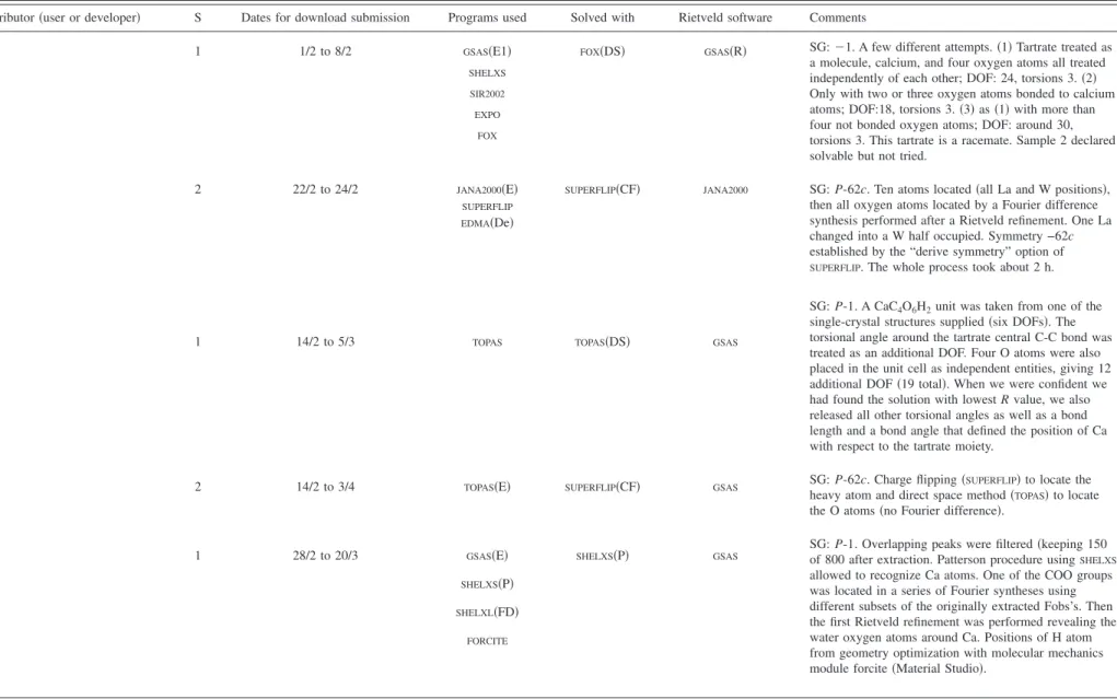
TABLE III. Summary of the results from the SDPDRR-3 contributors for the two samples共S兲. Note that C1 to C9: contributor number; CF: charge flipping; D: developer/conceiver; De: density analysis; DS: direct space; E: extraction of intensities; E1: extraction of intensities by the Le Bail method; E2: extraction of intensities by the Pawley method; FD: Fourier difference; P: Patterson; R: restraints on bond lengths and angles used at the Rietveld refinement stage; S: Sample; and U: User. Dates for download submission in 2008 are given as day/month.
Contributor共user or developer兲 S Dates for download submission Programs used Solved with Rietveld software Comments
C1 1 1/2 to 8/2 GSAS共E1兲 FOX共DS兲 GSAS共R兲 SG:⫺1. A few different attempts. 共1兲 Tartrate treated as
a molecule, calcium, and four oxygen atoms all treated independently of each other; DOF: 24, torsions 3.共2兲 Only with two or three oxygen atoms bonded to calcium atoms; DOF:18, torsions 3.共3兲 as 共1兲 with more than four not bonded oxygen atoms; DOF: around 30, torsions 3. This tartrate is a racemate. Sample 2 declared solvable but not tried.
共U兲 SHELXS
SIR2002 EXPO
FOX
C2 2 22/2 to 24/2 JANA2000共E兲 SUPERFLIP共CF兲 JANA2000 SG: P-62c. Ten atoms located共all La and W positions兲, then all oxygen atoms located by a Fourier difference synthesis performed after a Rietveld refinement. One La changed into a W half occupied. Symmetry −62c established by the “derive symmetry” option of
SUPERFLIP. The whole process took about 2 h.
共D兲 SUPERFLIP
EDMA共De兲
C3 1 14/2 to 5/3 TOPAS TOPAS共DS兲 GSAS
SG: P-1. A CaC4O6H2unit was taken from one of the
single-crystal structures supplied共six DOFs兲. The torsional angle around the tartrate central C-C bond was treated as an additional DOF. Four O atoms were also placed in the unit cell as independent entities, giving 12 additional DOF共19 total兲. When we were confident we had found the solution with lowest R value, we also released all other torsional angles as well as a bond length and a bond angle that defined the position of Ca with respect to the tartrate moiety.
共U兲
2 14/2 to 3/4 TOPAS共E兲 SUPERFLIP共CF兲 GSAS SG: P-62c. Charge flipping共SUPERFLIP兲 to locate the
heavy atom and direct space method共TOPAS兲 to locate
the O atoms共no Fourier difference兲.
C4 1 28/2 to 20/3 GSAS共E兲 SHELXS共P兲 GSAS
SG: P-1. Overlapping peaks were filtered共keeping 150 of 800 after extraction. Patterson procedure usingSHELXS
allowed to recognize Ca atoms. One of the COO groups was located in a series of Fourier syntheses using different subsets of the originally extracted Fobs’s. Then the first Rietveld refinement was performed revealing the water oxygen atoms around Ca. Positions of H atom from geometry optimization with molecular mechanics module forcite共Material Studio兲.
共U兲 SHELXS共P兲 SHELXL共FD兲 FORCITE 259 259 Powder Dif fr. , V ol. 24, No. 3, September 2009 Third structure d etermination b y p owder dif fractometry ...
https://doi.org/10.1154/1.3200881 Downloaded from
https:/www.cambridge.org/core
. University of Basel Library
, on
30 May 2017 at 15:07:18
, subject to the Cambridge Core terms of use, available at
TABLE III. 共Continued.兲
Contributor共user or developer兲 S Dates for download submission Programs used Solved with Rietveld software Comments
C5 1 2/2 to 21/4 FOX FOX共DS兲 GSAS
SG: P-1. Started as Z matrix from Pb-tartrate example. Start with one rigid body tartrate molecule plus five free atoms: 1Ca and 4O. Later on: release 11 torsion angles one by one within window of 10° around starting model angle. Next Z matrix converted to molecule description for finding final solution.
共U兲
2 2/2 to 21/4 FOX FOX共DS兲 GSAS
SG: C2cm. Started with set of La atoms and WO6octahedra.
Fourier map shows missing heavy atoms.FOXmerges atoms when too many atoms have been entered. Sometimes La atoms and W octahedra had to be interchanged. Initially only the X-ray pattern was used to find heavy atom positions. Later on the neutron pattern was added to position O atoms. The finalFOXsolution had 11 La atoms and 5 W octahedra. C6 1 2/2 to 29/4 FULLPROF共E1兲 FOX共DS兲 TOPAS SG: P-1. 1 semirigid molecule 2R, 3R C4H4O6with internal
DOF and restraints as used in FOX. Four rigid molecules H2O. One free atom of Ca. A total of 33 DOFs+ internal DOF
of the C4H4O6molecule.
共D兲 FOX
2 2/2 to 29/4 FULLPROF共E1兲 FOX共DS兲 TOPAS SG: P31c. 6 free atoms of La, 9 free atoms of W, 18 free
atoms of O, 1 octahedron WO6. A total of 105 DOFs. Both
data sets used jointly for structure solution and refinement.
C7 1 7/2 to 30/4 EXPO200X EXPO200X EXPO200X
SG: P-1. Solved by direct method and simulated annealing DM: whole structure共non-H atoms兲 recognized at the third phasing trial. SA: Six independent molecules/atoms: one tartrate, four water molecules, one Ca atom. Total number of DOFs= 38共33 external DOFs and 5 torsion angles兲. Sample 2 declared solvable but probably additional chemical
information is needed. 共D兲
C8 1 4/2 to 30/4 FULLPROF共E1兲 ESPOIR共DS兲 FULLPROF SG: P-1. One CaO8polyhedron, a C4molecule with free
torsion angle and 2O atoms. Best solution with R = 0.205 at test 91.
共U兲 ESPOIR
C9 1 20/4 to 1/5 TOPAS共E2兲 PSSP共DS兲 TOPAS
SG: P1. Based on description of the sample as tartrate共rather than mesotartrate兲, space group P1 was selected. Two independent tartrates, each with Ca tethered in a likely location to two oxygen atoms. eight independent oxygen atoms. The Ca and carboxyl torsions were varied over a limited range relative to planar. So each Ca tartrate was described by three translations, three rotations, and three torsions. Eight independent oxygens have three translations each for a grand total of 42 parameters. Questionable refinement was ascribed to low resolution data. Sample 2 not really tried. 共D兲 PSSP 260 260 Powder Dif fr. , V ol. 24, No. 3, September 2009 Le Bail et al.
https://doi.org/10.1154/1.3200881 Downloaded from
https:/www.cambridge.org/core
. University of Basel Library
, on
30 May 2017 at 15:07:18
, subject to the Cambridge Core terms of use, available at
The fact is that participants may end in multiple solu-tions共P1 or P-1 for sample 1 and P-62c, P31c, or C2cm for sample 2兲. We can only suppose that these differences would have vanished at the Rietveld structure refinement after more efforts and symmetry-checking ultimate stages共Spek, 2003兲, which were not included into the SDPDRR-3 targets and so not always realized共this was not a Rietveld round robin兲. For instance, applying thePLATONsymmetry check to the P31c model suggests that it fits at 91% in P-62c共the disordered W atom in P-62c can be ordered in P31c兲; for the C2cm de-scription, PLATONsuggests the hexagonal symmetry with ␥ = 119.98°; for the P1 tartrate description, PLATONdoes not find the inversion center, saying “no obvious space group change needed.” For these reasons, no metrics will be pre-sented for comparing the structure models with the “known” solutions. Moreover, if the tartrate structure, finally refined from single-crystal data, can be considered as hardly disput-able, the lanthanum tungstate structure could be certainly discussed, and some participants could well have tried to publish their own results sometimes differing a bit from the structure determined by the organizers, and even some mod-ern crystallographers would possibly have applied some four-dimensional algorithm to this disordered and clearly modulated 共but commensurable兲 structure. Anyway, given the low level of success, it appears too early to propose a round robin which would address the accuracy issue, com-paring the models obtained at the solution stage with the final structures. Nevertheless, when available from the par-ticipants, the comparison between the atomic coordinates at the structure solution and at the Rietveld stage shows little differences. For instance in the case of the tartrate 共partici-pant C1, see Table III兲 on a plot of the before/after refine-ment coordinates共Figure 7兲, the largest discrepancy in posi-tion is observed for the H atoms involved in C-H bonds 共other H atoms not located兲. This round robin is limited to give answers to simple questions “can this be done?” or “is it routine?” Moreover, if the minimal model allowing then to refine and complete the structure in the calcium tartrate case is close to 80% of the atomic positions共including Ca, ex-cluding H兲, it is limited to the finding of the heavy atoms 共W, La兲 in the tungstate case from the synchrotron data even if some computer programs are able to locate most of the oxy-gen atoms altogether by combining the synchrotron and the neutron data.
VI. DISCUSSION AND CONCLUSIONS
One should note that for crystallographers, either from powder or single-crystal data, structure solution is realized by expert algorithms embedded in computer programs not by human brains. Human intervention is limited to the choice of the computer program共looking adapted to the problem type兲 and to the preparation of the data and instructions required by the program. The question why 166 of the 175 partici-pants共⬃95%兲 did not sent any result may be answered in a simple way: most of the powder diffraction computer pro-grams for structure solution have not attained the level of automatism of the single-crystal software which beneficiate generally of high-quality three-dimensional intensity data. Very probably, the same problems as those presented in this round robin would have been solved by⬎95% of the par-ticipants from single-crystal data. The monodimensionality of a powder pattern implies that this routine character of a single-crystal approach共unless twinning or other difficulties兲 is lost because the solution is much less easily recognized, consequently to the data overlapping, and even the recording of an optimum data set requires special care. We could first advice to the software developers to include more efficiency and user friendliness in their computer programs than was done up to now. Second the advice to the powder diffraction expert is to increase his training level in crystallography and this would perhaps improve the current poor SDPD success rate. The SDPDRR-3 establishes that when properly applied by capable users, a variety of algorithmic methods is effec-tive for solving structures from powder diffraction data as provided by round-robin organizers. In the previous SDPDRR-1 and SDPDRR-2, software developers were the only contributors. In comparison with previous RR, solving round-robin structures was found to be feasible by users of existing packages, though developers of structure solving packages predominated共some being able to find a solution in a few hours using their own software兲. This round robin does not pretend to the generalization to any SDPD from a sample of two structures, and there is no new scientific knowledge to extract from it. It just shows that more science or more user friendliness has to be included in SDPD software if one ex-pects to see more共complex兲 crystal structures solved by sci-entists moderately trained in crystallography interested in such problems.
Adil, K., Le Bail, A., Dujardin, G., and Maisonneuve, V.共2007兲. “A new 1D hybrid fluoroaluminate templated by an original tetramine,” Polyhedron
26, 2493–2497.
Allen, F. H.共2002兲. “The Cambridge structural database: A quarter of a million crystal structures and rising,” Acta Crystallogr., Sect. B: Struct. Sci. 58, 380–388.
Altomare, A., Caliandro, R., Camalli, M., Cuocci, C., Giacovazzo, C., Mo-literni, A. G. G., and Rizzi, R.共2004兲. “Automatic structure determina-tion from powder data withEXPO2004,” J. Appl. Crystallogr. 37, 1025– 1028.
Andreev, Y. G., Lightfoot, P., and Bruce, P. G.共1997兲. “A general Monte Carlo approach to structure solution from powder-diffraction data: Ap-plication to poly共ethylene oxide兲3: LiN共SO2CF3兲2,” J. Appl. Crystallogr.
30, 294–305.
Baerlocher, Ch., McCusker, L., and Palatinus, L.共2007兲. “Charge flipping combined with histogram matching to solve complex crystal structures from powder diffraction data,” Z. Kristallogr. 222, 47–53.
Balzar, D., Audebrand, N., Daymond, M. R., Fitch, A., Hewat, A., Lang-ford, J. I., Le Bail, A., Louër, D., Masson, O., McCowan, C. N., Popa, N. C., Stephens, P. W., and Toby, B. H. 共2004兲. “Size-strain line-broadening analysis of the ceria round-robin sample,” J. Appl. Crystal-Figure 7.共Color online兲 Projection along the c axis of the superposition of
the tartrate structure as obtained before and after Rietveld refinement 共con-tributor C1兲.
logr. 37, 911–924.
Belsky, A., Hellenbrandt, M., Karen, V. L., and Luksch, P.共2002兲. “New developments in the inorganic crystal structure database共ICSD兲: Acces-sibility in support of materials research and design,” Acta Crystallogr., Sect. B: Struct. Sci. 58, 364–369.
Bergmann, J., Le Bail, A., Shirley, R., and Zlokazov, V.共2004兲. “Renewed interest in powder diffraction data indexing,” Z. Kristallogr. 219, 783– 790.
Boese, R. and Heinemann, O.共1993兲. “Crystal-structure of calcium tartrate tetrahydrate, C4H4O6Ca共H2O兲4,” Z. Kristallogr. 205, 348–349.
Brenner, S., McCusker, L. B., and Baerlocher, C.共2002兲. “The application of structure envelopes in structure determination from powder diffrac-tion data,” J. Appl. Crystallogr. 35, 243–252.
Brixner, L. H., Chen, H. Y., and Foris, C. M.共1982兲. “Structure and lumi-nescence of some rare-earth halotungstates of the type Ln3WO6Cl3,” J. Solid State Chem. 44, 99–107.
Brodski, V., Peschar, R., and Schenk, H.共2005兲. “ORGANA: A program pack-age for structure determination from powder diffraction data by direct-space methods,” J. Appl. Crystallogr. 38, 688–693.
Chambrier, M. H., Le Bail, A., Kodjikian, S., Suard, E., and Goutenoire, F. 共2009兲. “Structure determination of La18W10O57,” Inorg. Chem. 48,
6566–6572.
Clearfield, A., Reibenspies, J. H., and Bhuvanesh, N.共2008兲. Principles and
Applications of Powder Diffraction共Blackwell, Oxford兲.
Clegg, W. and Teat, S. J.共2000兲. “Tetracycline hydrochloride: A synchrotron microcrystal study,” Acta Crystallogr., Sect. C: Cryst. Struct. Commun.
56, 1343–1345.
Coelho, A. A.共2000兲. “Whole-profile structure solution from powder dif-fraction data using simulated annealing,” J. Appl. Crystallogr. 33, 899– 908.
David, W. I. F.共2007兲. “CPD Chairman’s Message,” Commission on Pow-der Diffraction IUCr Newsletter, Vol. 35, p. 2, 共http://www.iucr.org/ resources/commissions/powder-diffraction/newsletter兲.
David, W. I. F., Shankland, K., McCusker, L. L., and Baerlocher, Ch. 共2002兲. Structure Determination from Powder Diffraction Data 共Oxford University Press, Oxford兲.
David, W. I. F., Shankland, K., van de Streek, J., Pidcock, E., Motherwell, W. D. M., and Cole, J. C.共2006兲. “DASH: A program for crystal structure determination from powder diffraction data,” J. Appl. Crystallogr. 39, 910–915.
Day, G. M., Motherwell, W. D. S., Ammon, H. L., Boerrigter, S. X. M., Della Valle, R. G., Venuti, E., Dzyabchenko, A., Dunitz, J. D., Sch-weizer, B. P., van Eijck, B. P., Erk, P., Facelli, J. C., Bazterra, V. E., Ferraro, M. B., Hofmann, D. W. M., Leusen, F. J. J., Liang, C., Pan-telides, C. C., Karamertzanis, P. G., Price, S. L., Lewis, T. C., Nowell, H., Torrisi, A., Scheraga, H. A., Arnautova, Y. A., Schmidt, M. U., and Verwer, P.共2005兲. “A third blind test of crystal structure prediction,” Acta Crystallogr., Sect. B: Struct. Sci. 61, 511–527.
Dinnebier, R. E. and Billinge, S. J. L.共2008兲. Powder Diffraction—Theory
and Practice共RSC, Cambridge兲.
Engel, G. E., Wilke, S., Konig, O., Harris, K. D. M., and Leusen, F. J. J. 共1999兲. “POWDERSOLVE: A complete package for crystal structure solution
from powder diffraction patterns,” J. Appl. Crystallogr. 32, 1169–1179. Favre-Nicolin, V. and Cerny, R.共2002兲. “FOX: Free objects for crystallogra-phy: A modular approach to ab initio structure determination from pow-der diffraction,” J. Appl. Crystallogr. 35, 734–743.
Feng, Z. J. and Dong, C.共2007兲. “GEST: A program for structure determina-tion from powder diffracdetermina-tion data using a genetic algorithm,” J. Appl. Crystallogr. 40, 583–588.
Harris, K. D. M., Johnston, R. L., and Kariuki, B. M.共1998兲. “The genetic algorithm: Foundations and applications in structure solution from pow-der diffraction data,” Acta Crystallogr., Sect. A: Found. Crystallogr. 54, 632–645.
Harris, K. D. M., Tremayne, M., Lightfoot, P., and Bruce, P. G.共1994兲. “Crystal-structure determination from powder diffraction data by Monte-Carlo methods,” J. Am. Chem. Soc. 116, 3543–3547.
Hawthorne, F. C., Borys, I., and Ferguson, R. B.共1982兲. “Structure of cal-cium tartrate tetrahydrate,” Acta Crystallogr., Sect. B: Struct. Crystal-logr. Cryst. Chem. 38, 2461–2463.
Hill, R. J. and Cranswick, L. M. D.共1994兲. “International Union of
Crys-tallography. Commission on Powder Diffraction. Rietveld refinement round robin. II. Analysis of monoclinic ZrO2,” J. Appl. Crystallogr. 27,
802–844.
Jansen, J., Peschar, R., and Schenk, H. 共1993兲. “Application of direct-methods to powder data—A weighting scheme for intensities in the op-timal symbolic addition programSIMPEL88,” Z. Kristallogr. 206, 33–43.
Kovalevsky, A. V., Kharton, V. V., and Naumovich, E. N.共1999兲. “Oxygen ion conductivity of hexagonal La2W1.25O6.75,” Mater. Lett. 38, 300–304.
Le Bail, A.共2001兲. “ESPOIR: A program for solving structures by Monte Carlo analysis of powder diffraction data,” Mater. Sci. Forum 378–381, 65–70.
Le Bail, A.共2003兲. COD1000005—http://www.crystallography.net/ Le Bail, A.共2004兲. “Monte Carlo indexing withMCMAILLE,” Powder Diffr.
19, 249–254.
Le Bail, A.共2005兲. “Whole powder pattern decomposition methods and applications: A retrospection,” Powder Diffr. 20, 316–326.
Le Bail, A., Bazin, D., Daudon, M., Brochot, A., Robbez-Masson, V., and Maisonneuve, V.共2009兲. “Racemic calcium tartrate tetrahydrate 共form-II兲 in rat urinary stones,” Acta Crystallogr., Sect. B: Struct. Sci. 65, 350–354.
Le Bail, A. and Cranswick, L. M. D.共2001兲. “Revisiting the 1998 SDPD round robin,” Commission on Powder Diffraction IUCr Newsletter, Vol. 25, pp. 7–9 共http://www.iucr.org/resources/commissions/powder-diffraction/newsletter兲.
Le Bail, A. and Cranswick, L. M. D.共2003兲. “SDPD Round Robin 2002 results,” Commission on Powder Diffraction IUCr Newsletter, Vol. 29, pp. 31–34 共http://www.iucr.org/resources/commissions/powder-diffraction/newsletter兲.
Madsen, I. C., Scarlett, N. V. Y., Cranswick, L. M. D., and Lwin, T.共2001兲. “Outcomes of the International Union of Crystallography Commission on Powder diffraction round robin on quantitative phase analysis: Samples 1a to 1h,” J. Appl. Crystallogr. 34, 409–426.
McCusker, L. B., Von Dreele, R. B., Cox, D. E., Louër, D., and Scardi, P. 共1999兲. “Rietveld refinement guidelines,” J. Appl. Crystallogr. 32, 36– 50.
Oszlányi, G. and Sütő, A.共2004兲. “Ab initio structure solution by charge flipping,” Acta Crystallogr., Sect. A: Found. Crystallogr. 60, 134–141. Palatinus, L. and Chapuis, G.共2007兲. “SUPERFLIP: A computer program for
the solution of crystal structures by charge flipping in arbitrary dimen-sions,” J. Appl. Crystallogr. 40, 786–790.
Parise, J. B. and Brixner, L. H.共1983兲. “Refinement of the structure of trilanthanum trichlorohexaoxotungstate, La3WO6Cl3, from neutron
pow-der diffraction data,” Acta Crystallogr., Sect. C: Cryst. Struct. Commun.
39, 1326–1328.
Pawley, G. S.共1981兲. “Unit-cell refinement from powder diffraction scans,” J. Appl. Crystallogr. 14, 357–361.
Pecharsky, V. K. and Zavalij, P. Y.共2003兲. Fundamentals of Powder
Dif-fraction and Structural Characterization of Materials共Springer, New
York兲.
Polyanskaya, T. M., Borisov, S. V., and Belov, N. V.共1969兲. “Crystal struc-ture of Pr3WO6Cl3,” Dokl. Akad. Nauk SSSR 187, 1043–1046.
Putz, H., Schon, J. C., and Jansen, M.共1999兲. “Combined method for ab
initio structure solution from powder diffraction data,” J. Appl.
Crystal-logr. 32, 864–870.
Rietveld, H. M.共1969兲. “A profile refinement method for nuclear and mag-netic structures,” J. Appl. Crystallogr. 2, 65–71.
Rius, J.共2004兲. “Advances and some recent applications of the origin-free modulus sum function,” Z. Kristallogr. 219, 826–832.
Rodriguez-Carvajal, J.共1993兲. “Recent advances in magnetic-structure de-termination by neutron powder diffraction,” Physica B 192, 55–69. Spek, A. L.共2003兲. “Single-crystal structure validation with the program
PLATON,” J. Appl. Crystallogr. 36, 7–13.
Stephens, P. W. and Huq, A.共2002兲. “PSSP: An open source powder structure solution program for direct space simulated annealing,” Trans. Am. Crystallogr. Assoc. 37, 127–144.
Yanovskii, V. K. and Voronkova, V. I.共1975兲. “Crystallography and proper-ties of lanthanum oxytungstate, La2WO6,” Sov. Phys. Crystallogr. 20,
354–355.
Zhu, J. H., Wu, H. X., and Le Bail, A. 共1999兲. “Structure of 关Co共NH3兲5CO3兴NO3. H2O,” Solid State Sci. 1, 55–62.