© 1993 Oxford University Press
Enhanced positive selection of a transgenic
TCR by a restriction element that does not
permit negative selection
Pamela S. Ohashl, Rolf M. Zlnkernagel
1, Immanuel Leuscher
2,
Hans Hengartner
1, and Hanspeter Plrcher
1Ontario Cancer Institute, Department of Medical Biophysics, University of Toronto, 500 Sherbourne Street, Toronto, Ontario, Canada M4X 1K9
'Institute of Experimental Immunology, Department of Pathology, University of Zurich, Schmelzberstrasse 12, 8091 Zurich, Switzerland
2Ludwig Institute for Cancer Research, Lausanne Branch, 1066 Epalinges, Switzerland
Key words: bm13 bm14, mutant MHC molecules, ontogeny, thymocyte, tolerance
Abstract
Very little is known about the conformational properties of the MHC molecules that are able to signal positive selection of a given TCR. To try to understand these parameters and to determine whether these requirements are shared with Interactions during negative selection and antigen recognition, we have studied selection and antigen recognition of a transgenic TCR (specific for lymphocytlc chorlomenlngitls virus glycoproteln and H-2D") In the context of two D" mutants,
H.2bmi3 a n d H-21"1114. The data showed that the transgenic TCR was not positively selected by the H.2bmi4 haplotype but, Interestingly, enhanced positive selection was seen In H-2bm13 mice. The
transgenic TCR could not be negatively selected In H-2"11113 animals persistently Infected with the
virus (neonatal virus carrier mice), nor could the transgenic TCR be activated by H^*"1 3 Infected
cells in vivo or in vitro. These experiments show that although a TCR may be selected by a mutant MHC molecule, the corresponding viral antigen cannot be recognized In the context of the mutant MHC molecule, as judged by both negative selection and T cell reactivity In vivo and In vitro. The 'enhanced' positive selection occurring In the context of D""113 suggests that a
different conformation of the MHC molecule Is able to select the same TCR and also that various TCR - llgand avidities may permit positive selection.
Introduction
The TCR expressed on T lymphocytes is able to transmit signals for three different developmental or functional events: positive selection, tolerance induction, and activation by antigen. The stringent requirements for TCR - ligand interaction have been well characterized for antigen recognition indicating that a defined peptide is presented by a defined MHC molecule with limited variability (for a review see 1). In several systems certain amino acid residues of the peptide antigen (2,3) or of the MHC molecule ( 4 - 7 ) have been shown to be essential for T cell recognition (8 and reviewed in 9). In general, TCR recognition of a given peptide presented by a particular MHC is relatively strict.
Several studies have dealt with the interactions that are able to signal negative selection. Studies using superantigens such as Mis and Staphylococcal enterotoxin B (SEB) have shown that a 'gradient' of the clonal deletion may be defined depending on the presenting haplotype (10-13). Using a TCR transgenic
model specific for the alloantigen H-2Ld, Sha etal. (14) have
shown that a single amino acid change in the Kb molecule,
which is required for positive selection, resulted in the deletion of T cells. In general, however, it is difficult to judge the flexibility of the interacting ligands during negative selection in classic antigen-MHC-restricted recognition. Recently, Pircheref al. (15) showed that a variant peptide that is able to induce clonal deletion of T cells expressing a transgenic receptor is not able to activate the transgenic T cells. This suggests that antigen recognition by the TCR is demanding higher avidity interactions than tolerance induction.
Other studies have evaluated the TCR - ligand interactions during positive selection and suggest that a peptide may be involved in positive selection (14,16-19). Using TCR transgenic mice restricted to D*5, Jacobs etal. (18) showed that positive
selection does not occur in the presence of D*3"113 and D1*"14. Correspondence to: P. S. Ohashi
Sha etal. (14) demonstrated that a transgenic Kb-restricted
receptor cannot be positively selected in three of the four K6"1
mutants that could be analysed, suggesting that the interaction between the TCR and the selecting complex is relatively rigid. Based on these experiments, the precise fit, flexibility, and conformational requirements (i.e. the avidity) of a given TCR-MHC-peptide interaction during positive selection, negative selection, and antigen activation is difficult to define at present. Therefore a lymphocytic choriomeningitis virus (LCMV)-C^-specific transgenic TCR for positive and negative selection
in vivo, as well as for antigen recognition in vivo and in vitro, in
mice expressing mutant CP molecules has been evaluated.
Methods
Animals
Inbred mice were purchased from the Institut fur Zuchthygiene, Tierspital, University of Zurich, Switzerland. 36.0-^-2*™™ (H.2bmi3) a n d B.eC-H^1"1114 ( H ^1 4) animals (20,21) were
kindly provided by Dr Cornelis J. Melief. The TCR transgenic mouse line 327 (22) was crossed into the H-2txn13 and H^5"114
strains using the following strategy. TCR transgenic H-2Wd
animals were developed by mating the TCR transgenic H-2*3*
animals with BALB/c mice (H-2dW). It has been previously
demonstrated that this transgenic TCR could not be positively selected in a predominantly BALB/c background (23). TCR transgenic positive H-2b/d animals were then bred with
homozygous H^6"113 or H-2bm14 animals. Offspring from these
breeding pairs were either H-2b/bm or H-2d/bm that were positive
or negative for the transgene. The MHC haplotype was determined by staining peripheral blood lymphocytes with an H-^-specific antibody (24).
Flow cytometry analysis
Cells (1 xiO6) were incubated with various mAbs in 100 ^1
volume balanced salt solution (BSS); 2% fetal calf serum (FCS); 0 . 1 % sodium azide for 30 min at 4°C. The cells were washed and incubated with fluoresceinated antibodies. Analysis of blood lymphocytes was performed after lysis of red blood cells. Viable cells (10,000) were analysed on an EPICS profile analyser for single parameter analysis and 20,000 viable cells were analysed in 2-3-colour histograms. The mAbs used were KT3 (rat anti-CD3; 25), KJ16 (rat anti-V^.1, 8.2; 26), B20.1 (rat anti-V^; 27), and anti-CD4 conjugated with phycoerythrin (PE) Dickinson, Belgium), FITC conjugated anti-CD8 (Becton-Dickinson). For 3-cok>ur analysis, bbtinylated anti-CD8 was used together with streptavidin-RED613 (Gibco BRL, Basel, Switzerland). The percentages given for 2-colour analysis were obtained by defining quadrants based on the staining of thymocytes from negative littermate controls. The figures have been cropped to show the relevant data.
Cytotoxb assays
Animals were primed with 200 p.f.u. of the WE strain of LCMV (obtained from Dr F. Lehmann-Gruber Hamburg, Germany).
Eight days later the spleen was removed and the effector cells were incubated, together with target cells, at a ratio of 70:1, 23:1, 8:1, and 3:1. Target cells were either fibroblast lines MC57G
(KbCF), B10.A(5R) (WD*), or B10.HTG (I^D"), or concanavalin
A (Con A) induced spleen cell blasts. The blast cells were
prepared by culturing spleen cells with 3 /ig/ml Con A (Pharmacia, Uppsala, Sweden) for 48 h and purification over Ficoll gradients. Targets were incubated with 51Cr, with or
without peptides, for 2 h. The peptide antigen used in these experiments was IKAVYNFATCG. The effector cells were incubated with target cells for 4 - 5 h and the supernatant was removed and counted. Per cent specific release was calculated as described previously (28).
Proliferation assays
Lymph node cells (i x 10s) from TCR transgenic H-2b mice and
from negative littermate controls were incubated with 2 x 10⁴
LCMV-infected macrophages or uninfected macrophages from H-213 and H-S*"113 mice. Stimulator macrophages were irradiated
with 2000 rad. After 48 h [3H]thymidine was added overnight.
The wells were harvested and incorporated radioactivity was counted. Data show the average counts from triplicate samples.
Generation of virus carrier animals
Newborn mice from ( H - ^ transgenic x H^6"1) F, or control
H-2b transgenic mice were injected with 3 x 10s p.f.u. of LCMV
i.p. Mice (3- to 4-week-old) were sacrificed and thymocytes were analysed by flow cytometry. Mice were confirmed to be virus carriers by injection of 30 pi of their blood into the footpad of mice. Footpad swelling was seen 8 - 9 days after challenge with the blood, indicating that the virus could be transferred from the car-rier animals.
Results
H-2CP is required for positive selection of the transgenic TCR
TCR transgenic mice have been generated from a cytotoxic T cell clone specific for the LCMV glycoprotein (GP) (amino acids 32-42) presented in the context of Db (22). To formally
demonstrate that the TCR transgenic receptor requires the D*> molecule for positive selection, H-26 TCR transgenic mice were
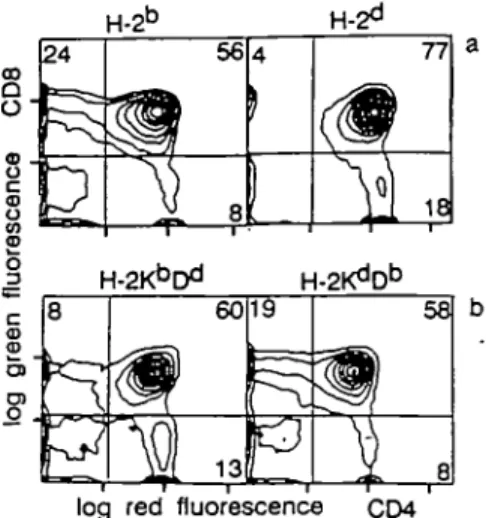
bred with B10.HTG ((^D") and B10.A(5R) (WD*) mice. Positive selection of class I restricted transgenic TCRs has previously been characterized by a skewing of thymocytes to the CD8 lineage (22,29,30). Thymocytes from TCR transgenic B10.HTG and B10.A(5R) animals were stained with CD4 and CD8 and compared with TCR transgenic animals in the selecting H-2b
(19.0% CD8+ ± 2.4, n = 7) and non-selecting H-26
back-ground (6.0% CD8+ ± 1.4, n = 4) (Fig. 1). Prominent skewing
to the CD8 lineage was seen only in the presence of D" (24.5% CD8+ ± 0.7, n = 2). Further analysis has shown that the
transgenic a and /3 TCR are present at high levels on CD8+
cells only in animals expressing H ^ and not H-26 (31,32).
Taken together, these data show that positive selection of the transgenic TCR occurs only in the presence of Db.
The effects of changes in the groove of the MHC or TCR - MHC interaction during positive selection, negative selection, and antigen recognition was examined using two Db mutant mouse
strains called BG.C-H-^1 3 (H-2*m13; (^D*™13) and B e . C - H - ^1 4
(H-2bm14; Kt'D1*1'14) (20). H-2bm13 has three amino acid
substitu-tions on the 0-pleated sheet of the D" class I molecule (residues 114 Leu - Glu, 116 Phe - Tyr, 119 Glu - Asp), while H^*"1 4 has
a single amino acid change on the or-helix of Db (residue 70
H-2D 24 -m» . 8 * \ 56
}L 8
60W 13
4 | 9 -> 77VJ 18
,
2KdDb 58PI
V »
LITTERMATE b/bm13-T d/bm13-T§
O8-i
ID o OT £ O 3log red fluorescence CD4
Fig. 1. Positive selection of the transgentc TCR by CP. Thymocytes from
TCR transgenic mice (KdCfi, BALB/c origin) bred with C57BU6 (t^D6), B10.A(5RKKbDd), and B10.HTG (KdCP)] were analysed using mAbs
specific for CD4 and CD8. At least two animals were analysed in each haplotype and a representative analysis is shown. The following percentages were obtained. KbDb: 19.0% CD8+ ± 2.4, 63%
CD4 + CD8+ ± 6.6, 4.6% CD4+ ± 2.5, n •= 7; KdDd: 6.0%
CD8+ ± 1.4, 69.0% CD4 + CD8+ ± 10.9, 15.8% CD4+ ± 5.5, n = 4;
KbDd: 9.5% CD8+ ± 2.2, 54.5% CD4 + CD8+ ± 6.3, 22%
CD4+ ± 5.6, n = 2; KdDb, 24.5% CD8+ ± 0.7, 56.0%
CD4 + CD8+ ± 0.0, 8% CD4+ ± 0.0, n = 2.
changes that have been predicted to alter the peptide binding pocket, as determined by computer modelling (33).
I-I_2iy>mi3 permits positive selection of the transgenic TCR
Positive selection of the transgenic TCR was studied in H^6™13
and H-2tu1114 mice using two criteria: a skewing to CD8+
thymocytes (22,29,30) and the presence of a subpopulation of thymocytes expressing CD3 at intermediate levels which has been previously correlated with positive selection (23,34,35). Because positive selection of the transgenic TCR does not occur in H-26 mice, H-2d/bm mice were examined for the positive
selection of the transgenic TCR. TCR transgene positive H^6*"1
mice were examined as controls to ensure that the D6"1 alleles
did not negatively affect maturation of the TCR transgene positive thymocytes. Thymocytes stained with CD4 and CD8 revealed skewing of the transgenic TCR towards CD8+ thymocytes in
H-2d*m13 mice (32.0% CD8+ ± 3.4%, n = 6) but not H-2d/bm14
mice (7.8% CD8+ ± 1.0%, n = 4) (Fig. 2A and C). An unusual
population of CD4+CD8b cells was often seen in H-2d/bm13 mice,
but how they fit into T cell ontogeny in these animals is not known. CD3 single parameter analysis of thymocytes showed the presence of TCR CDS1"1 peaks in mice carrying the H - ^ allele
or in H-2dybm13, but not the H-2d/bm14 TCR transgenic mice (Fig.
2B and D). Comparable results are also seen when the transgenic Vo- or ^-specific antibodies were used for analysis (data not
shown). These results indicated that positive selection occurred in H-2bm13, but not H ^1 4, mice.
Enhanced positive selection by Z-/-26""3
Interestingly examination of positive selection of the transgenic TCR by D ^1 3 compared with Db consistently showed a more
a
a 5 77 29 62
3 31
log1 red lluorescehco' CD4'
48
^ 9
I" H
Tog fluorescence intensity CD3 LITTERMATE b/bm14-T d/bm14-T
log red fluorescence CD4
log fluorescence intensity CD3
Fig. 2. Positive selection of the transgenic TCR by H - ^1 3 but not H.2bmi4 Thymocytes from H-a5*"1 and H-2d/bm transgenic mice, as well
as nontransgenic littermate controls, were analysed in the H-2bm13 (A
and B) and H - ^1 4 (C and D) mutant phenotypes using mAbs specific
for CD4, CD8 and CD3 (KT3) (25). (B and D) Broken lines represent cells stained with the second stage antibody alone. The vertical bar at the top of the profiles indicates the TCR -CD3 intermediate population and the triangle indicates TCR - CD3W cells. Four to six animals of each type
were analysed and a representative analysis is shown. The following percentages were obtained. KbDbm13: 32.0% CD8+ ± 3.4, 44.3%
CD4 + CD8+ ±10.7, 13.9% CD4+ ± 7.5, n = 6; ^ D6"1 1 4: 7.8%
CD8+ ± 1.0, 60.7% CD4 + CD8+ ±10.1, 14.2% CD4+ ± 7.8, n = 4.
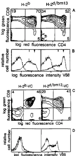
prominent skewing to the CD8+ thymocyte population (Rg. 3A).
In Db animals the CD8+ population in the thymus is 19.0%
( ± 2.4%, n = 7). The CD8+ thymocyte population in D""113
animals is - 3 4 . 0 % ( ± 3.4%, n = 6). In addition, an increased proportion of TCR CD3N-bearing cells was seen, compared with
the number of T cells expressing the TCR CD3 at intermediate densities (Fig. 3B). Examination of TCR transgenic thymocytes in the presence of D*5 and D5"113 by 3-colour cytofluorometric
analysis was also performed to study the TCR CD3 levels during the progression from double positive thymocytes to CD8+
thymocytes. In the CD8+ thymocytes, a high level of CD3
expression was seen, as expected in a mature subpopulation of T cells in both the D13 and D""113 transgenic animals (Fig. 4a).
The CD4b/CD8+ subset contained a majority of CD3W in the D"
transgenic animals, compared with the predominant CDS'* cells in the Dbm'13 mice (Fig. 4b). The CD4+CD8+ population
contained very few CD3W cells in Db mice, while the D13"113
8
o
CD O 19 70 5 34if
5 log red fluorescence CD4log fluorescence intensity V68
H-2b-VC H - 2d/b m 1 3- V C
log red fluorescence CD4
log fluorescence intensity VB8
Fig. 3. Positive and negative selection of the transgenic TCR in mice
carrying the H-2bfn13 alleles. (A) Thymocytes from H-2b TCR transgenic mice or TCR transgenic H-2d™m13 mice were compared using CD4 and CD8 (B) or KJ16 that reacts with the transgenic receptor V«8.1 (26). (C and D) Clonal deletion of the transgenic TCR was examined in H-213 and H.2bmi3 virus carrier mice. (Q Thymocytes from 3- to 4-week-o(d mice were analysed with CD4 and CD8 or (D) KJ16 mAbs. Profiles for (B) and (D) are as described in Fig. 2. A representative animal chosen from the three analysed is shown in the figure.
(Fig. 4c). This suggested that the transgenic TCR may be more efficiently selected to express TCR CD3 at high levels in the
H.2bm13 hap|0type.
To examine whether efficient selection resulted in an increase of peripheral T cells, the number of T lymphocytes in the blood and lymph nodes was investigated. The peripheral blood lymphocytes from TCR transgenic H^6"113 mice had an
increased number of T cells (52 ± 4%, n = 8) compared with transgene negative littermate controls (31 ± 2 % , n = 8) or TCR transgenic H-2b mice (36 ± 4%, n = 8) (Table 1). Lymph
nodes also contained elevated levels of T cells, with a predominant skewing to CD8+ cells in both Db and D6™13
transgenic animals (Table 1). Transgenic H-2bm13 mice, ranging
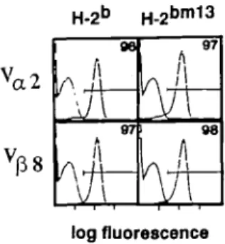
Tn age from 20 days to 6 months; show similar distributions of CD3, CD4, and CD8 positive cells. Analysis of the expression of the transgenic TCR on CD8+ lymph node cells was also
performed (Fig. 5). Two-colour anlaysis, using either B20.1 (V^) and CD8 or KJ16 (V^) and CD8, indicate that virtually all CD8+
CD4 CD3
Fig. 4. Comparison of TCR-CD3 expression from double positive to
single CD8 positive thymocytes in H-2b and H^6"1 1 3 transgenic mice. Thymocytes from H ^ and H-2bm13 were triple stained with CD3, CD4, and CD8 antibodies. The CD4CD8 profile is shown (left), and parts a, b, and c (right) show the levels of CD3 on thymocytes expressing different levels of CD4 and CD8, as shown in the corresponding boxed areas a, b, and c. (a) CO3 expression on CD8+ thymocytes; (b) CD3 expression on C O r e D S * thymocytes; and (c) CD3 expression on CD4+CD8+ thymocytes. The vertical bar indicates thymocytes expressing TCR - CD3 at intermediate levels and the triangle indicates TCR - CD3 high levels. It should be noted that in these animals CD3 expression parallels the expression of the transgenic TCR as assessed by staining with KJ 16 and B20.1 V region-specific antibodies (23 and data not shown). Two animals have been examined and similar results were obtained.
Table 1. Analysis of T cells in the blood and lymph node
Mice Blood Lymph node cells
H-2b H-2',bm13 tg-tg+ tg" tg+ %CD3 32 ± 5 36 ± 4 31 ± 2 52 ± 4 <M>CD3 55 ± 4 59 ± 4 55 ± 3 68 ± 5 %CD4 38 ± 1 11 ± 2 39 ± 2 7 ± 1 %CD8 18 ± 3 57 ± 7 19 ± 5 69 ± 5
cells express the transgenic Va and V^. Taken together these
data suggest that D""113 was able to more efficiently select the
transgenic TCR compared with the original Db molecule. It is
possible that the increase in T cells is also caused by peripheral expansion of T cells.expressing the transgenic receptor.
Cbnal deletion of the transgenic TCR does not occur in H-26""3 mice neonatalty infected with LCMV
Negative selection of LCMV-spectfic transgenic T cells was examined in transgenic H-2bm13 animals. Previous experiments
have shown that injection of newborn TCR transgenic mice with LCMV results in tolerance to the virus by clonal deletion of the CD4+CD8+ double positive TCR transgenic cells in H-2b mice
(22), but not H-2d mice (data not shown). To determine if clonal
deletion of the transgenic TCR also occurs in H-2bm13, H-2? TCR
transgenic mice were bred with H^6"113 mice and offspring
were neonatally infected with LCMV. Thymocytes were examined by 2-colour cytofluorometry at 3 - 4 weeks of age using anfj-CD4, anti-CD8, and V^-specific mAbs. Thymocytes from H-2d/bm13
TCR transgenic carrier mice showed a skewing to the CD8+
mature subset (Fig. 3C). Analysis with V^S-specific antibodies showed that the transgenic TCR was expressed on the majority of H-2d/bm13 thymocytes at intermediate and high levels,
H-2b H-2b m 1 3 96 97
A
u
A "
98/I
log fluorescenceFig. 5. Transgenic TCR expression on CD8+ lymph node cells. Lymph nodes cells from TCR transgenic H - ^ and H-2~" animals were stained with B20.1 and CD8 or KJ16 and CD8. The V.,2 expression (B20.1) or Vfl8 expression (KJ16) on CD8+ T cells is shown in the figure. This figure is representative of three animals.
stimulators or targets
1
- + - + LCMV \ It 9 • \Fig. 6. Transgenic TCR does not recognize LCMV presented in the
context of H-2tm'13. Proliferation assays (top panel) using unprimed
transgenic lymph node cells and either infected ( + ) or uninfected ( - ) macrophages from H-213 or H-2bm13 animals were performed. For lower values the SEM was between 30 and 50 c.p.m., while the positive response had an SEM of 10.4 x 10~3. Lymph node cells from transgene negative littermates were used as negative controls. The thymidine incorporation from these well3 ranged from 300 to 600 c.p.m. (not shown in this figure). This figure is representative of four animals. Cytotoxicity assays (bottom panel) using effector spleen cells from H-Z* TCR transgenic mice ( • ) , H - ^1 3 (A), or C57BU6 mice ( • ) primed with LCMV. Spleen cell blasts from the H-2b or H^""113 mice were used as target cells and labelled with 51Cr with ( • , • ) or without (O, • ) the GP peptide (32-42). Effector to target ratios began at 70:1, with 3-fokJ dilutions.
suggesting that clonal deletion did not occur in
transgenic carrier mice (Fig. 3D). Also, 8 0 - 9 0 % of peripheral CD8+ T cells were found to express both transgenic Vo and Vp
receptors (data not shown). This suggests that either LCMV determinants presented together with H-2bm13 were not
recognized by the transgenic TCR thymocytes and/or that the glycoprotein peptide is not efficiently presented by D6"113 in vivo.
Transgenic T cells are not activated by LCMV-infected DP™13
cells
Experiments were performed to see whether mature peripheral transgenic TCR could recognize LCMV-infected H - ^1 3 cells.
When T cell competent mice are infected intracerebrally with LCMV, they succumb to a CD8 T cell mediated immunopatho-logical disease. However, if mice cannot mount a strong antiviral cytotoxic T lymphocyte (CTL) response at the optimal time after infection, the mice may survive (36 - 38). To determine whether antigen recognition occurs in vivo, TCR transgenic H-2d/bm13
and H-2b/bm13 mice were injected intracerebrally with 2X10⁶
p.f.u. of LCMV. The majority of transgenic mice that expressed the H-2b allele died within 3 - 7 days (5/6, 4 ± 1.7), while none
of the H-2d/bm13 (0/4) transgenic animals died. This indicated that
the transgenic TCR cannot efficiently recognize LCMV-infected H_2bmi ggHg jn VIVO
The ability of the transgenic receptor to recognize LCMV-infected or peptide-coated H-2bm13 cells was also evaluated by
two in vitro assays. Proliferation assays were performed by cutturing TCR transgene positive lymph node cells ( H ^ with uninfected or LCMV-infected macrophages isolated from H-2b
or H^"™13 mice. Lymph node cells from non-transgenic
littermates were used as negative control cells. Proliferation measured by thymidine uptake was seen only when transgenic lymph node cells were stimulated with LCMV-infected H-2b
macrophages (Fig. 6), but not infected H-2bm13 macrophages.
CTL assays, using effector cell populations from TCR transgenic mice, C57BL/6, and H-2bm13 mice primed with LCMV, were
tested on H-2b or H-2bm13 target cells that were either
preincubated for 2 h with or without the peptide GP(32-42). Figure 6 indicates that transgenic effector cells could only recognize target cells expressing the GP peptide plus H-213, and
not the peptide presented in the context of H-2bm13. Taken
together, these experiments demonstrate that the transgenic TCR cannot recognize LCMV-infected H-2bm13 cells.
I-I_2bmi3 an(j peptjde GP(32-42) presentation
Results from the CTL assay showed that H-2bm13 mice
could generate a CTL response specific for the peptide GP(32-42). To determine whether the cytotoxic response was mediated by peptide GP(32-42) binding to K5 or D5™13, the
following assays were performed, based on the assumption that Derestricted effector T cells would cross react with the GP peptide presented on D""113 and vice versa. Cross reactivity of
Derestricted clones on Db mutants expressing the appropriate
antigen has been previously demonstrated (4,5), and therefore an in vivo polyclonal response could possibly generate a cross reactivity D"- or D^^-restricted response (as shown for the Kb
mutants; 7). Effector cells from C57BL/6 (r^D"), B10.A(5R)
(Kf>&), B10.A(4R) (HCD*), and H - ^1 3 (^D5"113) mice primed
with LCMV were tested on a variety of target cells that were incubated with or without the peptide GP(32-42). Results suggest that D5"113 does not present GP(32-42) efficiently
because LCMV-infected H-2bm13 mice did not generate effector
cells that can recognize the peptide on B10.HTG (r^D*3) target
cells bearing the relevant D" molecule (Table 2). In the corollary assay, B10.A(4R) ( r W ) effector cells did not recognize peptide-coated H-2bm13 (r^D6™13) target cells (Table 3). These assays
also showed that the Kb molecule could efficiently present the
Table 2. H-2bm13 does not mount an H-2Db-restricted GP(32-42)-specrfic cytotoxic response
Effectors Targets (% specific release) MC57G (KbDb + 88 71 43 23 52 41 24 10 ) — 5 5 1 3 15 16 7 4 B10.A(5R) (KbD + 88 64 36 17 44 42 25 13 -20 13 7 4 7 12 5 0 B10.HTG (K^D13) + 74 54 24 13 18 21 8 7 16 9 6 3 17 12 8 3 C57BU6 i bm13
Target cells were incubated with ( + ) or without ( - ) GP(32-42). Spontaneous release was below 25%. Effector to target ratios shown are 70:1, 23:1, 8:1, and 3:1.
H-2bm13 targets incubated with GP(32-42) and vice versa (4)
(Tables 2 and 3). These CTL assays suggest that the presentation of GP(32 - 42) by D*™13 is very inefficient in vivo because of the
lack of a measurable CTL response specific for this peptide antigen.
Discussion
In this study we wanted to determine whether one particular TCR was sensitive to small changes in the MHC with respect to positive selection, negative selection, and antigen recognition. Such studies may help to determine the interactions between the TCR and its ligand during these processes and give insights into the mechanism of signalling for the different events. The transgenic LCMV/Db-specific TCR bred into H-2bm13 and H^""114 mice
revealed the following results. The transgenic TCR cannot be positively selected in the presence of Kb or D13"114. D13™13 allows
positive selection of the transgenic TCR, but negative selection does not occur in the presence of LCMV. The transgenic TCR cannot recognize LCMV-infected cells expressing D6"113, as
demonstrated by the intracerebral virus challenge, proliferation, and cytotoxicity assays (Fig. 6). The lack of recognition is consistent with the interpretation that the peptide antigen p32 - 42 is not efficiently presented by D1*"13 and remains below a
density sufficient to induce effector function or recognition in vivo. Interestingly, positive selection of the HP-restricted transgenic TCR is apparently even more efficient in the presence of the Qbmi3 aiieig jnjs w a s demonstrated by an increased skewing
to CD8+ mature thymocytes, an increased proportion of CD3W
thymocytes compared with the CD3 intermediate, and low subpopulations. The increased positive selection was also reflected by increased numbers of T cells in peripheral blood and lymph nodes. This increase in efficiency of positive selection is not caused by new putative selecting peptides introduced from the BALB/c background, because previous work with (C57BL/6 x BALB/cJF, does not show a similar increased positive selec-tion (23). Detailed modelling studies of the D*™13 molecule
suggest that the changes in the £-sheet of the D**1113 molecule
should not directly alter the conformation of the class I molecule and should only affect the peptide binding (33). However, subtle changes may exist and therefore it is possible that the increase in positive selection is caused directly by the shape of the
Table S. does not efficiently present GP(32-42)
Effectors C57BU6 (KbD' B10.A(4R) (Kk[ B10.A(5R) (KbI Targets (% MC57G (Kb + =) 92 86 54 31 D13) 60 34 18 6 3d) 42 43 21 12 specific release) — 19 5 7 1 11 5 2 2 14 11 7 4 bm13 (KbC + 61 75 52 22 5 5 0 0 55 49 28 10 — 8 2 4 5 8 5 1 4 5 1 0 0 Target cells were incubated with (+) or without ( - ) GP(32-42). Spontaneous release was below 30%. Effector to target ratios shown are 70:1, 23:1, 8:1, and 3:1.
MHC molecule itself. Alternatively, an altered conformation of the self peptide - MHC complex may result in an enhanced positive selection of the transgenic TCR.
Others have used similar approaches to analyse TCR - MHC interaction required for positive selection. Using a transgenic TCR specific for the male H-Y antigen, presented in the context of D13,
Jacobs et al. (18) showed that the transgenic TCR could neither be selected by H-2bm13 or H-2bm14 nor could the H-Y antigen be
recognized in the context of the mutant H-2 types. A comparison with our data is interesting because both transgenic receptors are restricted to Db, but in contrast to H-Y the LCMV-specific
receptor is positively selected by D1"113. This suggests that the
selecting MHC complex has different requirements for different receptors. One possibility is that the conformation of a peptide presented by D**"13 fulfills the necessary requirements for
positive selection of the LCMV-specific receptor, t>ut~not the H-Y-specific receptor.
Berg era/. (19) examined positive selection of a cytochrome c-l-E*-restrict.ed transgenic TCR in mice expressing l-E^.Ept,.
This transgenic TCR could weakly recognize cytochrome c in the context of the hybrid I-E molecule, and correspondingly a weak positive selection of the transgenic TCR could also be demonstrated. In studies using a Kb-restricted transgenic TCR, Sha et al. (14) have also shown a reduction in positive selection in K1""8 mice; this mutant has substitutions on the bottom of the peptide binding groove. The authors speculated that the reduced positive selection is caused by an altered selecting peptide conformation, because the TCR should not be able to directly 'see' the changes in K^"18. These data, together with the results from our transgenic D**"13 and D*1 animals, suggest that a gra-dient of binding avidities exists for thymocyte selection. At one end no selection occurs and thymocytes die. A range of affinities appears to exist that results in poor to very efficient positive selec-tion. However, if the interaction between the TCR and the MHC ligand in the thymus is too 'strong', negative selection may occur by clonal deletion. It is possible that the avidity of our TCR towards Qbmi3 m ay QQ Qp tn e border of affinities leading to either positive and/or negative selection. The increase in mature T cells in transgenic D6™13 mice may then be a result of peripheral expansion induced by low level stimulation of the non-deleted transgenic T cells.
The data presented here demonstrate that positive selection of the transgenic TCR is more 'efficient' in D""113 animals compared with the original selecting element ZP. This suggests that the interaction between the TCR and MHC ligand in the thymus during positive selection is somewhat flexible and has different structural requirements than the TCR-peptide-MHC interactions required for negative selection or antigen recognition in vivo. Differences in TCR- MHC avidity may influence the ability of a given TCR to undergo positive selection and therefore may account for some of the subtle differences seen in the various TCR transgenic mouse models (23,39) and nontransgenic models (34,35).
Acknowledgements
We would like to thank Alana Althage for expert technical assistance with the cytotoxic assays, Luca Bolliger for help in the initial stages of this work, and Drs U. Kalinke, B. N. S. Oehen, and D. Speiser for helpful comments on the manuscript. Special thanks to Dr C. Melief for providing the bm13 and bm14 animals. P.S.O. is supported by the Medical Research Council of Canada and H.P. is supported by the Stiftung Prof. M. Cloetta. This work was funded by the Swiss National Fund, the Radiumstiftung Zurich, and Finanzpool 3R.
Abbreviations BSS Con A CTL FCS LCMV Mis PE SEB References
balanced salt solution concanavalin A cytotoxic T lymphocyte fetal calf serum
lymphocyte choriomeningitis virus mixed lymphocyte stimulating phycoerythrin
staphylococcal enterotoxin B
1 Matis, L. A. 1990. The molecular basis of T-cell specificity. Annu. Rev. Immunol. 8:65.
2 Hedrick, S. M., Matis, L. A., Hecht, T. T., Samelson, L. E., Longo, D. L , Heber-Katz, E., and Schwartz, R. H. 1982. The fine
hybridoma clones specific for cytochrome c. Cell 30:141. 3 Reske-Kunz, A. B. and Rude, E. 1985. Insulin-specific T cell
hybridomas derived from (H-2b x H-2k) F1 mice preferably employ F1 -unique restriction elements for antigen recognition. Eur. J. Immunol. 15:1048.
4 Byrne, J. A., Ahmed, R, and OkJstone, M. B. A. 1984. Biology of cloned cytotoxic T lymphocytes specific for lymphocytic chorio-meningitis virus. J. Immunol. 133:433.
5 Tevethia, S. S., Lewis, M., Tanaka, Y., Milici, J., Knowles, B., Matoy, W. L, and Anderson, R. 1990. Dissection of H^D^estricted cytotoxic T-lymphocyte epitopes on simian virus 40 T antigen by the use of synthetic peptides and H^D5"1 mutants. J. Virol. 64:1192. 6 Ajitkumar, P., Geier, S. S., Kesari, K. V., Borriello, F., Nakagawa, M.,
Bluestone, J. A., Saper, M. A., Wiley, D. C , and Nathenson, S. G. 1988. Evidence that multiple residues on both the a-helices of the class I MHC molecule are simultaneously recognized by the T cell receptor. Cell 54:47.
7 Nikolic-Zugic, J. and and Carbone, F. R. 1990. The effect of mutations in the MHC class I peptide binding groove on the cytotoxic T lymphocyte recognition of the Kb-restricted ovalbumin determinant. Eur. J. Immunol. 20:2431.
8 Sette, A., Buus, S., Colon, S., Smith, J. A., Miles, C , and Grey, H. M. 1987. Structural characteristics of an antigen required for its interaction with la and recognition by T cells. Nature 328:395.
9 Townsend, A. and Bodmer, H. 1989. Antigen recognition of class l-restricted T lymphocytes. Annu. Rev. Immunol. 7:601.
10 Kappler, J. W., Staerz, U. D., White, J., and Marrack, P. 1988. Self tolerance eliminates T cells specific for Mis-modified products of the major histocompatibility complex. J. Immunol. 332:35.
11 MacDonaJd, H. R., Schneider, R., Lees, R. K., Howe, R. C , Acha-Orbea, H., Festenstein, H., Zinkernagel, R. M., and Hengartner, H. 1988. T cell receptor Vfl use predicts reactivity and tolerance of M^-encoded antigens. Nature 332:40.
12 Pullen, A. M., Marrack, P., and Kappler, J. W. 1988. The T cell repertoire is heavily influenced by tolerance to polymorphic self-antigens. Nature 335:796.
13 White, J., Herman, A., Pullen, A. M., Kubo, R., Kappler, J. W., and Marrack, P. 1989. The Vjj-specific superantigen staphylococcal enterotoxin B: stimulation of mature T cells and clonal deletion in neonatal mice. Cell 56:27.
14 Sha, W. C , Nelson, C. A., Newberry, R. D., Pullen, K. J., Pease, L R., Russell, J. H., and Loh, D. Y. 1990. Positive selection of transgenic receptor-bearing thymocytes by Kb antigen is altered by Kb mutations that involve peptide binding. Proc. Nati Acad. Sci. USA 87:6186. 15 Pircher, H. P., Hoffmann Rohrer, U., Moskophidis, D.,
Zinkernagel, R. M., and Hengartner, H. 1991. Lower receptor avidity required for thymic clonal deletion than for effector T cell function. Nature 351:482.
16 Singer, A., Mizuochi, T., Munitz, T. I., and Gress, R. 1986. Role of self antigens in the selection of the developing T cell repertoire. In Cinader, B. and Miller, R. B., eds, Progress in Immunology VI, p. 60. Academic Press Inc., Orlando, FL.
17 Nikolic Zugic, J. and Bevan, M. J. 1990. Role of self-peptides in positively selecting the T-cell repertoire. Nature 344:65.
18 Jacobs, H., von Boehmer, H., Melief, C. J. M., and Berns, A. 1990. Mutations in the major histocompatibility complex class I antigen presenting groove affect both negative and positive selection of T cells. Eur. J. Immunol. 20:2333.
19 Berg, L J., Pullen, A. M., Fazekas de St.Groth, B., Mathis, D., Benoist, C , and Davis, M. M. 1989. Antigen/MHC-specificT cells are preferentially exported from the thymus in the presence of their MHC ligand. Cell 58:1035.
20 Morgan, G. M., Deltas, H., McKenzie, I. F. C , Metvold, R. W., and Bailey, D. W. 1980. Studies of two H-2& mutants: B e . C - H - ^1 3 and B6.C-H-2bm14. Immunogenetics 11:341.
21 Hemmi, S., Geliebter, J., Zeff, R. A., Metvold, R. W., and Nathenson, S. G. 1988. Three spontaneous H^D** mutants are generated by genetic miac-recomtDtnation (gene conversion) events. J. Exp. Med. 168:2319.
22 Pircher, H., Burki, K., Lang, R., Hengartner, H., and Zinkernagel, R. 1989. Tolerance induction in double specific T-cell receptor transgenic mice varies with antigen. Nature 342:559.
Hengartner, H. 1990. Distinct sequence of negative or positive selection implied by thymocyte T cell receptor densities. Nature 346:861.
24 Ozato, K., Mayer, N. N., and Sachs, D. H. 1982. Monoclonal antibodies to mouse major histocompatibility complex antigens. IV. A series of hybridoma clones producing anti-H-2d antibodies and an examination of expression of H-26 antigens on the surface of these
cells. Transplantation 34:113.
25 Tomonari, K. 1988. A rat antibody against a structure functionally related to the mouse T-cell receptor - T3 complex. Immunogenetics 28:455.
26 Haskins, K., Hannum, C , White, J., Roehm, N., Kubo, R., Kappler, J. W., and Marrack, P. 1984. The antigen-specific, major histocompatibility complex-restricted receptor on T cells VI. An antibody to a receptor allotype. J. Exp. Med. 160:452.
27 Pircher, H., Rebai, N., Groettrup, M., Gregoire, C , Speiser, D. E., Part Happ, M., Palmer, E., Zinkernagel, R. M., Hengartner, H., and Malissen, B. 1992. Preferential positive selection of VJ2.+ CD8+ T cells in mouse strains expressing both H-2* and T cell receptor Vaa haplotypes: determination with a V ^ specific monoclonal antibody. Eur. J. Immunol. 22:399.
28 Pircher, H., Baenziger, J., Schilham, M., Sado, T., Kamisaku, H., Hengartner, H., and Zinkernagel, R. M. 1987. Characterization of virus-specific cytotoxic T cell clones from allogeneic bone marrow chimeras. Eur. J. Immunol. 17:159.
29 Teh, H. S., Kisielow, P., Scott, B., Kishi, H., Uematsu, Y., BIGthmann, H., and von Boehmer, H. 1988. Thymic major histo-compatibility complex antigens and the afi T cell receptor determine the CD4/CD8 phenotype of T cells. Nature 335:229.
30 Sha, W. C , Nelson, C. A., Newberry, R. D., Kranz, D. M., Russell, J. H., and Loh, D. Y. 1988. Positive and negative selection of an antigen receptor on T cells in transgenic mice. Nature 336:73.
31 Pircher, H., Hoffmann-Rohrer, U., Moskophidis, D., Zinkernagel, R. M., and Hengartner, H. 1991. Lower receptor avidi-ty required for thymic clonal deletion than for effector T cell function. Nature 351:482.
32 Crompton, T., Ohashi, P. S., Schneider, S. D., Pircher, H., and MacDonald, H. R. 1992. A cortisone sensitive CD310* subset of CD4+CD8" thymocytes represents an intermediate stage of intrathymic repertoire selection. Int. Immunol. 4:153.
33 Joyce, S., Garrett, T. P. J., Geliebter, J., Sun, R., and Nathenson, S. G. 1991. Structural correlates of MHC class l-restricted antigen-specific and alloreactive immunity. In J. McCluskey, ed., Antigen Processing and Recognition, p. 109. CRC Press Inc., Boca Raton, FL. 34 Guides, C.J., Danska, J. S., Fathman, C. G., and Weissman, I. L.
1990. T ceil receptor-mediated negative selection of autoreactive T lymphocyte precursors occurs after commitment to the CD4 or CD8 lineages. J. Exp. Med. 172:835. - '
35 Hugo, P., Boyd, R. L, Waanders, G. A., Petrie, H. T., and Scollay, R. 1990. Timing of deletion of autoreactive V ^ * cells and down-modulation of either CD4 or CD8 on phenotypically distinct CD4+8+ subsets of thymocytes expressing intermediate or high levels of T cell receptor. Int. Immunol. 3:265.
36 Hotchin, J. 1962. The biology of lymphocytic choriomeningitis infection: virus induced immune disease. Cold Spring Harbor Symp. Quant. Bhl. 27 :479.
37 Lehmann-Grube, F. 1971. Lymphocytic choriomeningitis virus. Virol. Monogr. 10:1.
38 Buchmeier, M. J., Welsh, R. M., Dutko, F. J., and Oldstone, M. B. A. 1980. The virology and immunobiology of lymphocytic chorio-meningitis virus infection. Adv. Immunol. 30:275.
39 Huesmann, M., Scott, B., Kisielow, P., and von Boehmer, H. 1991. Kinetics and efficacy of positive selection in the thymus of normal and T cell receptor transgenic mice. Cell 66:533.


