Clinical case
Clonal analysis of a B-cell lymphoma with recurrent spontaneous remissions
and evolution into chronic lymphocytic leukaemia
S. Nagel,
1B. Borisch,
2A. von Rohr,
4A. Tobler
1-
3& M. F. Fey
1-
41Laboratory for Clinical and Experimental Research, 2Department of Pathology, ^Central Hematology Laboratory, 'Institute of Medical
Oncology, Inselspital and University of Berne, Berne, Switzerland
Key words: B-CLL, clonality, immunogenotype, immunophenotype, lymphoma
Introduction
Lymphomas are elegant models to develop and test new diagnostic approaches in oncology. We here dis-cuss a patient with a lymphoproliferative syndrome with an unusually long clinical evolution marked by repetitive periods of spontaneous remissions over many years before finally turning into chronic B-cell lymphocytic leukaemia. We believe that this case pro-vides an interesting basis to review several distinct and yet related clinical and biological problems in lym-phoma.
Case report
In 1984, a 49-year-old previously healthy woman suf-fered from abdominal pain, fever, general weakness, splenomegaly and enlarged lymph nodes. A viral infec-tion was suspected. The abnormalities subsided rapidly and fully without any treatment, and the patient de-clined any further investigations at that stage. In 1988, generalized lymphadenopathy and splenic enlargement accompanied by fever recurred, and the patient agreed to have a first lymph node biopsy taken which was thought to be inconclusive at that time. In subsequent years her clinical course was characterized by repetitive bouts of 2-3 weeks duration with fever, night sweats, generalized lymphomas measuring up to several cm, and marked splenomegaly. Subsequent lymph node biopsies yielded the diagnosis of a low grade B-cell lymphoma. In between these episodes all symptoms regularly subsided without any treatment, and lympho-mas as well as the spleen completely regressed to their normal size. Typically, the patient did well for periods of up to 2-3 months until the full clinical picture re-turned. She received no therapy for several years, but later a few of these many episodes were treated with short pulses of low dose prednisone to shorten the
periods of discomfort due to fever and night sweats. She usually responded with clinical remission of both symptoms and physical signs of disease, but her physi-cians felt it was difficult to decide whether such im-provement was due to treatment or not. In 1994, 10 years after her first symptoms and clinical signs due to lymphoma had appeared, she developed chronic B-cell lymphocytic leukaemia with peripheral blood lympho-cytosis, bone marrow infiltration, multiple lymphomas, splenomegaly, and a typical immunophenotype (stage Rai H/Binet B). She was treated with chlorambucil and prednisone because of her B-type symptoms and large lymphoma masses. She responded with a partial remis-sion. Treatment results were assessed both clinically and radiologically documenting a significant regression of lymphomas and splenic size as well as a reduction (but no eradication) of lymphoid cellular infiltrates in the marrow. Treatment was stopped after 8 chemother-apy cycles because of progressive disease. At that stage she developed acute pancreatitis of unknown cause and septicaemia, and she died in March 1995. An autopsy was not permitted by her family.
Lymph node biopsies were taken in 1988, 1989, 1991 and in 1993 with bone marrow biopsies. In 1988, no conclusive morphological diagnosis was made. The pathologist described reactive alterations compatible with a viral infection characterized with lymphoid hyperplasia, but also effacement of normal lymph node structure suspicious of lymphoma. However, in an en-larged lymph node excised in 1989 and in later biop-sies a low grade stage IVB non-Hodgkin's lymphoma was diagnosed with diffuse infiltration of the bone mar-row. Serological searches for HTV, cytomegalovirus, and toxoplasmosis were always negative. Interestingly, in 1988 there was serological evidence of acute infec-tious mononucleosis, and both the clinical picture as well as the lymph node morphology were thought to be compatible with acute EBV-infection at that stage. During later years serological follow-up tests showed an IgG pattern typical for a previous EBV-infection
without any signs of re-activation. Serum protein elec-trophoresis showed polyclonal elevation of IgG for many years, but as of 1994 small additional peaks of monoclonal paraproteins (IgM K and IgG X) appeared.
Diagnostic work-up
Serial lymph node biopsies and blood leukocyte sam-ples taken over several years gave us the opportunity to employ various standard and novel diagnostic tech-niques to study this lymphoma and its evolution over time.
Morphology and immunological analysis
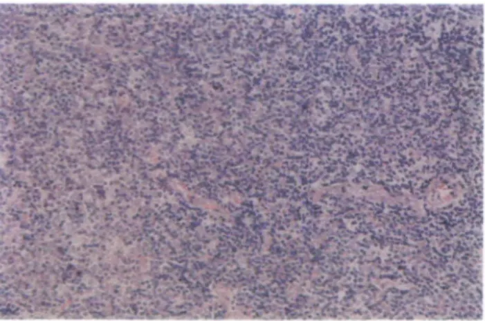
Upon morphological review (performed by B.B.) all lymph node biopsies (1988, 1989, 1991, and 1993) showed effacement of lymph node structure by small lymphoid cells homogeneously distributed all over the nodes (Figure 1). Paraffin and cryostat sections were immunophenotyped with anti-B-cell antibodies (CD10, CD19, CD20, CD22, CD23, CD45R, KiB3, IgG, IgM, IgD, IgA, and K as well X) and anti-T-ceil antibodies (UCHL, CD2, CD3, CD4, CD5, CD8, MT1 and CD45RO). Other markers employed were EBV-LMP (Epstein Barr Virus latent membrane protein), IL2-R (activation marker), Ki67 and MIBl (proliferation markers), and CD21 (follicular dendritic cell marker). On immunohistochemistry the cells were of B-phenotype and admixed with reactive T-cells in all the biopsies. Hardly any remnants of networks of fol-licular dendritic cells were found. The tumour cells exclusively expressed surface Ig K light chains but vir-tually no X chains (Figure 2). The proportion of prolif-erative cells (Ki67, MIBl) was low. Thus, the diagnosis
Figure 1. Giemsa stain of a lymph node section. There is total
effacement of the normal lymph node structure and a dense infiltra-tion by small lymphoid cells homogeneously distributed all over the node. The histological appearance corresponds to a low grade non-Hodgkin's lymphoma (International Working Formulation: Type A, small lymphocytic; R.E.A.L. classification: peripheral B-cell neo-plasm, small lymphocytic lymphoma/B-cell chronic lymphocytic leukaemia) [1,2].
Figure 2. Immunohistochemical staining of a lymph node section
with an anti-Ig K light chain antibody (avidin-biotin method with diaminobenzidine as a chromogenic substance). Virtually all small lymphoid cells show light brown staining on the cell curface with the antibody, indicating exclusive monoclonal expression of K light chains (no X light chain positivity. X light chain stain not shown) [13-15].
of a low grade B-cell non-Hodgkin's lymphoma was confirmed (International Working Formulation: type A, small lymphocytic; R.EA.L. classification: periph-eral B-cell neoplasm, small lymphocytic lymphoma/B-cell chronic lymphocytic leukaemia [1, 2]). The contro-versial biopsy taken in 1988 was carefully reviewed and the material typed with additional markers. Although it had initially been reported as representing a possibly polyclonal lymphoproliferation of perhaps viral aetiol-ogy, this view was revised and the histology found to be fully compatible with the diagnosis of the same malig-nant lymphoma established in later biopsies. The find-ing of monoclonal immunoglobulin (Ig) gene rear-rangements in this material further strengthens this view (see Discussion). In the final phase of the illness, marked lymphocytosis was present in the peripheral blood. Immunophenotyping of peripheral blood lym-phocytes by flow cytometry revealed predominant B-cells with co-expression of CD5 (Table 1 and Figure 3). In addition, a minority of B-cells weakly expressed cytoplasmic X chains but no K chains (X/K ratio >6:1). Based on the finding of a B-cell expansion with partial loss of CD20, co-expression of CD5, and predomi-nance of Ig X light chain expression typical B-cell chro-nic lymphocytic leukaemia was diagnosed (Table 2). Molecular analyses
DNA of all lymph node biopsies and of peripheral blood leukocytes (before and after CLL was diag-nosed) was examined by Southern blotting and the polymerase chain reaction (PCR) to detect Ig heavy chain and T-cell receptor (TCR) gene rearrangements (Figure 4) [3-6]. All DNA samples taken between 1988 and 1995 showed identical Ig heavy chain gene rearrangements by Southern blotting indicating the presence of a clonal population of lymphoid cells
Table 1. Immunophenotype results by flow cytometry of the pa- Discussion
dent's peripheral blood lymphoid cells at a late stage when morpho-logically CLL was present.
T-cell markers CD2 CD3 CD5 B-cell markers CD10 CD19 CD22 CD23 FMC7
Surface Ig light chains Cytoplasmic Ig K light chains Cytoplasmic Ig X light chains Co-expressions CD19/CD10 CD19/CD5 Results % posi-tive cells 8 8 74 6 76 43 53 <1 <1 <1 6 <1 63 Intensity of stain ++ + ++
% positive cells - percentage of cells in the gate expressing a single marker or co-expressing a marker combination. Intensity of marker expression is indicated as + (weak) to +++ (strong).
a o J I 1
i
2 in ~1 U 100 0 j : i ! 0. 1 1 1 1 I I 1 ' * A 3 2 -1000 CD19 CO19Figure 3. Immunophenotype analysis by flow cytometry of
periph-eral blood lymphoid cells. The cells were labelled with monoclonal anti-CD5 and anti-CD19 antibodies. Lymphoid cells were selected for analysis by electronic gating based on cell size and granularity. The two antibodies were coupled with different dyes permitting separate analysis in the flow cytometer (CD 19: green fluorescence; CD5: orange fluorescence). Left panel: peripheral blood lymphoid cells from a normal person. Note that a population of B-cells (posi-tive for CD 19; quadrant B4) and a separate population of T-cells (positive for CD5; quadrant Bl) are present. Less than 1% of lym-phoid cells are double positive for CD19 and CDS (quadrant B2). A minority of lymphoid cells (corresponding largely to natural killer cells) express neither CD19 nor CD5 (quadrant B3). Right panel: peripheral blood lymphoid cells from the patient in a sample where CLL was suspected based on morphology. The vast majority of lym-phoid cells co-express CD19 and CD5 (quadrant B2) typical of B-cell CLL. Note that a small population of normal T-cells positive for CD5 only is also present (quadrant Bl).
(Figure 5). This result was confirmed by amplification of rearranged Ig heavy-chain gene fragments by immu-nogenotype PCR (Figure 6). No TCR gene rearrange-ments or deletions were detected. Analyses (Southern blot and PCR) for the presence of EBV genome in the neoplastic celJs were negative.
The clinical course of our patient was noteworthy for its relatively favourable evolution over many years characterised by spontaneously waxing and waning lymphomas and splenomegaly. Cases of clonal low grade malignant non-Hodgkin's lymphoma with a clini-cally benign course and spontaneous regressions have been repeatedly described [7, 8]. Clonal lymphoprolif-erations need not necessarily show malignant behavi-our clinically as illustrated by several distinct clonal yet benign lymphoproliferative syndromes well known to dermatologists [9-12]. In our patient, a favourable long-term evolution and the pathologist's morpho-logical diagnosis of a cancer at times seemed a bit diffi-cult to be reconciled, but the review of the morphologi-cal findings in the successive lymph node biopsies in conjunction with immunogenotype findings, and the clinical evolution into B-CLL clearly argue in favour of a malignant lymphoma.
Recently, a very interesting case of mantle zone lym-phoma reminiscent of our patient's story and similarly characterized by recurrent cycles of acute illness alter-nating with spontaneous remissions was reported [8]. Two B-cell (sub-)clones were identified that had origi-nated from a single malignant clone. One of them was stably present whereas the other one (marked by an individual-specific Ig gene rearrangement) appeared during acute phases only. Presumably these neoplastic cells were eliminated through programmed cell death activated in vivo at the peaks of the acute phases. The generation of autoantibodies with anti-idiotypic activ-ity may perhaps play a role in such cases. Chronic viral infections might provide signals to induce initiation of lymphoma growth, followed by apoptosis and clinical remission. EBV infection was documented in our pa-tient by typical antibody patterns in her serum, and might perhaps have provided such signals to her clonal B-cells. However, this lymphoma was not an EBV-as-sociated B-cell lymphoma since no EBV genomic material was found in her tumour cells.
A few comments on the immunophenotype analysis of chronic B-cell lymphoproliferative disorders
The demonstration of Ig light chain restriction indi-cates a monoclonal lymphoid population, i.e., originat-ing from a soriginat-ingle B-cell either expressoriginat-ing K or X chains but not both [13-17]. Reactive polyclonal accumula-tions of B-cells in an inflammatory infiltrate would ex-press K and X Ig light chains at roughly a 2:1 to 1:1 ratio [13, 17]. Ig light chain restriction is considered the single most useful criterion for the immunophenotypic diagnosis of B-cell malignancies [3, 17, 18] although monoclonal Ig light chain expression has also been reported in rare cases of reactive lymphoid hyperplasia [19, 20]. It can easily be investigated by flow cytometry
Table 2. Low-grade B-cell lymphomas: immunohistological and genetic features in differential diagnosis [1,16,22].
Lymphoma Ig Igcyto- CD5 CD10 CD19 CD23 Typical chromosomal surface plasmic (CALLA) aberration
B-CLL or small lymphocytic lymphoma + Lymphoplasmacytoid lymphoma or immunocytoma + Mantle cell lymphoma or centrocytic lymphoma + Follicle center lymphoma or follicular lymphoma + Marginal zone lymphoma +
Trisomy 12 Not known t(14;18) Trisomy 3 (extranodal NHL) EcoRI V. J , . .
XTXIZLT
EcoRII
IX
II I ILL
"rrrr
~>
EooRS EcoRI v o. B 1kt> JtVprot* FR3A UHfrtrwFigure 4. Schematic representation of the detection of Ig gene rearrangements by Southern blot and PCR [3-6, 11, 32, 33]. A) Ig heavy
chain gene on chromosome 14 in the germline configuration. V - variable regions; D - diversity regions; J - joining regions; C(i - constant region. The restriction enzyme EcoRI cuts a 16 kb germline DNA fragment recognized by an Ig heavy-chain joining region probe (JH-probe). B) Hypothetical Ig heavy-chain gene rearrangement resulting in specific V-D-J joining. Note that the size of the EcoRI fragment described in A is altered to a smaller fragment also recognized by the JH-probe. C) JH-probe commonly used to detect the germline Ig heavy-chain joining region and its rearrangements with the Southern blot technique. D) Primers used for immunogenotype PCR. The primer pair can be used to amplify most rearrangements at this locus. Note that an Ig heavy-chain gene rearrangement results in a much smaller dis-tance between the FR3A (framework 3A) consensus primer and the LJH primer which is amenable to copying by the polymerase.
and immunohistochemistry both on the surface and in the cytoplasm of cells [21-24]. However, light chain expression may be very weak (as in our patient) or even absent in neoplastic cells, rendering analysis of mono-clonality by flow cytometry difficult or impossible. The interpretation of the results in our patient seems diffi-cult at first sight. The lymphoma cells showed exclusive expression of Ig K light chains whereas the B-CLL cells predominantly expressed Ig X light chains. A glance at the 1994 serum electrophoreses provides a possible clue: in addition to the former single monoclonal Ig K light chain peak an additional separate clonal X peak was now present, indicating two B-cell subclones. Such clonal evolution of lymphomas over time implies that patterns of clonal markers, for example monoclonal paraprotein peaks identified in a serum protein electro-phoresis may change over time.
In order to differentiate between B-CLL and other B-cell neoplasms particular patterns of immunological
markers may be helpful (Table 2) [1, 14, 17]. A scoring system considers three immunological markers (CD5, CD23, and FMC7) and the intensity of surface Ig and surface CD22 expression. The sum of the scoring points correlates with the likelihood that a diagnosis of CLL is correct [25] (Table 3). Our patient had a score of 5 in the leukaemic phase which is virtually diagnostic of B-CLL. Double-labelling with monoclonal anti-bodies revealed co-expression of the T-cell associated marker CD5 by the clonal B-cell population alongside with typical B-cell markers, for example CD19 (Table 1 and Figure 3). CD5 is also expressed by B-cell sub-populations in an age-dependent manner, particularly in cord blood, fetal lymph nodes and liver [26, 27]. In healthy adults CD5-positive B-lymphocytes constitute 1% to 30% of the total B-cell population in the spleen, lymph nodes and peripheral blood, but are virtually absent in bone marrow. A monoclonal expansion of CD5-positive B-cells is typical for either B-CLL or
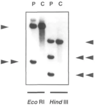
P C P C
EcoR! HintfWX
Figure 5. Southern blot detection of clonal Ig gene rearrangements.
P - patient's DNA from B-CLL cells; C - normal control DNA. Single arrows indicate germline restriction fragments, double arrows indicate clonally rearranged fragments. Blot hybridized with a DNA probe (JH) recognizing a fragment from the joining region of the Ig heavy-chain gene |4-6). The restriction enzyme EcoRI (left side of the panel) cuts a 16 kb fragment out of the gene (see also map in Figure 4A). The rearrangement process produces a smaller frag-ment in the CLL cells. This finding is confirmed using another enzyme Hindm (right side of panel) which normally yields a 10.5 kb germline fragment, but in B-CLL DNA shows two rearranged bands derived from a clonally rearranged Ig gene. The germline bands are either derived from contaminating normal cells, or represent a non-rearranged allele in the neoplastic cells, or both.
mantle cell lymphoma [25]. It is noteworthy in this respect that infectious mononucleosis was present in our patient six years before CLL was diagnosed. An in-creased number of polyclonal (or oligoclonal) CD5-positive B-cells has been reported in patients with infectious mononucleosis or previous EBV infection [28], with the hypothesis that clonal lymphomas might eventually evolve from these reactive disorders. An in-creased number of CD5-positive B-cells may also be present after allogeneic bone marrow transplantation [29] or in rheumatoid arthritis [30].
Table 3. Scoring system for the differential diagnosis of CLL and
other B-cell lymphoproliferative disorders after Matutes et al. |25|.
Score 1 0 Surface Ig CD5 CD23 FMC7 Surface CD22 Weak Positive Positive Negative Negative/weak Moderate/strong Negative Negative Positive Moderate/strong
1 2 3 4 5
190
bp
Figure 6. Amplification by PCR of a clonally rearranged Ig
heavy-chain gene fragment [32, 33]. Samples: 1) DNA from normal poly-clonal lymphocytes; 2) patient's lymphoma DNA from 1993; 3) lymphoma DNA from 1991; 4) lymphoma DNA from 1989; 5) molecular fragment size marker. All patient samples reveal the same 190 bp PCR fragment representing a clonal Ig heavy-chain gene rearrangement that remains constant over time. Note that DNA from polyclonal lymphocytes does not produce distinct PCR ampli-fication products but a smear (lane 1). In the germline gene the two primer annealing sites are too far apart, and therefore the polymer-ase fails to copy the DNA stretch between them (see Figure 4 for map).
Assessment of lymphoma clonality with the help of molecular biology
The detection of antigen receptor gene rearrangements with molecular probes for Ig and TCR genes has pro-vided a new powerful tool to assess the clonality of lymphoproliferative disorders [3-6, 31]. In their germ-line configuration Ig and TCR genes are composed of distinct DNA subsegments or regions (constant, diver-sity, joining and variable regions). Germline genes are non-functional. In order to produce a functional gene for the production of a specific antibody or T-cell receptor protein a lymphoid cell must reassemble the molecular units of its Ig or TCR genes during matura-tion (Figure 4). This mechanism is the molecular basis for the immense diversity of antibodies and TCR pro-teins which the immune system requires to do its job. Ig and TCR gene rearrangements are highly specific markers for a given B- or T-cell passed on to its pro-geny. A polyclonal lymphoid cell population is com-posed of myriads of different cells with individually sized gene rearrangements. On a Southern blot these various fragments are not detected because each single rearrangement is far below the sensitivity threshold of the method. However, if at least 5%-10% of the cells contribute the very same DNA rearrangement, a dis-tinct new non-germline band becomes detectable on autoradiography (Figure 5). Since the chances that
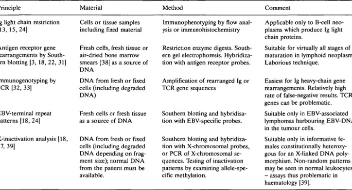
Table 4. Assessment of clonality in lymphoproliferations.
Principle Material Method Comment
Ig light chain restriction [13,15,24]
Antigen receptor gene rearrangements by South-ern blotting [3, 18, 22, 31] Immunogenotyping by PCR [32, 33] EBV-terminal repeat patterns [18, 24] X-inactivation analysis [18, 37, 39]
Cells or tissue samples including fired material Fresh cells, fresh tissue or air-dried bone marrow smears [38] as a source of DNA
DNA from fresh or fixed cells (including degraded DNA)
Fresh cells or fresh tissue as a source of DNA DNA from fresh or fixed cells (including degraded DNA depending on frag-ment size); normal DNA from the patient must be available.
Immunophenotyping by flow anal-ysis or immunohistochemistry Restriction enzyme digests. South-ern gel electrophoresis. Hybridiza-tion with antigen receptor probes. Amplification of rearranged Ig or TCR gene sequences
Southern blotting and hybridisa-tion with EBV-specific probes. Southern blotting and hybridiza-tion with X-chromosomal probes, or PCR of X-chromosomal se-quences. Testing of inactivation patterns by examining allele-spe-cific methylation.
Applicable only to B-cell neo-plasms which produce Ig light chain proteins.
Suitable for virtually all stages of maturation in lymphoid neoplasms. Laborious technique.
Easiest for Ig heavy-chain gene rearrangements. Relatively high rate of false-negative results. TCR genes can be problematic. Suitable only in EBV-associated lymphomas harbouring EBV-DNA in the tumour cells.
Suitable only in informative fe-males constitutionally heterozy-gous for an X-linked DNA poly-morphism. Non-random patterns may be seen in normal leukocytes - assays thus problematic in haematology [39].
many unrelated lymphoid cells would independently produce the very same Ig or TCR gene rearrangement are exceedingly small, a rearranged band points to a clonal population of lymphoid cells. This phenomenon may be likened to an 'in vivo' PCR amplification of a particular gene fragment through clonal expansion of neoplastic B-cells! A thought or two should be given to the possible pitfalls of the method as summarized in Table 5 [5,6]. For example, partial enzyme digestion or a DNA polymorphism may cause difficulties in inter-preting the results. In our patient Southern blot analysis of DNA from all lymph node biopsies and peripheral blood revealed two non germ-line rearranged bands that were constant over time, supporting the view that over ten years (including the final phase of B-CLL) a clonal lymphoid disorder was present.
The Southern blot findings in our case were con-firmed by immunogenotype PCR which detected an Ig heavy chain gene rearrangement constant throughout the lymphoma samples tested [32, 33]. However, no germline Ig gene fragment was seen on PCR analysis (Figure 6). The reason is that in the germline Ig gene the annealing sites of the primers encompass a frag-ment far too long to be amplified (the maximum length of DNA fragments amenable to PCR is in the range of several hundred to perhaps 1,000 to 2,000 base pairs) [32]. A somatic rearrangement, however, creates a new much smaller Ig gene fragment suitable for PCR ampli-fication, since many DNA segments located between the primer annealing sites that are not used to create a functional gene are deleted. A major limitation of PCR in detecting B-cell clonality is the high rate of false-negative results ranging from 10%-40% of these cases. Appropriate choice of the primers and careful control
of gel electrophoresis conditions to visualise clonal Ig gene fragments appear to be crucial to detect rear-ranged Ig gene sequences [33, 34]. Once these prob-lems are satisfactorily addressed PCR will soon be-come an essential routine tool to examine the clonality of lymphoid proliferations whilst the Southern blot technique is too cumbersome and laborious for every day practice outside specific research protocols. In practice morphological examination of good-quality lymph node biopsies and immunohistochemistry with an appropriate panel of diagnostic antibodies will pro-vide an accurate diagnosis of a lymphoid tumour in almost 95% of cases. Molecular analysis of clonality by Southern blotting of immunogenotype PCR will only be required to solve diagnostic problems in a rather small proportion of cases [23].
Two other approaches to assess clonality were tested in this case. Since the patient had serological evidence of EBV-infection, her lymphoma DNA was tested for the presence of clonal patterns of genome. EBV-specific probes can visualize virus-EBV-specific polymor-phic sequences as separate bands on Southern blots [34-36]. Monoclonal EBV-associated lymphoid tu-mours harbour one such EBV-terminal-repeat frag-ment only whereas polyclonal lymphocyte populations infected by different virions show multiple different bands. The lymphoma DNA of our patient was nega-tive for EBV-genomic material. Assessment of clonal-ity by X-inactivation analysis is based on the fact that in each somatic female cell only one of the two X-chro-mosomes is active. In polyclonal cell populations maternal and paternal X-chromosome copies are in-activated at a ratio of about 1:1 (random) whereas in clonal tumours all neoplastic cells keep the same
Table 5. Southern blots to detect clonal lymphoid proliferations.
Using any combination of restriction enzymes and antigen receptor gene probes the following possible patterns may be seen on a South-ern blot autoradiograph [3-6).
1. Germline bands only — no novel bands
No monoclonal gene rearrangement can be detected. Hence, the specimen must contain
a) non-lymphoid normal or tumour tissue;
b) polyclonal lymphoid cells, for example in inflammatory conditions;
c) a monoclonal lymphoid population below the detection lim-its of the Southern method (in the order of magnitude of a few % of all nucleated cells);
d) a monoclonal lymphoid population with Ig or TCR gene rearrangements that may not be apparent because their fragment size is similar to the size of germline fragments. This problem occurs mainly with large fragments as these are less well resolved during electrophoresis than smaller ones. Use of additional restriction enzymes creating differ-ent fragmdiffer-ents will usually offer a solution to this problem; e) a monoclonal lymphoid cell population with a
rearrangement that leads to biallelic deletion of the gene (this is rare -an example are TCR 6 gene deletions in acute lymphoblas-tic leukaemia). Examination of several antigen receptor genes will usually do the trick;
f) another patient's sample that must have been mixed up in the mail or in the laboratory due to logistic and technical errors.
2. Both germline and rearranged non-germline bands
This may be seen when
a) monoclonal lymphoid tissue is admixed with normal or non-lymphoid tissue contributing the germline band; b) the rearrangement in the clonal cells affects only one allele
of the Ig or TCR gene retaining the other one in the germ-line configuration.
c) rearranged bands are faked because the restriction enzyme digest is incomplete leading to 'novel' fragments. Such 'novel' bands are typically larger than the germline fragment predicted from the restriction map;
d) novel bands are due to a constitutional DNA polymor-phism. Use of several restriction enzymes, testing of consti-tutional DNA from the patient (which should contain the polymorphic bands but no somatic rearrangements) as well as family studies (tracing the inheritance of polymorphic alleles) usually solve this problem.
3. Novel rearranged bands only
This pattern occurs in samples that exclusively contain monoclonal lymphoid populations with biallelic rearrangements of an Ig or TCR gene.
parental copy in the active state (non-random use of one X-chromosome) [18, 38]. X-chromosomal poly-morphisms (constitutional variations in the DNA sequence within the X-chromosome) suitable for these clonality tests have been found in the hypoxanthine phosphoribosyl transferase gene, the phosphoglycerate kinase gene, an X-chromosomal locus termed M27|3 and in a polymorphic locus within the human androgen receptor gene. Unfortunately our patient was not infor-mative for any of these polymorphisms and thus not suitable for this approach.
Conclusions
This patient offers the opportunity to comment on sev-eral interesting features of lymphomas. The clinical course is unusual with many episodes of lymphoma regression without any treatment over several years. Such clinical data should be taken into account when results of lymphoma treatment are interpreted. Al-though the causes of this phenomenon in our patient are unknown, programmed cell death or apoptosis trig-gered by exogenous factors might have played a role in these conditions [8]. Molecular techniques detected an Ig gene rearrangement which served as an individual-specific tag to show that a constant clonal lymphoid proliferation was present over many years including the final phase of B-CLL. This illustrates the point that molecular diagnostics have expanded our armamen-tarium for the characterisation of such cases [40], al-though morphology, immunohistochernistry and immu-nophenotyping are still the diagnostic cornerstones to study lymphoma. During our work-up of the case the clonal marker was also detected in the first biopsy which initially had created some diagnostic difficulties. The diagnosis of a monoclonal lymphoproliferation would have helped to overcome these problems at this early stage. Finally the question arises of whether ear-lier treatment with cytostatic drugs could have prevent-ed or postponprevent-ed the eventually fatal final leukaemic phase. This seems unlikely with a standard regime such as chlorambucil and prednisone, but the impact of newer treatments including purine analogues in this respect remains to be established.
Acknowledgment
This work was funded by Swiss National Foundation grants 37577.93 (M.F.F.), 28744.90 (B.B.), 31-32524.91 (A.T.) and 31-40763.94 (A.v.R.). S.N. was supported by the Bernische Krebsliga. We would like to thank M. Tinguely for the PCR assays and M. Oest-reicher for technical assistance.
References
1. Harris NL, Jaffe ES, Stein H et al. A revised European-Ameri-can classification of lymphoid neoplasms: A proposal from the Internationa] Lymphoma Study Group. Blood 1994; 84: 1361-92.
2. Non-Hodgkin's Lymphoma Pathologic Classification Project. National Cancer Institute sponsored study of classifications of non-Hodgkin's lymphomas. Summary and description of a Working Formulation for clinical usage. Cancer 1982; 49: 2112-35.
3. Fey MF, Wainscoat JS. Molecular diagnosis of haematological neoplasms. Blood Rev 1988; 2: 78-87.
4. Fey MF, Tobler A, Stadelmann B et al. Immunogenotyping with antigen receptor gene probes as a diagnostic tool in child-hood acute lymphoblastic leukemia. Eur J Haematol 1990; 45: 215-22.
immuno-globulin and T-cell receptor genes. Part I: Basic and technical aspects. Clin Chim Acta 1991; 198: 1-92.
6. van Dongen JJM, Wolvers-Tettero ILM. Analysis of immu-noglobulin and T-cell receptor genes. Part II: Possibilities and limitations in the diagnosis and management of lymphoprolif-erative diseases and related disorders. Clin Chim Acta 1991; 198:93-174.
7. Krikorian JG, Portlock CS, Cooney P, Rosenberg SA. Spon-taneous regression of non-Hodgkin's lymphoma: A report of nine cases. Cancer 1980; 46: 2093-9.
8. Kaufmann Y, Many A, Rechavi G et al. Brief report: Lym-phoma with recurrent cycles of spontaneous remission and re-lapse - possible role of apoptosis. N Engl J Med 1995; 332: 507-10.
9. Weiss LM, Wood GS, Trela M et al. Clonal T-cell populations in lymphomatoid papulosis: Evidence of a lymphoproliferative origin for a clinically benign disease. N Engl J Med 1986; 315: 475-9.
10. Fishleder A, Tubbs R, Hesse B, Levine H. Uniform detection of immunoglobulin-gene rearrangements in benign lymphoepi-thelial lesions. N Engl J Med 1987; 316:1118-21.
11. Griesser H, Tkachuk D, Reis M, Mak TW. Gene rearrange-ments and translocations in lymphoproliferative diseases. Blood 1989; 73:1402-15.
12. Shearer WT, Ritz J, Finegold MK et al. Epstein-Barr virus associated B-cell proliferations of diverse clonal origins after bone marrow transplantation in a 12 year old boy with severe combined immunodeficiency. N Engl J Med 1985; 312: 1151-9.
13. Levy R, Warnke R, Dorfman RF, Haimovich J. The mono-clonality of human B-cell lymphomas. J Exp Med 1977; 145: 1014-28.
14. Freedman AS. Cell surface antigens in leukemias and lympho-mas. Cancer Invest 1996; 14: 252-76.
15. Preud'homme JL, Seligman M. Surface bound immunoglobu-lins as a cell marker in human lymphoproliferative diseases. Blood 1972; 40: 777-94.
16. van Dongen JJM, Adriaansen HJ, Hoojkaas H. Immunopheno-typing of leukaemias and non-Hodgkin's lymphomas: Immuno-logical markers and their CD codes. Neth J Med 1988; 33: 298-314.
17. General Haematology Task Force of BCSH. Immunopheno-typing in the diagnosis of chronic lymphoproliferative dis-orders. J Clin Pathol 1994; 47: 871-5.
18. Wainscoat JS, Fey MF. Assessment of clonality in human tu-mors: A review. Cancer Res 1990; 50:1355-60.
19. Levy N, Nelson J, Meyer P et al. Reactive lymphoid hyper-plasia with single class (monoclonal) surface immunoglobulin. Am J Clin Pathol 1983; 80: 300-8.
20. Lush CJ, Vora AJ, Campbell AC, Wood JK. Polyclonal CD5+ B-lymphocytosis resembling chronic lymphocytic leukemia. Br J Haematol 1991; 79:119-20.
21. Johnson A, Olofsson T. Flow cytometric clonal excess analysis of peripheral blood, routine handling, and pitfalls in interpreta-tion. Cytometry 1993; 14: 188-95.
22. Henni T, Gaulard P, Divine M et al. Comparison of genetic probe with immunophenotype analysis in lymphoproliferative disorders: A study of 87 cases. Blood 1988; 72: 1937-43. 23. Kamat D, Laszewski MJ, Kemp JD et al. The diagnostic utility
of immunophenotyping and immunogenotyping in the patho-logic evaluation of lymphoid proliferations. Modern Pathol 1990; 3: 105-12.
24. Letwin BW, Wallace PK, Muirhead KA et al. An improved
clonal excess assay using flow cytometry and B-cell gating. Blood 1990; 75:1178-85.
25. Matutes E, Catovsky D. The value of scoring systems for the diagnosis of biphenotypic leukemia and mature B-cell dis-orders. Leuk Lymph 1994; 13 (Suppl 1): 11-4.
26. Bofill M, Janossy G, Janossa M et al. Human B-cell develop-ment: Subpopulations in the human fetus. J Immunol 1985; 134:1531-7.
27. Gadol N, Ault KA. Phenotypic and functional characterization of human LEU1 (CD5) Bcells. Immunol Rev 1986; 93: 2 3 -34.
28. Hasson J, Feighery C, Bresnihan B, Whelan A. Elevated T-cell receptor cells in patients with infectious mononucleosis. Br J Haematol 1991; 77:255-6.
29. Antin JH, Ault KA, Reppepart JM, Smith BR B-lymphocyte reconstitution after human bone marrow transplantation: Leu-1 antigen defines a distinct population of B-lymphocytes. J Clin Invest 1987; 80: 325-32.
30. Plater-Zyberk C, Maini RN, Lam K et al. A rheumatoid arthri-tis B-cell subset expresses a phenotype similar to that in chronic lymphocytic leukemia. Arthritis Rheum 1985; 28: 971-6.
31. Arnold A, Cossman J, Bakhshi A et al. Immunoglobulin-gene rearrangements as unique clonal markers in human lymphoid neoplasms. N Engl J Med 1983; 309:1593-9.
32. Trainor KJ, Brisco MJ, Story CJ, Morley A. Monoclonality in B-lymphoproliferative disorders detected at the DNA level. Blood 1990; 75: 2220-2.
33. Tbakhi A, Tubbs RR. Utility of polymerase chain reaction in detecting B-cell clonality in lymphoid neoplasms. Cancer 1996; 77: 1223-5.
34. Lozano MD, Tierens A, Greiner TC et al. Clonality analysis of B-lymphoid proliferations using the polymerase chain reaction. Cancer 1996; 77:1349-55.
35. Raab-Traub N, Flynn K. The structure of the termini of Epstein-Barr virus as a marker of clonal cellular proliferation. Cell 1986; 47: 883-9.
36. Borisch B, Finke J, Hennig I et al. Distribution and localization of Epstein-Barr virus subtypes A and B in AIDS-related lym-phomas and lymphatic tissue of HIV+ patients. J Pathol 1992; 168:229.
37. Fey MF, Peter HJ, Hinds HL et al. Clonal analysis of human tumours with M27p", a highly informative polymorphic X-chro-mosomal probe. J Clin Invest 1992; 89:1438-44.
38. Fey MF, Pilkington SP, Summers C, Wainscoat JS. Molecular diagnosis of haematological disorders using DNA from stored bone marrow slides. Br J Haematol 1987; 67:489-92. 39. Fey MF, Liechti-Gallati S, von Rohr A et al. Clonality and
X-inactivation patterns in haematopoietic cell populations detected by the highly informative M27|5 probe. Blood 1994; 83:931-8.
40. Jack AS, Johnson R, Morgan GJ. The detection and clinical significance of monoclonality in lymphoproliferative disorders. Current Diagn Pathol 1995; 2: 181-94.
Received 23 November 1995; accepted 18 July 1996.
Correspondence to:
Prof. M. F. Fey
Institute of Medical Oncology Inselspital
CH-3010 Berne Switzerland

![Table 2. Low-grade B-cell lymphomas: immunohistological and genetic features in differential diagnosis [1,16,22].](https://thumb-eu.123doks.com/thumbv2/123doknet/14922387.663387/4.885.102.822.104.648/table-grade-lymphomas-immunohistological-genetic-features-differential-diagnosis.webp)