Proceedings
of
the
Nutrition
Society
The Winter meeting of the Nutrition Society was held at the Royal College of Physicians, London on 6–7 December 2011
70th Anniversary Conference on ‘Body weight regulation – food, gut
and brain signalling’
Symposium I: Food–gut interactions
The receptive function of hypothalamic and brainstem centres to
hormonal and nutrient signals affecting energy balance
Thomas Riediger
Institute of Veterinary Physiology and Centre of Integrative Human Physiology, University of Zurich, 8057 Zurich, Switzerland
The hypothalamic arcuate nucleus (ARC) and the area postrema (AP) represent targets for hormonal and metabolic signals involved in energy homoeostasis, e.g. glucose, amylin, insulin, leptin, peptide YY (PYY), glucagon-like peptide 1 (GLP-1) and ghrelin. Orexigenic neuro-peptide Y expressing ARC neurons are activated by food deprivation and inhibited by feeding in a nutrient-dependent manner. PYY and leptin also reverse or prevent fasting-induced acti-vation of the ARC. Interestingly, hypothalamic responses to fasting are blunted in different models of obesity (e.g. diet-induced obesity (DIO) or late-onset obesity). The AP also responds to feeding-related signals. The pancreatic hormone amylin acts via the AP to control energy intake. Amylin-sensitive AP neurons are also glucose-responsive. Furthermore, diet-derived protein attenuates amylin responsiveness suggesting a modulation of AP sensitivity by mac-ronutrient supply. This review gives an overview of the receptive function of the ARC and the AP to hormonal and nutritional stimuli involved in the control of energy balance and the possible implications in the context of obesity. Collectively, there is consistency between the neurophysiological actions of these stimuli and their effects on energy homoeostasis under experimental conditions. However, surprisingly little progress has been made in the develop-ment of effective pharmacological approaches against obesity. A promising way to improve effectiveness involves combination treatments (e.g. amylin/leptin agonists). Hormonal altera-tions (e.g. GLP-1 and PYY) are also considered to mediate body weight loss observed in obese patients receiving bariatric surgery. The effects of hormonal and nutritional signals and their interactions might hold the potential to develop poly-mechanistic therapeutic strategies against obesity.
Arcuate nucleus: Area postrema: Food intake: Obesity
Driven by the search for therapeutic treatment strategies against obesity and associated metabolic disorders, con-siderable knowledge has accumulated about the control mechanisms involved in the maintenance of energy bal-ance. The redundancy of control mechanisms, the capacity of these systems to compensate for pharmacological effects and the existence of obesity-related hormonal insensitivities make it difficult to achieve sufficient
therapeutic efficiency for the reduction of body weight over longer periods of time. Evidently, at least under non-laboratory conditions, so-called non-homoeostatic factors (hedonic properties and availability of food, food pre-ferences, social factors, eating habits, etc.) and central reward mechanisms override homoeostatic hormonal and metabolic signals that are considered to reflect and to control the body’s energy status.
Abbreviations: AMPK, adenosine monophosphate-activated protein kinase; AP, area postrema; APX, area postrema lesion; ARC, arcuate nucleus; CCK,
cholecystokinin; DIO, diet-induced obesity; FAS, fatty acid synthase; GLP-1, glucagon-like peptide-1; GLP-1R, glucagon-like peptide-1 receptor; mTOR, mammalian target of rapamycin; NED, non-energy diet; NPY, neuropeptide Y; NTS, nucleus of the solitary tract; OXM, oxyntomodulin; POMC, pro-opiomelanocortin; PYY, peptide YY; STAT, signal transducer and activator of transcription.
Corresponding author:Thomas Riediger, fax + 41 44 635 8932, email [email protected]
Proceedings
of
the
Nutrition
Society
In contrast to the currently available pharmacological treatment options, bariatric surgery (e.g. Roux-en-Y gastric bypass) is very effective in reducing body weight and improving glucose homoeostasis in patients with type-2 diabetes mellitus(1,2). Apparently, bariatric surgery alters gastrointestinal physiology in a way that changes nutrient-induced gastrointestinal hormone responses, which pre-sumably contribute to these therapeutic effects. Hence, the signalling mechanisms of the gut–brain axis and the inter-action between its nutritional and hormonal feedback sig-nals are not only essential for our understanding of the body’s energy homoeostasis but also for the ultimate desire to develop pharmacological approaches against the obesity epidemic. Some of the hormones (e.g. glucagon-like pep-tide 1 (GLP-1) and peppep-tide YY (PYY)) discussed in this review with regard to their central actions are presumed as mediators of weight loss after bariatric surgery. Although the exact mechanisms behind bariatric weight loss surgery and their potential to be targeted pharmacologically remain to be identified, there are other promising therapeutic approaches that are based on hormonal combination treat-ments. Both pre-clinical studies in obese rodents and clin-ical trials in human subjects have established a beneficial effect of combined treatment with the anorexigenic pan-creatic hormone amylin with the fat-derived hormone lep-tin. Amylin treatment seems to reverse leptin insensitivity in obese subjects, in which amylin and leptin exert a synergistic action to reduce body weight(3,4).
Brain circuits involved in the control of energy balance also function as fuel sensors responding to diet-derived macronutrients. Nutrients not only alter neuronal activity in feeding regulatory brain areas, they also modulate neu-ronal responsiveness to hormonal stimuli. Two important brain sites that are targeted by hormonal and nutrient sig-nals involved in the control of energy balance are the hypothalamic arcuate nucleus (ARC) and area postrema (AP) of the brainstem. The current review focuses on the receptive function of the ARC and the AP to feeding-related signals and alterations of neuronal responsiveness that are associated with obesity. It is beyond the scope of this review to describe similar hormone and nutrient-dependent signalling mechanisms in other brain areas that also contribute to the overall control of energy homo-eostasis.
The hypothalamic arcuate nucleus
The ARC is considered as one of the most important hypothalamic target structures for blood-borne hormonal and metabolic factors involved in the regulation of food intake and energy homoeostasis. The best-characterised hormone supposed to act via the ARC is the lipostatic and anorectic hormone leptin, a 16 kDa protein that is secreted from white adipose tissue. Genetic deficiency of leptin or its receptor in rodents causes excessive food intake, body weight gain and an obese phenotype(5,6). Leptin acts via the ARC, which contains two functionally antagonistic populations of neurons with respect to the control of food intake. One population contains the orexigenic neuropeptide Y (NPY) and agouti gene-related peptide(7).
The other distinct population synthesises the anorectic pro-opiomelanocortin (POMC) gene product a-melanocyte stimulating hormone(8). Leptin up-regulates POMC neu-rons while it inhibits NPY/agouti gene-related peptide neurons in the ARC(9,10). Similar to leptin, insulin fulfils the criteria of an adiposity signal because circulating insulin levels increase in relation to body adiposity. Fur-thermore, insulin has access to the brain and it influences food intake and body weight in a similar way as leptin via the ARC(11).
Catalysed by the high importance of the ARC for body weight control and the failure of leptin therapy as an effective anti-obesity treatment because of leptin resis-tance(12), the scientific interest in other factors acting via the ARC and its downstream signalling pathways began to increase. The well-established concept that the ARC is targeted by the adiposity signals leptin and insulin has been extended by numerous studies demonstrating ARC-dependent actions of hormones, neuropeptides and meta-bolites that change dynamically according to the status of energy intake. Notably, leptin itself shows dynamic short-term changes as a function of food intake(13,14).
Ghrelin
One of the hormones targeting the ARC is the orexigenic gastrointestinal hormone ghrelin(15). Plasma levels of ghrelin are modulated by the nutritional state. During fasting and shortly before meals circulating ghrelin con-centrations are elevated, while feeding or nutrient intake reduce blood-ghrelin levels(16–18). Various electro-physiological and immunohistological studies char-acterised ghrelin’s effects on neuronal activity of ARC neurons that have been suggested to represent target cells for ghrelin in the brain(19–22). Ghrelin binds to the growth hormone secretagogue receptor that is highly abundant in the ARC(23). The growth hormone secretagogue receptor is predominantly expressed in NPY neurons (94 % co-localisation), while it is found in only 8 % of POMC neu-rons(24). Ghrelin exerts opposite effects to leptin on the activity of ARC neurons. Ghrelin or growth hormone secretagogue receptor agonists excite leptin-inhibited NPY neurons via a direct postsynaptic effect(19,25–27). In addition to this action, ghrelin indirectly inhibits neurons of the lateral ARC, where POMC neurons are located(20,28). The excitatory effect of ghrelin on ARC neurons and the NPYergic phenotype of these cells have been confirmed in vivo by immunohistochemical studies using c-Fos as a marker for neuronal activation(22,29).
Whether ghrelin can be considered a physiological sig-nal in the control of energy homoeostasis has not yet been confirmed based on the general criteria that have been suggested to define a physiological relevance(30). Owing to the weak consequences of ghrelin antagonism for feeding behaviour or body weight phenotype and because of the ineffectiveness of physiological ghrelin doses to pro-duce feeding or body weight effects, the physiological relevance of ghrelin has not yet been fully established(31). Based on the leptin counter-regulatory action of ghrelin, antagonism of ghrelin signalling has been proposed as a therapeutic approach against obesity. However, the role of
Proceedings
of
the
Nutrition
Society
ghrelin in the pathogenesis of obesity is not clear. Ghrelin levels in obese human subjects are lower than under nor-mal weight conditions(32) limiting the usefulness and suc-cess of ghrelin antagonism as an anti-obesity strategy(31). Interestingly, weight loss in obese subjects seems to be associated with an increase in ghrelin levels suggesting that a blockade of ghrelin action might facilitate the body weight reducing effect of other therapeutic interven-tions(32). Notably, there is cumulating evidence for a role of ghrelin in reward mechanisms that involve extra-hypo-thalamic pathways originating in the ventral tegmental area(33). The potential of pharmacological ghrelin-depen-dent approaches with respect to this mechanism has not yet been sufficiently explored in clinical studies.
Peptide YY/glucagon-like peptide 1
The two gut hormones PYY and GLP-1 act on ARC neu-rons. They have received special attention in the context of energy homoeostasis and as potential targets for the treat-ment of overweight. Both hormones might contribute to weight loss after gastric bypass surgery because Roux-en-Y gastric bypass patients have increased GLP-1 and PYY levels and blockade of gastrointestinal hormone release (including GLP-1 and PYY) by the somatostatin analogue octreotide led to increased appetite(34–36). A spe-cific role for PYY is suggested by the absence of weight loss in PYY knockout mice subjected to gastrointestinal bypass surgery(37).
PYY and PYY(3–36) are two biologically active pep-tides arising from endocrine L-cells of the ileum and colon in response to food intake(38). Although PYY binds to several subtypes of Y receptors, PYY(3–36) exhibits rela-tive specificity for the Y2 receptor(39). Both PYY isoforms inhibit food intake after peripheral administration; but PYY(3–36) appears to be more potent possibly due to its specificity to the Y2 receptor that is decisive for the anorexigenic response(40,41). However, the minimal doses that were required to suppress food intake in these studies
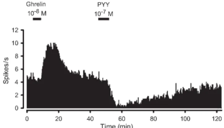
were supraphysiological when compared with meal-induced changes of total PYY blood levels. Therefore, the question whether PYY(3–36) acts as a physiological satiating signal is controversial. Direct site-specific injec-tions of PYY(3–36) identified the ARC as possible central target structure(42). With respect to the electrophysiological effects of PYY(3–36) in the ARC, conflicting data exist in the literature. In mice, PYY(3–36) has been demonstrated to indirectly (presynaptically) excite phenotypically iden-tified POMC neurons(42), while another electro-physiological study demonstrated direct postsynaptic inhibitions in POMC neurons of mice(43). Among others, the reasons for these seemingly contradictory findings have not yet been identified, but might be related to experi-mental and technical differences concerning the slice pre-paration and selection of cells(43). In rats, full-length PYY exerts direct inhibitory effects on ghrelin-exited ARC neurons (Fig. 1) suggesting that the NPYergic system is also affected by PYY signalling(44). This is consistent with the demonstration that PYY(3–36) inhibits NPY release from ARC explants in addition to stimulating a a-melanocyte stimulating hormone response(42).
Basal and postprandial PYY(3–36) levels are decreased in obese human subjects and rodents(45). The absence of PYY(3–36) resistance in obese subjects is in favour of using PYY agonists as anti-obesity drugs. Chronic periph-eral administration of PYY(3–36) decreases body weight in different species including rhesus macaques(42,46,47). Whe-ther PYY agonists can be Whe-therapeutically used in human subjects to reduce body weight awaits further evaluation. Interestingly, PYY seems to synergise with other hormones or hormone agonists affecting the control of energy bal-ance and glucose homoeostasis, including the GLP-1 ago-nist exendin-4 and the pancreatic hormone amylin(48,49). The neuronal correlates of these synergies are unknown.
Similar to PYY, GLP-1 is released in response to food intake from gastrointestinal L-cells(50,51). GLP-1 plays an important role in glucose homoeostasis (incretin) and gas-trointestinal function(52). In addition to its peripheral expression, GLP-1 is a neuropeptide expressed in a dis-crete population of enteroceptive neurons located in the brainstem(53). It binds to the GLP-1 receptor (GLP-1R) that is also activated by the agonist exendin-4, a peptide that has been isolated from the saliva of the lizard Heloderma suspectum. The truncated form of exendin (9–39) (also termed exendin-9) acts as a competitive antagonist at the GLP-1R. The ARC, and other hypothalamic and brainstem structures, including the paraventricular nucleus and the AP, show high GLP-1R expression(54,55). Central and per-ipheral injection GLP-1R agonists decrease food intake in rodents(56–59). Furthermore, numerous studies have demonstrated a GLP-1-mediated reduction in energy intake in human subjects(60). Similar to ghrelin and PYY, it has not yet been confirmed that endogenous GLP-1 physiolo-gically controls food intake.
The neuronal mechanisms mediating the action of GLP-1 on food intake have not yet been clearly identified, which might be due to the fact that different pathways seem to be activated depending on the experimental conditions, particularly the route of administration and the potency of the stimulus. While subdiaphragmatic deafferentation
Fig. 1. Continuous rate meter recording of a spontaneously active neuron from the medial arcuate nucleus of the rat. Consecutive superfusions of ghrelin and the anorectic hormone peptide YY (PYY) at the indicated times caused opposite effects on neuronal activity. While ghrelin induced a strong excitatory response, PYY effectively decreased the discharge rate. Reproduced with
Proceedings
of
the
Nutrition
Society
blocked the anorexigenic effect of hepatic portal vein GLP-1 infusion, subdiaphragmatic deafferentation did not blunt the feeding suppressive effect of GLP-1 infused into the vena cava(61). In contrast, the effect of the long-acting GLP-1 agonist exendin-4 was not blocked in rats with subdiaphragmatic deafferentation(62). There is some evi-dence that the ARC represents a target site not only for GLP-1 but also for the structurally related hormone oxy-ntomodulin (OXM), which is co-secreted with GLP-1 and exerts an anorexigenic effect via the GLP-1R after central injection(63,64). GLP-1R activation by intra-arcuate injec-tion of OXM decreased food intake and the OXM-medi-ated suppression of intraperitoneal OXM administration was blocked by injection of the GLP-1R antagonist exen-din(9–39)(58). Another study did not observe a feeding suppressive effect after intra-arcuate GLP-1 injection, but hepatic glucose production was reduced under these con-ditions(65). However, GLP-1 consistently reduced feeding after site-specific injection into the paraventricular nucleus(65,66).
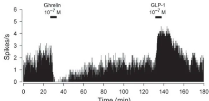
The effects of GLP-1 and OXM on neuronal activity in the ARC are similar. In rats, both hormones induced exci-tatory responses in ghrelin-inhibited cells(67)(see example in Fig. 2) suggesting a stimulation of POMC signalling, which is consistent with the high expression of the
GLP-1R in POMC neurons(65). GLP-1 and OXM induced
heterogeneous excitatory and inhibitory effects in ghrelin-excited ARC neurons, but the functional and neurochem-ical phenotype of these cells have not yet been identified. The absence of GLP-1R expression in NPY neurons seems to exclude the NPY system as a primary target for GLP-1 in the ARC. Despite the existing in vivo and in vitro evi-dence for a potential role of ARC-dependent GLP-1 sig-nalling in energy and glucose homoeostasis further support for a physiological role of endogenous GLP-1R ligands acting via hypothalamic circuits needs to be established. Although beneficial effects on body weight have been reported for anti-diabetic GLP-1 agonists(68–70), moderate effectiveness and undesired side effects limit the potential of these drugs at least as a mono-therapy against obesity.
Feeding and nutrient-related effects on neuronal function Numerous studies have confirmed the reciprocal regulation of NPY and POMC gene expression in response to
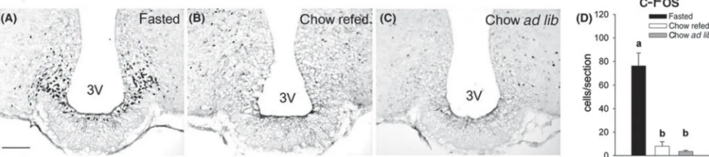
alterations in energy intake(71). Fasting not only leads to alterations in neuropeptide expression but also to altera-tions in neuronal activity. Food deprivation for 24 h elicits an activation of ARC neurons in mice as reflected by an increase in c-Fos expression(72–74). Refeeding of 12 h food-deprived mice completely reverses the fasting-induced ARC activation (Fig. 3) indicating that feeding-related signals exert a strong inhibitory effect on ARC neurons that are activated under negative energy balance(44,75). Hence, ARC neurons appear to respond to negative and positive changes in energy intake. As demonstrated by phenotyping studies, fasting-induced ARC activation spe-cifically occurs in NPY neurons, but not in POMC neurons under these conditions(75,76). While this observation sug-gests increased hypothalamic NPY signalling as a result of fasting, it does not exclude a parallel inhibition of POMC neurons in the ARC. The induction of a fasting-induced ARC activation has also been demonstrated in rats, although considerably longer periods of food deprivation (3 d) are typically required for an immunohistochemically detectable response in this species, at least when c-Fos is used as a marker. Interestingly, there seems to be a shift of neuronal activation from the NPY system in the fasted state to the melanocortin system following refeeding(77,78). This response is consistent with the function of the NPY system to promote energy intake under negative energy balance and with the counter-regulatory function of the POMC system to limit energy intake.
Feeding of palatable non-energy diet (NED; vanilla-fla-voured cellulose) with or without selective nutrient sup-plementation has proven to be a useful approach to explore the role of diet-derived nutrients in feeding-related mod-ulation of neuronal ARC activity. Mice receiving NED for 12 h instead of standard chow exhibit a strong c-Fos expression in the ARC that is only slightly lower than in 12 h fasted animals(75). The same is true for fasted mice that were refed with NED. Therefore, nutrient-independent stimuli associated with feeding activity (e.g. gustatory, oropharyngeal cues and gastric distension) appear to exert a minor, but significant influence on the ARC because they only weakly counteract the stimulation of ARC activation triggered by short-term energy deprivation. When refeed-ing with NED that was selectively supplemented with each of the different macronutrients (carbohydrates, protein or fat), a stronger reversal of fasting-induced ARC activation was observed than that following refeeding with non-supplemented NED(75). When the intake of each nutrient was matched to specific nutrient intake of chow-fed ani-mals, the carbohydrates were more effective than protein and fat. However, increasing the protein content in the diet or refeeding with pure fat (lard) resulted in a similar reversal of fasting-induced c-Fos expression as observed for refeeding with carbohydrate. Therefore, the relative contribution of each macronutrient to the inhibition of ARC activity in this experimental setting appeared to depend on the amount of intake. Whether isoenergetic intakes of the different macronutrients are equipotent to reverse fasting-induced ARC activation has not yet been investigated.
The mechanism underlying the fasting and feeding-related effects on neuronal activity in the ARC are likely to
Fig. 2. Representative recording showing the excitatory effect of glucagon-like peptide-1 (GLP-1) on a ghrelin-inhibited neuron of the arcuate nucleus. Reproduced with permission (published in Riediger
et al.(67)
Proceedings
of
the
Nutrition
Society
involve not only direct effects of metabolites but also indirect actions mediated by gastrointestinal hormones. ARC neurons have been shown to respond to glucose in excitatory and inhibitory ways(79). Phenotypically identi-fied NPY neurons of mice and rats decrease their firing rate with increasing glucose concentrations (glucose-inhibited cells)(80,81). Conversely, POMC neurons have been demonstrated to be glucose-excited because they decrease their discharge rate when the extracellular glucose con-centration is decreased(82,83). The intracellular mechanisms transducing changes in the glucose concentration into altered neuronal activity are well understood for glucose-excited neurons, while the molecular correlates of inhibi-tory glucose signalling are less clear. Glucose sensitivity in glucose-excited neurons has obvious parallels to glucose responsiveness of pancreatic b-cells, in which glucose metabolism leads to increased ATP:ADP ratios. As a con-sequence ATP induces a closure of the Kir6.2 pore-forming subunit of the ATP-sensitive K+ channel, leading to a depolarisation of the cell. Genetic manipulations that cause a loss of function of Kir6.2 subunit are associated with a loss of glucose responsiveness in hypothalamic neurons, including POMC neurons of the ARC(83,84). Although the ATP-sensitive K+ channel is required for the glucose sensitivity of glucose-excited neurons, it is also expressed in glucose-insensitive cells suggesting that other molecular transducers are decisive for neuronal glucose responsiveness. One of these transducers is the enzyme glucokinase, the rate-limiting enzyme for glucose metabo-lism in glucose sensitive neurons(85). Another enzyme that seems to function as a master switch for intracellular metabolic pathways including glucose metabolism is the enzyme adenosine monophosphate-activated protein kinase (AMPK). AMPK responds to changes in the ADP:ATP ratio and induces counter-regulatory metabolic responses through a variety of downstream targets(86). Hypothalamic AMPK activity in the ARC and other hypothalamic struc-tures is inhibited not only by leptin and insulin but also by refeeding in fasted mice and by intraperitoneal and intra-cerebroventricular glucose administration(87). Genetic manipulation that results in constitutive hypothalamic AMPK activity was associated with increased body weight, while reduced AMPK activity resulted in reduced body weight. These effects were partly attributed to changes in
food intake(87). Genetic disruption of AMPK in POMC and NPY neurons was paralleled by a loss of glucose respon-siveness of these cells in the ARC supporting of a role of AMPK in glucose sensing(82). In line with the well-established glucose sensitivity of ARC neurons, an increase in circulating glucose after intraperitoneal glucose administration reversed the fasting-induced ARC activa-tion to a similar degree as refeeding(88). These studies do not exclude indirect hormonal or vagal afferent signalling mechanisms that might be engaged in parallel to a direct action of glucose on ARC neurons.
Not only glucose but also hypothalamic fatty acid sig-nalling contributes to the control of energy balance and glucose homoeostasis because intracerebroventricular infusion of oleic acid inhibits food intake and hepatic glucose production(89,90). Besides other hypothalamic structures, the ARC has been identified as a fatty acid responsive area(91). Lipid sensitivity has been demonstrated for POMC neurons that were inhibited by oleic acid(92). In analogy to the importance of intracellular glucose meta-bolism, intracellular lipid metabolism has been linked to altered neuronal function and to changes in food intake and body weight. Pharmacological blockade fatty acid synthase (FAS) pathway by central or peripheral injection of the FAS inhibitor C75 inhibited feeding and reduced body weight in mice(93). Among other hypothalamic sites, FAS is expressed in NPY neurons of the ARC(94). Peripherally applied C75 prevented the fasting induced c-Fos expres-sion in the medial ARC and in hypothalamic downstream targets of the ARC supporting a role of lipid signalling in fasting-induced ARC activation(95). Different intracellular mechanisms have been proposed to mediate the effects of FAS inhibition on energy homoeostasis. These mechan-isms include an increase in the FAS substrate malonyl CoA, which represents an intracellular metabolic signal inhibiting food intake and body weight. This view is con-sistent with the development of obesity and increased food intake when malonyl CoA degradation in the mediobasal hypothalamus is enhanced by overexpression of the enzyme malonyl CoA decarboxylase(96). A regulatory function for cellular lipid sensing has also been postulated for the enzyme carnitine palmitoyltransferase-1 that con-trols long-chain fatty acid transport into the mitochondria. Pharmacological or genetic inhibition of hypothamalic
Fig. 3. Refeeding with chow completely reversed the fasting-induced c-Fos expression in the arcuate nucleus (ARC). Representative ARC
sections immunostained for c-Fos of 14-h fasted (A), chow-refed (B) andad libitum chow-fed (C) mice. Bar charts show the quantitative
results of c-Fos expression (D). a,b Different letters indicate significant differences between groups (P< 0.05). 3 V: 3rd ventricle. Scale bar:
Proceedings
of
the
Nutrition
Society
carnitine palmitoyltransferase-1 reduced food intake and glucose production(97). Despite the cumulating evidence for a role of hypothalamic lipid sensing further confirma-tion is required for the physiological relevance of these processes in the context of feeding-related or metabolically induced changes in circulating mediators involved in lipid signalling.
Similar to lipid signalling a role of hypothalamic amino acid signalling in the control of food intake and body weight has emerged. The serine-threonine kinase mamma-lian target of rapamycin (mTOR) functions as nutrient sensor implicated in cell growth and proliferation(98). Par-ticularly branched amino acids such as leucine activate the mTOR pathway. Components of the mTOR pathway have been detected in 90 and 45 % of ARC NPY and POMC neurons, respectively(99). In analogy to the changes of neuronal activity in the ARC as a result of fasting and refeeding, activity of the mTOR pathway in the ARC is down-regulated in the fasted state, while it increases in response to refeeding. Both intracerebroventricular and site-specific injection of leucine into the mediobasal hypothalamus inhibited food intake(99,100). POMC neurons of the ARC increase their electrical activity in response to stimulation with leucine, which is consistent with in vivo studies demonstrating a melanocortin receptor involvement in the anorexigenic effect of mediobasal hypothalamic leucin injection(100).
Fasting and the ingestion of each of the single macro-nutrients lead to hormonal responses adjusting energy homoeostasis and gastrointestinal function to the metabolic and digestive requirements of the body. Some attempts have been made to explore the involvement of hormones in the feeding-related changes in ARC activity. Based on the increase in ghrelin levels under fasting conditions, ghrelin might contribute to the fasting-induced ARC activation in mice. In rats, neutralisation of ghrelin by a site-specific injection of anti-ghrelin Ig into the ARC attenuated food intake in fasted animals during refeeding(101). An alter-native approach to neutralise ghrelin is the use of an L-RNA-based hormone antagonist, a so-called Spiegelmer, that specifically binds and inactivates the bioactive form of ghrelin(102,103). Owing to its long-lasting action a single intravenous infusion of the anti-ghrelin Spiegelmer NOX-B11-3 blocked the c-Fos response in the ARC of ad libitum fed mice induced by exogenous ghrelin injected periph-erally 12 h after administration of the antagonist(21). In the same study, ghrelin antagonism did not significantly attenuate the fasting-induced c-Fos response in the ARC suggesting that endogenous ghrelin is not necessary for the activation of the ARC under these experimental conditions. Among the feeding-related hormones that are likely to act via the ARC, ghrelin is the only hormone that increases during fasting. Not only increases but also decreases in hormone levels are likely to reflect alterations in energy homoeostasis and metabolism. One experimental approach to investigate this consists in the compensation of decreased hormone levels by exogenous administration of the respective hormone. Superimposed to the well-described differences in leptin levels related to body adip-osity, leptin plasma levels decrease considerably (5-fold) in fasted v. ad libitum fed lean mice(75). Preventing the
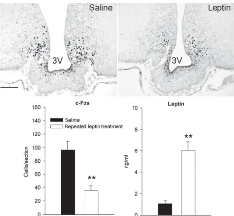
fasting-induced decrease in leptin by repeated low-dose leptin injections during the fasting period markedly attenuated the c-Fos expression of in the ARC of food-deprived mice (Fig. 4)(104). Despite the limitation of this approach to precisely mimic physiological leptin levels by exogenous leptin administration, this finding supports the contribution of decreased leptin signalling to fasting-induced ARC activation. Studies in leptin deficient ob/ob mice seem to complement these findings because fasting-induced ARC activation is exaggerated in ob/ob mice compared with wild-type littermates(104). Together these studies underscore that leptin signalling has an important impact on the ARC in the context of feeding-related modulation of ARC neurons.
Using the fasting/refeeding paradigm the possible involvement of other hormones in feeding-related changes of ARC activity has been studied. Similar to the ineffec-tiveness of an acute leptin treatment to reverse fasting-induced ARC activation, insulin did not attenuate c-Fos expression in fasted animals regardless of whether a glu-cose-lowering dose of insulin was used or a dose that did not produce a reduction in blood glucose(75). Besides insulin the anorexigenic hormones PYY, amylin and cho-lecystokinin (CCK) were tested for their ability to reverse fasting-induced c-Fos expression in the ARC. Only PPY, but not amylin or CCK attenuated the stimulation of ARC neurons in food-deprived mice(75,105). The lack of effec-tiveness was probably not due to ineffective doses, because in the same animals both amylin and CCK elicited c-Fos expression in the AP and the nucleus of the solitary tract (NTS), respectively. As amylin and CCK primarily act via the brainstem (see later) and because the ARC is sidered as a target site for PYY, these findings are con-sistent with the presumed central signalling mechanisms for these hormones. Despite the failure of CCK and amylin to reverse fasting-induced ARC activation, at least CCK partially prevented ghrelin-induced c-Fos expression in the ARC in ad libitum fed mice(75) indicating some CCK-dependent inhibitory input to the ARC. Collectively, these findings suggest diverse roles for the investigated hor-mones in feeding-related changes of neuronal function in the ARC. However, the use of knockout models or specific hormone antagonists is required to confirm the relevance of endogenous hormone actions.
Altered neuronal arcuate nucleus responses in obese rodent models
Obesity is associated with hormonal and metabolic per-turbations, including hyperleptinaemia, hyperinsulinaemia, leptin and insulin resistance, dyslipidaemia and hypergly-caemia and others(12,106,107). In rodent models of diet-induced obesity (DIO) neuronal to responsiveness of ARC neurons to leptin is disrupted. DIO mice show a reduced phosphorylation of the signal transducer and activator of transcription 3 (STAT3), a marker for leptin receptor acti-vation(108,109). Furthermore, the leptin-dependent release of NPY and a-melanocyte stimulating hormone(110) is
decreased. As recently demonstrated, obesity also seems to be associated with ghrelin resistance in the ARC because peripheral or central ghrelin injection did not induce food
Proceedings
of
the
Nutrition
Society
intake and ARC activation in DIO mice(111). Moreover, ghrelin failed to induce the release of NPY and agouti gene-related peptide in hypothalamic explants from DIO mice in this study. Not only hormonal resistance but also impaired responsiveness of the ARC to metabolic stimuli has been described(112).
It has been demonstrated in different obesity models that obese mice show blunted neuronal responses of the ARC to alterations in energy intake. Age-related obese (late-onset obesity) and DIO mice show completely absent or a strongly attenuated fasting-induced activation of NPY neurons in the ARC depending on the duration of food deprivation (12 h or 24 h, respectively). The blunted responses in these animals were not due to age per se or due to high-fat diet feeding because age-matched controls and diet-resistant lean controls showed intense fasting-induced c-Fos responses in the ARC(104,113). The absence of fasting-induced ARC activation was associated with the absence of refeeding hyperphagia that only occurred in lean mice after fasting but not in late-onset obesity mice. The blunted ARC activation in these hyperleptinaemic obesity models contrasts with the exaggerated fasting response in the ARC of obese leptin-deficient ob/ob mice(113). Measurements of metabolic (blood glucose and lipids) and hormonal parameters (insulin, ghrelin and lep-tin) were conducted in fasted and ad libitum fed obese
mice and in respective lean controls(104,113). Among these parameters, the only factor that consistently predicted the altered neuronal responses across the different obesity models was leptin because neuronal ARC activation is enhanced under leptin deficiency, prevented by exogenous leptin and blunted under hyperleptinaemia. However, the idea that obesity-related hyperleptinaemia prevents the response of the ARC to fasting is challenged by well-described leptin resistance in hyperleptinaemic obesity. Factors that limit central effectiveness of circulating leptin involve reduced transport across the blood–brain barrier, increased intracellular inhibition of leptin-signalling and reduced leptin receptor expression. Each of these factors are affected by feeding in a way that promotes leptin responsiveness under fasting conditions. Fasting increases the rate of leptin transport into the brain(114,115), reduces intracellular leptin-antagonistic signalling(116,117) and increases leptin receptor expression(118) and leptin binding to the receptor(119). Hence, leptin responsiveness is dynamic and linked to the feeding status. In line with this assumption, fasting not only increases the leptin-induced STAT3 phosphorylation in lean mice but also in late-onset obesity mice that did not show a significant STAT3 response in the ad libitum fed state(104). Consequently, the combination of increased leptin levels and increased leptin responsiveness might prevent the activation of ARC
Fig. 4. Effect of exogenous leptin-induced hyperleptinaemia during fasting on arcuate nucleus (ARC) activation in lean mice. Representative ARC sections immunostained for c-Fos of young lean mice treated with saline or leptin every 3 h during a 14-h food-deprivation period. Leptin treatment significantly increased the leptin plasma concentration and atte-nuated the fasting-induced c-Fos expression in the ARC (**P< 0.01 for both effects). 3 V: 3rd
ventricle. Scale bar: 100mm. Reproduced with permission (published in Becskei et al.)(104)
Proceedings
of
the
Nutrition
Society
neurons under negative energy balance. Together, these findings suggest that dynamic changes in leptin sensitivity under hyperleptinaemia might interfere with the receptive function of the ARC for stimuli reflecting short-term excursions in energy intake.
The area postrema
The AP is a sensory circumventricular organ located in the dorsal vagal complex of the brainstem. Owing to the lack of a functional blood–brain barrier AP neurons can be reached by humoral factors including peptide hormones that do not passively cross the blood–brain barrier in other areas of the brain. The AP is neuronally interconnected with structures of the visceral enteroceptive neuroaxis including the NTS and the lateral parabrachial nucleus
(120)
. Among other neuronal efferents the AP also projects to the dorsal motor nucleus of the vagus that controls visceral motor functions. The AP plays an important pro-tective role as a so-called chemoreceptor trigger zone that mediates aversive and emetic responses induced by nox-ious or toxic stimuli. This function is for example reflected by the absence of lithium chloride induced taste aversion after ablation of the AP-lesioned (APX) in rats(121).
Observations from APX studies suggested an AP invol-vement in the control of food intake and body weight. APX rats are hypophagic and show lower body weight compared with sham-lesions animals, although they over-consume highly palatable food(122,123). Furthermore, APX leads to loss of hypoglycaemic or glucoprivic feeding responses induced by insulin, 2-deoxy-glucose or 5-thio-glucose(122,124), suggesting a role of the AP in central nervous glucose sensing.
Area postrema-mediated effects of amylin on food intake The AP has been identified as a target structure for the anorexigenic hormone amylin, which is widely accepted to contribute to the physiological control of food intake. Amylin is co-secreted with insulin from pancreaticb-cells in response to meal-related stimuli(125). Amylin reduces food intake at near physiological doses via a reduction of meal size(126–128). Amylin antagonism not only blocks the anorexigenic effect of exogenous amylin but also increases food intake, supporting a physiological role of endogenous amylin in energy intake(129). Numerous studies provided evidence that the AP mediates amylin’s suppressive effect on food intake. While vagal afferents are not required for amylin’s anorexigenic action(130), APX blocks feeding inhibitory action of amylin(131–133).
The effects of amylin on neuronal function in the AP and the transmission of amylin signalling to other brain structures involved in the control of energy homoeostasis are well established. The amylin receptor consists of a functional complex of the calcitonin receptor and so-called receptor activity modifying proteins (receptor activity modifying protein 1 and receptor activity modifying protein 3)(134). The calcitonin receptor is densely ex-pressed in the AP(135), where receptor activity modifying protein 3 gene expression has also been described(136). In
electrophysiological studies, amylin exerted strong excita-tory effects in 44 % of the recorded spontaneously active AP neurons(137). These effects were blocked by the amylin receptor antagonist AC187 and appeared to be mediated by the intracellular second messenger cyclic GMP. The excitatory action of amylin on AP neurons is in line with studies demonstrating an amylin-induced c-Fos expression in the AP(137,138). Not only exogenous but also feeding-related endogenous amylin activates AP neurons because refeeding of fasted rats triggered a c-Fos response in the AP that was attenuated by amylin antagonism(139). About 50 % of neurons showing amylin-induced c-Fos are nora-drenergic and specific neurotoxic lesion of these cells blunted amylin’s feeding inhibitory action(140).
Interaction of amylin and nutrient signalling Glucose. In support of the afore-mentioned evidence for a role of the AP as central glucose sensor, electro-physiological studies identified glucose-responsive neurons in the AP(141). These studies were extended by the demonstration that amylin-sensitive neurons in the AP are specifically excited by glucose (Fig. 5), whereas amylin-insensitive neurons are unresponsive to changes in the ambient glucose concentration(142). Based on the impor-tance of amylin-sensitive AP neurons for the physiological control of feeding behaviour, these findings further support a role of the AP in the metabolic control of food intake. The AP is not the only glucose sensitive structure in the brainstem, but it is located in a brainstem area where local injection of 5-thio-glucose induced the strongest gluco-privic feeding responses(143).
The glucose sensitivity of the AP has also been postu-lated as a protective mechanism against excursions in blood glucose values. Hypoglycaemia induces a counter-regulatory increase in the rate of gastric emptying(144) that compensates the decreased blood glucose via increased gastrointestinal nutrient absorption. This mechanism inter-feres with hormonal and pharmacological effects that delay gastric emptying because glucose appears to be a permis-sive factor. Amylin potently inhibits gastric emptying via an AP-dependent action, which is overridden by hypogly-caemia(145). Our group recently demonstrated a similar permissive effect of glucose for amylin’s feeding suppres-sive effect because amylin failed to significantly inhibit feeding under hypoglycemic conditions (C Neuner-Boyle, unpublished results). The inhibitory effect of the amylin agonist pramlintide on gastric emptying is an important therapeutic mechanism to lower postprandial glucose levels in diabetic patients. As treatment-induced hypogly-caemia is a severe risk in diabetic patients, the permissive effect of glucose on gastric emptying is considered as a fail-safe mechanism that overrides the anti-diabetic action of amylin agonism under hypoglycaemic conditions (see(146,147)for review).
Protein. Amylin’s satiating and effect and amylin-induced AP activation are modulated by diet-derived macronutrients, in particular by protein. An influence of diet-derived nutrients on amylin signalling was inferred from the observation that amylin-induced activation of the AP is stronger in fasted v. ad libitum fed rats and also in
Proceedings
of
the
Nutrition
Society
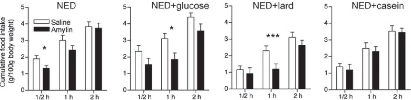
rats that received nutrient-deficient NED(148). Selective supplementation of NED with protein but not with glucose or fat strongly attenuated the amylin-induced AP activation suggesting that protein counteracts anorexigenic amylin signalling. In light of the well-established satiating action of protein this assumption appeared unexpected, but it was confirmed by feeding studies in which amylin failed to inhibit protein supplemented NED (Fig. 6)(148). The inhi-bitory effect of diet-derived protein on amylin action has been corroborated by immunohistological and feeding studies using isoenergetic diets of different protein content. Amylin more potently decreased intake of 1 and 8 % pro-tein chow than standard chow containing 18 % propro-tein. Similarly, amylin-induced c-Fos expression was higher in rats receiving less protein(149). These effects were not sec-ondary to altered energy intake that was similar across the experimental groups. While blood glucose levels were also similar, blood amino acid levels decreased in rats fed low-protein diet suggesting that circulating amino acids might contribute the protein-dependent attenuation of amylin-induced AP activation. Pre-treatment of rats with
an intraperitoneal administration of a mixed amino acid solution also attenuated amylin’s c-Fos response in the AP, which supports this hypothesis(149). However, the exact neuronal mechanism underlying the protein-dependent modulation of amylin signalling remains to be identified. The relationship between reduced amylin-responsiveness and protein intake might bear both physiological and therapeutic implications. Amylin’s function in the control of nutrient and energy intake might be adapted to the nutrient composition of the diet. At least under undisturbed energy homoeostasis protein is metabolically less impor-tant than carbohydrate or fat, which might suggest that amylin’s contribution to the overall control of energy intake is stronger when the intake of protein relative to glucose or fat is low. Another mutually not exclusive implication might consist of an AP-sensitising effect pro-duced by short-term reduction of food intake or food availability in a protein-dependent manner, pre-adapting AP responsiveness to a higher level in the pre-prandial state before meal onset. In addition to the possible phy-siological relevance, the effectiveness of pharmacological
Fig. 6. Effect of amylin (5mg/kg s.c.) on food intake in rats kept on different test diets (NED, non-energy diet, vanilla-flavoured
cellulose) for 24 h prior to injection. Bars represent group means andSEM(n 12). *P< 0.05 and ***P < 0.001, significantly different
from respective control (saline) group (paired Student t test). Modified and reproduced with permission (published in Michel
et al.(148)).
–
Fig. 5. Co-sensitivity of an area postrema (AP) neuron to glucose and amylin. Decreasing the glucose concentration in the superfusion solution from the standard concentration of 10 mM to 2, 4 and 6 mM during the indicated time caused concentration-dependent decreases of the spontaneous discharge rate. Superfusion of amylin elicited a strong excitatory effect. Reproduced with permission (published
Proceedings
of
the
Nutrition
Society
approaches based on amylin agonism might be influenced in a nutrient-dependent way.
Fat. There is not much experimental evidence for a role of the AP in lipid sensing. Although lipoprivic feeding induced by the fatty acid oxidation inhibitor mercaptoace-tate is blunted in rats after AP/NTS lesion, this effect has been attributed to the destruction of vagal terminal fields in the NTS(150). Accordingly, lipoprivic feeding is also blocked in vagotomised rats, confirming a mediation of vagal afferent signals(150). Similarly, c-Fos expression in the AP/NTS region after intraduodenal lipid infusion is attenuated by capsaicin-induced destruction of vagal afferents and by CCK receptor antagonism. The latter finding indicates an indirect effect of intestinal lipid sen-sing via CCK-mediated activation the vagal afferents sig-nalling(151). It is reasonable to assume that amylin signalling and vagally transmitted lipid/CCK dependent signals converge at the level of the AP/NTS region. Fur-thermore, the existence of a satiogenic synergy between amylin and CCK(152) might imply that lipid-dependent signals indirectly enhance amylin’s feeding suppressive effect via a CCK-dependent action. Unlike the develop-ment of hormonal resistance to leptin and insulin as a
consequence of prolonged high-fat diet feeding,
neither short-term nor long-term high-fat diet feeding leads to amylin resistance at least when amylin’s inhibitory action on food intake is used as an index of amylin responsiveness(153).
Hormonal interactions involving area postrema and brainstem mechanisms
The dorsal vagal complex is a site of convergence for vagal and blood-borne signals controlling food intake. Brainstem structures including amylin-sensitive sites are reciprocally interconnected with hypothalamic and extra-hypothalamic areas involved in energy homoeostasis(154). For some hormones known to act via the brainstem synergistic effects have been described, although the exis-tence of a synergy is not always based on equal criteria in the literature. Synergy is often inferred from the observa-tion that the effect of two combined stimuli is higher than the sum of the effects when each stimulus is applied alone. Sophisticated statistical models (e.g. isobolographic ana-lysis) have been used to detect and describe synergistic actions. Several synergistic effects have been demonstrated for amylin in combination with other hormones(155). In addition to the synergy between amylin and CCK men-tioned earlier, there is a synergistic effect of amylin and leptin to reduce body weight in obese and lean subjects(3,4). Several neuronal mechanisms have been associated with the synergy between amylin and leptin. Amylin seems to enhance neuronal leptin responsiveness as reflected by the reversal of the blunted STAT3 response to leptin in the ventromedial hypothalamus. Amylin also affects leptin signalling in the brainstem because amylin knockout mice tend to show reduced leptin-induced STAT3 phosphoryla-tion in the NTS, and pre-treating leptin-resistant DIO rats with amylin increased leptin-dependent STAT3 signalling in the AP(3,4).
Anorexigenic synergy has also been reported for amylin and PYY(3–36), whereas body weight reduction was additive(49). The neuronal correlate of this anorexigenic synergy has not yet been identified. While the AP was not required for PYY(3–36)-induced suppression of feeding in one study(156,157), another study suggested an involvement of the AP in the PYY-dependent effects on gastric acid secretion(158). Binding sites for PYY of the Y receptor family are abundant in the AP(159–161). Interestingly, a dose of full-length PYY that did not trigger a c-Fos response in the AP alone, increased amylin-induced c-Fos expression in the AP(105) suggesting a possible interaction between amylin and PYY in the AP.
A recent study conducted in non-human primates demonstrated anorexigenic synergy between the potent amylin receptor agonist salmon calcitonin and the GLP-1R agonist exendin-4(162). The mechanism underlying this synergy also remains to be identified. There is evidence that GLP-1 inhibits feeding via an AP-dependent effect after hepatic portal vein infusion(163). However, the anor-exigenic effect of exendin-4 is not blocked in APX rats(164). Although amylin and GLP-1 activate AP neurons, these hormones seem to have different target cells in the AP. Similar to the aversive stimulus lithium chloride, GLP-1 and exendin-4 almost exclusively activate AP neurons that do not express the calcitonin receptor, which is con-sidered as a marker for the primary target cells for amylin in the AP(149). The functional implication of this dissocia-tion has not yet been specifically investigated. GLP-1R-mediated activation of the AP has been linked to the induction of aversion(165). Therefore, it is particularly important to exclude aversive mechanisms in approaches based on GLP-1R activation.
Conclusions
Extensive evidence has accumulated demonstrating
that the ARC and the AP are responsive to hormonal and nutrient-related signals involved in the homoeostatic feed-back control of energy homoeostasis. Overall, the neuro-physiological effects that have been described by a multitude of different in vivo and in vitro approaches are consistent with the effects of these signals on food intake, body weight or metabolism. However, for many of the discussed mechanisms, the physiological relevance remains to be confirmed. Even when physiological relevance is established (e.g. amylin) or postulated (ghre-lin, PYY and GLP-1) the predictive value of these physiological mechanisms for the effectiveness of mono-therapeutic anti-obesity approaches targeting these mechan-isms appears to be weak. Nevertheless, the impressive effectiveness of bariatric surgery suggests the existence of signalling mechanisms that potently reduce body weight and improve metabolic disorders without being compensated by the body. Obviously, the interaction between the different feedback signals has been underestimated, which is reflected by the promising improvements in the therapeutic outcome of combination treatments. The concept of the homoeostatic feedback control of energy homoeostasis needs to be
Proceedings
of
the
Nutrition
Society
integrated into the increasing knowledge about the non-homoeostatic controls affecting energy balance.
Acknowledgements
The author thanks T.A. Lutz (Institute of Veterinary Phy-siology, University of Zurich) for the critical reading of the manuscript. The research presented in this work has been continuously supported by the Swiss National Science Foundation, the University of Zurich and the Centre of Integrative Human Physiology, University of Zurich. The author declares no conflicts of interest.
References
1. Ferchak CV & Meneghini LF (2004) Obesity, bariatric surgery and type 2 diabetes – a systematic review. Diabetes Metab Res Rev 20, 438–445.
2. Ahn SM, Pomp A & Rubino F (2010) Metabolic surgery for type 2 diabetes. Ann N Y Acad Sci 1212, E37–E45. 3. Roth JD, Roland BL, Cole RL, et al. (2008) Leptin
responsiveness restored by amylin agonism in diet-induced obesity: evidence from nonclinical and clinical studies. Proc Natl Acad Sci USA 105, 7257–7262.
4. Turek VF, Trevaskis JL, Levin BE, et al. (2010) Mechan-isms of amylin/leptin synergy in rodent models. Endocri-nology 151, 143–152.
5. Zhang Y, Proenca R, Maffei M, et al. (1994) Positional cloning of the mouse obese gene and its human homologue. Nature 372, 425–432.
6. Chua S, Chung W, Wu-Peng X, et al. (1996) Phenotypes of mouse diabetes and rat fatty due to mutations in the OB (leptin) receptor. Science 271, 994–996.
7. Broberger C, Johansen J, Johansson C, et al. (1998) The neuropeptide Y/agouti gene-related protein (AGRP) brain circuitry in normal, anorectic, and monosodium glutamate-treated mice. Proc Natl Acad Sci USA 95, 15043–15048. 8. Ellacott K & Cone R (2004) The central melanocortin
sys-tem and the integration of short- and long-term regulators of energy homeostasis. Recent Prog Horm Res 59, 395–408. 9. Cowley M, Smart J, Rubinstein M, et al. (2001) Leptin
activates anorexigenic POMC neurons through a neural network in the arcuate nucleus. Nature 411, 480–484. 10. Spanswick D, Smith M, Groppi V, et al. (1997) Leptin
inhibits hypothalamic neurons by activation of ATP-sensitive potassium channels. Nature 390, 521–525. 11. Woods SC & Seeley RJ (2000) Adiposity signals and the
control of energy homeostasis. Nutrition 16, 894–902. 12. Heymsfield SB, Greenberg AS, Fujioka K, et al. (1999)
Recombinant leptin for weight loss in obese and lean adults: a randomized, controlled, dose-escalation trial. JAMA 282, 1568–1575.
13. Boden G, Chen X, Mozzoli M, et al. (1996) Effect of fast-ing on serum leptin in normal human subjects. J Clin Endocrinol Metab 81, 3419–3423.
14. Weigle DS, Duell PB, Connor WE, et al. (1997) Effect of fasting, refeeding, and dietary fat restriction on plasma leptin levels. J Clin Endocrinol Metab 82, 561–565. 15. Kojima M, Hosoda H, Date Y, et al. (1999) Ghrelin is a
growth-hormone-releasing acylated peptide from stomach. Nature 402, 656–660.
16. Tschop M, Smiley D & Heiman M (2000) Ghrelin induces adiposity in rodents. Nature 407, 908–913.
17. Asakawa A, Inui A, Kaga T, et al. (2001) Ghrelin is an appetite-stimulatory signal from stomach with structural resemblance to motilin. Gastroenterology 120, 337–345. 18. Shiiya T, Nakazato M, Mizuta M, et al. (2002) Plasma
ghrelin levels in lean and obese humans and the effect of glucose on ghrelin secretion. J Clin Endocrinol Metab 87, 240–244.
19. Traebert M, Riediger T, Whitebread S, et al. (2002) Ghrelin acts on leptin-responsive neurones in the rat arcuate nucleus. J Neuroendocrinol 14, 580–586.
20. Riediger T, Traebert M, Schmid HA, et al. (2003) Site-specific effects of ghrelin on the neuronal activity in the hypothalamic arcuate nucleus. Neurosci Lett 341, 151–155. 21. Becskei C, Bilik KU, Klussmann S, et al. (2008) The anti-ghrelin Spiegelmer NOX-B11-3 blocks anti-ghrelin- but not fasting-induced neuronal activation in the hypothalamic arcuate nucleus. J Neuroendocrinol 20, 85–92.
22. Wang L, Saint-Pierre DH & Tache Y (2002) Peripheral ghrelin selectively increases Fos expression in neuropeptide Y – synthesizing neurons in mouse hypothalamic arcuate nucleus. Neurosci Lett 325, 47–51.
23. Guan X, Yu H, Palyha O, et al. (1997) Distribution of mRNA encoding the growth hormone secretagogue receptor in brain and peripheral tissues. Brain Res Mol Brain Res 48, 23–29.
24. Willesen M, Kristensen P & Romer J (1999) Co-localization of growth hormone secretagogue receptor and NPY mRNA in the arcuate nucleus of the rat. Neuroendocrinology 70, 306–316.
25. Hewson AK, Viltart O, McKenzie DN, et al. (1999) GHRP-6-induced changes in electrical activity of single cells in the arcuate, ventromedial and periventricular nucleus neurones [correction of nuclei] of a hypothalamic slice preparation in vitro. J Neuroendocrinol 11, 919–923.
26. Salome N, Haage D, Perrissoud D, et al. (2009) Anorexi-genic and electrophysiological actions of novel ghrelin receptor (GHS-R1A) antagonists in rats. Eur J Pharmacol 612, 167–173.
27. Tung YC, Hewson AK & Dickson SL (2001) Actions of leptin on growth hormone secretagogue-responsive neu-rones in the rat hypothalamic arcuate nucleus recorded in vitro. J Neuroendocrinol 13, 209–215.
28. Cowley MA, Smith RG, Diano S, et al. (2003) The dis-tribution and mechanism of action of ghrelin in the CNS demonstrates a novel hypothalamic circuit regulating energy homeostasis. Neuron 37, 649–661.
29. Dickson SL & Luckman SM (1997) Induction of c-Fos messenger ribonucleic acid in neuropeptide Y and growth hormone (GH)-releasing factor neurons in the rat arcuate nucleus following systemic injection of the GH secretagogue, GH-releasing peptide-6. Endocrinology 138, 771–777.
30. Geary N (2004) Endocrine controls of eating: CCK, leptin, and ghrelin. Physiol Behav 81, 719–733.
31. Kirchner H, Heppner KM & Tschop MH (2012) The role of ghrelin in the control of energy balance. Handb Exp Phar-macol, 161–184.
32. Cummings DE, Weigle DS, Frayo RS, et al. (2002) Plasma ghrelin levels after diet-induced weight loss or gastric bypass surgery. N Engl J Med 346, 1623–1630.
33. Dickson SL, Egecioglu E, Landgren S, et al. (2011) The role of the central ghrelin system in reward from food and chemical drugs. Mol Cell Endocrinol 340, 80–87.
34. Borg CM, le Roux CW, Ghatei MA, et al. (2006) Pro-gressive rise in gut hormone levels after Roux-en-Y gastric bypass suggests gut adaptation and explains altered satiety. Br J Surg 93, 210–215.
Proceedings
of
the
Nutrition
Society
35. le Roux CW, Aylwin SJ, Batterham RL, et al. (2006) Gut hormone profiles following bariatric surgery favor an anor-ectic state, facilitate weight loss, and improve metabolic parameters. Ann Surg 243, 108–114.
36. le Roux CW, Welbourn R, Werling M, et al. (2007) Gut hormones as mediators of appetite and weight loss after Roux-en-Y gastric bypass. Ann Surg 246, 780–785. 37. Chandarana K, Gelegen C, Karra E, et al. (2011) Diet and
gastrointestinal bypass-induced weight loss: the roles of ghrelin and peptide YY. Diabetes 60, 810–818.
38. Grandt D, Schimiczek M, Beglinger C, et al. (1994) Two molecular forms of peptide YY (PYY) are abundant in human blood: characterization of a radioimmunoassay recognizing PYY 1-36 and PYY 3-36. Regul Pept 51, 151–159.
39. Dumont Y, Fournier A, St-Pierre S, et al. (1995) Char-acterization of neuropeptide Y binding sites in rat brain membrane preparations using [125I][Leu31,Pro34]peptide YY and [125I]peptide YY3-36 as selective Y1 and Y2 radioligands. J Pharmacol Exp Ther 272, 673–680. 40. Degen L, Oesch S, Casanova M, et al. (2005) Effect of
peptide YY3-36 on food intake in humans. Gastro-enterology 129, 1430–1436.
41. Sloth B, Holst JJ, Flint A, et al. (2007) Effects of PYY1-36 and PYY3-36 on appetite, energy intake, energy expendi-ture, glucose and fat metabolism in obese and lean subjects. Am J Physiol Endocrinol Metab 292, E1062–E1068. 42. Batterham RL, Cowley MA, Small CJ, et al. (2002) Gut
hormone PYY(3-36) physiologically inhibits food intake. Nature 418, 650–654.
43. Ghamari-Langroudi M, Colmers WF & Cone RD (2005) PYY3-36 inhibits the action potential firing activity of POMC neurons of arcuate nucleus through postsynaptic Y2 receptors. Cell Metab 2, 191–199.
44. Riediger T, Bothe C, Becskei C, et al. (2004) Peptide YY directly inhibits ghrelin-activated neurons of the arcuate nucleus and reverses fasting-induced c-Fos expression. Neuroendocrinology 79, 317–326.
45. Batterham RL, Cohen MA, Ellis SM, et al. (2003) Inhibi-tion of food intake in obese subjects by peptide YY3-36. N Engl J Med 349, 941–948.
46. Adams SH, Lei C, Jodka CM, et al. (2006) PYY[3-36] administration decreases the respiratory quotient and reduces adiposity in diet-induced obese mice. J Nutr 136, 195–201.
47. Koegler FH, Enriori PJ, Billes SK, et al. (2005) Peptide YY(3-36) inhibits morning, but not evening, food intake and decreases body weight in rhesus macaques. Diabetes 54, 3198–3204.
48. Talsania T, Anini Y, Siu S, et al. (2005) Peripheral exendin-4 and peptide YY(3-36) synergistically reduce food intake through different mechanisms in mice. Endocrinology 146, 3748–3756.
49. Roth JD, Coffey T, Jodka CM, et al. (2007) Combination therapy with amylin and peptide YY[3–36] in obese rodents: anorexigenic synergy and weight loss additivity. Endocrinology 148, 6054–6061.
50. Orskov C, Wettergren A & Holst JJ (1996) Secretion of the incretin hormones glucagon-like peptide-1 and gastric inhibitory polypeptide correlates with insulin secretion in normal man throughout the day. Scand J Gastroenterol 31, 665–670.
51. Kreymann B, Williams G, Ghatei MA, et al. (1987) Glu-cagon-like peptide-1 7-36: a physiological incretin in man. Lancet 2, 1300–1304.
52. Holst JJ (2007) The physiology of glucagon-like peptide 1. Physiol Rev 87, 1409–1439.
53. Larsen PJ, Tang-Christensen M, Holst JJ, et al. (1997) Distribution of glucagon-like peptide-1 and other pre-proglucagon-derived peptides in the rat hypothalamus and brainstem. Neuroscience 77, 257–270.
54. Merchenthaler I, Lane M & Shughrue P (1999) Distribution of pre-pro-glucagon and glucagon-like peptide-1 receptor messenger RNAs in the rat central nervous system. J Comp Neurol 403, 261–280.
55. Alvarez E, Roncero I, Chowen JA, et al. (1996) Expression of the glucagon-like peptide-1 receptor gene in rat brain. J Neurochem 66, 920–927.
56. Turton MD, O’Shea D, Gunn I, et al. (1996) A role for glucagon-like peptide-1 in the central regulation of feeding. Nature 379, 69–72.
57. Rodriquez de Fonseca F, Navarro M, Alvarez E, et al. (2000) Peripheral versus central effects of glucagon-like peptide-1 receptor agonists on satiety and body weight loss in Zucker obese rats. Metab Clin Exp 49, 709–717. 58. Dakin CL, Small CJ, Batterham RL, et al. (2004) Peripheral
oxyntomodulin reduces food intake and body weight gain in rats. Endocrinology 145, 2687–2695.
59. Reidelberger RD, Haver AC, Apenteng BA, et al. (2011) Effects of exendin-4 alone and with peptide YY(3-36) on food intake and body weight in diet-induced obese rats. Obesity (Silver Spring) 19, 121–127.
60. Verdich C, Flint A, Gutzwiller JP, et al. (2001) A meta-analysis of the effect of glucagon-like peptide-1 (7-36) amide on ad libitum energy intake in humans. J Clin Endocrinol Metab 86, 4382–4389.
61. Ruttimann EB, Arnold M, Hillebrand JJ, et al. (2009) Intrameal hepatic portal and intraperitoneal infusions of glucagon-like peptide-1 reduce spontaneous meal size in the rat via different mechanisms. Endocrinology 150, 1174–1181.
62. Punjabi M, Arnold M, Geary N, et al. (2011) Peripheral glucagon-like peptide-1 (GLP-1) and satiation. Physiol Behav 105, 71–76.
63. Le Quellec A, Kervran A, Blache P, et al. (1992) Oxynto-modulin-like immunoreactivity: diurnal profile of a new potential enterogastrone. J Clin Endocrinol Metab 74, 1405–1409.
64. Baggio LL, Huang Q, Brown TJ, et al. (2004) Oxyntomo-dulin and glucagon-like peptide-1 differentially regulate murine food intake and energy expenditure. Gastro-enterology 127, 546–558.
65. Sandoval DA, Bagnol D, Woods SC, et al. (2008) Arcuate glucagon-like peptide 1 receptors regulate glucose homeostasis but not food intake. Diabetes 57, 2046–2054.
66. Dakin CL, Gunn I, Small CJ, et al. (2001) Oxyntomodulin inhibits food intake in the rat. Endocrinology 142, 4244– 4250.
67. Riediger T, Eisele N, Scheel C, et al. (2010) Effects of glucagon-like peptide 1 and oxyntomodulin on neuronal activity of ghrelin-sensitive neurons in the hypothalamic arcuate nucleus. Am J Physiol Regul Integr Comp Physiol 298, R1061–R1067.
68. Klonoff DC, Buse JB, Nielsen LL, et al. (2008) Exenatide effects on diabetes, obesity, cardiovascular risk factors and hepatic biomarkers in patients with type 2 diabetes treated for at least 3 years. Curr Med Res Opin 24, 275–286. 69. Rosenstock J, Klaff LJ, Schwartz S, et al. (2010) Effects of
exenatide and lifestyle modification on body weight and glucose tolerance in obese subjects with and without pre-diabetes. Diabetes Care 33, 1173–1175.
70. Vilsboll T, Christensen M, Junker AE, et al. (2012) Effects of glucagon-like peptide-1 receptor agonists on
Proceedings
of
the
Nutrition
Society
weight loss: systematic review and meta-analyses of ran-domised controlled trials. BMJ 344, d7771.
71. Schwartz MW, Woods SC, Porte D Jr, et al. (2000) Central nervous system control of food intake. Nature 404, 661–671.
72. Wang L, Martinez V, Barrachina MD, et al. (1998) Fos expression in the brain induced by peripheral injection of CCK or leptin plus CCK in fasted lean mice. Brain Res 791, 157–166.
73. Ueyama E, Morikawa Y, Yasuda T, et al. (2004) Attenuation of fasting-induced phosphorylation of mitogen-activated protein kinases (ERK/p38) in the mouse hypotha-lamus in response to refeeding. Neurosci Lett 371, 40–44. 74. Mistry AM, Helferich W & Romsos DR (1994) Elevated
neuronal c-Fos-like immunoreactivity and messenger ribo-nucleic acid (mRNA) in genetically obese (ob/ob) mice. Brain Res 666, 53–60.
75. Becskei C, Lutz TA & Riediger T (2009) Diet-derived nutrients mediate the inhibition of hypothalamic NPY neu-rons in the arcuate nucleus of mice during refeeding. Am J Physiol Regul Integr Comp Physiol 297, R100–R110. 76. Coppola A, Liu ZW, Andrews ZB, et al. (2007) A central
thermogenic-like mechanism in feeding regulation: an interplay between arcuate nucleus T3 and UCP2. Cell Metab 5, 21–33.
77. Sanchez E, Singru PS, Acharya R, et al. (2008) Differential effects of refeeding on melanocortin-responsive neurons in the hypothalamic paraventricular nucleus. Endocrinology 149, 4329–4335.
78. Singru PS, Sanchez E, Fekete C, et al. (2007) Importance of melanocortin signaling in refeeding-induced neuronal acti-vation and satiety. Endocrinology 148, 638–646.
79. Wang R, Liu X, Hentges ST, et al. (2004) The regulation of glucose-excited neurons in the hypothalamic arcuate nucleus by glucose and feeding-relevant peptides. Diabetes 53, 1959–1965.
80. Fioramonti X, Contie S, Song Z, et al. (2007) Character-ization of glucosensing neuron subpopulations in the arcuate nucleus: integration in neuropeptide Y and pro-opio mela-nocortin networks? Diabetes 56, 1219–1227.
81. Muroya S, Yada T, Shioda S, et al. (1999) Glucose-sensitive neurons in the rat arcuate nucleus contain neuro-peptide Y. Neurosci Lett 264, 113–116.
82. Claret M, Smith MA, Batterham RL, et al. (2007) AMPK is essential for energy homeostasis regulation and glucose sensing by POMC and AgRP neurons. J Clin Invest 117, 2325–2336.
83. Parton LE, Ye CP, Coppari R, et al. (2007) Glucose sensing by POMC neurons regulates glucose homeostasis and is impaired in obesity. Nature 449, 228–232.
84. Miki T, Liss B, Minami K, et al. (2001) ATP-sensitive K+ channels in the hypothalamus are essential for the main-tenance of glucose homeostasis. Nat Neurosci 4, 507–512. 85. Levin BE, Routh VH, Kang L, et al. (2004) Neuronal
glu-cosensing: what do we know after 50 years? Diabetes 53, 2521–2528.
86. Kahn BB, Alquier T, Carling D, et al. (2005) AMP-activated protein kinase: ancient energy gauge provides clues to modern understanding of metabolism. Cell Metab 1, 15–25.
87. Minokoshi Y, Alquier T, Furukawa N, et al. (2004) AMP-kinase regulates food intake by responding to hormonal and nutrient signals in the hypothalamus. Nature 428, 569–574.
88. Becskei C, Lutz TA & Riediger T (2008) Glucose reverses fasting-induced activation in the arcuate nucleus of mice. Neuroreport 19, 105–109.
89. Morgan K, Obici S & Rossetti L (2004) Hypothalamic responses to long-chain fatty acids are nutritionally regu-lated. J Biol Chem 279, 31139–31148.
90. Obici S, Feng Z, Morgan K, et al. (2002) Central adminis-tration of oleic acid inhibits glucose production and food intake. Diabetes 51, 271–275.
91. Wang R, Cruciani-Guglielmacci C, Migrenne S, et al. (2006) Effects of oleic acid on distinct populations of neu-rons in the hypothalamic arcuate nucleus are dependent on extracellular glucose levels. J Neurophysiol 95, 1491–1498. 92. Jo YH, Su Y, Gutierrez-Juarez R, et al. (2009) Oleic acid directly regulates POMC neuron excitability in the hypo-thalamus. J Neurophysiol 101, 2305–2316.
93. Loftus TM, Jaworsky DE, Frehywot GL, et al. (2000) Reduced food intake and body weight in mice treated with fatty acid synthase inhibitors. Science 288, 2379–2381. 94. Kim EK, Miller I, Landree LE, et al. (2002) Expression of
FAS within hypothalamic neurons: a model for decreased food intake after C75 treatment. Am J Physiol Endocrinol Metab 283, E867–E879.
95. Miller I, Ronnett GV, Moran TH, et al. (2004) Anorexi-genic C75 alters c-Fos in mouse hypothalamic and hind-brain subnuclei. Neuroreport 15, 925–929.
96. He W, Lam TK, Obici S, et al. (2006) Molecular disruption of hypothalamic nutrient sensing induces obesity. Nat Neu-rosci 9, 227–233.
97. Obici S, Feng Z, Arduini A, et al. (2003) Inhibition of hypothalamic carnitine palmitoyltransferase-1 decreases food intake and glucose production. Nat Med 9, 756–761. 98. Jacinto E & Hall MN (2003) Tor signalling in bugs, brain
and brawn. Nat Rev Mol Cell Biol 4, 117–126.
99. Cota D, Proulx K, Smith KA, et al. (2006) Hypothalamic mTOR signaling regulates food intake. Science 312, 927–930.
100. Blouet C, Jo YH, Li X, et al. (2009) Mediobasal hypotha-lamic leucine sensing regulates food intake through activa-tion of a hypothalamus-brainstem circuit. J Neurosci 29, 8302–8311.
101. Bagnasco M, Tulipano G, Melis MR, et al. (2003) Endo-genous ghrelin is an orexigenic peptide acting in the arcuate nucleus in response to fasting. Regul Pept 111, 161–167. 102. Helmling S, Maasch C, Eulberg D, et al. (2004) Inhibition
of ghrelin action in vitro and in vivo by an RNA-Spie-gelmer. Proc Natl Acad Sci USA 101, 13174–13179. 103. Kobelt P, Helmling S, Stengel A, et al. (2006)
Anti-ghrelin Spiegelmer NOX-B11 inhibits neurostimulatory and orexigenic effects of peripheral ghrelin in rats. Gut 55, 788–792.
104. Becskei C, Lutz TA & Riediger T (2010) Reduced fasting-induced activation of hypothalamic arcuate neurons is associated with hyperleptinemia and increased leptin sensi-tivity in obese mice. Am J Physiol Regul Integr Comp Physiol 299, R632–R641.
105. Riediger T, Michel S, Becskei C, et al. (2007) Diet-derived nutrients and PYY modulate the effects of amylin on c-Fos expression in the area postrema and on food intake. 2007 Neuroscience Meeting Planner. San Diego, CA: Society for Neuroscience, 2010. Online. Program No. 299.7/UU3. 106. Halaas JL, Boozer C, Blair-West J, et al. (1997)
Physiolo-gical response to long-term peripheral and central leptin infusion in lean and obese mice. Proc Natl Acad Sci USA 94, 8878–8883.
107. Bjorntorp P (1997) Body fat distribution, insulin resistance, and metabolic diseases. Nutrition 13, 795–803.
108. Munzberg H, Flier JS & Bjorbaek C (2004) Region-specific leptin resistance within the hypothalamus of diet-induced obese mice. Endocrinology 145, 4880–4889.