Comparison of the effects of GnRH-I and GnRH-II on HCG synthesis and secretion by first trimester trophoblast
7
0
0
Texte intégral
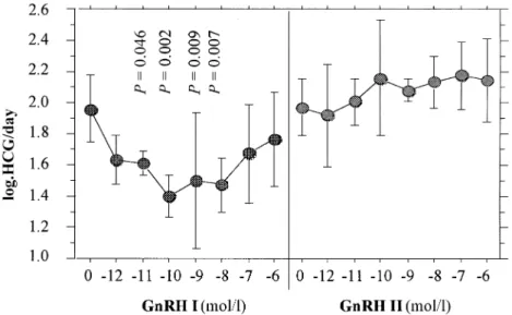
(2) D.Islami et al.. have also been detected in the goldfish brain and pituitary. Although these receptors share 71% identity, there are marked differences in their ligand selectivity (Illing et al., 1999). Since the physiological effects of GnRH-II have not yet been described, we undertook the present study to see whether GnRH-II could be involved in the secretion and synthesis of HCG in first trimester trophoblast.. Materials and methods A total of 32 placentae were obtained by elective pregnancy terminations, carried out in the first trimester (6–11 weeks, legal abortions). Preparation and culture of cytotrophoblastic cells (CTB) CTB were prepared according to an already published protocol (Bischof et al., 1991). Briefly, first-trimester trophoblasts, obtained by legal abortions, were dissected out and rinsed to remove the blood. The tissue was minced and digested four times with trypsin and layered onto a discontinuous Percoll gradient. After centrifugation, the layer of CTBs was aspirated and the cells immunopurified using an antibody to CD45 (Dako, Glostrup, Denmark) to eliminate the contaminating leukocytes. Experiment 1 CTB (5⫻105/ well) were incubated in duplicate in serum-free Dulbecco’s modified Eagle’s medium (DMEM; Gibco, Basel Switzerland), containing penicillin (100 IU/ml), streptomycin (100 µg/ml), gentamycin (100 µg/ml) and fungison (2.5 µg/ml), for 96 h with or without varying concentrations (10–12 to 10–6 mol/l) of GnRH-I or GnRH-II (a generous gift from Ferring, Zu¨ rich, Switzerland). Media were changed on day 2 and 4 and the cultures stopped on day 4. At least three experiments (including the appropriate controls) were performed for each culture condition, using different CTB preparations. Experiment 2 CTB were prepared as for experiment 1 and the same incubation protocol was used, except that GnRH-I or GnRH-II were incubated for only 4 h and the media harvested at 4, 8 and 24 h. Cultures were stopped at 24 h. Preparation of placental explants and superfusion culture A superfusion apparatus, consisting of a peristaltic pump driving culture medium through a thermostatic (37°C) cell culture chamber to a fraction collector, was used to study the dynamics of HCG secretion. Placental explants of 250–300 mg wet weight were dissected out from abortion products, placed in a large excess of sterile 0.9% NaCl solution to remove all blood elements and finally rinsed in antibiotic solution (penicillin 200 IU/ml and streptomycin 200 µg/ml). Subsequently, tissues were placed into the culture chambers of the superfusion apparatus and eluted with pre-gassed (5% CO2 in air) HEPES-buffered DMEM (with the components as described for DMEM preparation above). After collecting fractions (1 ml) of medium every 3 min for 120 min, a pulse (10–8 mol/l in 200 µl of medium) of GnRH-I or GnRH-II was added to the elution medium and sampling was continued for another period of 2 h. The experiment was repeated five times with GnRH-I and GnRH-II. Both forms of GnRH were systematically tested in parallel on different explants of the same placenta. Measurement of HCG We have considered a pulse of HCG as that part of the graph containing at least two ascending and one descending points of HCG values. Total HCG was measured in all samples by an automated. 4. enzyme-linked immunosorbent assay (ELISA, Kryptor; CisBio International, Saclay, France). In the experiments using CTB, the daily production of HCG was calculated according to the following formula: (HCG day2 ⫹ HCG day4) 4 where HCG is the concentration of HCG in the media expressed in mIU/ml. Statistical analysis In order to normalize the distribution, statistical evaluation was performed on log-transformed values, using the Statview Programme (Abascus) in a personal computer. Tests included one-way analysis of variance (ANOVA), descriptive statistics, paired Student’s t-test and correlation analysis when appropriated. P ⬍ 0.05 was considered to be statistically significant. All results were expressed as mean ⫾ SEM. For the superfusion experiments, we calculated the area under the curve (AUC) and the amplitude of HCG pulses. These parameters were expressed according to the explants weight. AUC was calculated according to the following formula: [Σ(HCG1 – HCG2)/2⫻(t2 – t1)]n weight where HCG1 is the HCG value of the first point of a given pulse; HCG2 is the HCG value of the next point (after 3 min); t2 – t1 is the time interval between these two points; and n is the number of pulses.. Results Effect of GnRH-I or GnRH-II on HCG secretion from CTB The concentrations of HCG released by CTBs, incubated for 4 or 96 h with different concentrations of GnRH-I or GnRH-II, are shown in Figures 1, 2 and 3. These data represent the mean results of six different experiments. The data shown in Figure 1 indicate that when CTBs were incubated for 96 h with GnRH-I or GnRH-II, GnRH-I significantly (P ⫽ 0.046 to P ⫽ 0.002) down-regulated HCG secretion over four different concentrations. In contrast, GnRH-II had no effect on HCG secretion under these conditions. When GnRH-I or GnRH-II were incubated for 4 h with CTBs, a significant (P ⫽ 0.021 to P ⬍ 0.0001) and dosedependent increase of HCG values was observed for GnRH-I and GnRH-II at 8 h (Figure 2). GnRH-I was more potent than GnRH-II, since a significant (P ⫽ 0.021) increase of HCG was observed with 10–8 mol/l of GnRH-I, whereas HCG increased significantly (P ⫽ 0.003) only with 10–7 mol/l of GnRH-II (Figure 2). When measured at 24 h, HCG was significantly (P ⫽ 0.024, P ⫽ 0.046) increased by GnRH-I (10–7 and 10–6 mol/l respectively), whereas GnRH-II had no effect (Figure 3). Effect of GnRH-I and GnRH-II on HCG secretion from placental explants The data shown in Figures 4 and 5 show the effect of GnRH-I and GnRH-II on the pulsatile pattern of HCG secretion. Each figure shows two representative experiments with different explants. Both GnRHs induced an important immediate (within 6 min).
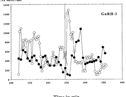
(3) Different effects of GnRH-I and-II on HCG secretion. Figure 1. Log concentrations of human chorionic gonadotrophin (HCG)/day for cytotrophoblastic cells (CTB) incubated for 96 h with different concentrations of gonadotrophin-releasing hormone (GnRH)-I or GnRH-II. Statistical analysis was carried out by analysis of variance; P values refer to differences compared with controls (GnRH-0).. Figure 2. Log concentrations of human chorionic gonadotrophin (HCG)/day for cytotrophoblastic cells (CTB) at 8 h, when incubated for 4 h with different concentrations of gonadotrophin-releasing hormone (GnRH)-I or GnRH-II. Statistical analysis was carried out by analysis of variance; P values refer to differences compared with controls (GnRH-0).. pulse of HCG after their injection into the system (Figures 4 and 5). However, the mean amplitude of all pulses was not changed either by GnRH-I, or by GnRH-II (results not shown). The results from Table I indicate that GnRH-I induced a significant (P ⫽ 0.010) increase in the AUC, when compared with controls (media without GnRH-I). In contrast, GnRH-II had no effect on the AUC (Table I). The pulse frequency was significantly (P ⫽ 0.008, P ⫽ 0.02) decreased by both GnRH-I and II (Table I).. Discussion It has been well established that most hormonal secretions are episodic, when followed in the peripheral circulation with an appropriate time of sampling. Recent studies have observed that the placenta continues to secrete HCG in a pulsatile. fashion, even when separated from uterus, and that this secretion is stimulated by GnRH in vivo (Iwashita et al., 1993) and in vitro (Siler-Khodr, 1987; Currie and Leung, 1993; Li et al., 1994; Leung and Peng, 1996). The spontaneous HCG secretion by placental explants is pulsatile, and this is enabled by cell–cell contact or communication, as shown previously (Barnea et al., 1992). The lack of pulsatility in isolated cells demonstrates that it requires the functional integrity of the tissue, where both cyto- and syncytiotrophoblast elements appear to have an important role. Indeed, it has been suggested that the cytotrophoblast secretes GnRH-I, while the syncytiotrophoblast secretes HCG (Barnea et al., 1992). However, the presence of GnRH-II in the placenta has not yet been reported. Our results are in line with previous observations and show that HCG release by first trimester placental explants is 5.
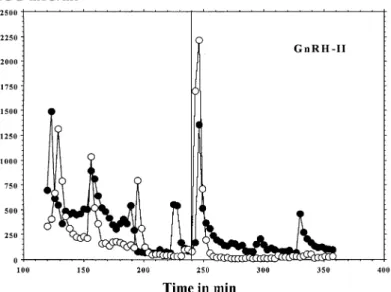
(4) D.Islami et al.. Figure 3. Log concentrations of human chorionic gonadotrophin (HCG)/day for cytotrophoblastic cells (CTB) at 24 h, when incubated for 4 h with different concentrations of gonadotrophin-releasing hormone (GnRH)-I or GnRH-II. Statistical analysis was carried out by analysis of variance; P values refer to differences compared with controls (GnRH-0).. Figure 4. Representative pulsatile human chorionic gonadotrophin (HCG) secretion by two different placental explants before and after injection of 10–8 mol/l gonadotrophin-releasing hormone (GnRH)-I, given at the 240th minute. Open and filled circles represent two different experiments.. pulsatile, and that this episodic secretion is enhanced by GnRH. The spontaneous HCG pulses (in absence of exogenous GnRH) had a similar amplitude, with a mean frequency of every 30.5 min. After GnRH treatment, the mean frequency was about every 39 min for both forms of GnRH. Thus both GnRH-I and GnRH-II caused a decrease in HCG pulse frequency from placental explants. The amplitude of the HCG pulses was not changed by either form of GnRH, whereas only GnRH-I induced an increase in the AUC. In previous studies (Barnea and Kaplan, 1989; Barnea et al., 1991a,b), a similar increase in AUC has been observed. In contrast to our data, these authors have described an increase of pulse 6. frequency and amplitude after GnRH treatment. These different results can probably be explained by the diverse protocols used. In our experiments, we added GnRH only once to first trimester placental explants, while in the other authors’ protocol, GnRH was added several times at different intervals (range 30–72 min). In our experiments with placental explants, we observed a rapid effect of GnRH on HCG secretion. This rapid effect resembles that seen in the pituitary (Loumaye and Catt, 1983) and could be independent of receptors (cell depolarization), since it is observed within 6 min after GnRH administration. The immediate peak after GnRH treatment is unique in its.
(5) Different effects of GnRH-I and-II on HCG secretion. Figure 5. Representative pulsatile human chorionic gonadotrophin (HCG) secretion by two different placental explants before and after injection of 10–8 mol/l gonadotrophin-releasing hormone (GnRH)-II, given at the 240th minute. Open and filled circles represent two different experiments.. Table I. Pulse characteristics of peak (HCG) secretion by placental explants, before and after GnRH treatment Amplitude (mIU/mg). Medium alone (n ⫽ 37) GnRH-I (n ⫽ 19) GnRH-II (n ⫽15). Area under the curve (cm2/mg). Interval between pulses (min). Mean. Range. Mean. Range. Mean. Range. 2.1 3.0 2.7. 0.5–6.1 0.3–14.6 0.4–11.9. 17.3 31.3*** 17.3. 1.3–46.3 1.1–91.7 1.3–76.9. 30.5 39.1* 38.6**. 17–40 30–60 30–45. HCG ⫽ human chorionic gonadotrophin; GnRH ⫽ gonadotrophin-releasing hormone. *Significantly different (P ⫽ 0.02), compared with medium alone (GnRH-0). **Significantly different (P ⫽ 0.008), compared with medium alone (GnRH-0). ***Significantly different (P ⫽ 0.01), compared with medium alone (GnRH-0).. amplitude, and almost similar in amplitude for both forms of GnRH. Previous studies have shown that placental tissue restores its ability to store HCG within a short time and specific HCG storage granules appear in first trimester placenta, but not in term placentae (Morrish et al., 1987). This could suggest that the granules are needed for the pulsatile HCG secretion. This pulsatility would reflect the release of granules, which pass from the synthesis/storage phase to that of release (Yen, 1986). The factors responsible for this seem to be paracrine, and GnRH is a likely candidate. Although our study may suggest a rapid receptor-independent secretion of HCG, the effect of GnRH on HCG secretion has also been shown to last for 30 min, suggesting that GnRH binds locally to produce a prolonged effect (Barnea et al., 1991a). Furthermore, a rapid, receptor-independent effect has so far only been reported for steroids, including progesterone (Barnea et al., 1991b). In our hands, GnRH-I seems to be more effective than GnRH-II. It is well-known that most species express more than one isoform of GnRH (Sherwood et al., 1993; King and Millar, 1995). GnRH isoforms have been described in mammals (Rissman et al., 1995; Kasten et al., 1996; Jimenenz-Linan. et al., 1997; Lescheid et al., 1997; Quanbeck et al., 1997), including humans (White et al., 1998), and cows (Duello and Boyle, 1997). Furthermore, other GnRH-like factors are present in human reproductive tissues (Li et al., 1987), and posttranslationally modified forms of GnRH are active in the human placenta (Gautron et al., 1981, 1992; Currie et al., 1993). Both authentic GnRH forms (Osathanondh and Elkind-Hirsch, 1981; Tan and Rousseau, 1982; Zhuang et al., 1991; Raga et al., 1999) and larger GnRH-like peptides (Mathialagan and Rao, 1986a,b; Zhuang et al, 1991), have been reported to be present in human placental tissues. Our results provide evidence for different effects of the two GnRH isoforms (GnRH-I and GnRH-II) on HCG secretion and synthesis, in both CTB and placental explants. In explant experiments, GnRH-I caused an increase in the AUC of HCG pulses, while GnRH-II did not. Other parameters, e.g. mean frequency of pulses and their amplitude, were very similar for both forms of GnRH. We would speculate that GnRH-I induces both HCG secretion and synthesis in CTB, thus behaving as a placental GnRH, whereas GnRH-II induces HCG secretion, but not its synthesis. This speculation remains to be investigated using reverse transcription–polymerase chain reaction (RT– PCR) in order to test the effects of the different GnRH species 7.
(6) D.Islami et al.. on HCG mRNA. It must be admitted that the effects of both forms of GnRH on CTB are somewhat surprising, since GnRH receptors have been found only in syncytiotrophoblast so far, and that under our culture conditions (absence of fetal calf serum), no syncytium formation is observed during the 96 h of incubation. The mechanism of action of these two isoforms of GnRH is not known. We would, however, speculate that the different isoforms bind differently to placental receptors, or to different receptor subtypes. This could imply that two different types of placental GnRH receptors exist. However, so far, distinct forms of GnRH-receptor have not been found in human placenta (Bramley et al., 1999). Another possible explanation for the different effects of GnRH-I and GnRH-II could be the existence of different pathways of GnRH degradation. Many studies have described GnRH-degrading activities in hypothalamus, pituitary, serum and several other tissues. Degradation of GnRH occurs also in human placental tissue (Bramley and Menzies, 1996; Bramley et al., 1999), and this is certainly an important regulator of GnRH action. It has been shown that human placental cytosol fractions contain three distinct peptidase activities. Enzyme (i) is a cathepsin D-like enzyme, optimal at acid pH, with a broad specificity for GnRH isoforms and analogues. Enzyme (ii) is a metallothiol peptidase and is shown to degrade preferentially GnRH-II, though with some activity against other GnRH isoforms and agonists (Bramley and Menzies, 2000). Its optimal pH is 8–9. Enzyme (iii) is shown to act specifically on GnRH-II at neutral pH (pH 6.0–7.5), and has minimal activity towards other GnRH isoforms and agonists. We conclude that pulsatile HCG secretion can be measured in trophoblast explants of the first trimester, and that GnRH-I and GnRH-II can act rapidly on this secretion. We would suggest that GnRH-I induces both HCG secretion and synthesis, thus behaving as a placental GnRH and that GnRH-II induces only HCG secretion, but not synthesis. To explain the different actions of the two GnRHs, we suggest that these two isoforms of GnRH could have different receptor affinities, could bind to different placental receptors, or could undergo different pathways of degradation.. Acknowledgements We thank Christine Wuillemin and Claire Gruffat for their skilful technical help. D.Islami is the recipient of a scholarship from the Ernst Schering Research Foundation and FIGO.. References Barnea, E.R. and Kaplan, M. (1989) Spontaneous gonadotrophin-releasing hormone-induced, and progesterone-inhibited pulsatile secretion of human chorionic gonadotrophin in the first trimester placenta in vitro. J. Clin. Endocrinol. Metab., 69, 215–217 Barnea, E.R., Kaplan, M. and Naor, Z. (1991a) Comparative stimulatory effect of gonadotrophin releasing hormone (GnRH) and GnRH agonist upon pulsatile human chorionic gonadotrophin secretion in superfused placental explants: reversible inhibition by a GnRH antagonist. Hum. Reprod., 6, 1063–1069. Barnea, E.R., Feldman, D. and Kaplan, M. (1991b) The effect of progesterone upon first trimester trophoblastic cell differentiation and human chorionic gonadotrophin secretion. Hum. Reprod., 6, 905–909.. 8. Barnea, E.R., Shurtz-Swirski, R. and Kaplan, M. (1992) Factors controlling spontaneous human chorionic gonadotrophin in superfused first trimester placental explants. Hum. Reprod., 7, 1022–1026 Belisle, S., Guevin, J.F., Bellabarba, D. and Lehoux, J.G. (1984) Luteinising hormone-releasing hormone binds to enriched human placental membranes and stimulates in vitro the synthesis of bioactive human chorionic gonadotrophin. J. Clin. Endocrinol. Metab., 59, 119–124 Bischof, P., Friedli, E., Martelli, M. and Campana, A. (1991) Expression of Extracellular matrix-degrading metalloproteinases by cultured human cytotrophoblastic cells: effects of cell adhesion and immunopurification. Am. J. Obstet. Gynecol., 165, 1791–1801. Boadi, W.Y., Shurtz-Swirski, R., Barnea, E.R., et al. (1992) Secretion of human chorionic gonadotrophin in superfused young placental tissue exposed to cadmium. Arch. Toxicol., 66, 95–99. Bramley, T.A. and Menzies, G.S. (1996) Measurement of luteal and placental gonadotrophin-releasing hormone (GnRH) binding sites: role of inactivation of GnRH tracer. Mol. Hum. Reprod., 2, 535–539. Bramley, T.A. and Menzies, G.S. (2000) Human placental gonadotrophinreleasing hormone-like factors: an artefact of human placental peptidases? Mol. Hum. Reprod., 6, 113–126. Bramley, T.A., Boyle, H.P. and Menzies, G.S. (1999) Human placental GnRHlike factors: Parallel displacement in GnRH immuno- and receptor-binding assays can be caused by degradation of radiolabelled GnRH tracers. Mol. Hum. Reprod., 5, 1095–1106. Canfield, R.E., Morgan, F.J., Kammerman, S., Birken, S. et al. (1973) Studies of human chorionic gonadotrophin. Recent Prog. Horm. Res., 27, 121–139. Chen, A., Yahalom, D., Ben-Aroya, N. et al. (1998) A second form of gonadotrophin-releasing hormone is present in the brain of human and rodents. FEBS Lett., 18, 199–203 Currie, W.D. and Leung, P.C.K. (1993) Gonadotrophin releasing hormone (GnRH) action in the ovary and placenta. In Bouchard, P., Caraty, A., Coelingh-Bennink, H.J.T. and Pavlou, S.N. (eds), GnRH, GnRH Analogs, Gonadotrophins and Gonadal Peptides. Parthenon, Carnforth, UK, pp. 135–156. Currie, W.D., Steele, G.L., Yuen, B.H. et al. (1993) LHRH- and LHRHstimulated hCG secretion from perifused first-trimester placental cells. Recent Prog. Horm. Res., 48, 505–509. Duello, T.M. and Boyle, T.A. (1997) A second bovine placental pro-GnRH mRNA in bovine placenta. Biol. Reprod., 56 (No. S1), p553. Gautron, J.P., Pattou, E. and Kordon, C. (1981) Occurrence of higher molecular weight forms of LHRH in fractionated extracts from rat hypothalamus, cortex and placenta. Mol. Cell Endocrinology, 24, 1–15. Gautron, J.P., Leblanc, P., Bluet-Pajot, M.T. et al. (1992) A second endogenous form of mammalian hypothalamic LHRH, (hydroxyproline9) LHRH, releases luteinising hormone and follicle-stimulating hormone in vitro and in vivo. Mol. Cell Endocrinology, 85, 97–107. Gestrin, E.E., White, R.B. and Fernald, R.D. (1999) Second form of gonadotrophin-releasing hormone in mouse: immunocytochemistry reveals hippocampal and periventricular distribution. FEBS Lett., 9, 289–291. Illing, N, Troskie, B.E., Nahorniak, C.S. et al. (1999) Two gonadotrophinreleasing hormone receptor subtypes with distinct ligand selectivity and differential distribution in brain and pituitary in the goldfish. Proc. Natl Acad. Sci. USA, 96, 2526–2531. Iwashita, M, Kudo, Y., Shizonaki, Y. and Takeda, Y. (1993) Gonadotrophinreleasing hormone increases serum human chorionic gonadotrophin in pregnant women. Endocr. J., 40, 539–544. Jimenenz-Linan, M., Rubin, B.S. and King, J.C. (1997) Examination of guinea pig luteinising hormone-releasing hormone gene reveals a unique decapeptide and existence of two transcripts in the brain. Endocrinology, 138, 4123–4130. Kasten, T.L., White, S.A., Norton, T.T. et al. (1996) Characterisation of two new preproGnRH messenger RNAs in the tree shrew – first direct evidence for mesencephalic GnRH gene expression in a placental mammal. Gen. Comp. Endocr., 104, 7–19. King, J.A. and Millar, R.P. (1995) Evolutionary aspects of gonadotrophinreleasing hormone and its receptor. Cell. Mol. Neurobiol., 15, 5–23. Lescheid, D.W., Terasawa, E., Abler, L.A. et al. (1997) A second form of gonadotrophin-releasing hormone (GnRH) with characteristics of chicken GnRH-II is present in the primate brain. Endocrinology, 138, 5618–5629. Leung, P.C.K. and Peng, C. (1996) Gonadotrophin-releasing hormone receptor: gene structure, expression and regulation. Biol. Signals, 5, 63–69. Li, C.H., Ramasharma, K., Yamashiro, D. and Chung, D. (1987) Gonadotrophin-releasing peptide form human follicular fluid: isolation, characterisation and chemical studies. Proc. Natl Acad. Sci. USA, 84, 959–962..
(7) Different effects of GnRH-I and-II on HCG secretion Li, W., Olofsson, J.I., Jeung, E.-B. et al. (1994) Gonadotrophin-releasing hormone (GnRH) and cyclic AMP positively regulate inhibin subunit messenger RNA levels in human placental cells. Life Sci., 55, 1717–1724. Loumaye, O.H. and Catt, K.H. (1983) Agonist-induced regulation of pituitary receptors for gonadotrophin releasing hormone. J. Biol. Chem., 258, 12002–12004. Mathialagan, N. and Rao, A.J. (1986a) Gonadotrophin-releasing hormone in the first trimester human placenta: isolation, partial characterisation and in vitro biosynthesis. J. Biosci., 10, 429–441. Mathialagan, N. and Rao, A.J. (1986b) Gonadotrophin-releasing hormone (GnRH) stimulates both secretion and synthesis of human chorionic gonadotrophin (hCG) by first trimester human placental minces in vitro. Biochem. Int., 13, 757–765. Morrish, D.W., Marusyk, H. and Siy, O. (1987) Demonstration of specific secretory granules for human chorionic gonadotrophin in placenta. J. Hystochem. Cytochem., 35, 93–101. Nekda, M.V., Horwath, A., Ge, L.J. et al. (1982) Supression of ovulation in the rat by an orally active antagonist of luteinising hormone-releasing hormone. Science, 218, 160–165. Osathanondh, R. and Elkind-Hirsch, K.E. (1981) Presence of immunoreactive luteinising hormone-releasing factor in hydatiform mole as compared with normal human trophoblastic tissue. Placenta (Suppl. 3), 257–261. Owens, O.M., Ryan, K.J. and Tulchinsky, D. (1981) Episodic secretion of human chorionic gonadotrophin in early pregnancy. J. Clin. Endocrinol. Metab., 53, 1307–1310. Padmanabhan, V., Sonstein, J., Olton, P.L. et al. (1989) Serum bioactive follicle-stimulating hormone-like activity increases during pregnancy. J. Clin. Endocrinol. Metab., 69, 968–973. Pohl, C.R., Richardson, D.A., Hutchison, J.S. et al. (1984) Hypophysiotropic signal frequency and the functioning of the pituitary ovarian system in the rhesus monkey. Neuroendocrinology, 39, 256–263. Popkin, R., Bramley, T.A., Currie, A. et al. (1983) Specific binding of luteinising hormone-releasing hormone to human luteal tissue. Biochem. Biophys Res. Commun., 114, 750–754. Quanbeck, C., Sherwood, N.M., Millar R.P. and Terasawa, E. (1997) Two populations of luteinising hormone-releasing hormone neurons in the. forebrain of the rhesus macaque during embryonic development. J. Comp. Neurol., 380, 293–309. Raga, F., Casan, E.M.Wen, Y. et al. (1999) Indipendent regulation of matrix metalloproteinase-9, tissue inhibitor of metalloproteinase-1 (TIMP-1), and TIMP-3 in human endometrial cells by gonadotrophin-releasing hormone: Implications in early human implantation. J. Clin. Endocrinol. Metab., 84, 636–642. Rissman, E.F., Alones, V.E., Craig-Veit, C.B. and Millam, J.R. (1995) Distribution of chicken-II gonadotrophin-releasing hormone in mammalian brain. J. Comp. Neurol., 357, 524–531. Sherwood, N.M., Lovejoy, D.A. and Coe, I.R. (1993) Origin of mammalian gonadotrophin-releasing hormones. Endocr. Rev., 14, 241–254. Siler-Khodr, T.M. (1987) Placental LHRH-like activity. In Vickery, B.H. and Nestor, J.J., Jr (eds), LHRH and its Analogs: Contraceptive and Therapeutic Applications, Part 2. MTP Press, Lancaster, pp. 161–178. Siler-Khodr, T.M. and Khodr, G.S. (1979) Extrahypothalamic luteinising hormone releasing factor (LRF); release of immunoreactive LRF in vitro. Fertil. Steril., 39, 294–299. Tan, L. and Rousseau, P. (1982) The chemical identity of the immunoreactive LHRH-like peptide biosynthesised in the human placenta. Biochem. Biophys Res. Commun., 109, 1061–1071. Urbanski, H.F., White, R.B., Fernald, R.D. et al. (1999) Regional expression of mRNA encoding a second form of gonadotrophin-releasing hormone in the macaque brain. Endocrinology, 140, 1945–1948 White, R.B., Eisen, J.A., Kasten, T.L. and Fernald, R.D. (1998) Second gene for gonadotrophin-releasing hormone in humans. Proc. Natl Acad. Sci. USA, 95, 305–309. Yaholom, D., Chen, A., Ben-Aroya, N. et al. (1999) The gonadotrophinreleasing hormone family of neuropeptides in the brain of human, bovine and rat: identification of a third isoform. FEBS Lett, 463, 289–294. Yen, S.S.C. (1986) Neuroendocrine control of hypophyseal function. In: Yen, S.S.C. and Jaffe, R. (eds), Reproductive Endocrinology. W.S.Saunders, Philadelphia, USA, pp. 3–33. Zhuang, C.L., Cheng, L.R., Wang, H. et al. (1991) Neuropeptides and neurotransmitters in human placental villi. Neuroendocrinology, 53 (Suppl. 1), 77–83. Received on April 20, 2000; accepted on October 3, 2000. 9.
(8)
Figure
Documents relatifs
whale shark the GnRH genes correspond to GnRH1, GnRH2, and GnRH3, while in elephant shark they correspond to GnRH1a and GnRH1b, two copies of the GnRH1 gene, plus GnRH2..
the ovarian response, recruitment (number of follicles) and selection (rate of atresia) to superovulatory treatments is affected by the population of follicles present
This asynchronous development of the ovaries is characteriyed by great variations ln GtH (plasma and pituitary) and in GnRH (pituitary and hypothalamus) contents
L’archive ouverte pluridisciplinaire HAL, est destinée au dépôt et à la diffusion de documents scientifiques de niveau recherche, publiés ou non, émanant des
Le fait que la suppression génétique de la neuropiline-1 dans les neurones produisant la GnRH entraîne une puberté précoce chez la souris suggère que certains variants
To determine whether BMP-4 was capable of modifying gonadotrophin production and secretion in LbT2 gonado- trophs, cells given activin Ga daily 1 h pulse of GnRH for three days
Each of the cell lines were treated by a fixed concentration of GnRH corresponding to the three-fold higher dose than the calculated EC 50 (3 × EC 50 ; a concentration optimized
[r]
