Article No. lich. 1999.0220
Available online at http://www.idealibrary.com on IDEKL
PCR PRIMERS FOR THE AMPLIFICATION OF
MITOCHONDRIAL SMALL SUBUNIT RIBOSOMAL
DNA OF LICHEN-FORMING ASCOMYCETES
Stefan ZOLLER,* Christoph SCHEIDEGGER* and
Christoph SPERISEN*
Abstract: Four primers for the amplification of mitochondrial DNA of
lichen-forming ascomycetes are presented. The primers match the conserved regions U2, U4, and U6, respectively, of mitochondrial small subunit (SSU) ribosomal DNA (rDNA). Polymerase chain reaction using different combinations of the primers produced single amplification products from DNA of eight lichen-forming fungal species but did not amplify DNA of two axenic cultured algal species. The amplification product obtained from Lobaria pulmonaria was sequenced and the 894-bp sequence was compared with the mitochondrial SSU rDNA sequence of
Podospora anserina. The two sequences revealed more than 76% identity in the
conserved regions U3 to U5 demonstrating that we amplified mitochondrial DNA. The primers matching U2 and U6 yielded amplification products of 800-1000 bp depending on the species examined. The variation observed suggests that mito-chondrial SSU rDNA may be useful for phylogenetic analyses of lichen-forming ascomycetes. © 1999 The British Lichen Society
Introduction
Analysis of nuclear ribosomal DNA (rDNA) has become an important tool in
molecular studies of lichens. These studies have used rDNA to provide insight
into the origin of lichens (Gargas et al. 1995), phylogenetic relationships
(Lutzoni & Vilgalys 1995; Tehler 1995), and population structures (DePriest
1993). Application of mitochondrial rDNA for lichen studies has not been
described although mitochondrial rDNA of fungal species often reveals high
levels of nucleotide substitutions and length mutations (Hibbett & Donoghue
1995; Gryta et al. 1997; Bruns et al. 1998; Gonzales & Labarere 1998;
Johnson 1999). In the basidiomycete order Boletales the variation of
mito-chondrial rRNA genes was even higher than that of nuclear rRNA genes
(Bruns & Szaro 1992).
In this study, we present primers for the amplification of mitochondrial
small subunit (SSU) rDNA of lichen-forming fungi. The primers match
the universal regions U2, U4 and U6, respectively. These regions are part of
eight universally conserved regions that form the minimal core secondary
structure of mitochondrial SSU rRNA (Gray et al. 1984; Schnare et al. 1986;
Cummings et al. 1989). We show that the novel primers produce amplification
*Swiss Federal Institute for Forest, Snow and Landscape Research, Zurcherstrasse 111, CH-8903 Birmensdorf, Switzerland, e-mail: stefan.zoller@wsl.ch
Forward Forward Reverse Reverse U2 U4 U4 U6 TABLE 1. Primer sequences for amplification of mitochondria! SSU rDNA
Homologous Primer Primer sequence Forward/reverse to U region mrSSUl 5'-AGCAGTGAGGAATATTGGTC-3'
mrSSU2 5'-CTGACGTTGAAGGACGAAGG-3' mrSSU2R 5'-CCTTCGTCCTTCAACGTCAG-3' mrSSU3R 5'-ATGTGGCACGTCTATAGCCC-3'
products from a broad range of lichen-forming ascomycetes and demonstrate
that the amplification products reveal length differences among different
species.
Materials and Methods
Lichen and algal material
The lichen species investigated were collected in Switzerland: Lobaria pulmonaria and Peltigera
praetextata in Vordernwald, Canton of Aargau (co-ordinates 47°17 ± 20"N, 7°52 ± 30"E); Lecanora allophana and Cladonia digitata in Gurnigel, Canton of Bern (46°45 ± 00" N, 7°26 ±
20" E); Leptogium saturninum and Graphis scripta in Wagital, Canton of Schwyz (47°04 ± 00" N, 8°54 ± 40" E); Parmelia pastillifera and Parmelia sulcata in Bondo, Canton of Graubunden (46°20 ± 00" N, 9°32 ± 00" E).
The algae Trebouxia species and Dictyochloropsis reticulata, the photobiont of L. pulmonaria, were isolated using the procedure developed by Yamamoto (1987) and cultured on Bold's Basal Medium BBM (Bischoff & Bold 1963).
Extraction of total DNA
Total DNA of lichens was extracted as described by Ziegenhagen et al. (1993) and purified using the QIAamp DNA Blood Mini Kit (QIAGEN). Air-dried thallus pieces (up to 70 mg) were cleaned by hand, transferred into a 2-ml precooled microfuge tube containing an agate ball (7 mm in diameter), and ground to fine powder in a shaking mill (Micro-Dismembrator II, Braun) for 2 min at full speed. The powder was dispersed in 350 ul of extraction buffer [100 mM sodium acetate pH 5-5, 50 mM EDTA, 500 mM NaCl, 2% (w/v) polyvinyl pyrrolidone, 1-4% (w/v) sodium dodecyl sulphate supplemented with 0-5% (w/v) sodium bisulphite], incubated at 65°C for at least 20 min, and centrifuged for 15 min at 20 000^. The supernatant (c. 300 ul) was transferred to a new 1-5-ml tube and 300 ul of AL buffer (provided with the kit) was added. The mixture was thoroughly shaken and incubated at 70°C for 10 min. Three hundred microlitres of absolute ethanol were added and the mixture was transferred onto a spin column placed on a 2-ml collection tube (provided with the kit) and centrifuged for 1 min at 6800 g. The column was washed by addition of 400 ul of AW washing buffer (provided with the kit) and centrifugation for
1 min at 6800 g. The washing step was repeated with a final centrifugation for 1 min at 20 000 g. The spin column was placed onto a new 1-5 ml microfuge tube and incubated for 10 min at 70°C to dry the membrane. DNA was eluted by addition of 300 ul TE (1 mM EDTA, 10 mM Tris-HCl pH 9-0, preheated to 70°C), incubation for 5 min at 70°C in an oven, and centrifugation for 1 min each at 6800 g and 20 000 g. Finally, 5 ul RNAse A (10 mg ml ~ ') was added to each sample.
Primer construction and PCR
Four primers for amplification of fungal mitochondrial SSU rDNA were designed (Table 1). The primer sequences were derived from conserved regions among the ascomycetes Neurospora
crassa (GenBank accession number: J05254), Aspergillus terricola (U29212), Trichophyton rubrum
(X88896), Beauveria bassiana (U91338), Petromyces albertensis (U29229), and Podospora anserina (X14734). A BLAST search on the GenBank database was performed to verify specificity to
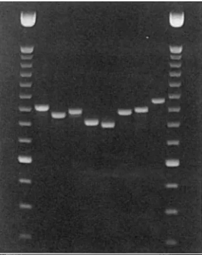
FIG. 1. Agarose-gel electrophoresis of PCR products obtained with primers matching the conserved regions U2 and U6 of the mitochondrial SSU rDNA. Lanes 1 and 10: 100-bp DNA ladder with brighter 600-bp band (GIBCOBRL). Lanes 2-9 contain 5 nl of PCR product of the following taxa: Cladonia digitata (2); Peltigera praetextata (3), Graphis scripta (4), Lecanora allophana (5), Leptogium satuminum (6), Lobaria pulmonaria (7), Parmelia pastillifera (8), and
Parmelia sulcata (9).
fungal sequences. Amplification of fungal and algal DNAs was performed in 50 |jl reaction volumes [1 x PCR buffer (GIBCOBRL), 1 mM dNTPs, 2 mM MgCl2, 2 nM of each primer,
2-5 U Taq DNA polymerase (GIBCOBRL), 2 ul DNA extract] in a thermal cycler (PTC-100, MJ RESEARCH, INC.) using a standard cycling protocol: denaturation at 94°C for 3 min; 35 cycles with 94°C denaturation for 1 min, annealing at 52°C for 1 min, and extension at 72° for 1 min; followed by a final extension at 72°C for 7 min. PCR products were analysed on 2% TBE-agarose gels. The two primers MSI and MS2 (White et al. 1990) were tested using the same PCR procedure with DNA of L. pulmonaria.
Sequence analysis
The PCR-product of L. pulmonaria was ethanol precipitated and sequenced with the Applied Biosystems Dye Terminator Cycle Sequencing Ready Reaction Kit (ABI). Nucleotide base detection was performed on an Applied Biosystems 310 genetic analyser. Cycle sequencing was carried out with the PCR primers. Sequences were assembled using Sequencher 3-0 (Gene Codes Corporation) and manually adjusted. The alignment of L. pulmonaria and P. anserina sequences was done with GAP of the GCG 9-1 software package (Genetics Computer Group) by using a gap creation penalty of 5-00 and a gap extension penalty of 0-30. An unambiguous alignment of the two sequences was not possible. We did not do any visual adjustments although in several regions more matches could have been obtained.
Results and Discussion
The primers MSI and MS2, previously described for the amplification of
fungal mitochondrial small subunit rDNA (White et al. 1990) did not amplify
DNA of Lobaria pulmonaria. However, the primers we designed on the basis of
conserved ascomycete sequences yielded single amplification products from
all eight lichen-forming fungal species examined. The primers did not amplify
DNA from axenic cultures from the two algae Trebouxia species and
P. L. pulmonaria P. L. p P. anserina L. P. L. AClM^ACTGAG^TATTan^a>ATCCCCTAAC«Xrr<aUttTCGTAACTTGGA 70 AATAGCCTAACGCCTCUUICCAGCMCTTCGAGCAATGAAAGCGCAATA AGAATCGCTCCATAAAAATCXYUSTTAATCGATTRATCGAATAAA 1 4 0 ii i i j mi inn i n n GCTGATTffl^TTAAATTTAAAATTAGAGTTrTTnTATrrTTAATCTAGTTACTACITTGTAGTTTAAAT U3 ATTCTGTATACATACCTATAAT(»CGGTATCTAI»TCTT.CjrCTTCy>CC.AATTGCCfKXX:A(XyiOTCOC 2 1 0 TTCTAGGTAGAATTATcaTGATGACATTATCTAWTATAOCKrrTGACTXAATTACGTGCCAGCAGTCGC U3 <XXaUlTAC<WAAC»(a«rrAGT<rrTATK»TCTTAAATAGGTTTAAAGGOTACTrAGACG 280 GGTAATACGTAGAAGACAAC?rcTTATTCATCnT'ATTAGGTTTAAAGAGTACCAAGACGGTTAATAAAGC .GTAGAAriTGCCTTAACGGGAACAATTTTACTAGAGTTTTATATAAG AAGATCTTACTCTAGAC 3 5 0 I I I I I I 1 1 1 I M I I I I I M M U I I I I I I l l l I I I I CCAAAATACATGAGCACAGGGGACACArrAACTAGAGTTATATGAAAGGMGCATTAAGGTACTCTTGAG TGTAGAGTTGTAATTCATTAATACC TAGGGGACTGGTAAAGGCGAAGGCA.ATCTTTTATATAAA A 9 0
11 II III i nun nun i it n i i i i inn in i i mi in
TCn'AGAGATAAAATTCAGTCATACCAGATGAAAGCKACAmTTTAT^KGAAO^ATCTTTCTTATGTAAT 04 ^ 490 AACT<»CCTTGTAG<»CT»AGGCTTTTCAAGTCAAAA«^TrAGATACC<n^^ TAT<^TGCCATAG<7rrAGATATAGTrAAATACAAACAAGGCCGTAACXrrO^ATTAAATTrTACX^TCT 5 6 0 , , . Mini i m i i i I I i n I I i n
L. pulmonaria GATGAATCTTA TAGGTTTAATATATACAGCCCAAAAAAAAAAGGGTCAATGTTTTATTG U5 ACTCTATAAATGAAAGT<rrAAGCATTTCACCTCAAGAGTAATCreGaUlCGCAaSAACTGAAATCACTAO 6 3 0 i n n i i i i i n n i n i i i i n i n u n i n n i i i n i i i i n n i n n i i n n TAA p. L. P. L._ P. anserina avATATAAAAAAATGCTATCTACTACCATCACTTATrrAAAGCTAGCCGCGCCGGGCGGCGAAAGCTCCT 7 7 0 L. pulmonaria P. anserina CCT<XKrrCTAAATrrAAC?rGTTAGGC«^\AGCTCTAAGATATATAGCl«GCTTAAATATTATTTAGCTAA 8 4 0 L. pulmonaria P. L. P. L._ P. anserina L._ IT5SU3R CACCTCATAAT i n n i i i i i n n i n i i i i n i n u n i n n i i i n i i i i n n i n i i n GGTCTATAMTGAAATTGTAAGCATeeCTCCTCAAAATTAATTTGGCAACAriKX^CTGAAATCATTAA ACCGTTTC«aiCACCAGCA(m»AGTATGTTATTTAATTC<»TGACCCACGAAAAACCTTAaaiCAACTT 7 0 0 111tn11111 inn i n n n n n n m i n i inn in i n i i m i i n n n ACCGTTTCTGAAACCAGTAGTGAACTATCTTGTTTAArrAC^TGATACACAAAAAACCTTACCACAAT.. CTATGCTTTTAGGTTATAGATTAATGCAAGT ATTATACAWTIXrrTGCAOXKriXrr^rrTCAGTTGATGTTG 9 1 0 I t l l l l I I I I MM MMII I I I I I I I I I I I H I M I I I I I MM I TTGAATATATATCATTAATGATATT.TAOU«XX?ITGCAC<WXrrGTCTTCAGTTAATGTCG TGAAACTGTGGTTCGGTCCATGGMTTAACGAAAACCCTTCCTTTATTTGTA^ 980 H I M I M i l l I M i l l 1 1 M I I 1 1 I M I I I i I I M I M I M M M M I I I I TGAAATTTT«nTAGTTCCATAAAATTAACGTAAACarKXrrTTTATTTATAAA. . . TAAAATTTACAAT TTCACCTGTATATGGGTTATGATAAAAGGGATCAAGACAAGTCCTCA'KSGCCTTGATGTTT^ i i n i i I I in i i i i i i i innni I I nn CT(7nTCTAAAATTTGATTTAAAAAAGG..OACTAAAACAATTCACATGGCCTAAATATTGT 1050
r. anserina EACGTGCCACATCTACCTAAACAAAGAG io?e
FIG. 2. Sequence comparison of mitochondrial SSU rDNA of Podospora anserina (GenBank Accession XI4734) and Lobaria pulmonaria. The mitochondrial sequence of L. pulmonaria was deposited in the GenBank database (Accession Nr. AF069541). U2 to U6 denote the universally
conserved regions and are shaded. Primer sequences are given in boxes.
search did not reveal any significant similarities between the primer and algal
sequences.
Primers matching the universally conserved regions U2 and U6 (see
Table 1) produced single amplification products of approximately
800-1000 bp depending on the species examined (Fig. 1). Primers homologous to
U2 and U4, and U4 and U6, respectively, resulted in amplification products
of 400-500 bp. Five of the seven species could clearly be distinguished based
on PCR-fragment-length differences. The two species Graphis scripta and L.
pulmonaria gave similar fragment lengths.
The amplification product of L. pulmonaria was sequenced and the 894-bp
sequence was compared with the mitochondrial SSU rDNA sequence of
to 89% identity in the regions U3 to U5 demonstrating that mitochondrial
DNA was amplified (Fig. 2). From U2 and U6, only segments were amplified
revealing 88% and 71% identity, respectively. The sequences between the
conserved regions showed considerable length differences. To align these
sequences, a total of nine gaps of 1 to 131 bp length had to be introduced
(Fig. 2). Parts of these regions could not be aligned unambiguously.
Our findings are consistent with results obtained from basidiomycetes, often
showing species-specific length mutations in mitochondrial rDNA (Bruns &
Szaro 1992; Hibbett & Donoghue 1995; Bruns et al. 1998; Gonzales &
Labarere 1998). This variation together with nucleotide substitutions has
been proved to be useful for phylogenetic studies. By analogy with the
basidiomycetes the findings of this study shows promise that mitochondrial
SSU rDNA may also be useful for studying the phylogeny of
lichen-forming ascomycetes. Mitochondrial rDNA may be valuable for phylogenetic
analyses for several reasons. First, mitochondrial rDNA appears to reveal high
rates of nucleotide substitutions in many fungal species, in the order Boletales
higher than that observed in its nuclear counterpart (Bruns & Szaro 1992). A
reduced constraint on mitochondrial rRNA has been proposed as an
import-ant factor contributing to the high rates of nucleotide substitutions observed
although increased mutation rates could not be excluded. Second,
mitochon-drial DNA is uniparentally inherited in most ascomycetes studied so far (Rohr
et al. 1999). Variation uniparentally inherited is more prone to genetic drift
and founder events than variation of nuclear DNA, both resulting in reduced
diversity and higher rates of molecular divergence (Moritz et al. 1987). Finally,
concerted evolution might be rather different in mitochondrial rDNA because
the mitochondrial genomes are partitioned into many mitochondria and are
separated mitotically. In summary, mitochondrial rDNA appears to show
different modes and rates of evolution than nuclear rDNA and may thus
represent an additional marker to the widely used nuclear rDNA.
We thank Beat Frey for providing the axenic cultures of algae, Urs Buchler, Kurt Eichenberger and Gabor Matyas for technical assistance, and Francois Lutzoni (The Field Museum, Chicago) for his help with primer construction.
REFERENCES
Bischoff, H. W. & Bold, H. C. (1963) Physiological studies: IV. Some soil algae from enchanted rock and related algal species. University of Texas Publications No. 6318, Physiological Studies 4: 1-95.
Bruns, T. D. & Szaro, T. M. (1992) Rate and mode differences between nuclear and mitochondrial small-subunit rRNA genes in mushrooms. Molecular Biology and Evolution 9: 836-855.
Bruns, T. D., Szaro, T. M., Gardes, M., Cullings, K. W., Pan, J. J., Taylor, D. L., Horton, T. R., Kretzer, A., Garbelotto, M. & Li, Y. (1998) A sequence database for the identification of ectomycorrhizal basidiomycetes by phylogenetic analysis. Molecular Ecology 7: 257-272. Cummings, D. J., Domenico, J. M., Nelson, J. & Sogin, M. L. (1989) DNA sequence, structure
and phylogenetic relationships of the small subunit rRNA coding region of mitochondrial DNA from Podospora anserina. Journal of Molecular Evolution 28: 232-241.
DePriest, P. T. (1993) Small subunit rDNA variation in a population of lichen fungi due to optional group I introns. Gene 134: 67-71.
Gargas, A., DePriest, P. T., Grube, M. & Tehler, A. (1995) Multiple origins of lichen symbioses in fungi suggested by SSU rDNA phylogeny. Science 268: 1492-1495.
Gonzales, P. & Labarere, J. (1998) Sequence and secondary structure of the mitochondrial small-subunit rRNA V4, V6, and V9 domains reveal highly species-specific variations within the genus Agrocybe. Applied and Environmental Microbiology 64: 4149-4160.
Gray, M. W., Sankoff, D. & Cedergren, R. J. (1984) On the evolutionary descent of organisms and organelles: a global phylogeny based on a highly conserved structural core in small subunit ribosomal RNA. Nucleic Acids Research 12: 5837-5852.
Gryta, H., Debaud, J. C , Effosse, A., Gay, G. & Marmeisse, R. (1997) Fine-scale structure of populations of the ectomycorrhizal fungus Hebeloma cylindrosporum in coastal sand dune forest ecosystems. Molecular Ecology 6: 353-364.
Hibbett, D. S. & Donoghue, M. J. (1995) Progress toward a phylogenetic classification of the Polyporaceae through parsimony analysis of mitochondrial ribosomal DNA sequences.
Canadian Journal of Botany 73(Suppl. 1): 853-861.
Johnson, J. (1999) Phylogenetic relationships within Lepiota sensu lato based on morphological and molecular data. Mycologia 91: 443-458.
Lutzoni, F. & Vilgalys, R. (1995) Omphalina (Basidiomycota, Agaricales) as a model system for the study of coevolution in lichens. Cryptogamic Botany 5: 71-81.
Moritz, C.j Dowling, T. E. & Brown, W. M. (1987) Evolution of animal mitochondrial DNA: relevance for population biology and systematics. Annual Review of Ecology and Systematics 18: 269-292.
Rohr, H., Kiies, U. & Stahl, U. (1999) Recombination: Organelle DNA of plants and fungi: Inheritance and recombination. In Progress in Botany 60 (K. Esser, J. W. Dadereit, U. Liittge & M. Runge, eds): Berlin: Springer.
Schnare, M. N., Heinonen, T. Y. K., Young, P. G. & Gray, M. W. (1986) A discontinuous small subunit ribosomal RNA in Tetrahymena pyriformis mitochondria. Journal of Biological
Chemistry 261: 5187-5193.
Tehler, A. (1995) Arthoniales phylogeny as indicated by morphological and rDNA sequence data.
Cryptogamic Botany 5: 82-97.
White, T. J., Bruns, T., Lee, S. & Taylor, J. W. (1990) Amplification and direct sequencing of fungal ribosomal RNA genes for phylogenetics. In PCR Protocols, a Guide to Methods and
Applications (M. A. Innis, D. H. Gelfan, J. J. Sninsky & T. White, eds): 315-322. San Diego:
Academic Press.
Yamamoto, Y. (1987) Tissue culture of lichens. In Tissue Cultures of Lichen and Bryophyte (Y. Yamada et al., eds): 14-25. Osaka: Nippon Paint Company.
Ziegenhagen, B., Guillemaut, P. & Scholz, F. (1993) A procedure for mini-preparations of genomic DNA from needles of silver fir {Abies alba Mill.). Plant Molecular Biology Reports 11: 117-121.