HAL Id: hal-01894125
https://hal.archives-ouvertes.fr/hal-01894125
Submitted on 12 Oct 2018
HAL is a multi-disciplinary open access
archive for the deposit and dissemination of
sci-entific research documents, whether they are
pub-lished or not. The documents may come from
teaching and research institutions in France or
abroad, or from public or private research centers.
L’archive ouverte pluridisciplinaire HAL, est
destinée au dépôt et à la diffusion de documents
scientifiques de niveau recherche, publiés ou non,
émanant des établissements d’enseignement et de
recherche français ou étrangers, des laboratoires
publics ou privés.
Low-biofouling membranes prepared by liquid-induced
phase separation of the PVDF/polystyrene-b-poly
(ethylene glycol) methacrylate blend
Antoine Venault, Yi-Hung Liu, Jia-Ru Wu, Hui-Shan Yang, Yung Chang,
Juin-Yih Lai, Philippe Aimar
To cite this version:
Antoine Venault, Yi-Hung Liu, Jia-Ru Wu, Hui-Shan Yang, Yung Chang, et al.. Low-biofouling
membranes prepared by liquid-induced phase separation of the PVDF/polystyrene-b-poly
(ethy-lene glycol) methacrylate blend. Journal of Membrane Science, Elsevier, 2014, 450, pp.340-350.
�10.1016/j.memsci.2013.09.004�. �hal-01894125�
OATAO is an open access repository that collects the work of Toulouse
researchers and makes it freely available over the web where possible
Any correspondence concerning this service should be sent
to the repository administrator:
tech-oatao@listes-diff.inp-toulouse.fr
This is an author’s version published in:
http://oatao.univ-toulouse.fr/20270
To cite this version:
Venault, Antoine and Liu, Yi-Hung and Wu, Jia-Ru and Yang,
Hui-Shan and Chang, Yung and Lai, Juin-Yih and Aimar, Philippe
Low-biofouling membranes prepared by liquid-induced phase
separation of the PVDF/polystyrene-b-poly (ethylene glycol)
methacrylate blend. (2014) Journal of Membrane Science, 450.
340-350. ISSN 0376-7388
Low-biofouling membranes prepared by liquid-induced phase
separation of the PVDF/polystyrene-b-poly (ethylene glycol)
methacrylate blend
Antoine Venault
a,n, Yi-Hung Liu
a, Jia-Ru Wu
a, Hui-Shan Yang
a, Yung Chang
a,b,n,
Juin-Yih Lai
a,b, Pierre Aimar
caDepartment of Chemical Engineering, Chung Yuan Christian University, Chung-Li 32023, Taiwan bR&D Center for Membrane Technology, Chung Yuan Christian University, Chung-Li 32023, Taiwan cLaboratoire de Génie Chimique, Université Paul Sabatier, 31062 Toulouse Cedex 9, France
a r t i c l e
i n f o
Keywords: PVDF membranes PS-b-PEGMA copolymer Membrane formation Low-biofouling LIPS processa b s t r a c t
In the present work, the focus is laid on the formation, and low-biofouling properties of polyvinylidene fluoride (PVDF) membranes modified using an amphiphilic copolymer additive: polystyrene-b-poly (ethylene glycol) methacrylate (PS-b-PEGMA). PVDF was blended with PS-b-PEGMA and membranes were prepared by liquid-induced phase separation. The additive played a significant role on membrane formation, slightly decreasing surface porosity, reducing the shrinkage during phase separation, and increasing both the size and porosity of macrovoids. Owing to its numerous hydrophilic moieties, the copolymer was believed to promote solvent and nonsolvent exchanges during phase inversion. In addition, it significantly enhanced surface hydrophilicity and matrix hydration capability. Indeed, water was easily trapped by the PEGylated chains spread onto the surface and within the matrix, and then stored in the larger macrovoids. It led to an important reduction of protein adsorption, including bovine serum albumin (65%) and lysozyme (89%). Bacterial attachment tests revealed that adhesion of Escherichia coli and Staphylococcus epidermidis was almost totally prevented (over 99% reduction of attachment), which demonstrates the excellent efficiency of PS-b-PEGMA copolymer to provide PVDF membranes with low-biofouling properties.
1. Introduction
Formation and application of low-biofouling membranes have been widely investigated over the past 10 years [1–6]. Still, it remains challenging to find both an appropriate formulation and a preparation method that would be cost-effective, quick to carry out, and efficient regarding the desired properties. Existing meth-ods can be classified into three categories. The first one gathers methods aiming at modifying the surface top-layer using a surface chemical reaction [7–9], induced for instance by plasma, UV, or ozone treatment. These methods are efficient, since they permit to achieve very low-biofouling properties. However, their scale-up is difficult to control and expensive. The second type of methods concerns the modification by coating [10,11]. Coating is readily preformed and only requires immersing the membrane in a
solution containing the copolymer additive. Physical interactions occur, but they are less strong than covalent bonds, which may lead to stability issues. Supplementary disadvantages of coating methods concern the scale-up of membranes too. In addition, to ensure a high coating density, an important amount of additive must be used. So, the process becomes expensive at larger scale. Furthermore, modification of the physical structure, especially the surface porosity, may be responsible for permeability decline. Finally, the third type of method involves the blending method associated to a phase separation process[12–14]. These methods are convenient and readily carried out. Also, the scaling of membranes is facile, since only one unit operation is involved. Nonetheless, issues concern (i) potential modification of porous structure and, most of all, (ii) lack of stability: the additive may leach out of the matrix since it is often very hydrophilic and solubilized in the aqueous nonsolvent used during membrane formation, or in the feed during membrane filtration. But despite these disadvantages explaining why few results are reported using the blending method, compared to other techniques, it is believed to be a very promising path.
n
Corresponding authors at: Department of Chemical Engineering, Chung Yuan Christian University, Chung-Li 32023, Taiwan. Tel.: þ886 3 265 4113.
E-mail addresses: avenault@cycu.edu.tw (A. Venault),
ychang@cycu.edu.tw (Y. Chang).
When a phase separation process is chosen in the design of antifouling membranes, the additive may affect their final structure, phenomenon that cannot be neglected. In this respect, works need to be carried out to fully understand the impact of polymer additives on membrane formation. Several investigations were performed in the past with common additives such as PVP or PEG systems, which led to important results[14–16]. Indeed, they were often shown to act as pore formers, therefore increasing membrane permeability. Conse-quently, additives have both a chemical influence and a physical effect on membranes' properties. However, it is hard to predict the real impact of a polymer additive, since it may also depend on its concentration in the casting solution. Therefore, a systematic study should be carried out each time a new formulation is tested.
As aforementioned, PEG-like systems are commonly employed to prepare low-biofouling membranes. But if used in blend with a polymer, part of the additive is washed out of the matrix during membrane formation by wet-immersion process (or liquid-induced phase separation – LIPS). Indeed, the non-solvent is almost always water, which solubilizes many PEG systems. From this consideration, if one wants to keep investigating this direction, there are two possible choices: either using another phase separation process, vapor-induced phase separation for instance, or employing an additive copolymer water insoluble. Changing the preparation process leads to membranes with different morphologies and this is not always suitable as far as the final application is concerned. Therefore, the focus is now laid on the development of appropriate amphiphilic additives possessing anchoring groups totally insoluble in water, able to interact with common hydrophobic membrane polymers and containing hydrophilic moieties[18–21].
Among the popular hydrophobic polymers used in membrane technology, polyvinylidene fluoride (PVDF) exhibits good chemical, thermal and mechanical properties. But its use as a membrane material is limited by its hydrophobic nature. Indeed, water does not wet spontaneously PVDF membrane. Consequently, this material shows adsorption tendency, which has a highly negative influence on the flux through the creation of a supplementary resistance to mass transfers. Therefore, hydrophilic polymers are sometimes more interesting for the design of porous membranes. Yet, PVDF offers very good bulk properties, which makes it an ideal polymer material for application of membranes in water treatment. So, efforts need to be made to successfully modify PVDF membranes with a suitable additive and results of major investigations using various additives among which amphiphilic copolymers have been summarized in Liu et al.'s review paper[22]. Despite all of these extensive works and studies performed till then, there is still a need for the preparation of stable anti-biofouling PVDF matrices, considering their potential wide range of applications – from water treatment to blood filtration – in which biofoulants of various natures (proteins, cells or bacteria) can be found. Recently, Li et al. reported the preparation of anti-fouling PVDF membranes, by grafting sulfobetaine-based zwitterio-nic polymers [23]. Their process – a two-step polymerization process- led to very good results concerning antifouling properties, but it is a time-consuming process which appears to be complicated to scale up. A promising set of results was also published very recently by Pezeshk et al. They prepared PVDF ultrafiltration flat-sheet membranes by blending the polymer with hydrophilic poly-urethane additive that they named L2MM[24]. Results highlighted that L2MM additive acted both as a pore former and a surface modifier, eventually leading to an increase of water permeability. The wetting of PVDF membranes was dramatically improved and it is accepted that increasing hydration capability of membranes usually leads to a decreasing of fouling tendency. Then, Pezeshk and cow-orkers presented a second work on the potential fouling resistance of these modified PVDF ultrafiltration membranes in water treatment applications [25]. Fouling was assessed by filtration/fouling tests, and results suggested that modified L2MM helped improving the
fluxes, but (i) a decreasing of fouling was only reported in some cases and (ii) a significant cake formation of NOM was observed for all membranes. This study is very promising since the method is easy to carry out, and the additive is water-insoluble: no removal occurs during phase inversion. However it still remains challenging to try to further decrease the fouling tendency of PVDF membranes. Espe-cially, water contains lots of proteins and bacteria that can easily interact with membrane material. There is clearly a lack of investiga-tion on the development of low-biofouling PVDF membranes intended to resist bacteria adhesion. In addition, the type of bacteria, whether gram-positive or gram-negative, can also affect the fouling tendency. Consequently, extensive research is still needed to obtain a membrane combining the very good bulk properties of PVDF and a reduced fouling/biofouling tendency, whatever the nature of the proteins/bacteria.
In our group, we have synthesized polystyrene-b-poly (ethylene glycol) methacrylate copolymer block[26]. This molecule is made of a strong anchorage group – polystyrene (PS) – that may easily interact with hydrophobic membrane materials such as PVDF, and a hydrophilic segment – poly (ethylene glycol) methacrylate (PEGMA) – able to bind water molecules and therefore increase the wett-ability and membrane resistance to fouling. What is more, the PS to PEGMA ratio can be controlled, that is, the hydrophilic–lipophilic balance can be tuned. This additive is soluble in solvent for PVDF such as N-methylpyrrolidone (NMP) and water-insoluble. Also, preliminary experiments showed that homogeneous solution was obtained when blending this amphiphilic copolymer with PVDF in NMP. Therefore, it might be an appropriate copolymer to form low-biofouling membrane by LIPS process. The scope of this manuscript is to present our first results concerning the elaboration of low-biofouling membrane with this type of additive. After fully char-acterizing the membranes using physical and chemical tools, and carefully investigating its role on PVDF membrane formation during phase separation, a special attention will be given to its effect on membranes' low-biofouling properties. Especially, adsorption of protein will be tested using several proteins, and attachment of bacteria assessed with micro-organisms of different nature.
2. Experimental
2.1. Materials
Polyvinylidene fluoride (PVDF) (Mw¼150,000 g mol#1) was supplied by Kynars. It was washed with methanol and deionised
water before use. PS-b-PEGMA copolymer was synthesized in our group. It contains 30 repeat units of hydrophobic anchorage group, polystyrene (PS), and 68 repeat units of hydrophilic segment, poly (ethylene glycol) methacrylate (PEGMA). Its molecular weight and polydispersity index are 35,656 g mol#1 and 1.28, respectively.
N-methylpyrrolidone (NMP), purchased from Tedia, was used as solvent without further purification. Deionized water used in the experiments was purified using a Millipore water purification system with a minimum resistivity of 18.0 M
Ω
cm.2.2. Methods
2.2.1. Preparation of solutions
Solutions were prepared blending PVDF and PS-b-PEGMA in NMP solvent at 40 1C. Total weight of solutions was kept constant to 20 g. Solvent content was always 75 wt%. PVDF concentration varied between 20 wt% and 25 wt% while PS-b-PEGMA was in the range 0–5 wt%. Solutions were stirred for at least 24 h, until homogeneous blend was obtained, and then allowed to rest until they stopped bubbling.
2.2.2. Preparation of membranes
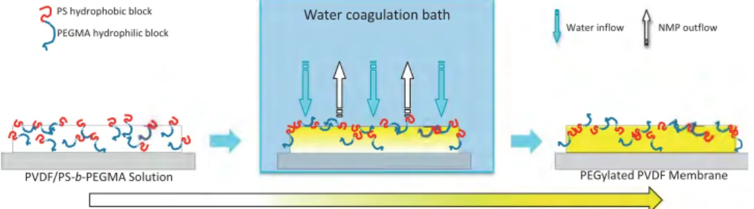
Membranes were prepared using the liquid-induced phase separation process (sometimes referred as wet-immersion process or nonsolvent-induced phase separation process) as presented byFig. 1. Solutions were cast on a glass plate, using an appropriate metal casting knife allowing to control the initial thickness of the film to 300
μ
m. Afterwards, glass plates were immersed in DI water, to induce phase separation of the system. The newly-formed membranes were kept in water for 24 h, and water was changed once during that period of time, to ensure a total removal of residual solvent. Thereafter, membranes were dried at ambient temperature for at least 24 h before use.In solution, it is expected that PEGMA segments of copolymer molecules located at the upper interface will be oriented toward the liquid, which provides a more hydrophilic environment. Hydro-phobic PS groups should be in contact with air. But once phase separation has been initiated, copolymer molecules are turned up-side down. Indeed, the polymer system becomes more hydro-phobic owing to a solvent outflow. In the end, PS groups interact with PVDF while PEGMA segments should be oriented toward the upper interface. Also, additive molecules initially in the bulk solution may migrate toward the interface during phase separation, which would ensure a more stable configuration[24]. However, one should note that this motion is limited because phase separation occurs quickly in LIPS process.
2.2.3. Surface characterization
Chemical composition of membranes was assessed using two methods. In a first time, ATR FT-IR spectrophotometry (Perkin-Elmer Spectrum One) with ZnSe as an internal reflection element was used. Measurements were realized perpendicular to the surface of the membranes. Each spectrum was captured by averaging 16 scans at a resolution of 4 cm#1. In addition, X-ray photoelectron spectroscopy
(XPS) was performed using a PHI Quantera SXM/Auger spectrometer with a monochromated Al KR X-ray source (1486.6 eV photons). The energy of emitted electrons was measured with a hemispherical energy analyzer at pass energies ranging from 50 to 150 eV. All the data were collected at photoelectron take off angles of 451 with respect to the sample surface. The binding energy (BE) scale was referenced by setting the peak maximum in the C1s spectrum to 284.6 eV. A high-resolution C1s spectrum was fitted using a Shirley background subtraction and a series of Gaussian peaks. The data analysis software was from Service Physics, Inc.
Porous structure of virgin PVDF membrane and modified PVDF membranes was characterized using microscopy techniques. Scan-ning electronic microscopy (SEM) analysis was performed using a Hitachi S-3000 instrument. Accelerating voltage varied in the range 7 keV–10 keV, depending on the observation carried out. Prior to observation, membranes were mounted on sample stages, using double-sided adhesive tape, and sputter-coated with gold for 150 s. Furthermore, membranes were examined by atomic force micro-scopy (AFM). The AFM images were acquired with a JPK Instruments AG multimode NanoWizard (Germany). The instrument is equipped
with a NanoWizard scanner and operated in air. For tapping-mode AFM, a commercial Si cantilever (TESP tip) of $320 kHz resonant frequency from the JPK instrument was used. The relative humidity was less than 40%.
2.2.4. Assessment of wettability and low-biofouling properties Water contact angles were measured with an angle-meter (Automatic Contact Angle Meter, Model CA-VP, Kyowa Interface Science Co., Ltd. Japan) at 25 1C. The DI water was dropped on the sample surface at 10 different sites and the average of the measured values was taken as the membrane water contact angle. Hydration capabilities (mg/cm3) of membranes were also
deter-mined. Dry weights of 0.85 cm diameter membranes were first recorded using a 10#5g precision balance (Mettler, Toledo Pac Rim
AG, Taiwan Branch). Afterwards, membranes were immersed in deionized water for 24 h. Then, water was gently wiped out with tissue, and membranes weighed. Hydration capability was evaluated taking the difference of dry and wet weights per unit volume. The volume considered was calculated after measuring the thickness of the membranes at 10 different positions. For each membrane, five independent measurements were performed and the average of the obtained values was taken as the final value for membrane hydration capability.
The adsorption of bovine-serum-albumin (BSA, MwE 66,000 g mol#1; Sigmas) was performed. Membranes were
placed in a 24-well plate and immersed in 1 mL of pure ethanol for 30 min. Subsequently, membranes were soaked in phosphate-buffered saline (PBS) for 2 h at 25 1C. PBS was removed and membranes were incubated with 1 mL of 1 mg mL#1 BSA for
2 h at 25 1C. The absorbance at 280 nm was measured using a UV–vis spectrophotometer (PowerWave XS, Biotech). Each test was performed three times on each membrane. A similar method was used for adsorption of lysozyme from chicken egg white (Mw¼14,300 g mol#1) provided by Sigmas. After immersing
membranes in ethanol (30 min) and in PBS (2 h), they were incubated with 1 mL of 1 mg mL#1
lysozyme for 2 h at 25 1C and absorbance was measured at 280 nm. A control test was per-formed on membrane containing 5 wt% additive to ensure that there was no release of additive that would interfere with the reading of absorbance.
Staphylococcus epidermidis (Gram-positive) and Escherichia coli (Gram-negative) were used to investigate bacterial adhesion behavior on the surface of membranes. S. epidermidis and E. coli were cultured in a medium containing 3.0 mg mL#1 beef extract
and with 5.0 mg mL#1
peptone. Cultures were incubated at 37 1C and shaken at 100 rpm until the stationary phase was reached. For S. epidermidis, final concentration was 109 cells/ml after 15 h of incubation whereas for E. coli, it reached a concentration of about 106 cells/ml after 12 h. Membranes were washed three times with PBS in a 24-well plate. Afterward, one milliliter of bacteria suspension was added to each well, and incubation at 37 1C lasted for 3 h. Bacterial solution was then removed and membranes were washed with PBS three times at 37 1C. Bacteria adhering to the
sample surfaces were observed by SEM (Hitachi S-3000 instru-ment) using an accelerating voltage of 7 keV, and after sputter-coating surfaces with gold for 150 s.
3. Results and discussion
3.1. Chemicophysical characterization of membranes – effect of PS-b-PEGMA on PVDF membrane formation
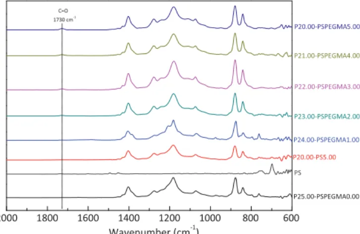
Chemical state of virgin and PEGylated membranes were first investigated using FT-IR, and related spectra are displayed on Fig. 2. Spectrum of a PVDF-PS membrane prepared from a blend containing 20 wt% PVDF and 5 wt% polystyrene (PS) is also displayed and referred as P20.00-PS5.00. Characteristic absorption bands of PVDF membranes are provided in literature and will not be reminded herein[27–29]. Our purpose concerned absorption bands attributed to PS-b-PEGMA copolymer. Spectra of virgin PVDF membrane (P25.00-PSPEGMA0.00) and of P20.00-PS5.00 do not show major differences. Many characteristic absorption bands due to the benzene rings of polystyrene should be located in the range 1450–3060 cm#1[30]while main absorption bands of
PVDF are found in the range 700–1400 cm#1. Herein, the
concentration of additive was low (5 wt%) compared to that of PVDF (20 wt%). In addition, as all PS groups were probably not all found at the surface, but dispersed within the whole matrix, it may explain why stretching bands of PS could not be detected and major difference not found between two spectra. Similarly, absorption bands attributed to PEGMA segments were difficult to identify, especially at very low additive concentration. Nonethe-less, it could be seen that when increasing PS-PEGMA concentra-tion, a broad signal appeared at about 1730 cm#1. It was attributed
to the stretching of carbonyl function, as mentioned in literature in the preparation of heparinized polypropylene films grafted with PEGMA [31], or in the preparation of poly(phthalazinone ether sulfone ketone) (PPESK) backbones containing poly-(poly(ethylene glycol) methyl ether methacrylate) (P(PEGMA)) side chains[32]. The increasing of this signal intensity with additive content tended to prove the presence of copolymer at the surfaces of membranes. XPS was subsequently used and related spectra are shown on Fig. 3. As for the virgin membrane, literature is very complete, permitting to identify the peaks of the curve-fitted C1s core-level spectrum[20,23,33–36]. Quickly, the peak at binding energy (BE) of 284.6 eV corresponds to neutral CH species, that at BE of 286.2 eV is associated to CH2species while the final large peak
at BE of 290.6 eV is due to CF2bonds. One should note that the
presence of the shoulder at 284.6 eV is not always reported. As for the spectrum of the PEGylated membrane, it also evidenced two main peaks and one shoulder. If the peak at 290.3 eV and that at 284.4 eV are owed to CF2and CH bonds, respectively, the large
peak observed at 285.9 eV is due to both CH2and C–O function
contributions. Indeed, Li and coworkers prepared membranes by blending PVDF with PVDF-g-PSBMA and PEG, and clearly showed that the peak due to C–O bonds was overlapped by that of CH2[23]. Furthermore, as shown byFig. 4when adding
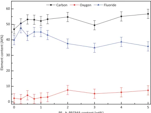
PS-b-PEGMA, the relative oxygen content slightly increases with the additive content, due to C–O functions carried by PS-b-PEGMA backbone. On the other hand, fluoride content logically decreased since PS-b-PEGMA does not contain fluoride atoms.
Trends obtained from chemical analyses were very slight. Owing to a fast phase separation, polymer additive is rapidly entrapped when matrix solidifies, which prevents possible motion toward the upper interface and the complete change of orientation of additive molecules initially located at the surface. Consequently, probably not all hydrophilic moieties could be found at the surface. However, they Fig. 2.FT-IR chemical analysis of virgin PVDF membrane, polystyrene, PVDF/PS
membrane and PEGylated PVDF membranes.
Fig. 4.XPS chemical analysis of virgin PVDF membrane and PEGylated PVDF membranes.
Fig. 5. SEM characterization of the surfaces of virgin PVDF membrane and PEGylated PVDF membranes.
at least remained in the membrane matrix, since PS-b-PEGMA copolymer is totally water insoluble.
Structures were then investigated by performing SEM observa-tions. Images obtained related to surfaces and cross-sections are presented byFigs. 5and6, respectively. First, images highlight typical structures obtained by LIPS process, that is, finger-like structures with a porous skin top-layer. Such morphologies arise from fast phase separation rate. Indeed, systems exhibiting instantaneous demixing usually show macrovoids whereas when phase separation is delayed, these formations are usually absent. Second, results evidenced that PS-b-PEGMA affected membrane formation mechan-isms. Concerning the surfaces, they tended to be denser when adding copolymer. According to fundamentals related to the formation of phase inversion membranes, a decreasing of surface porosity occurs when polymer concentration in the upper layer is increased. In the present case, amphiphilic copolymer additive was added in the casting solution. For thermodynamic considerations, migration of this additive toward the surface could be expected, to ensure equilibrium of the system. Therefore, a polymer concentration gradient may have been created in the system. If this assumption is verified, then the total polymer concentration (PVDFþPS-b-PEGMA) should be higher at the surface than in the deeper layers, arising in a denser surface, compared to the virgin membrane for which there is no copolymer, and therefore not a similar concentra-tion gradient. The fact that the trend was not clear could be explained by the fast phase inversion rates involved in LIPS process inhibiting optimal motion of the copolymer. Moreover, expansion of polymer-lean phase in the bulk when adding copolymer was evidenced by cross-section images. Indeed, PEGylated membranes exhibited larger finger-like structures. In addition,Fig. 7, reporting the evolution of membrane's thickness as a function of additive content, evidenced that shrinkage during membrane formation tended to be prevented for PEGylated membranes. Undoubtedly, the additive has an important impact on membrane formation: from SEM pictures ofFig. 6, it can be seen that to the width of the largest macrovoids is increased from about 10
μ
m for virgin PVDF mem-brane to about 45μ
m for PEGylated PVDF membrane containing 5 wt% additive. Assuming a same trend in the three dimensions of the space, the polymer-lean phase leading to the final membrane porosity had been greatly expanded, which was also supported by the thickness evolution.When phase separation is instantaneous, Mulder reminds that first droplets of polymer-lean phase are formed underneath the surface[37]. Their growth depends on concomitant solvent flow, coming from the polymer-rich phase right underneath the nuclei, and non-solvent diffusion from the top-layer. Growth of macrovoids stops when the polymer concentration at the interface between the polymer-lean phase and the polymer solution below is so high that solvent/nonsolvent contribution to the expansion of the macrovoids cannot balance forces responsible for polymer solidification. Adding PS-b-PEGMA in the casting solution clearly contributed to expand the polymer-lean phase domains, which meant that growth of nuclei was favored. It was possible either because copolymer contributed to better solvent/nonsolvent exchanges or because no new nuclei were formed, below the first set of growing nuclei from the upper interface, that could have inhibited macrovoids growth. The effect of polymer-additive on membrane formation has been reported many times, but is not clearly elucidated. The fast phase separation involved makes on-line experimental analyses complicated and challenging and there seems to be no general rule. Indeed, Guillen and coworkers recently presented a review on membranes formed by nonsolvent induced phase separation (termed LIPS in the present paper)[38]. They reported that PEG additives mostly acted as a pore-forming agent but it also depended on the additive molecular weight since small molecular weight PEG additives tended to work as
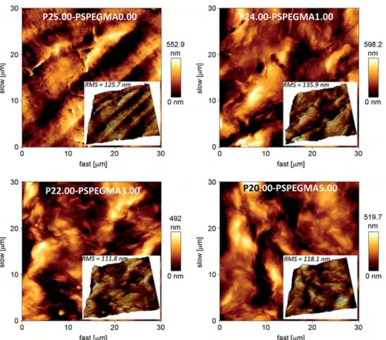
pore-reducing agent. Still, mechanisms could be proposed by considering PS-b-PEGMA nature. It is an amphiphilic copolymer, soluble in NMP solvent, and insoluble in water. PEGMA blocks did entrap water molecules (this is the main goal of this work) while solubility in NMP was due to both blocks (PS and PEGMA). Therefore, it was reasonable to assume that inflows of solvent and nonsolvent in the nucleus were enhanced, compared to those occurring in a totally hydrophobic matrix. Especially supplemen-tary hydrogen bonds were formed between additive and water, compared to PVDF and water, owing to the presence of polyethy-lene glycol groups, leading to improved mass transfers and in the end, to larger macrovoids. There was also an impact on the wall of these macrovoids, as shown by the 3 K magnification of the cross-sections inFig. 6. If virgin membrane exhibits rough dense walls, those of PEGylated PVDF membranes present many small pores. Thus, PS-b-PEGMA favored pore-formation in the system studied. AFM characterization was performed to obtain further informa-tion concerning the potential effect of copolymer additive on the surface structuring. In addition, the roughness (RMS) coefficient can be related to the homogeneity and surface coverage of top-surfaces[39]. Images are displayed onFig. 8, and RMS coefficients are reminded inTable 1. Relatively smooth surfaces were obtained, owing to the preparation process and to the nature of nonsolvent. Indeed, LIPS using water as a nonsolvent favors the obtaining of dense smooth top-layers. It was worth noting that in each case, the roughness coefficient was in a similar range. The change observed was not important enough to be attributed to a major effect of additive on membrane surface structuring. This also confirmed SEM results that mainly showed an important effect of the additive on the macrovoids morphology while only a slight change of surface porosity was evidenced. Globally, the change of struc-ture may have two consequences on the membranes properties: (i) mechanical properties should be reduced in terms of tensile strength or Young's modulus (ii) permeability should be increased. As for the mechanical properties, they will be assessed in sub-sequent works prior to applying the membranes in filtration processes. A priori, it was not noticed any reduction of mechanical properties of the modified membranes compared to those of virgin PVDF matrix. None of them was torn apart upon handling.
To conclude, contrary to water soluble PEG-system additives also acting as pore-formers [40], PS-b-PEGMA is not washed out of the membrane during phase inversion. Chemical analyses did not evidence important changes when comparing virgin PVDF membrane to PEGylated PVDF membranes, but this additive, water-insoluble, did affect both surface chemistry and morphology of membranes. In this
respect, it was expected to influence membranes hydrophilic proper-ties via both physical and chemical contributions.
3.2. Effect of PS-b-PEGMA on hydration properties of PVDF membranes
The additive was used to increase the hydrophilicity of PVDF membranes. Indeed, it is currently admitted that adsorption of biofouling compounds (proteins, bacteria) is often facilitated on rough and hydrophobic surfaces. As membranes were prepared by LIPS, they are quite smooth (see also RMS coefficients). So, the chemical effect is expected to be the major parameter affecting protein adsorption and bacterial attachment. Hydrophilicity of virgin and PEGylated membranes were measured by evaluating their water contact angle and hydration capacity, and results are
presented inFig. 9and inTable 1. As for the water contact angle, it is about 85711 for virgin PVDF membrane, evidencing a quite hydrophobic surface. By adding amphiphilic copolymer additive, it decreased until reaching a plateau at about 591. This result proves that hydrophilicity of surfaces is enhanced by incorporating additive copolymer. In the meantime, hydration capacity of poly-mer matrices was increased from 0 to 226.7 727.1 mg/cm3when
adding 5 wt% copolymer additive. In this respect, the whole wettability of membranes was improved. Entrapment of water inside the polymer structure became possible owed to the presence of ethylene-glycol groups dispersed in the bulk and establishing hydrogen bonds with water molecules. Furthermore, it was shown in Fig. 7that the thickness was expanded when adding copolymer and Fig. 6 evidenced that the polymer-lean phase, that is the overall porosity of the membranes, was more Table 1
Preparation of membranes and their physicochemical characterization.
Membrane ID PVDF (wt%) PS (wt%) PS-b-PEGMA (wt%) NMP (wt%)
Atom content Water contact angle (deg) Hydration capability (mg/cm3) Thickness (μm) RMS coefficient (nm) C (%) O (%) F (%) P25.00-PSPEGMA0.00 25.00 – – 75 55.37 4.47 40.16 8571 0 148 712 125.7 P20.00-PS5.00 20.00 5.00 – 75 – – – – – 18075 – P24.75-PSPEGMA0.25 24.75 – 0.25 75 50.63 1.80 47.56 8071 51.976.6 187713 – P24.50-PSPEGMA0.50 24.50 – 0.50 75 53.19 4.27 42.55 8071 56.3730.5 265722 – P24.25-PSPEGMA0.75 24.25 – 0.75 75 53.00 1.91 45.09 8071 83.1 716.2 271731 – P24.00-PSPEGMA1.00 24.00 – 1.00 75 52.16 2.46 45.07 7971 106.7746.1 269726 135.9 P23.75-PSPEGMA1.25 23.75 – 1.25 75 53.35 3.07 43.08 7871 107.0727.8 29179 – P23.00-PSPEGMA2.00 23.00 – 2.00 75 54.77 7.64 37.59 73 71 112.1 730.0 280728 – P22.00-PSPEGMA3.00 24.00 – 3.00 75 49.45 5.26 34.88 72 71 233.0735.2 250718 111.8 P21.00-PSPEGMA4.00 21.00 – 4.00 75 55.14 6.15 38.71 5973 233.1735.4 284715 – P20.00-PSPEGMA5.00 20.00 – 5.00 75 56.77 7.44 35.78 5972 226.7727.1 299721 118.1
important for PEGylated membranes. This physical effect of the additive also contributed to allow the entrapment of more water molecules within the matrix. Also, a plateau is reached, possibly due to (i) saturation of membranes' surfaces with PS-b-PEGMA copolymer, and to (ii) a maximum thickness obtained from a 1 wt% additive content. Hence, no more water molecules could be entrapped within the structure using the LIPS process, and wettability (swelling) of membranes was optimum from a 3 wt% additive content.
This result supports the efficient modification of PVDF mem-brane. In previous section, it was pointed the difficulty to ascertain an effective change of surface chemistry. But water contact angles were reduced, mostly attributed to a change of surface chemistry. Combined to the results of hydration capability measurements, the new membranes are clearly more hydrophilic and may possibly reduce biofouling, which is to be investigated in next section. 3.3. Effect of PS-b-PEGMA on resistance of PVDF membranes to protein adsorption
In membrane applications such as water or blood treatment, feed often contains numerous proteins, able to interact with hydrophobic membrane material. Adsorption of proteins leads to biofouling, but also favors adsorption of micro-organisms such as bacteria, via hydrophobic–hydrophobic interactions with proteins constituting their outer cell wall. In this respect, it is of major concern to design membranes able to resist protein adsorption. BSA is a common protein tested to evaluate membrane resistance to biofouling by carrying out typical adsorption tests[41]or filtration experiments [42]. Therefore, it was used in the present study, as well as lysozyme, found in many secretions and that may contribute to biofouling in biological applications of membranes.
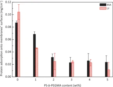
Fig. 10presents the adsorption of BSA and LY as a function of the copolymer additive content. The amphiphilic additive permits to reduce BSA adsorption from about 0.087 mg/cm2 for the virgin
membrane to 0.024 mg/cm2for the PEGylated membrane
contain-ing 5 wt% additive, while LY attachment was reduced from 0.104 mg/ cm2to 0.012 mg/cm2. Hence, more than 65% and about 89% of BSA
and LY adsorption, respectively, was prevented thanks to the amphiphilic copolymer, showing its efficiency in the preparation of low-biofouling surface. Binding water around ethylene glycol groups and favoring water entrapment within the very porous matrix led to a sharp decrease of protein adsorption. As reported earlier, better
results were obtained with LY, probably due to the fact that it possesses a molecular weight lower than that of BSA (66,000 vs. 14,300 g mol#1). Therefore, it contains less hydrophobic segments
on its backbone able to interact with hydrophobic sites available on membrane surface and within the matrix[43].
It was also important to compare these results with previously published data. Wang et al. used a membrane prepared by similar process as that in our study, but blending polyethersulfone (PES) (18 wt%), PEG 2000 (15 wt%) and P123-b-PEG copolymer (prepared from Pluronics123 and PEG by substitution reaction) (0.8 wt%) in
DMF solvent[17]. For this initial formulation, BSA adsorption was about 2
μ
g/cm2. Compared to their control membrane preparedusing PES (18 wt%) and PEG 2000 (15 wt%) for which BSA adsorp-tion was 6.8
μ
g/cm2, protein adsorption was reduced to about 70%,which compares to the present work. Wang and coworkers mana-ged to obtain even better results, but they actually used two additives, including one (PEG 2000) in large amount compared to the main polymer content. Furthermore, Boributh and coworkers modified PVDF membranes by introducing chitosan into the casting solution in the range 0.5–2 wt%[44]. Indeed, chitosan is a hydro-philic polymer which can be efficient to inhibit protein adsorption. Fig. 9.Hydrophilic properties of virgin PVDF membrane and PEGylated PVDF membranes.
Fig. 10.Protein adsorption tests performed onto virgin PVDF membrane and PEGylated PVDF membranes.
BSA adsorption tests at neutral pH (our conditions) revealed that chitosan permitted to reduce protein adsorption from about 58
μ
g/ cm2to 22μ
g/cm2, that is, a decreasing of 62% was reported. We alsocompared these results to those we previously reported. Using vapor-induced phase separation (VIPS) process applied to a PVDF/ Pluronics
(poly(ethylene oxide)–poly(propylene oxide)–poly(ethy-lene oxide)) blend, BSA and LY adsorption were reduced to about 65% and 95% the limitation of the virgin membrane[43]. In that study, VIPS process was applied, which means that more additive copolymer was found onto the surface, owing to a facilitated motion during phase separation, compared to LIPS. Indeed, protein adsorp-tion tests using fibrinogen as a model protein, polysulfone as a bulk polymer and Pluronics
F108 as an additive, have shown that VIPS process allowed preparing better low-biofouling membranes than LIPS process[45]. One may wonder why LIPS was chosen in this study and not VIPS. Actually, the present research work aimed at preparing microfiltration membranes. Applying VIPS to a PVDF solution system will more likely lead to a nodular morphology, which will not present optimum mechanical properties, from our knowledge. In addition, using this polymer system, it seems rather difficult to obtain a bi-continuous structure by VIPS suitable for MF applications. Indeed, low dissolution temperature (32 1C) is required to obtain a bi-continuous PVDF membrane by VIPS[46], but for such a temperature, it is not possible to totally solubilize the PVDF/PS-b-PEGMA system in a reasonable period of time. Therefore LIPS was used rather than VIPS, process that is not believed to lead to a copolymer surface density as high as that obtainable in VIPS, but still good enough to permit an important decrease of protein adsorption, as demonstrated herein.
Globally, and even though comparisons cannot always be accurately performed between research studies, present data show that PEGylated PVDF membranes were very efficient to resist BSA and LY adsorption.
3.4. Effect of PS-b-PEGMA on resistance of PVDF membranes to bacterial adhesion
Proteins adsorption usually favors bacteria adhesion. Once the first microorganisms have stuck to a membrane's surface, this surface may undergo bacteria colonization, phenomenon respon-sible for biofouling. Our target is to limit biofouling of membranes by creating a hydrophilic interface. It was shown in previous sections that results regarding protein adhesion tests were encouraging, with an important decreasing of BSA and LY adsorp-tion. Therefore, further biofouling tests were conducted, using
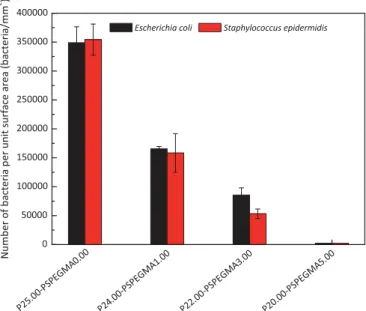
Escherichia coli and Staphylococcus epidermidis incubated with the membranes for 3 h. These two bacteria have different nature, size and shape. Moreover, EC is regularly used to assess low-biofouling properties of membranes [3,4,47–49]. The use of fluorescence technique and confocal microscope technique are reported to analyze adhesion of bacteria onto surfaces [43,45]. In this work, noise attributed to the PVDF surface background was too impor-tant. So, we made use of SEM to analyze bacterial attachment on membranes, as displayed inFig. 11. Each test was performed five times. To obtain quantitative data for each condition, bacteria were counted on five SEM images obtained at a 5 K magnification. It corresponded to the scan of a 437.5
μ
m2 surface area.The average number of bacteria per unit surface area, obtained from five different membranes, was then reported inFig. 12.
Both E. coli and S. epidermidis easily attached to virgin PVDF membranes. Indeed, hydrophobic surfaces favor interactions with bacteria hydrophobic outer cell-wall. Despite smooth sur-faces earlier evidenced by AFM images, and therefore preventing bacterial attachment by physical interaction as with rough interfaces, the number of bacteria onto virgin membranes was important (about 350,000 bacteria/mm2in each case). As shown
Fig. 11.Bacterial attachment tests performed onto virgin PVDF membrane and PEGylated PVDF membranes.
Fig. 12.Quantitative attachment of bacteria onto virgin PVDF membrane and PEGylated PVDF membranes after a 3-h incubation time in bacteria solution.
earlier, the surface morphology was not drastically affected by PS-b-PEGMA copolymer and the surface roughness was in the same range. In this respect, the surface chemistry is the key point permitting to explain the results. SEM images presented onFig. 11, and related quantitative data displayed on Fig. 12, showed that copolymer tested in this study allowed providing an efficient chemical modification of the surface in order to resist bacterial adhesion. From a 5 wt% additive, almost no bacteria remained onto the surface (adhesion was reduced by 99%, compared to the virgin surfaces), whether from EC tests or SE ones, which was a good indication of low-biofouling properties of the membranes pre-pared in this work.
Literature also provides interesting data on the efficiency of PEG-like systems to resist bacteria. For instance, Miller and coworkers recently reported on the use of polydopamine-g-poly(ethylene glycol) surface coatings to control biofouling of membranes and feed spacers. Their short-term static adhesion tests ran for 1 h evidenced that the PEG-like systems permitted reducing adhesion of P. aeruginosa to about 75% the limitation of the virgin polysulfone membrane[50]. Recently, we also reported on the potential low-biofouling properties of membranes prepared by LIPS or VIPS process[43,45]. The additive used was Pluronics F108 which is water-soluble. Reduction of
bacterial attachment (EC and SE) was only observed when mem-branes where prepared by VIPS. Indeed, during membrane formation by LIPS process, part of F108 was washed out of the matrix, therefore reducing the potential low-biofouling properties of the membranes. Herein, we obtained as good results as those early presented with membranes prepared by VIPS, but using LIPS and a PEG additive water insoluble. Moreover, regardless the intended application, one can find that literature is well provided concerning PEGylation of surfaces aiming at resisting bacterial attachment. Indeed, it is a challenge in many fields to design anti-biofouling surfaces. For instance, Li et al. designed silicone films grafted with both PEG-derivative polymer to improve their hemocompatibility [51]. They showed that grafted silicone substrates reduced the adhesion of bacteria including Staphylococcus aureus, Escherichia coli, and Staphy-lococcus epidermidis by over 90%, showing the efficiency of PEG-systems, further supporting the results of the present work. Further-more, Weber et al. recently presented their study on the effect of PEGylation of aminopropylated surfaces on bacteria adhesion and evidenced that EC adhesion was inhibited[52]. Of course, it is again hard to compare data of other authors with those presented in this work, since the polymer matrix, the bacteria tested or the additive copolymer are different, but it is important noting that PEG-systems in general, and the present system in particular, have excellent low-biofouling properties. To the best of our knowledge, the results presented in this study constitute among the best reported on the design of PVDF membranes resisting bacterial attachment.
4. Conclusions
In this manuscript, we reported the use of PS-b-PEGMA amphiphilic additive in the preparation of PVDF membrane by LIPS process. The main goal was to decrease biofouling of PVDF membranes, and the major conclusions are listed as follows:
%
Membrane structure was drastically affected by the copolymeradditive, with a decreasing of surface porosity, probably owed to the establishment of a polymer concentration gradient during membrane formation, and an expansion of the polymer lean-phase, due to an improvement of solvent outflow and non-solvent inflow during membrane formation. The walls of the characteristic macrovoids tended to be more porous with the additive content, and the shrinkage usually occurring
during PVDF membrane formation was inhibited by the copo-lymer additive;
%
Membrane surface hydrophilicity was improved, thanks to thepresence of PEGMA segments onto membranes' surface;
%
Membrane hydration capability was importantly enhanced,attributed to both chemical and physical effects of the copoly-mer additive;
%
Protein adsorption including BSA and LY, was decreased in animportant extent;
%
Bacterial attachment onto the surfaces was inhibited using twomodel bacteria, Escherichia coli and Staphylococcus epidermidis. Subsequent studies will concern filtration tests using membranes presented in this work. Indeed, evaluation of low-biofouling proper-ties in practical filtration operation is now a major priority.
Acknowledgments
The authors wish to express their sincere gratitude to the Center-of-Excellence (COE) Program on Membrane Technology from the Ministry of Education (MOE), ROC., the project of Outstanding Professor Research Program in the Chung Yuan Christian University, Taiwan (CYCU-107090022), the 2013–2015 NSC-ANR Blanc Interna-tional II Programme (Taiwan–France Project: Super-NAM), the National Science Council (NSC 102-2923-E-033-001-MY3 and NSC 102-2218-E-033-001), and Chung Yuan Christian University (CYCU campus plan 107011072) for their financial support.
References
[1]H. Ivnitsky, I. Katz, D. Minz, E. Shimoni, Y. Chen, J. Tarchitzky, R. Semiat, C. G. Dosoretz, Characterization of membrane biofouling in nanofiltration pro-cesses of wastewater treatment, Desalination 185 (2005) 255–268. [2]X. Ma, Y. Su, Q. Sun, Y. Wang, Z. Jiang, Preparation of
protein-adsorption-resistant polyethersulfone ultrafiltration membranes through surface segrega-tion of amphiphilic comb copolymer, J. Membr. Sci. 292 (2007) 116–124. [3]C.X. Liu, D.R Zhang, Y. He, X.S. Zhao, R. Bai, Modification of membrane surface
for anti-biofouling performance: effect of anti-adhesion and anti-bacteria approaches, J. Membr. Sci. 346 (2010) 121–130.
[4]Y.F. Yang, Y. Li, Q.L. Li, L.S Wan, Z.K. Xu, Surface hydrophilization of micro-porous polypropylene membrane by grafting zwitterionic polymer for anti-biofouling, J. Membr. Sci. 362 (2010) 255–264.
[5]C.H. Worthley, K.T. Constantopoulos, M. Ginic-Markovic, R.J. Pillar, J.G. Matisons, S. Clarke, Surface modification of commercial cellulose acetate membranes using surface-initiated polymerization of 2-hydroxyethyl methacrylate to improve membrane surface biofouling resistance, J. Membr. Sci. 385–386 (2011) 30–39. [6]M. Zhang, K. Zhang, B. De Gussene, W. Verstraete, Biogenic silver nanoparticles
(bio-Ag1) decrease biofouling of bio-Ag1/PES nanocomposite membranes, Water Res. 46 (2012) 2077–2087.
[7]Z. Zhang, Z. Wang, J. Wang, S. Wang, Enhancing chlorine resistances and anti-biofouling properties of commercial aromatic polyamide reverse osmosis membranes by grafting 3-allyl-5,5-dimethylhydantoin and N,N′-Methylene-bis(acrylamide), Desalination 309 (2013) 187–196.
[8]Y. Chang, T.Y Cheng, Y.J. Shi, K.R. Lee, J.Y. Lai, Biofouling-resistance expanded poly(tetrafluoroethylene) membrane with a hydrogel-like layer of surface-immobilized poly(ethylene glycol) methacrylate for human plasma protein repulsions, J. Membr. Sci. 323 (2008) 77–84.
[9]F. Razi, I. Sawada, Y. Ohmukai, T. Maruyama, H. Matsuyama, The improvement of antibiofouling efficiency of polyethersulfone membrane by functionaliza-tion with zwitterionic monomers, J. Membr. Sci. 401–402 (2012) 292–299. [10]A. Nguyen, S. Azari, L. Zou, Coating zwitterionic amino acid l-DOPA to increase
fouling resistance of forward osmosis membrane, Desalination 312 (2013) 82–87.
[11]Y.C. Chiang, Y. Chang, C.J. Chuang, R.C. Ruaan, A facile zwitterionization in the interfacial modification of low bio-fouling nanofiltration membranes, J. Membr. Sci. 389 (2012) 76–82.
[12]H.D.W. Roesink, M.A.M. Beerlage, W. Potman, Th. van den Boomgaard, M.H.V. Mulder, C.A. Smolders, Characterization of new membrane materials by means of fouling experiments adsorption of BSA on polyetherimide— polyvinylpyrrolidone membranes, Colloids Surf. 55 (1991) 231.
[13]Y.Q. Wang, T. Wang, Y.L. Su, F.B. Peng, H. Wu, Z. Jiang, Remarkable reduction of irreversible fouling and improvement of the permeation properties of poly (ether sulfone) ultrafiltration membranes by blending with Pluronic F127, Langmuir 21 (2005) 11856–11862.
[14]W. Zhao, Y. Su, C. Li, Q. Shi, X. Ning, Z. Jiang, Fabrication of antifouling polyethersulfone ultrafiltration membranes using Pluronic F127 as both sur-face modifier and pore-forming agent, J. Membr. Sci. 318 (2008) 405–412. [15]H.J. Li, Y.M. Cao, J.J. Qin, X.M. Jie, T.H. Wang, J.H. Liu, Q. Yuan, Development and
characterization of anti-fouling cellulose hollow fiber UF membranes for oil– water separation, J. Membr. Sci. 279 (2006) 328–335.
[16]A. Higuchi, K. Shirano, M. Harashima, B.O. Yoon, M. Hara, M. Hattori, K. Imamura, Chemically modified polysulfone hollow fibers with vinylpyrro-lidone having improved blood compatibility, Biomater 23 (2002) 2659–2666. [17]Y.Q. Wang, Y.L. Su, Q. Sun, X.L. Ma, Z.Y. Jiang, Y. Chang, S. Chen, Q. Yu, Z. Zhang, Generation of anti-biofouling ultrafiltration membrane surface by blending novel branched amphiphilic polymers with polyethersulfone, J. Membr. Sci. 286 (2006) 228–236.
[18]F. Ran, S. Nie, W. Zhao, J. Li, B. Su, S. Sun, C. Zhao, Biocompatibility of modified polyethersulfone membranes by blending an amphiphilic triblock co-polymer of poly(vinyl pyrrolidone)–b-poly(methyl methacrylate)–b-poly(vinyl pyrroli-done), Acta Biomater. 7 (2011) 3370–3381.
[19]Y.H. Zhao, Y.L. Qian, B.K. Zhu, Y.Y. Xu, Modification of porous poly(vinylidene fluoride) membrane using amphiphilic polymers with different structures in phase inversion process, J. Membr. Sci. 310 (2008) 567–576.
[20]F. Liu, Y.Y. Xu, B.K. Zhu, F. Zhang, L.P. Zhu, Preparation of hydrophilic and fouling resistant poly(vinylidene fluoride) hollow fiber membranes, J. Membr. Sci. 345 (2009) 331–339.
[21]C. Zhang, Y. Bai, Y. Sun, J. Gu, Y. Xu, Preparation of hydrophilic HDPE porous membranes via thermally induced phase separation by blending of amphi-philic PE-b-PEG copolymer, J. Membr. Sci. 365 (2010) 216–224.
[22]F. Liu, N. Awanis Hashim, Y. Liu, M.R. Moghaeh Abed, K. Li, Progress in the production and modification of PVDF membranes, J. Membr. Sci. 375 (2011) 1–27.
[23]Q. Li, Q.Y. Bi, B. Zhou, X.L. Wang, Zwitterionic sulfobetaine-grafted poly (vinylidene fluoride) membrane surface with stably anti-protein-fouling performance via a two-step surface polymerization, Appl. Surf. Sci 10 (2012) 4707–4717.
[24]N. Pezeshk, D. Rana, R.M. Narbaitz, T. Matsuura, Novel modified PVDF ultrafiltration flat-sheet membranes, J. Membr. Sci. 389 (2012) 280–286. [25]N. Pezeshk, R.M. Narbaitz, More fouling resistant modified PVDF ultrafiltration
membranes for water treatment, Desalination 287 (2012) 247–254. [26]Y.C. Chiag., Y. Chang, W.Y. Chen, R.C. Ruaan, Biofouling resistance of
ultra-filtration membranes controlled by surface self-assembled coating with PEGylated copolymers, Langmuir 28 (2012) 1399–1407.
[27]T. Boccaccio, A. Bottino, G. Capannelli, P. Piaggio, Characterization of PVDF membranes by vibrational spectroscopy, J. Membr. Sci. 210 (2002) 315–329. [28]A. Salimi, A.A. Yousefi, Conformational changes and phase transformation
mechanisms in PVDF solution-cast films, J. Polym. Sci. Part B: Polym. Phys. 42 (2004) 3487–3495.
[29]R. Gregorio Jr., Determination of theα,β, andγcrystalline phases of poly (vinylidene fluoride) films prepared at different conditions, J. Appl. Polym. Sci 100 (2006) 3272–3279.
[30]A. Siri, C. Alkan, A. Biçer, A. Karaipekli, Synthesis and thermal energy storage characteristics of polystyrene-graft-palmitic acid copolymers as solid–solid phase change materials, Sol. Energy Mater. Sol. Cells 95 (2011) 3195–3201. [31]J. Jin, W. Jiang, Q. Shi, J. Zhao, J. Yin, P. Stagnaro, Fabrication of
PP-g-PEGMA-g-heparin and its hemocompatibility: from protein adsorption to anticoagulant tendency, Appl. Surf. Sci. 258 (2012) 5841–5849.
[32]L.P. Zhu, Y.Y. Xu, H.B. Dong, Z. Yi, B.K. Zhu, Amphiphilic PPESK-graft-P(PEGMA) copolymer for surface modification of PPESK membranes, Mater. Chem. Phys. 115 (2009) 223–228.
[33]J.F. Hester, A.M. Mayes, Design and performance of foul-resistant poly(viny-lidene fluoride) membranes prepared in a single-step by surface segregation, J. Membr. Sci. 202 (2002) 119–135.
[34]G.M. Qiu, L.P. Zhu, B.K. Zhu, Y.Y. Xu, G.L. Qiu, Grafting of styrene/maleic anhydride copolymer onto PVDF membrane by supercritical carbon dioxide:
preparation, characterization and biocompatibility, J. Supercrit. Fluids 45 (2008) 374–383.
[35] Y. Sui, X. Gao, Z. Wang, C. Gao, Antifouling and antibacterial improvement of surface-functionalized poly(vinylidene fluoride) membrane prepared via dihydroxyphenylalanine-initiated atom transfer radical graft polymerizations, J. Membr. Sci. 394–395 (2012) 107–119.
[36] Y. Sui, Z. Wang, X. Gao, C. Gao, Antifouling PVDF ultrafiltration membranes incorporating PVDF-g-PHEMA additive via atom transfer radical graft poly-merizations, J. Membr. Sci. 413–414 (2012) 38–47.
[37]M. Mulder, Basic Principles of Membrane Technology, 2nd Edition, Kluwer Academic Publishers, Dordrecht, the Netherlands, 1997.
[38] G.R. Guillen, Y. Pan, M. Li, E.M.V. Hoek, Preparation and characterization of membranes formed by nonsolvent induced phase separation: a review, Ind. Eng. Chem. Res. 50 (2011) 3798–3817.
[39] Y. Chang, Y.J. Shih, R.C. Ruaan, A. Higuchi, W.Y. Chen, J.Y. Lai, Preparation of poly(vinylidene fluoride) microfiltration membrane with uniform surface-copolymerized poly(ethylene glycol) methacrylate and improvement of blood compatibility, J. Membr. Sci. 309 (2008) 165–174.
[40] J.H. Kim, K.H. Lee, Effect of PEG additive on membrane formation by phase inversion, J. Membr. Sci. 138 (1998) 153–163.
[41]Y. Zhou, Z. Wang, Q. Zhang, X. Xi, J. Zhang, W. Yang, Equilibrium and thermodynamic studies on adsorption of BSA using PVDF microfiltration membrane, Desalination 307 (2012) 61–67.
[42] M. Hashino, K. Hirami, T. Ishigami, Y. Ohmukai, T. Maruyama, N. Kubota, H. Matsuyama, Effect of kinds of membrane materials on membrane fouling with BSA, J. Membr. Sci. 384 (2011) 157–165.
[43] A. Venault, Y. Chang, D.M. Wang, J.Y. Lai, Surface anti-biofouling control of PEGylated poly(vinylidene fluoride) membranes via vapor-induced phase separation processing, J. Membr. Sci. 423–424 (2012) 53–64.
[44]S. Boributh, A. Chanachai, R. Jiraratananon, Modification of PVDF membrane by chitosan solution for reducing protein fouling, J. Membr. Sci. 342 (2009) 97–104.
[45] A. Venault, Y. Chang, D.M. Wang, D. Bouyer, A. Higuchi, J.Y. Lai, PEGylation of anti-biofouling polysulfone membranes via liquid- and vapor-induced phase separation processing, J. Membr. Sci. 403–404 (2012) 47–57.
[46] C.L. Li, D.M. Wang, A. Deratani, D. Quemener, D. Bouyer, J.Y. Lai, Insight into the preparation of poly(vinylidene fluoride) membranes by vapor-induced phase separation, J. Membr. Sci. 361 (2010) 154–166.
[47]E. Lee, H.K. Shon, J. Cho, Biofouling characteristics using flow field-flow fractionation: effect of bacteria and membrane properties, Bioresour. Technol. 101 (2010) 1487–1493.
[48] K. Zodrow, L. Brunet, S. Mahendra, D. Li, A. Zhang, Q. Li, P.J.J. Alvarez, Polysulfone ultrafiltration membranes impregnated with silver nanoparticles show improved biofouling resistance and virus removal, Water Res. 43 (2009) 715–723.
[49] R. Malaisamy, D. Berry, D. Holder, L. Raskin, L. Lepak, K.L. Jones, Development of reactive thin film polymer brush membranes to prevent biofouling, J. Membr. Sci. 350 (2010) 361–370.
[50] D.J. Miller, P.A. Araújo, P.B. Correia, M.M. Ramsey, J.C. Kruithof, M.C.M. von Loosdrecht, B.D. Freeman, D.R. Paul, M. Whiteley, J.S. Vrouwenvelder, Short-term adhesion and long-Short-term biofouling testing of polydopamine and poly (ethylene glycol) surface modifications of membranes and feed spacers for biofouling control, Water Res. 46 (2012) 3737–3753.
[51]M. Li, K.G. Neoh, L.Q. Xu, R. Wang, E.T. Kang, T. Lau, D.P. Olszyna, E. Chiong, Surface modification of silicone for biomedical applications requiring long-term antibacterial, antifouling, and hemocompatible properties, Langmuir 28 (2012) 16408–16422.
[52] T. Weber, Y. Gies, A. Terfort, Bacteria-repulsive polyglycerol surfaces by grafting polymerization onto aminopropylated surfaces, Langmuir 28 (2012) 15916–15921.