HAL Id: inserm-00723270
https://www.hal.inserm.fr/inserm-00723270
Submitted on 8 Aug 2012
HAL is a multi-disciplinary open access
archive for the deposit and dissemination of
sci-entific research documents, whether they are
pub-lished or not. The documents may come from
teaching and research institutions in France or
abroad, or from public or private research centers.
L’archive ouverte pluridisciplinaire HAL, est
destinée au dépôt et à la diffusion de documents
scientifiques de niveau recherche, publiés ou non,
émanant des établissements d’enseignement et de
recherche français ou étrangers, des laboratoires
publics ou privés.
Marie-Anne Félix, Fabien Duveau
To cite this version:
Marie-Anne Félix, Fabien Duveau. Population dynamics and habitat sharing of natural populations
of Caenorhabditis elegans and C. briggsae.. BMC Biology, BioMed Central, 2012, 10 (1), pp.59.
�10.1186/1741-7007-10-59�. �inserm-00723270�
natural populations of Caenorhabditis elegans and
C. briggsae
Félix and Duveau
Félix and Duveau BMC Biology 2012, 10:59 http://www.biomedcentral.com/1741-7007/10/59 (25 June 2012)
R E S E A R C H A R T I C L E
Open Access
Population dynamics and habitat sharing of
natural populations of Caenorhabditis elegans and
C. briggsae
Marie-Anne Félix
1,2*and Fabien Duveau
1,2Abstract
Background: The nematode Caenorhabditis elegans is a major model organism in laboratory biology. Very little is known, however, about its ecology, including where it proliferates. In the past, C. elegans was mainly isolated from human-made compost heaps, where it was overwhelmingly found in the non-feeding dauer diapause stage. Results: C. elegans and C. briggsae were found in large, proliferating populations in rotting plant material (fruits and stems) in several locations in mainland France. Both species were found to co-occur in samples isolated from a given plant species. Population counts spanned a range from one to more than 10,000 Caenorhabditis individuals on a single fruit or stem. Some populations with an intermediate census size (10 to 1,000) contained no dauer larvae at all, whereas larger populations always included some larvae in the pre-dauer or dauer stages. We report on associated micro-organisms, including pathogens. We systematically sampled a spatio-temporally structured set of rotting apples in an apple orchard in Orsay over four years. C. elegans and C. briggsae were abundantly found every year, but their temporal distributions did not coincide. C. briggsae was found alone in summer, whereas both species co-occurred in early fall and C. elegans was found alone in late fall. Competition experiments in the laboratory at different temperatures show that C. briggsae out-competes C. elegans at high temperatures, whereas C. elegansout-competes C. briggsae at lower temperatures.
Conclusions: C. elegans and C. briggsae proliferate in the same rotting vegetal substrates. In contrast to previous surveys of populations in compost heaps, we found fully proliferating populations with no dauer larvae. The temporal sharing of the habitat by the two species coincides with their temperature preference in the laboratory, with C. briggsae populations growing faster than C. elegans at higher temperatures, and vice at lower temperatures.
Background
The nematode Caenorhabditis elegans is a top model organism in biology. The wealth of data on its biology has greatly contributed to advancing knowledge in develop-mental, cellular and molecular biology. Yet the laboratory biology of C. elegans is disconnected from its natural con-text, and studies of natural populations are in their infancy [1]. One aim in deciphering the natural history of C. ele-gansis to provide an evolutionary and ecological context for the accumulated genomic and mechanistic knowledge. A complementary aim is to develop C. elegans as a model species for evolutionary and ecological studies, making use
of genetic and molecular tools and knowledge that this organism affords, and integrating the two facets of its biol-ogy. In addition to the experimental advantages of C. ele-gans, its mode of reproduction with selfing hermaphrodites and facultative outcrossing to males makes it a favorable system to study the regulation of outcrossing and its evolu-tionary consequences. Recent years have thus seen a surge in interest for using C. elegans in evolutionary studies [1-4]. Yet, even the habitat where it feeds and reproduces has so far not been well characterized.
C. elegansdevelops in three to four days at 20°C in the laboratory, going through a short embryonic development and four juvenile/larval stages separated by molts, followed by the reproductive adult [5]. In conditions of low food, crowding and high temperature, the young larvae of C. ele-gansdevelop into an alternative L3 diapause stage called
* Correspondence: felix@biologie.ens.fr
1
Institut Jacques Monod, CNRS - University of Paris VII, 15 rue H. Brion, 75205 Paris cedex 13, France
Full list of author information is available at the end of the article
© 2012 Félix and Duveau; licensee BioMed Central Ltd. This is an Open Access article distributed under the terms of the Creative Commons Attribution License (http://creativecommons.org/licenses/by/2.0), which permits unrestricted use, distribution, and reproduction in any medium, provided the original work is properly cited.
the dauer. Specification of dauer development occurs at the end of the L1 stage, which then molts into an L2d pre-dauer stage. Entry into the pre-dauer stage is still reversible at this stage if better conditions resume. If not, the L2d larva molts into a dauer larva, which can pause in its develop-ment for several months, without feeding, and is resistant to a range of stresses. If the dauer larva encounters a new favorable environment, it will start feeding, develop into post-dauer L4 and adult stages.
Previously, C. elegans has been sampled mostly in com-post heaps [6-9] where individuals were found predomi-nantly in the dauer stage [7,9] and also found in association with invertebrates, such as snails and isopods [7,9,10]. We occasionally found C. elegans in rotting fruits [9], which drove us to a more systematic survey of rotting vegetal material on different continents and islands. We and others thereby found many new Caenorhabditis species, most of them in tropical regions [11]. A genomic analysis was con-ducted on a worldwide set of C. elegans isolates and sug-gested the recent occurrence of several worldwide selective sweeps affecting large genomic regions [12].
Understanding the habitat, food, and pathogens that C. elegansexperiences may provide clues to the possible selective pressures that it encounters in the wild, which may have driven these intense selective sweeps.
Here we describe the systematic local sampling of C. ele-gansand C. briggsae in rotting fruits and stems. C. elegans and C. briggsae are the two most common Caenorhabditis species in mainland France. The approximate census size and frequency of developmental stages could be assessed by analyzing fresh samples that were examined in the laboratory within a few hours after collection. Rotting fruits in orchards and rotting herbaceous stems in wood and shrubland areas, support large proliferating popula-tions, some of them without any diapausing dauer larvae. Remarkably, C. elegans and C. briggsae often co-exist in the same locations and substrates. In order to study their relative abundance and distribution, we systematically sampled a large spatio-temporal structured set over four years in an apple orchard in Orsay and over three years in a wood in Santeuil. The two species present a strikingly different temporal distribution during the year, with C. briggsaedominating in summer and C. elegans appearing during the fall, as C. briggsae gradually fades out. This sea-sonal shift correlates with their respective temperature preferences in the laboratory, as shown in a laboratory competition experiment.
Results
C. elegansand C. briggsae proliferate in the same natural habitats in rotting fruits and stems and are found associated with the same invertebrates
Rotten vegetal habitats were sampled in different loca-tions in France (Figure 1) and assayed for the presence,
abundance and developmental stage distribution of Cae-norhabditis nematodes. Caenorhabditis populations were found on various substrates, which include rotting fruits and rotting stems (Table 1 and illustrated in Fig-ure 2). By exploring these habitats, we found for the first time Caenorhabditis populations that were in pro-liferative stages, without any dauer juveniles (Table 2 Additional Files 1 and 2).
C. briggsaeand C. elegans were both found on rotting fruits of the same species, such as apples, pears, plums, peaches, tomatoes and figs. A mix of both species could be found proliferating in the very same individual fruit. Plant species in the fruits of which we found only one of the Caenorhabditis species, but not the other (likely because of undersampling of these plant species, or sea-sonal patterns; see below) were C. briggsae on wild cher-ries and peaches in Gif-sur-Yvette and Le Blanc, respectively, and C. elegans once on wild sloe fruits in Plougasnou. Quantitative data on Caenorhabditis occur-rence in several samples at the same location are found in Additional Files 1 and 2. We focused on sampling rotting apples in the Orsay orchard [see Additional File 1]. Summing over the four years of sampling in Orsay, we found C. elegans and C. briggsae each at a frequency of around 20% of the rotten apples (n = 429; but see below for the heterogeneity among timepoints). Analysis
Ivry Ivry Orsay Santeuil Le Blanc Plougasnou 100 km C. briggsae C. elegans C. remanei C. sp. 13 Hermanville Obernai Merlet Fréchendets Montsoreau Toulaud Marseille Buzançais Bergerac BeauchêneParis Trépassés Nice Antibes
Figure 1 Sampling locations in France. Both C. elegans and C. briggsaewere found in the locations where extensive sampling was performed (large disks), except in the Western France (Plougasnou) location. Locations with names in black are those in Table 1. Others are from [9,52] or new locations that have been only occasionally and sparsely sampled (small symbols and fonts) (Credits: Bergerac: Victor Nigon [53]; Buzançais: Jean-Baptiste Pénigault; Marseille, Nice, Antibes: Christian Braendle).
of rotten fruits still hanging on the trees (Orsay, 6 and 14 October 2008, apples O70-77 and O98-O105; [see Additional File 1] rather than fallen on the ground under the same tree did not yield any Caenorhabditis, but did yield other nematodes such as fungi-eating aphelenchoides.
Proliferating Caenorhabditis populations were also found in a variety of rotting stems of herbaceous plants: Petasites, Heracleum, Arum, Symphytum species, and so on (Figure 2 and Table 1). Several of these are bi-annual plants with relatively thick stems that rot in summer or fall off during the second year of growth. Again, both C. briggsae and C. elegans were found proliferating in stems of the same species, and sometimes in the same individual stem. We focused on the Santeuil wood for rotting stems and sampled over three years in October [see Additional File 2 sheet 1]. Each year, C. elegans was found in Santeuil on about 50% of the rotten stems and C. briggsaeon about 10% of the rotten stems (n = 88 in total). Note however that these proportions are only indicated as an example of a habitat and location where Caenorhabditiswas abundant and that they may vary as a function of season and location [see Additional File 2]. Substrates negative for Caenorhabditis, but not for other nematodes, include rotting wood, decomposing grass and generally rotting leaves. Caenorhabditis can be only occasionally found in soil, for example immediately below a rotting apple [See Additional File 1, 14 October 2008] or a rotting stem [see Additional File 2, S174]. In contrast, Oscheius tipulae is very commonly found in soil in the dauer stage [13], and was here found for the first time in proliferative stages in rotting fruits and stems, like Caenorhabditis species.
Other nematodes could be commonly found prolifer-ating in rotting fruits and stems, sometimes in large populations: i) several species of the Eurhabditis clade [14] including O. tipulae and O. sp. 2, species with tube-waving dauers (resembling Rhabditis sp. SB347 [15]), and several others; ii) Pristionchus spp.; iii)
Panagrellus(fruits only) and Panagrolaimus (both fruits and stems) spp.; iv) Rhabditophanes sp. (cold seasons); v) Mesorhabditis sp.; and vi) fungi-eating aphelenchoides (fruits).
Besides nematodes, the biotic environment includes bacteria and phages, fungi, acellular and cellular slime molds, ciliates, slugs and snails, collembola, mites, insect larvae (for example, often Drosophila spp. in rotting fruits, but not in stems), isopods, myriapods, and so on. At a small scale, when observed under the high-power light microscope, the rotting fruits and stems are colo-nized by an assemblage of bacteria and fungi, the latter under both their yeast and hyphal forms. These microbes form growing colonies that may be separated by remains of plant walls and thus constitute heteroge-neous micro-environments (Figure 2, right panels).
Possible invertebrate carriers of nematodes were also sampled and analyzed for the presence of nematode spe-cies. C. elegans and C. briggsae were found on the same species of arthropod or mollusk hosts, often in the same local host population or even the same individual host. Specifically, as previously reported for C. elegans [9,10,16,17], we found C. elegans and C. briggsae on diverse isopods, millipedes, snails and slugs [see Addi-tional Files 1 and 2]. We also found once C. briggsae on a dead adult male firefly in Gif-sur-Yvette and on insects (insect larva and small coleopter) associated with a rot-ten apple with a large C. briggsae population (Orsay O634, 26 July 2010), and C. elegans on the surface of an annelid in a rotten apple with a large C. elegans popula-tion (Orsay 0843, 11 November 2011). Whether there is any higher specificity of association other than co-occur-rence of nematode and host in the same rotting material is unclear. Systematic sampling of the diverse set of potential invertebrate carriers outside rotting fruits/ stems still remains to be performed to determine whether insects, myriapods and annelids carry dauer lar-vae beyond one patch of rotting habitat. Drosophila spe-cies share the rotting fruit substrate with
Table 1 Habitats of C. elegans and C. briggsae in temperate regions of France
Location name and landscape type Coordinates (latit., longit.) Substrates
(proportion of Caenorhabditis positive)
Species
Orsay orchard 48.702, 2.172 Apples, pears; rhubarb stems Cel + Cbr
Orsay pond side 48.701, 2.180 Petasitesstems Cel + Cbr
Ivry-sur-Seine city garden with pond 48.809, 2.386 Petasitesstems (68%, n = 28); snails, slugs Cel + Cbr Le Blanc garden 46.629, 1.059 Compost; fruits (apples, pears, plums, tomatoes, peaches); snails Cel + Cbr
Le Blanc wood 46.639, 1.051 Arumstems, acorns, walnut skins Cel
Santeuil orchards 49.126, 1.962 Apples, grapes; slugs, snails Cel + Cbr
Santeuil wood along small stream 49.121, 1.951 Petasites, Heracleum, Symphytum, etc. stems; snails Cel + Cbr Plougasnou-Primel garden 48.709, -3.813 Compost; fruits; snails; Arum stems Cel Plougasnou coastal heath 48.705, -3,795 Arum, Heracleum, Pteridium stems; Tamus leaves/stem; sloe fruits Cel
Plougasnou wet shrubland Le Blanc wood
Apple and slug O831 Orsay orchard
Petasites stems
Ivry pondside
Arum stem
Santeuil wood Symphytum stems
Heracleum stems Waving dauer group Apple O145 Apple O151 Arum stem B10 Apple O150 1 mm 1 cm
Figure 2 Landscapes and substrates with proliferating Caenorhabditis populations. The left panels depict landscapes in mainland France locations, as referenced in Table 1 and shown on the map on Figure 1. The central panels show examples of the corresponding sampled substrates that yielded Caenorhabditis. The right panels, except the bottom-most one, are Nomarski pictures of samples, showing a very heterogeneous composition, with bacteria, fungi and unidentified structures. A young Caenorhabditis juvenile is visible in the second panel from the top, labeled “Apple O151”. The bottom panel is a juxtaposition of two focal planes of a large waving group of thousands of dauers, standing on rotten apple O673 (at the bottom of the picture). A few individual dauer larvae are indicated by arrowheads. The corresponding movie is available on request. Bars: 10 μm, unless otherwise indicated. On the Arum panel, the white arrow designates the rotting part of the stem. On the upperleft of the apple O831 panel, the small white animals are springtails.
Caenorhabditis. However, catching Drosophila adults never yielded Caenorhabditis, but did yield Panagrellus sp., both in Le Blanc and Orsay (M-AF and T. Bélicard). In Le Blanc, Drosophila flies were caught at a given spot of the vegetable garden spot with mouth aspiration tubes (courtesy of D. Anxolabéhère) or baits of vinegar-coated fly tubes and worm agar plates and then plated individually or by groups of five flies. Panagrellus sp. was isolated in four out of six five-fly sets and in two out of seven individual flies, suggesting a proportion of about 20% of the flies being carriers.
The Caenorhabditis developmental stage previously shown to be carried by isopods and terrestrial mollusks was the dauer larva for C. elegans and related species such as C. remanei [7,10,18]. We have observed dauer larvae being discharged from isopods [9]. Yet, so far we had not reported the developmental stage of Caenorhab-ditis individuals on mollusks, because we could only observe them on the plate several days after sampling. Here we succeeded in observing nematodes within hours of sampling, by better shearing and spreading the snail or slug tissues on large plates, thus allowing the
Table 2 Stage composition of Caenorhabditis populations
Sample ID
Substrate type
Substrate ID Sample weight (total)
Population in plated sample Total census (Log)
Population stage?
Species
O661 fruit Rosaceae - Malus domestica
52 (94) ~ 30, all stages 2 P1 Cbr (9/
9) O662 fruit Rosaceae - Malus
domestica
25 (39) ~ 500, 20 ad, 300 d, 100 L2d, 100 L1 3 P3 Cbr (20/20) O663 fruit Rosaceae - Malus
domestica
15 (23) ~ 20 (d?) 2 ? Cbr
(12/12) O664 fruit Rosaceae - Malus
domestica
10 (20) ~ 10,000, 250 ad, 7000 d, 3000 L2d 5 P3 Cbr (17/17) O665 fruit Rosaceae - Malus
domestica
19 (32) ~ 2,000, 60 ad, 600 d, 600 L2d, 600 L1 4 P3 Cbr (18/18) O666 fruit Rosaceae - Malus
domestica
27 (44) ~ 30 (d?) 2 ? Cbr (5/
5) O667 fruit Rosaceae - Malus
domestica
46 (83) 2 (1 ad, 1 L2d) 1 ? Cbr (2/
2) O668 fruit Rosaceae - Malus
domestica
26 (43) ~ 500-, 40 ad, 20 L4, 20 L3, 60 d, 200 L2, 100 L1
3 P2 Cbr
(18/18) O669 fruit Rosaceae - Malus
domestica
22 (37) ~ 1000+, 100 ad, 100 L4, 100 L3, 500 L2d, 500 L1,10 d
4 P2 Cbr
(20/20) O670 fruit Rosaceae - Malus
domestica
36 (62) 1 1 ? Cbr (1/
1) S101 stem Apiaceae - Heracleum
sphondylium
6 ~ 30, dauers, 1 L3, all stages 2 P Cel (6/
6)
S102 stem Asteraceae 4 ~ 30, non-dauer larvae 2 P1 Cel (6/
6) S103 stem Asteraceae - Cirsium
oleraceum?
8 ~ 2000 total, proportions ca. 30 ad, 200 d, 100 L2d, 100 L1
4 P3 Cel (6/
6) S104 stem Asteraceae - Petasites
sp.
1 2 dauers (L4 next day) 1 D Cel (3/
3) S105 stem Asteraceae - Cirsium
oleraceum?
5 ~ 1000 total, proportions ca. 5 ad, 10 L4, 5 L3, 50 L2d, 50 L1
3 P2 Cel (6/
6) S106 stem Apiaceae - Heracleum
sphondylium
1 ~ 500 total, proportions ca. 30 ad, 15 L4, 10 L3, 30 L2, 30 L1
3 P1 Cel (6/
6) S107 stem Asteraceae - Petasites
sp.
4 6, stage unclear 1 ? Cel (6/
6)
S108 3 snails 3 snails - 0 -
-S109 stem Asteraceae - Cirsium oleraceum?
2 few, at least 1 dauer, next day 1 L2 1 ? Cel (5/ 5) S110 stem Asteraceae - Petasites
sp.
6 ~ 50, 5 ad, 5 L4, 5 L3, 5 d ? 2 P Cel (6/
6)
Scoring of Caenorhabditis populations in ten Orsay apples (O661-670) and ten Santeuil samples (S101-S110). These sets were chosen to represent a variety of Caenorhabditis population size and type, and maximize the proportion of positive samples (that is, the presence of Caenorhabditis is less frequent than would be suggested by these sets; see Additional Files 1 and 2 and Figure 5). Only a part of each apple was analyzed, which is taken into account for in the total census. Weight is in grams. Total census is on a Log scale. d: dauer larva. ad: adult. The tentative stage of the population is indicated: D, dauer stage; P, proliferating; P1, early stage of proliferation (no dauers); P2, intermediate stage (L3 and L4 stages present, as well as L2d or dauers); P3, late stage (many L1, L2d, dauers). The Caenorhabditis species was identified for a set of individuals of each population, as indicated in the last column. ~, approximately; Cbr: C. briggsae; Cel: C. elegans.
worms to exit more easily. Surprisingly, C. elegans and C. briggsae were not restricted to the dauer stage but occurred as adults and mixed larval stages, indicating the presence of proliferating populations [see Additional File 2, Ivry and Santeuil S162A]. Although we do not know in which mollusk body part the nematodes were harbored, it is possible that the Caenorhabditis indivi-duals were inside the intestinal lumen rather than in the host body. In one instance (slug O831bis), we could observe that dauer juveniles were found both in intest-inal contents and under the mantle.
Populations differ by their developmental stage composition
Most of the populations were analyzed on the day of sampling, which enabled us to determine the population census and developmental stage of individual worms. The corresponding data are in Additional File 1 for Orsay and in Additional File 2 for the other locations. Table 2 shows examples of population scoring on ten Orsay apples (O661-670) with C. briggsae and ten San-teuil samples (S101-S110) with C. elegans.
Population sizes in a given sample spanned one to more than 10,000 Caenorhabditis individuals, and were expressed on a Log scale with an index of 1 for 1 to 10 individuals, 2 for 11 to 100, 3 for 102 to 103, 4 for 103 to 104, 5 for > 104. Some populations of small cen-sus size (index 1 or 2) contained individuals that were all in dauer, corresponding to non-proliferating popu-lations in migrating or pausing stage. Some popula-tions of moderate census (index 2 or 3) included all stages that would be expected from a proliferating population (adults and larval stages with no L2d or dauer stages, labeled P1 in Table 2). Samples with high census (index 4-5) always contained some L2d and dauer larvae (labeled P2), and some did not contain L3 and L4s at all (labeled P3), indicative of populations entering the dauer stage at the end of a proliferative stage. Note that some populations of smaller size were also found to include dauer and L2d larvae. This per-haps occurs when the environment is only able to sus-tain a smaller population and entry into dauer occurs at a lower abundance, or when most animals have already migrated out and/or when only some immi-grating dauers have resumed development.
We could not detect any difference between C. brigg-sae and C. elegans, nor between fruit and stem sub-strates, as to the general composition of populations or the overall density of Caenorhabditis in the sample [see Additional Files 1 and 2]. In the specific samples where both species were found together, their populations were composed of similar developmental stages (data not shown). The only notable exception is apple O738 on 8 November 2010, where the only dauer out of
about 50 animals was C. briggsae, the rest being C. ele-gans. At this late time in the season (see below), most C. briggsaewere found in the dauer stage.
Dauers can be seen waving individually or in groups at the surface of any pointed structure of the habitat. This nictation behavior is thought to help in finding an inver-tebrate host for migration [17,19]. We observed that the masses of dauer larvae can reach centimeter-size, with thousands of dauers oscillating en masse (Figure 2, bot-tom right panel). This behavior appears similar in C. ele-gansand C. briggsae.
Sex, food and infections
The isolation of Caenorhabditis individuals from fresh samples provides information about their life in the wild, especially concerning mating, food and pathogens.
C. elegans and C. briggsae both reproduce through selfing XX hermaphrodites and facultative X0 males. In agreement with previous reports [7,9], males were rarely observed in natural populations. We occasionally observed single adult males in large populations, some-times a few [see Additional File 2, Le Blanc B11-31 with C. briggsae], which could be the result of spontaneous X-chromosome non-disjunction. When we isolated L1-L3/dauer individuals from the population to determine their species, without knowing their sex [see Additional Files 1 and 2], we never isolated a male. An exception is the stem sample population P141 where we found two males out of 15 isolated C. elegans individuals (both in the dauer stage; there were no adults in this population) [see Additional File 2]. Mating of a male with a her-maphrodite results in half of the cross-progeny being male. By isolating hermaphrodite adults from the wild, it is thus possible to assess whether they were mated by scoring the male frequency in their progeny. Out of hundreds of isolated hermaphrodite adults of each spe-cies, we observed a F1 brood with a large proportion of males in one of 13 individual adults isolated from apple O111 for C. elegans and one in five adults in apple O170 for C. briggsae [see Additional File 1] (in stem S210, males were found in the progeny of an adult iso-lated four days after sampling, which renders unclear whether it had been mated in the wild). We thus con-firm that outcrossing does occur, albeit very infrequently in natural populations of C. elegans and we extend here the observation to C. briggsae.
Concerning nutrition, C. elegans is cultured in the laboratory on the bacterium E. coli OP50 and a supple-ment of cholesterol that the bacteria do not provide. Observations of C. elegans and C. briggsae animals freshly isolated from natural sources revealed that their intestinal lumen often contained eukaryotic cells, mostly yeasts (Figure 3A-D). The yeast cells sometimes appeared digested, with only cell wall remains,
suggesting that C. elegans and C. briggsae may feed off them as well as off bacteria.
Wild Caenorhabditis were observed to often harbor a live bacterial flora in their intestinal lumen, as shown in Figure 3. In some instances, the flora proliferates and a large plug of bacteria obstructs the whole intestinal tract (Figure 3F). In some freshly isolated wild animals, pumping has in addition been observed to be hindered
by bacteria that obstruct the anterior part of the alimen-tary tract, in front of the posterior bulb grinder (Figure 3G). The distinction between bacteria being food or pathogens is difficult to make in such cases.
We found several other types of pathogens in these natural populations. The first C. elegans natural virus was found during this survey in animals from Orsay apple O87 [20]. We previously reported microsporidia
A
B
C
D
E
F
G
intestinal cells intestinal lumen yeasts
bacteria bacterial plug
bacteria
yeasts
yeast cell wall rectum
yeasts bacteria rectum bacteria pharynx pharyngeal valve pharyngeal grinder intestinal cell depleted in fat granules
intestinal cell reduced to a thin sheet
Figure 3 Alimentary tracts of freshly sampled animals. (A) C. elegans late L3 larva, freshly isolated from a rotting apple (#7) in Santeuil, with bacteria and yeast in the intestinal lumen. (B) C. elegans adult in the same sample, with empty fungal cell walls in the intestinal lumen. (C). C. elegansadult freshly isolated from a rotting apple (O145) from Orsay. (D) C. briggsae adult in a rotting peach from Le Blanc. (E, F) C. briggsae adults in a rotting apple (O635) from Orsay, showing bacterial proliferation in the intestinal lumen, with total obstruction in (F). (G) C. elegans adult in a rotting Arum stem from Plougasnou, with defects in feeding, accumulating bacteria in the pharyngeal lumen. The posterior pharyngeal bulb is visible in (E-G). The ventral side of the animals is down in all pictures.
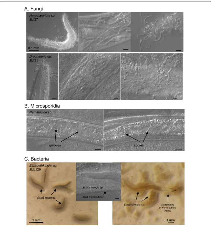
in C. elegans from an apple near Santeuil and in Mon-tsoreau [21]. Consistent with our previous observations that microsporidia are relatively common natural para-sites of C. elegans [20], microsporidia were observed in the surveyed C. elegans populations in Orsay apples in December 2009, October 2010 and November 2011 (one in six, 22 and ten Caenorhabditis populations, respectively [see Additional File 1; Figure 4B]), and in a stem in Plougasnou in August 2009 (one in seven popu-lations [see Additional File 2]). As predators, trapping fungi with adhering knobs or rings are commonly found (for example, Figure 2 in [1], showing fungus JUf26). In addition, we observed fungi that develop hyphae from spores that are either ingested by the nematode or adhere to its cuticle, such as Harposporium sp. or Drechmeria coniospora[22,23] (Figure 4A). Bacteria that appear pathogenic from their effect on Caenorhabditis morphology, behavior and rate of proliferation are fre-quent and most remain to be characterized. In the most dramatic infection (Figure 4C), the bacterium first induces worm bagging (hatching of the embryos inside their mother) and then dissolves the worm cuticle, kill-ing the entire population on the plate within about four days. The bacterium (strain JUb129) was isolated by cul-turing on C. elegans N2 and identified by 16S sequen-cing to be an Elizabethkingia sp. (Bacteroidetes; Flavobacteriaceae, formerly part of Chryseobacterium [24]). Bacteria of this genus are known to be able to digest keratins [25] and the present strain seems to be able to digest the collagens of the nematode cuticle.
Spatio-temporal differences in distribution: studies in the Orsay orchard
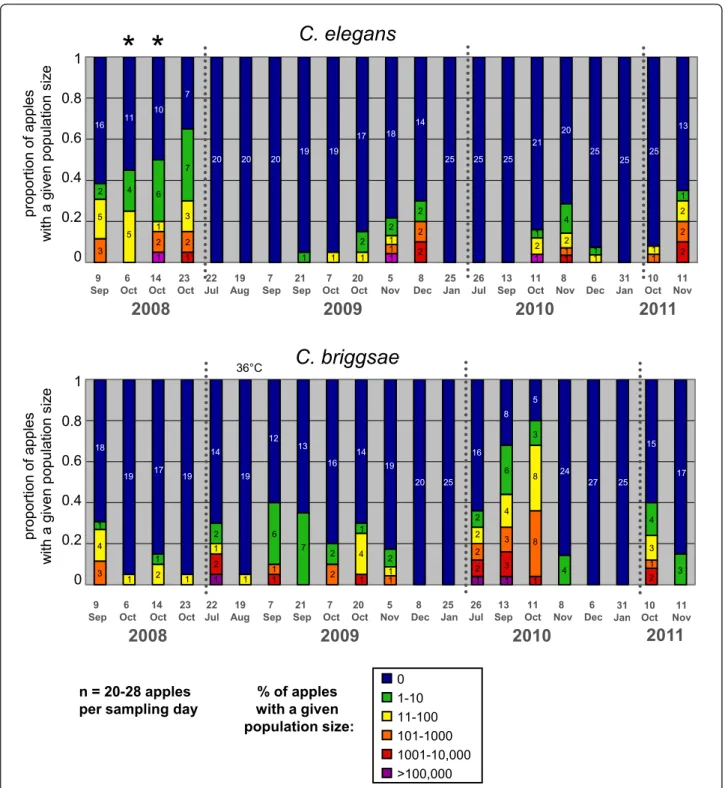
In order to assess the relative spatio-temporal distribu-tion of C. briggsae and C. elegans in a given locadistribu-tion, we undertook a systematic sampling of rotten apples in the Orsay orchard. We sampled 20 to 28 apples throughout the orchard at 19 time points over four seasons (2008 to 2011) and, within a few hours of sampling, analyzed the distribution of developmental stages of Caenorhabditis in each of them. In addition, we sampled adjacent groups of apples twice in October 2008 (see analysis below). From positive samples, we isolated 12 to 25 (when available) individual Caenorhabditis and deter-mined the species to which each belonged. During this survey, we also found a new male-female Caenorhabditis sp., C. sp. 13 [11].
Both C. briggsae and C. elegans were abundantly found every year in the orchard. The two species could be found in the same apple (11 out of 429 apples). How-ever, considering all individual apples, no positive corre-lation between the C. elegans and C. briggsae abundance in a given apple could be found (Spearman rank correla-tion, r = -0.057, P = 0.23). The temporal distribution of
C. briggsaeand C. elegans is presented in Figure 5, and their spatial distribution in Additional File 3.
For each species, both day of sampling and individual apple were significant explanatory factors (generalized linear model, for C. elegans, main effect day treated as factor: df = 18, c2= 192.19, P < 0.0001, apple nested in day: df = 19, c2 = 44.63, P = 0.0008; for C. briggsae, main effect day: c2 = 255.22, P < 0.0001, apple nested in day: c2 = 48.09, P = 0.0003). Each species showed a significant temporal distribution along the season, an effect of the year and a significant interaction term (Generalized Linear Model, for C. elegans, main effects year: df = 3, c2 = 67.36, P < 0.0001, month: df = 1, c2= 17.387, P < 0.0001, year*month: df = 3, c2 = 20.14, P = 0.0002; for C. briggsae, main effects year: c2 = 11.75, P < 0.0001, month: c2 = 127.69, P < 0.0001, year*month: c2 = 34.16, P < 0.0001). Indeed, the temporal distribution of the two species did not coincide (Spearman’s rank correlation of cumulative abundance indices at one time point, r = -0.075, P = 0.76). C. briggsae was found alone in summer and C. elegans only appeared in September to early October. C. briggsae faded out from October on. C. elegans was then predominant in November and finally faded out in December to January (Figure 5). The third species, C. sp. 13, was only found once in Septem-ber 2008, twice (on the Eastern orchard end) in Novem-ber/December 2010 and once in November 2011 [see Additional Files 1 and 3].
At a time when both species were present, we screened for C. elegans and C. briggsae at a smaller spatial scale, by sampling two sets of apples, each below one tree, in four groups of five apples. The distance between apples within one group was 0 to 20 cm, and the distance between groups was 0.6 to 2.5 m [see Additional File 4]. Each apple was cut in the field into four pieces (when its shape and rotting stage allowed it), namely top, bottom, lateral and inner sides, and each part was separately analyzed in the laboratory [see Additional File 1]. There was no effect of day on the abundance index of each species in this dataset, and a strong effect of group, at least for C. ele-gansfor which the sample size was larger (generalized linear model, for C. elegans, main effect day: df = 1, c2= 0.761, P = 0.38, group nested in day: df = 6, c2= 32.11, P < 0.0001; for C. briggsae, main effect day: c2= 1.33, P = 0.25, group nested in day: c2= 12.68, P = 0.048). Thus, at the scale of the apple groups, the distribution of Caenor-habditisindividuals was heterogeneous, indicating that migration among groups (at the scale of 1 m) was limit-ing. Molecular analysis of the isolated worms will even-tually provide more power to detect spatial patterns. Within apples that contained C. elegans, individuals were preferentially found at the bottom of the apple (towards the soil) compared to the top (Wilcoxon signed rank test, P= 0.0054, n = 15). Such a distribution was not found
B. Microsporidia
A. Fungi
C. Bacteria
0.1 mm 1 mm Harposporium sp. JUf27 Drechmeria sp. JUf31 Nematocida sp. Elizabethkingia sp. JUb129 dead worms Elizabethkingia sp.dead worm cuticle
0.1 mm
Elizabethkingia sp. last remains of worm cuticle
(head)
grooves spores
Figure 4 Pathogens in natural C. elegans and C. briggsae populations. (A) Fungal pathogens. Top three panels: The nematophagous fungus Harposporiumsp. JUf27 was isolated from this dead C. elegans individual O143.12 (29 October 2008). JUf27 can infect the intestine of C. elegans N2 and produce hyphae that invade the whole body, resulting in death within six to eight days (middle panel). New spores are then produced at the surface of the dead nematode (right panel). The host response that it provokes has been characterized transcriptionally [23]. Bottom three panels: Fungal pathogen JUf31 in Orsay apple O641 on C. briggsae, Drechmeria coniosporia. Another D. coniosporia strain JUf28 was isolated from apple O567 on a Pristionchus sp. and deposited as CBS129433 at the CBS Fungal Biodiversity Centre, Utrecht, The Netherlands by Nathalie Pujol. These spores also develop into hyphae that invade the nematode body (middle panel) and produce a new generation of spores at the surface of the dead nematode (right panel). (B) Microsporidial infection in C. elegans in Orsay apple O695. Two infected dauer juveniles, in the microsporidial groove stage (left) and in the spore stage (right) (see [21] for microsporidial stages). Both groove and spore stages were also seen in C. elegans L2 larvae in apple O575. (C) Bacterial pathogen Elizabethkingia sp. (strain JUb129), found on C. elegans in Orsay apple O675, here shown on C. elegans N2. The bacteria induce worm bagging and then dissolve their cuticle. On the right, a larva is seen within her mother’s corpse. Bars: 10 μm, except otherwise indicated.
9 Sep 23 Oct 7 Oct 5 Nov 6 Oct 14 Oct 22 Jul 20 Oct 21 Sep 19 Aug 7 Sep 8 Dec 25 Jan 26 Jul 13 Sep 11 Oct 8 Nov 6 Dec
2008
2009
2010
31 Jan 11 Nov 10 Oct 9 Sep 23 Oct 7 Oct 5 Nov 6 Oct 14 Oct 22 Jul 20 Oct 21 Sep 19 Aug 7 Sep 8 Dec 25 Jan 26 Jul 13 Sep 11 Oct 8 Nov 6 Dec2008
2009
2010
31 Jan 11 Nov 10 Oct2011
% of apples with a given population size: 0 1-10 11-100 101-1000 1001-10,000 >100,000 36rC* *
C. elegans
C. briggsae
p ro p o rt io n o f a p p le s w it h a g iv e n p o p u la ti o n s iz e2011
n = 20-28 apples per sampling day0 0.2 0.4 0.6 0.8 1 p ro p o rt io n o f a p p le s w it h a g iv e n p o p u la ti o n s iz e 0 0.2 0.4 0.6 0.8 1 3 2 5 16 11 10 7 20 20 20 19 19 17 18 14 25 25 25 25 25 25 13 21 20 4 5 1 1 1 1 1 1 1 1 1 1 11 1 1 1 1 1 1 1 1 1 1 1 1 1 1 1 1 11 1 1 1 1 2 2 2 2 2 2 2 2 2 2 2 2 2 2 2 2 4 19 20 25 27 25 3 7 6 4 3 18 19 17 19 2 2 2 14 12 6 7 13 16 2 2 1 4 14 19 2 16 8 6 4 3 3 5 3 8 8 24 4 15 4 3 2 3 17
Figure 5 Temporal distribution of C. elegans (top) and C. briggsae (bottom) abundance in Orsay apples along four consecutive years. The graphs show the proportion of apples with a given census size (expressed on a log scale) of C. elegans or C. briggsae populations. The number of apples with a given census size is indicated in each color-coded bar. The two time points labeled by stars (6 and 14 October 2008) were sampled under a single tree (see Additional File 4) and were not used with the others in the statistical analyses. 19 August 2008 was particularly hot (36°C maximum temperature, labeled above the graph). See statistical analyses in the Results.
for C. briggsae, not even a similar trend; however, sample size was low (P = 0.875, n = 4 apples with C. briggsae in this set).
Temperature affects the relative abundance of C. elegans versus C. briggsae in laboratory competition experiments
Several ecological parameters could account for the observed different seasonal distribution of C. elegans and C. briggsae in the Orsay orchard. Temperature is an obvious candidate as it varies between summer and win-ter in France (from an average of 19.2°C in August to 3.3°C in December over 2008 to 2011 in the Orsay area) and the range of temperature allowing fertility in the laboratory is lower in C. elegans compared to C. briggsae [26-28]. In order to show that temperature can affect the distribution of the two species in a given habitat, we performed competition assays between C. elegans and C. briggsaepopulations grown at 15°C, 21°C and 27°C in a controlled laboratory environment. Two different wild isolates of C. elegans and C. briggsae (either from Orsay or Santeuil) were competed in parallel at each tempera-ture. These two pairs of sympatric strains were chosen arbitrarily among the wild isolates of C. elegans and C. briggsaeto test for the effect of species, but not to study sampling site effects (to this end, more pairs of strains should have been compared). Overall, six different com-binations of temperature and genotype were assessed and for each one, five replicate cultures were grown in parallel and transferred to fresh plates at fixed intervals of time to avoid starvation. The number of transfers was
thus used as a measure of time in the analysis. Starting from an equal number of C. elegans and C. briggsae lar-vae, the proportion of both species was followed through time using quantitative pyrosequencing. As expected, for both pairs of genotypes (Orsay and San-teuil), the temporal dynamics of C. briggsae frequency depended on temperature (Figure 6). The competitive ability of C. briggsae versus C. elegans increased as the temperature increased from 15°C to 21°C (generalized linear model, main effect temperature: df = 1, c2 = 38.32, P < 0.0001, time nested in temperature: df = 1, c2 = 1.08, P = 0.298) and from 21°C to 27°C (generalized linear model, main effect temperature: df = 1, c2 = 8.15, P = 0.004, time nested in temperature: df = 1, c2 = 17.08, P < 0.0001). In addition, although the genotype of the wild isolates did not affect the direction of the tem-perature effect, it significantly changed the dynamics of species frequency (Figure 6). Indeed, for each tempera-ture condition, the competitive ability of C. briggsae ver-sus C. elegans is always higher for the Santeuil strains compared to the Orsay strains, resulting in a significant genotype effect and in a non-significant genotype × tem-perature interaction (generalized linear model, main effect genotype: df = 1, c2 = 4.86, P < 0.0001, genotype*-temperature: df = 1, c2 = 4.41, P = 0.086). Strikingly, the C. elegansstrain from Orsay was fixed in all replicate plates after ten transfers (360 hours) when competed against C. briggsae from Orsay at 21°C (Figure 6A), whereas at the same temperature, C. elegans from San-teuil disappeared in four out of five replicate plates
Pr o p o rti o n o f C . b ri g g s a e [m e a n s SE]
Time (number of transfers)
A
B
27rC 21rC 15rC
Orsay strains Santeuil strains
Figure 6 Experimental evolution of the proportion of C. briggsae individuals in competition with C. elegans at different growth temperatures. Two wild strains of C. elegans and C. briggsae from Orsay (A) or Santeuil (B) were competed against each other in the laboratory for several generations at three different temperatures (15°C, 21°C and 27°C). Starting from a frequency of 50% (ten C. elegans L4 larvae and ten C. briggsaeL4 larvae), the proportion of C. briggsae individuals was quantified at different time points with five replicate populations per treatment. The mean proportion across replicates is indictaed as a thick line, and error bars indicate standard errors over replicates. Replicates are indicated as thin dotted lines. Time is represented as the number of transfers of each population to a fresh culture plate from the beginning of the experiment. For each treatment, the experiment was continued until all replicates reached fixation of either C. briggsae or C. elegans. With the Santeuil strains at 21°C, C. briggsae outcompeted C. elegans in four out of five replicates.
when competed against C. briggsae from Santeuil (Fig-ure 6B).
Compared with C. briggsae, the C. elegans strains used in the competition assays reached sexual maturity at a lower age at 15°C, but at a higher age at 21°C and 27°C [see Additional File 5]. Therefore, phenotypic variation in minimal generation time between the two species might contribute to the effect of temperature on the temporal dynamics of mixed populations of C. elegans and C. briggsae. In contrast, the effect of the genetic background is not likely to be explained by differences in the age at maturity, as this trait does not vary between the two C. elegans strains or between the two C. briggsaestrains for all temperatures [see Additional File 5].
These results confirm that temperature is a relevant parameter to explain the seasonal distribution of C. ele-gans and C. briggsae and suggest that its fitness effect could partially be mediated through variation in minimal generation time. However, temperature is not sufficient to predict the relative abundance of the two species, since for intermediate temperatures the species found in the majority was shown to depend on the genetic back-ground of the strains.
Discussion
Natural habitat of C. elegans and C. briggsae
Here we provide the first detailed analysis of natural populations of the nematodes C. elegans and C. briggsae in a habitat where they feed and proliferate. We identi-fied rotting fruits and stems of herbaceous plants as nat-ural habitats where C. elegans can be found feeding and proliferating in large numbers. In contrast to tropical areas where large wild fruits are easily sampled [11], the main source of large fruits in mainland France is pro-vided by domesticated plant species. In temperate areas, rotting stems of herbaceous plants may be a more nat-ural habitat of C. elegans.
Individual and population stage
Scoring a large number of natural populations of C. ele-gansand C. briggsae in rotting fruits and stems provided a comprehensive overview of individual (that is, develop-mental stage) and population stages (as assessed by the presence and relative frequency of developmental stages). Populations in compost were previously found to be mostly composed of dauer larvae. Here in rotting fruits and stems, we found for the first time populations without any dauer larvae, usually with an intermediate census size of 10 to 1,000 individuals [See Additional Files 1 and 2]. Larger populations of 1,000 to 100,000 individuals included pre-dauers (L2d) and dauers. The stages of individuals in small populations could not always be assessed (because the individuals were not
always seen within a few hours of sampling), yet some were seen to be composed of one or a few dauer larvae.
The analysis of groups of adjacent apples below a given tree showed heterogeneity at this scale in the dis-tribution of C. elegans and/or C. briggsae. Thus, migra-tion among groups (at a 1 m scale) was likely limiting. The fact that nearby apples may contain widely different population census and stages [See Additional File 1, 6 October and 14 October 2008] suggests that proliferat-ing populations mostly remain in the same fruit.
From these population snapshots, one can infer the likely dynamics of these populations. Migrating dauer larvae initiate a new population in a given fruit or stem, either being discharged from a large invertebrate or migrating at small range through the leaf litter or soil. Depending on habitat and available food, the dauer lar-vae may start a new population by staying in place, exit-ing the dauer stage and reproducexit-ing. Exit from the dauer stage is a key decision. Upon food exhaustion and crowding, young L1 larvae in the population may enter the pre-dauer and dauer stage. Entry in the dauer stage seems to occur quite synchronously for all young larvae in a population [See Additional Files 1 and 2]. It is so far unclear how many generations are supported by a given fruit or stem, but from the larger observed popu-lations, at least two to three generations must be required before all larvae are in the dauer stage in good conditions. Dauers may then leave the substrate, and only a few will make it to a good food source. C. elegans and C. briggsae thus typically have booming population dynamics, which selects for a short generation time and fast progeny production.
Associated micro-organisms and resource for further studies
The organisms occurring in the habitats where C. ele-gansand C. briggsae feeding stages are found are likely to be important players in their ecology, either as poten-tial food or as pathogens. Pathogens may enter through the digestive tract or else adhere to the nematode cuticle.
We here show that wild-caught Caenorhabditis often harbor a large microbial fauna in their intestinal lumen. The N2 reference strain of C. elegans cultured on E. coli OP50 in standard laboratory conditions crushes bacteria in its pharyngeal grinder almost fully efficiently. In older laboratory adults (starting at two days of adulthood), some live E. coli may eventually succeed in entering the intestinal tract, escaping defecation and starting to colo-nize and proliferate in the intestinal lumen [29,30]. E. colican enter the nematode intestine much earlier in C. elegans phm-2 mutants with a defective grinder [29]. Some (human) pathogenic bacteria can pass the grinder of C. elegans N2 and colonize its intestinal lumen
[29,31-33], perhaps by regulating the production of enzymes that affect it [33,34]. The absence of live bac-terial cells in the intestinal lumen is thus considered the norm for laboratory C. elegans. In contrast, wild animals were observed to harbor a live bacterial flora in their intestinal lumen. The flow of bacteria is regulated by four dynamic components, namely, i) entry past the pharyngeal valve and grinder, ii) proliferation, iii) disap-pearance by digestion or host defences, and iv) exit through the rectum with worm defecation. In many cases, a healthy steady-state of bacterial flow appears to result from the balance between these four components. In some instances however, proliferation takes over and the obstructed intestinal tract can no longer support any flow (Figure 3F). Such weak and strong colonization phenotypes can sometimes be maintained over several generations and depend both on the microbial fauna and on the worm genotype: many wild C. elegans iso-lates were not as efficient in crushing E. coli OP50 as C. elegansN2 is, and many wild bacteria could colonize C. elegansN2 more readily than E. coli OP50 does (MAF, unpublished observations). The dynamics of microbe entry, growth, digestion, and exit will be interesting to study, as well as to determine the metabolic and patho-genic effects of different intestinal microbial communities.
C. elegans N2 cultured in standard laboratory condi-tions activates its pharyngeal muscles in a rapid cycle, pumping several times per second in the presence of food [35]. Pumping of wild-caught animals could occa-sionally be observed to be hindered by bacteria obstruct-ing the pharynx (Figure 3G). Such animals appeared malnourished, as indicated by the pale coloration of their intestine. The case in Figure 3G appears to result from an interaction between the bacteria in the samples and the C. elegans wild genotype, because neither C. ele-gans N2 fed on the corresponding wild bacteria nor the wild C. elegans genotype fed on E. coli OP50 displayed this defective swallowing phenotype.
Fungi are abundant in rotting fruits and stems, and could be seen in the intestines of wild-caught Caenor-habditis. Yeasts synthesize sterols such as ergosterol and dehydroergosterol (for example [36] for Saccharomyces cerevisiae), which can be used as a sterol source by C. elegans[37]. It is thus possible that C. elegans derives its sterols from fungi. Alternatively, it may ingest sterols from plant remains as well.
We here established a large collection of isolates of C. elegans, and to a lesser degree C. briggsae, which was generally frozen without bleaching, thus potentially retaining associated microbial fauna. We also directly froze a fraction of the original sample at -80°C for further analysis. This opens the way for evolutionary and ecological characterization, such as population
genetic analysis of the nematodes, metagenomics and isolation of the microbial fauna, and surveys of the occurrence of pathogens.
Habitat sharing: C. elegans likes it cool
C. elegans shares its habitat with C. briggsae, and also with a variety of other bacterivorous nematodes of var-ious genera in the families Rhabditidae (including diplo-gastrids such as Pristionchus) and Panagrolaimidae (Panagrellus, Panagrolaimus). These species are likely to compete for food, and some of them, such as Pris-tionchusspecies [38], may also act as predators of Cae-norhabditis species, although this remains to be demonstrated.
Most strinkingly, we find C. elegans and C. briggsae in the same type of habitat. However, several results sug-gest that C. elegans is preferentially found at cooler tem-peratures than C. briggsae. First and most strikingly, the seasonal pattern in the Orsay orchard is strong, with C. briggsaefound in summer and early fall and C. elegans in the late fall. Second, the Plougasnou area in Brittany was sampled over the years in many places, and particu-larly in high summer when only C. briggsae could be found in Orsay. This Western France region presents an oceanic (wet, cool, quite even throughout the year) cli-mate and only yielded C. elegans. Both species were found in all other tested regions of France, North, South and East.
Both species are found on several continents, but C. briggsaeis found in both tropical and temperate regions whereas C. elegans is found mostly in temperate regions, or in tropical regions at relatively high altitudes [11]. The latter might be explained by decreased tempera-tures or by other correlates of altitude, such as oxygen levels. Deeper sampling will be required to confirm the tendency of C. elegans to live at high altitude in tropical locations.
The laboratory competition assays performed in con-trolled environments clearly show an effect of growth temperature on the relative fitness of C. elegans and C. briggsaewhen sharing the same habitat. Consistent with the seasonal and worldwide patterns, C. briggsae wins at higher temperatures and C. elegans at lower tempera-tures (Figure 6). In these competition experiments, the effect of temperature is likely mediated in part by the difference in generation time between the two species [See Additional File 5]. Other temperature-sensitive traits of C. elegans and C. briggsae can be inferred from comparisons of independent studies in each species and may also contribute to the competition results. For example, self-brood size declines in C. elegans at tem-peratures above 18 to 20°C [28,39], whereas the decline occurs at higher temperatures in C. briggsae, with still a high brood size at 28°C [27], a temperature where C.
elegansis fully sterile. Dauer entry is another key devel-opmental decision that is differentially regulated by tem-perature in C. elegans and C. briggsae and thus could be involved in the spatio-temporal dynamics of the two species in wild populations. A growth temperature above 27°C is sufficient to trigger dauer entry in C. ele-ganswild isolates with various penetrances, whereas it seems to have no effect in C. briggsae [40,41].
Our data further indicate an effect of the genotype within a species in the competition experiment. Other competition experiments will be required to determine which species is involved in this genotype effect, which may be due to a difference of fitness between the two C. elegans strains, the two C. briggsae strains, an indepen-dent combination of both, or a specific interaction. This intraspecific difference between the two sets of geno-types is unlikely to be explained by a difference in gen-eration time [See Additional File 5]. Each species was shown to display intraspecific variation in the tempera-ture that maximizes lifetime fecundity of wild isolates [27,28,39]. In C. briggsae, the temperature resistance correlates with the latitude of strain isolation [27]. In C. elegans, a few quantitative trait loci (QTLs) involved in the thermal plasticity of fertility, age at maturity, growth rate and egg size have been mapped from a set of recombinant inbred lines (RILs) between N2 and the Hawaiian strain CB4856 [42]. The plasticity of gene expression to temperature was shown to vary between N2 and CB4856, primarily due to trans-acting eQTLs [43]. Finally, a third analysis from the same RILs led to the identification of a coding polymorphism in the tra-3 gene that affects body size response to temperature [44]. In the wild, besides an effect of these life history traits, the temperature-dependent distribution of C. elegans and C. briggsae could also be regulated through varia-tion in behavioral responses. Interestingly, when placed in a temperature gradient, different isolates of C. elegans present different thermotactic behavior and seem to show a preference for the temperature maximizing their fitness [28]. The reference strain N2 could have lost this adaptive thermophilic response after introduction to the stable temperature of the laboratory [45]. A better understanding of how thermotactic behaviors evolve in the wild will thus require a better characterization of the spatial and temporal dynamics of temperature in natural habitats of C. elegans.
Apart from temperature, the population dynamics of Caenorhabditis species is likely to be affected by other ecological parameters that are not yet identified. We note for instance that the distribution of Caenorhabditis species of the Elegans group in Europe is puzzling and does not quite fit a simple temperature pattern. Indeed, C. briggsae has only been found so far in France (not more Southern countries, such as Spain and Portugal,
where C. elegans was found). C. remanei instead is found in Germany, Switzerland and Hungary [46] and the North-East of France (Alsace; Figure 1). Whether this pattern might be due to limited migration or results from aspects of local adaptation other than temperature sensitivity remains to be determined.
Conclusions
C. elegansand C. briggsae were found to proliferate in the same rotting vegetal substrates. For the first time, we found proliferating populations without arrested dauer stages. The large populations, however, always contain pre-dauer or dauer stages. Together with the fact that these rotting substrates are temporary habitats, it appears thus likely that C. elegans and C. briggsae undergo successive population growth periods upon encountering a favorable rotting substrate, separated by periods of migration in the dauer stage. The discovery of habitats where C. elegans and C. briggsae feed opens the way for evolutionary and ecological characterization. Here we show that the temporal sharing of the habitat by C. elegans and C. briggsae coincides with their tem-perature preference in the laboratory, with C. elegans populations growing faster than C. briggsae at cooler temperatures, and C. briggsae faster at higher temperatures.
Methods
Nematode sampling and isolation
The samples were photographed and collected in plastic tubes (60 ml) or zipped bags. In the laboratory, they were weighed and cut into pieces (about 1 cm3). At least half of the sample was used for nematode isolation, while the rest was weighed and stored at -80°C for further use. Samples were analyzed on the day of sam-pling for all Orsay, Santeuil and Ivry time points. The day of sample deposition on plate and first analysis is indicated for Le Blanc and Plougasnou samples in Addi-tional File 2.
The samples were placed onto standard C. elegans Normal Growth Medium culture plates [47] of 90 or 55 mm diameter, previously seeded with an E. coli OP50 lawn in the center. The samples were spread around the lawn and 1 to 2 ml moisture (water or M9 solution) was added onto the samples. All plates were examined regu-larly in the dissecting microscope: several times within a few hours, once or twice on the next two days, and at least on days 4 and 7. Sampled arthropods and mollusks were usually first left free on the plate for a few hours to test for the presence of nematodes on their surface, then sacrificed with a scalpel. Nematodes that crawled out of the sample were identified to the genus (or family) level by morphological criteria [47] in the dis-secting microscope with trans-illumination. Animals
resembling Caenorhabditis were picked individually onto 55 mm culture plates. Because i) ‘sick’ wild animals do not always resemble the canonical healthy laboratory animal and ii) dauer larvae are difficult to distinguish, animals of dubious genus were also isolated for further identification. C. elegans and C. briggsae are both facul-tative selfers and require a single hermaphrodite larva to reproduce (males are very rare). Caenorhabditis sp. 13, however, reproduces through males and females. In this case, cultures were started from an adult female or with a pair of male and female larvae.
Species identification
C. elegansand C. briggsae resemble each other morpho-logically, even when observed with a high-power micro-scope. In the early part of this work, we systematically assessed the species using a cross and/or a molecular method.
For the biological species test, three to six hermaphro-dites of the tested isolate were placed with three to six males of reference laboratory strains C. elegans N2 or C. briggsaeAF16. The plates were observed three to four days later at 20° to 23°C to test for the presence of a large proportion (> 20%) of males in the progeny, an indication of a successful cross [48].
Molecular tests were as described in [47], using spe-cies-specific PCR primers. After testing hundreds of Caenorhabditiscultures, we noticed some morphological and behavioral criteria that were decisive for species identification, when assayed on populations, that is, not on the single wild parent, which remains hard to iden-tify: 1) C. elegans retains more embryos in its uterus (usually 10 to 50) than C. briggsae (usually none to five); 2) C. elegans has a dumpier body aspect ratio than C. briggsae; 3) C. elegans clumps on culture plates with food, whereas C. briggsae does not. The latter criterion was rendered decisive when it was understood that the non-clumping phenotypes of the C. elegans reference strain N2 was acquired through laboratory mutations [49]. All three of the above criteria depend on the envir-onment and are best scored in clean cultures, that is, with E. coli OP50 only. If the species identification was dubious, cultures were systematically either cleaned by bleaching or identified by crossing with males. All iden-tifications that were dubious on morphological/beha-vioral criteria proved correct with genetic or molecular criteria. With training, most identifications could be made using morphological/behavioral criteria.
Orsay orchard sampling
This orchard is located at 48.7015 North, 2.1725 East. This orchard contains 322 apple trees with a large diver-sity of apple varieties, which ripen and rot throughout the second half of the year (July to December), and a
large fraction of the apples are left to rot (except in 2011 when they were harvested). Schematically, the main grid has trees spaced every 3 meters in each (let-tered) North-South column and every 4 meters in each (numbered) West-East line, from A1 to S23, with an open space in the middle (222 trees in this grid). In addition, two columns of 2-meter-spaced trees, 2-meters apart, are found on the East side, after a 17 m wide empty area (100 trees).
At each of the 17 time points, 20 to 28 very rotten apples were sampled on the floor throughout the orch-ard, and the corresponding position noted. Apple pro-duction is very variable among trees and years, which may explain possible spatial patterns in sampling. Each apple was called “O” (for Orsay) followed by an identify-ing number. Similarly, Santeuil samples were labeled “S”, followed by a number.
For each of the 6 and 13 October 2008 collections, a tree with a large number of rotten apples below it was chosen. Four groups (within 50 cm of each other on the ground) of five rotten apples were then chosen. When possible (that is, the apple was not yet reduced to a very flat puree), each apple was cut into bottom (towards soil at sampling time), top, lateral and inner pieces. Each piece was separately weighed and tested for nematode presence and census.
C. briggsaeversus C. elegans competition assays
Six competition assays involving two different pairs of C. elegans and C. briggsae wild strains cultivated at three different temperatures were performed in parallel for several generations to assess the role of temperature and genotype on the relative growth of the two species. For each of the six experimental conditions, five replicate populations were grown in parallel on 6 cm diameter NGM dishes (2.5% agar) seeded with 50 μl of OP50. We chose to compete the C. elegans strain JU1530 against the C. briggsae strain JU1529 (both isolated the same day from two different rotten apples found below the same tree in Orsay), and the C. elegans strain JU1918 against the C. briggsae strain JU2138 (isolated the same day from two different rotten stems in Santeuil). These strains were derived from wild parents by four to six generations of selfing in the laboratory, to avoid possible within-line heterozygosity due to outcrossing in the wild. The experimental populations were founded from ten L4 C. elegans individuals and ten L4 C. briggsae individuals deposited on each plate in the center of the E. colilawn and grown at either 15°C, 21°C or 27°C. All strains were previously bleached and synchronized one generation prior to the experiment by 1.5 hour of egg laying of ten adults on eight plates.
To avoid starvation, a fraction of each population was transferred to a fresh culture plate at fixed intervals of
time: worms grown at 15°C were transferred every 144 hours, while those maintained at 21°C or 27°C were transferred every 36 hours. Animals from old plates were retrieved in 1 ml of M9 solution in sterile condi-tions and centrifuged at 3,000 rpm for five minutes. The pellet (1 μl containing approximately 100 to 400 mixed stage individuals) was then dropped in the middle of a fresh culture plate.
For each replicate population, the proportion of C. briggsae versus C. elegans was quantified at different time points by pyrosequencing, using a PyroMark Q96 ID instrument from Biotage (Uppsala, Sweden). One single-nucleotide divergence was identified between the two species in an otherwise well-conserved region of the eef-2 coding sequence (C/T at position I:9,166,508 in WormBase WS228 release), allowing for non-biased PCR. For DNA preparation, 1.5 μl of M9 suspensions containing about 500 mixed-stage individuals from each culture plate were mixed with 18.5 μl of worm lysis buf-fer and proteinase K (100 μg/ml). After 1.5 hours of lysis at 60°C and 15 minutes of inactivation at 95°C, 0.5 μl of worm lysate were used as PCR template. PCRs were performed in 50 μl reactions containing 0.25 μl of GoTaq DNA polymerase (Promega, Madison, Wiscon-sin, USA), 10 μl of 5x GoTaq buffer, 5 μl dNTPs at 2 mM, 1 μl of forward primer at 10 μM (5’-GCTTACTTGCCTGTCAACGAGTC-3’), 0.17 μl of reverse primer at 10 μM (5’-TAGCAGGATACGAC-TATCCGGTGTTGGAGCGGAGAT-3’) and 0.83 μl of a universal biotinylated primer at 10 μM (5’-[Btn]TAG-CAGGATACGACTATC-3’) [50]. The purification of single-stranded PCR amplicons and the pyrosequencing reactions were performed according to the manufac-turer’s instructions using the following sequencing pri-mer: 5’-CTTCGGATTCACCGC-3’. C. briggsae versus C. elegansallele frequencies were obtained from the height of the pyrogram peaks using the Allele Quantification tool supplied with the pyrosequencing software of Bio-tage. For each of the six experimental conditions, the assay was stopped when all replicate populations were sufficiently close to fixation of either C. elegans or C. briggsae (more than 95% of the more frequent eef-2 allele).
To estimate the precision of the quantification method, the frequency of eef-2 alleles was measured on worm lysates prepared from known proportions of C. briggsae and C. elegans individuals. The discrepancy between observed and expected frequencies was, on average, 3.66%.
Minimal generation time
The minimal generation time (or age from egg-laying to sexual maturity) was measured at 15°C, 21°C and 27°C for each C. elegans or C. briggsae strain used in the
competition assays. A first pilot experiment was carried out to get a broad estimation of the age at which her-maphrodites became fertile at the different tempera-tures. Then, more accurate values of the age at maturity were measured as follows. The strains were synchro-nized by one to two hours of egg laying of ten adults at 15°C, 21°C and 27°C. For each strain, five larvae were isolated on separate 55 mm culture plates 24 hours (21° C and 27°C) or 48 hours (15°C) after egg laying. From 36 hours (27°C), 58 hours (21°C) or 92 hours (15°C) post-synchronization, plates were checked regularly for laid eggs until all isolated hermaphrodites reached reproductive maturity. The age at maturity was calcu-lated as the difference between the time when the first eggs were observed and the time when the parent itself was laid (estimated as the middle time point of the one to two hour egg laying period).
Data analysis
From the census observations, we assigned an abun-dance index for each species in each apple on a Log scale with an index of 1 for one to 10 individuals, 2 for 11 to 100, 3 for 102 to 103, 4 for 103to 104, and 5 for > 104. At each time point, a cumulative abundance index for each species was the sum of the individual index of the corresponding apples. Statistical analysis was per-formed in R version 2.11.1 [51].
To test whether the species abundance index varied along and across years, we used a generalized linear model (GLM), assuming a Poisson response variable and a log link function. Effects included in the model were monthtreated as a number and year treated as a factor. Year corresponded to a rotting apple production season, that is, from July to January. Month was thus calculated starting in July, allowing for half-integer numbers in cases of two time points in a month. Tested dependent variables included the C. elegans cumulative abundance index and the C. briggsae cumulative abundance index.
To test whether the distribution of apples was homo-geneous among different groups of apples sampled below a given tree, we used a generalized linear model (GLM), assuming a Poisson response variable and a log link function. Effects included in the model were day treated as a factor and group nested in day, treated as a factor. Tested dependent variables included the C. ele-gans cumulative abundance index and the C. briggsae cumulative abundance index.
To test whether the abundance of the two species sig-nificantly differed, we used a Wilcoxon signed rank test on the weighted census of each species, corrected for the weights of each apple part. A Friedman rank sum test using the weighted census in all four apple parts (bottom, side, inside, top) was not significant for C. ele-gans(c2= 6.6, df = 3, P = 0.0858).