HAL Id: hal-01321293
https://hal.sorbonne-universite.fr/hal-01321293
Submitted on 25 May 2016
HAL is a multi-disciplinary open access
archive for the deposit and dissemination of
sci-entific research documents, whether they are
pub-lished or not. The documents may come from
teaching and research institutions in France or
abroad, or from public or private research centers.
L’archive ouverte pluridisciplinaire HAL, est
destinée au dépôt et à la diffusion de documents
scientifiques de niveau recherche, publiés ou non,
émanant des établissements d’enseignement et de
recherche français ou étrangers, des laboratoires
publics ou privés.
Distributed under a Creative Commons Attribution| 4.0 International License
interneurons
Bruno Cauli, Xiaojuan Zhou, Ludovic Tricoire, Xavier Toussay, Jochen F
Staiger
To cite this version:
Bruno Cauli, Xiaojuan Zhou, Ludovic Tricoire, Xavier Toussay, Jochen F Staiger. Revisiting enigmatic
cortical calretinin-expressing interneurons. Frontiers in Neuroanatomy, Frontiers, 2014, 8, pp.52.
�10.3389/fnana.2014.00052�. �hal-01321293�
Revisiting enigmatic cortical calretinin-expressing
interneurons
Bruno Cauli
1,2,3*, Xiaojuan Zhou
4, Ludovic Tricoire
1,2,3, Xavier Toussay
1,2,3and Jochen F. Staiger
4*
1Sorbonne Universités, UPMC University Paris 06, UM CR18, Neuroscience Paris Seine, Paris, France2Centre National de la Recherche Scientifique, UMR 8246, Neuroscience Paris Seine, Paris, France 3
Institut National de la Santé et de la Recherche Médicale, UMR-S 1130, Neuroscience Paris Seine, Paris, France 4
Institute for Neuroanatomy, UMG, Georg-August-University Göttingen, Göttingen, Germany
Edited by:
Zsofia Magloczky, Hungarian Academy of Sciences, Hungary
Reviewed by:
José A. Armengol, University Pablo de Olavide, Spain
Fiorenzo Conti, Universita Politecnica delle Marche, Italy
*Correspondence:
Bruno Cauli, CNRS-UPMC, Neuroscience Paris Seine, 9 Quai Saint Bernard, 75005 Paris, France e-mail: [email protected]; Jochen F. Staiger, Institute for Neuroanatomy, UMG,
Georg-August-University Göttingen, 37075 Göttingen, Germany e-mail: jochen.staiger@ med.uni-goettingen.de
Cortical calretinin (CR)-expressing interneurons represent a heterogeneous subpopulation
of about 10–30% of GABAergic interneurons, which altogether total ca. 12–20% of all
cortical neurons. In the rodent neocortex, CR cells display different somatodendritic
morphologies ranging from bipolar to multipolar but the bipolar cells and their variations
dominate. They are also diverse at the molecular level as they were shown to
express numerous neuropeptides in different combinations including vasoactive intestinal
polypeptide (VIP), cholecystokinin (CCK), neurokinin B (NKB) corticotrophin releasing
factor (CRF), enkephalin (Enk) but also neuropeptide Y (NPY) and somatostatin (SOM)
to a lesser extent. CR-expressing interneurons exhibit different firing behaviors such as
adapting, bursting or irregular. They mainly originate from the caudal ganglionic eminence
(CGE) but a subpopulation also derives from the dorsal part of the medial ganglionic
eminence (MGE). Cortical GABAergic CR-expressing interneurons can be divided in two
main populations: VIP-bipolar interneurons deriving from the CGE and SOM-Martinotti-like
interneurons originating in the dorsal MGE. Although bipolar cells account for the majority
of CR-expressing interneurons, the roles they play in cortical neuronal circuits and in the
more general metabolic physiology of the brain remained elusive and enigmatic. The aim
of this review is, firstly, to provide a comprehensive view of the morphological, molecular
and electrophysiological features defining this cell type. We will, secondly, also summarize
what is known about their place in the cortical circuit, their modulation by subcortical
afferents and the functional roles they might play in neuronal processing and energy
metabolism.
Keywords: neuropeptides, neocortex, neocortical circuits, embryonic and fetal development, neuroenergetics
INTRODUCTION: WHAT ARE THE CORTICAL
CALRETININ-EXPRESSING INTERNEURONS?
Cortical calretinin (CR) expressing interneurons represent a
heterogeneous subpopulation of about 10–30% of GABAergic
interneurons (
Kubota et al., 1994; Gonchar and Burkhalter,
1997; Tamamaki et al., 2003
). They display different
soma-todendritic morphologies ranging from bipolar to multipolar
(
Jacobowitz and Winsky, 1991; Kubota et al., 1994; Gonchar
and Burkhalter, 1997; Gonchar et al., 2008; Caputi et al., 2009
).
They are also diverse at the molecular level as they express
numerous neuropeptides in different combinations including
vasoactive intestinal polypeptide (VIP) (
Kubota et al., 1994; Cauli
et al., 1997
), cholecystokinin (CCK) (
Cauli et al., 1997; Gonchar
et al., 2008
), neurokinin B (NKB) (
Kaneko et al., 1998; Gallopin
et al., 2006
) corticotrophin releasing factor (CRF) (
Gallopin
et al., 2006; Kubota et al., 2011
), enkephalin (Enk) (
Taki et al.,
2000; Férézou et al., 2007
), but also to a lesser extent and in a
species-dependent manner neuropeptide Y (NPY) (
Cauli et al.,
1997; Wang et al., 2004; Gonchar et al., 2008
) and somatostatin
(SOM) (
Cauli et al., 1997; Wang et al., 2004; Xu et al., 2010b
).
CR-expressing interneurons also exhibit different firing behaviors
such as adapting, bursting or irregular (
Kawaguchi and Kubota,
1996; Cauli et al., 1997, 2000; Porter et al., 1998; Wang et al., 2002;
Karagiannis et al., 2009
). They mainly originate from the caudal
ganglionic eminence (CGE) (
Xu et al., 2004; Butt et al., 2005
) but
a subpopulation also derives from the dorsal part of the medial
ganglionic eminence (MGE) (
Fogarty et al., 2007; Xu et al., 2008
).
Cortical GABAergic CR
+ interneurons can be divided in two
main populations: VIP-bipolar interneurons deriving from the
CGE and SOM-Martinotti-like interneurons originating in the
dorsal MGE. In the rodent neocortex, bipolar cells account for
the majority of CR
+ interneurons, therefore, we consider them
as our main focus of the review. Due to the relatively few and
scattered studies of CR
+ interneurons, their inputs and outputs,
the roles they play in cortical neuronal circuits as well as
neu-roenergetics of the brain remained elusive and enigmatic until
quite recently. The aim of this review is, firstly, to provide a
comprehensive view of the morphological, molecular and
elec-trophysiological features defining cortical bipolar CR-expressing
interneurons. We will, secondly, also review their places in the
cortical circuit, their modulations by subcortical afferents and
the functional roles they might play in neuronal processing and
energy metabolism. We are convinced that the synthesis of recent
data on bipolar CR-expressing interneurons will show that they
are much less enigmatic than they appeared roughly 25 years ago
(
Peters and Harriman, 1988
).
EMBRYONIC ORIGINS
Unlike glutamatergic neurons which are born in the
ventricu-lar zone of the telencephalic vesicle and then migrate radially
(
Molyneaux et al., 2007; Rakic, 2007
), most of the GABAergic
interneurons derive from one proliferative region called
gan-glionic eminence (GE) located in the ventral part of the
telencephalon (
Wonders and Anderson, 2006; Batista-Brito and
Fishell, 2009; Bartolini et al., 2013
). Once born, interneuron
pre-cursors migrate first dorsally, guided by attracting and repulsive
molecular cues, then tangentially toward neocortex and
hip-pocampus along two main migratory streams (
Chedotal and Rijli,
2009; Marin, 2013
). They eventually acquire their final
lami-nar location by penetrating the cortical plate. Anatomically, the
GE is subdivided in three main regions, the medial, lateral, and
caudal GE (MGE, LGE, and CGE, Figure 1). Cortical and
hip-pocampal interneurons derive mainly from the MGE and CGE,
while the LGE is the major contributor of GABAergic
interneu-rons of striatum and basal forebrain structures. The preoptic area
(Figure 1) has recently been described as another source of
cor-tical interneurons (
Gelman et al., 2009
) but given that very few
CR+ interneurons derive from this zone, we will focus on the
MGE and CGE.
Lineage analyses using either grafts or genetic tools such as
Cre driver lines that label specifically a subfield of the GE have
shown that parvalbumin-expressing (PV+) and SOM+
interneu-rons are generated in the MGE at different time points (between
E9 and E15, Figure 1;
Xu et al., 2004; Butt et al., 2005; Miyoshi
et al., 2007; Tricoire et al., 2011
). In contrast, similar approaches
revealed that a large portion of the remaining interneuron
sub-types, including CR
+ interneurons, derive from the CGE and are
produced at later embryonic stages (between E12.5 and E16.5,
Figure 1;
Lee et al., 2010; Miyoshi et al., 2010; Tricoire et al.,
2011
). In addition to being generated by distinct progenitors,
CR
+ interneuron precursors use a different migratory
path-way. While SOM
+ interneurons take a more rostrolateral route,
CGE-derived interneurons take a caudal path for their tangential
migration (
Kanatani et al., 2008
). The late birth of CGE-derived
interneurons has the consequence that they are still migrating at
birth and reach their final destination later than MGE-derived
interneurons (
Miyoshi et al., 2010
). However, several refinements
have to be considered. SOM+ hippocampal neurons exhibit a
dual origin with one expressing type 3 serotonin receptor (5-HT3
R) and the other not (
Chittajallu et al., 2013
). In the neocortex,
such a dichotomy has not been reported yet.
Several transcription factors of CR
+ (and SOM+)
interneu-rons are necessary for the proper specification and migration
from the GE. Mice lacking the Dlx1 gene show reduction of
CR+ and SOM+ interneurons without affecting the PV+
pop-ulation (
Cobos et al., 2005
). Removal of the transcription factor
Nkx2.1 restricted at early embryonic stages to the MGE domain
(Figure 1) results in a molecular and cellular switch of
MGE-derived cortical interneurons (PV
+ and SOM+ subpopulations)
to CGE-derived neurons (VIP
+ and CR+ cells;
Butt et al., 2008
).
In contrast with SOM+ interneurons, little is known about
the regional and cell type specification of CGE in general and of
CR
+ bipolar interneurons in particular. Gsx2 (also called Gsh2)
is enriched in (but not restricted to) the LGE and CGE from
early development (Figure 1) and has been directly implicated in
promoting the CR+ interneuron identity (
Xu et al., 2010a
). The
orphan nuclear receptor COUP-TFII shows restricted expression
in the CGE (
Kanatani et al., 2008; Willi-Monnerat et al., 2008
)
and, together with COUP-TFI, is required for the caudal
migra-tion of cortical interneurons (
Tripodi et al., 2004
). Moreover, in
Nkx2.1 mutant mice, a higher number of CR
+ and VIP+
cor-tical interneurons are generated and COUP-TFII is ectopically
expressed in the MGE (
Butt et al., 2008
). Conversely, conditional
loss-of-function of COUP-TFII in subventricular precursors and
postmitotic cells leads to a decrease in VIP
+ and CR+
interneu-rons, compensated by the concurrent increase of MGE-derived
PV+ interneurons. Interestingly, COUP-TFI mutants are more
resistant to pharmacologically induced seizures (
Lodato et al.,
2011
). In addition to these genetic factors, electrical activity has
been also shown to regulate development of cortical neurons.
CR
+ but not VIP+ interneurons activity is required before
post-natal day 3 for correct migration and Elmo1, a target of Dlx1 and
expressed in CR+ neurons, is both necessary and sufficient for
this activity-dependent interneuron migration (
De Marco Garcia
et al., 2011
).
Over the past decade, many advances have been achieved in
the identification of the genetic factors that influence the
speci-fication of cortical and hippocampal interneurons especially for
MGE-derived interneurons. However, the program specifying the
identity of CGE-derived interneurons still needs to be unraveled.
For instance, the cues that regulate the final maturation of the
morphological, synaptic and electrophysiological properties have
to be determined. Indeed, although embryonic origin is a major
contributing factor, immature interneurons arriving at their final
destination are likely to encounter local factors such as guidance
molecules and specific levels of network activity that will instruct
them where to grow dendrites and axons.
ANATOMICAL PROPERTIES
LAMINAR, COLUMNAR, AND AREAL DISTRIBUTION PATTERN
The initial descriptions of the brain-wide distribution of
CR
+ neurons did not report obvious differences in the number or
type of neurons across the different cortical areas (
Jacobowitz and
Winsky, 1991; Resibois and Rogers, 1992; Gabbott et al., 1997
).
They observed that within a certain cortical area always
supra-granular layers II/III show the highest density of CR+ neurons
(Figure 2). A more fine-grained analysis, however, suggested that
there is a subtle gradient from rostral to caudal, with a higher
density of CR
+ neurons in the visual cortex (
Xu et al., 2010b
),
a finding similar to that of VIP
+ neurons, one of the major
subtypes of CR+ neurons (
Morrison et al., 1984; Rogers, 1992;
Gonchar et al., 2008
). Interestingly, in the mouse barrel cortex
where columnar modules are easily visualized, both, CR
+ and
VIP
+ interneurons showed a preference for septal compartments
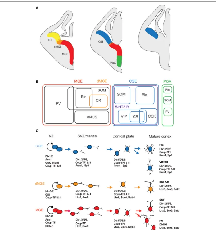
FIGURE 1 | Embryonic origins and genetic factors affecting the mature fate of cortical interneurons. (A) Diagram showing the subdivisions of the
embryonic telencephalon. The three regions where cortical and hippocampal interneurons originate are the medial ganglionic eminence (MGE) (including the dorsal MGE-dMGE), the caudal ganglionic eminence (CGE), and the preoptic area (POA). The lateral ganglionic eminence (LGE) gives rise, amongst other, to basal forebrain neurons. (B) Ball scheme of the major classes of cortical and hippocampal interneurons identified using
neurochemical markers and represented depending of their place of origin in the embryonic telencephalon. The MGE generates 50% of all cortical interneurons and includes mainly parvalbumin (PV)-expressing and
somatostatin (SOM)-expressing subtypes. In hippocampus, it also includes a large population expressing the neuronal isoform of nitric oxide synthase (nNOS). CGE-derived reelin (Rln)-expressing interneurons represent the neurogliaform cells. In both cortex and hippocampus, almost all CGE-derived interneurons express the type 3 serotonin receptor (5-HT3 R). In contrast with cortex, hippocampal SOM+ interneuron have a dual origin with a significant subset co-expressing 5-HT3 R. VIP; vasoactive intestinal polypeptide, CR, calretinin; CCK, cholecystokinin. (C) Genetic programs controlling neurogenesis, cell commitment, tangential, and radial migration and maturation of cortical interneuron.The subdivision of the neuroepithelium
FIGURE 1 | Continued
can be identified by combinatorial expression of transcription factors involved at different stages of cortical interneuron development. Some of these factors participate broadly in interneuron development such as Dlx and CoupTF gene families. Some transcription factors are unique to specific
domains and/or stages of differentiation: Nkx2-1 defines the MGE and activates a cascade of genes including Lhx6, Sox6, and Satb1; Nkx6-2 and GLI1 are enriched in the dMGE. Prox1 and SP8 are expressed in CGE-derived cortical interneurons at all stages of their development (adapted from
Kessaris et al., 2014).
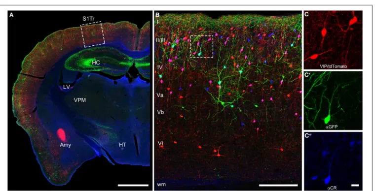
FIGURE 2 | Coronal brain section of a VIPcre/tdTomato/GIN mouse additionally stained for calretinin. Immunostaining of calretinin in a
coronal brain section of a VIPcre/tdTomato/GIN mouse shows VIP neurons in red, Martinotti cells in green and calretinin in blue in the primary somatosensory cortex (S1Tr). (A) Low magnification of one hemisphere depicting the hippocampus (HC), the lateral ventricle (LV), the amygdala (Amy), the thalamic nucleus ventralis posteromedialis (VPM), and the hypothalamus (HT); the dashed rectangle marks the selected area of (B); scale bar: 1000μm. (B) Close-up of the rectangle in (A) in a
maximum intensity projection; Roman numerals indicate cortical layers;
dashed rectangle marks the selected area of (C–C”); scale bar: 250μm. Please note that VIP neurons co-localizing CR appear pink whereas Martinotti cells that co-localize CR show a cyan-colored soma. (C) Red channel of the inset in (B), showing the tdTomato signal only; (C’) green channel of the inset in (B), showing the GFP only; (C”) blue channel of the inset in (B), showing the labeled calretinin antibody only; scale bar for (C–C”) 20μm. Please not that nearly all VIP and the single Martinotti cell are co-localizing CR.
over barrel-associated columns (
Zilles et al., 1993; Melvin and
Dyck, 2003
). Such a preference was not obvious for the rat
bar-rel cortex (
Bayraktar et al., 2000
), although no tangential sections
where used, which would have allowed a much better resolution
of columnar compartments. In summary, CR+ neurons show a
relatively uniform appearance across cortical areas in terms of
dis-tribution, numbers and cell types, suggesting that they perform a
basic and comparable function in neuronal processing or control
of energy supply.
CELLULAR MORPHOLOGY
Since the recognition of cortical interneurons as a distinct cell
class different from principal (i.e., pyramidal) cells (
Jones, 1975;
Fairen et al., 1984; Ramón y Cajal, 1995
), researchers have been
struck by the abundance of morphological features that are
already expressed at the somatodendritic level, let alone by the
manifold axonal ramification patterns when they later became
observable (
DeFelipe et al., 2013
). To agree upon a common
nomenclature on the somatodendritic patterns of GABAergic
FIGURE 3 | Somatodendritic morphology of CR+ interneurons. For
details see text.
interneurons, a minimal consensus paper was published (
Ascoli
et al., 2008
). However, we feel that for the appreciation of the
full diversity of observable somatodendritic configurations, this
terminology has to be extended and refined in the future. In the
following, we will elaborate on what we originally proposed for
VIP+ neurons in the rat barrel cortex (
Bayraktar et al., 2000
).
This classification is strictly focused on the origin of
the primary dendrites at the soma (Figure 3). Specific
examples in a histological preparation can be found in
Figure 2.
A. Bipolar: Two dendrites originating from opposite sides of a
spindle (to round)-shaped soma. It is not important in this
classification how close or far from the soma these dendrites
starts to branch into their terminal tufts.
B. Single tufted: A single dendrite on one side of the soma and
at least 2 dendrites originating individually from the opposite
side. We suggest that acceptable origins from where a dendrite
is allowed to emerge for the tufted category are
±20
◦from the
upper or lower pole of the soma. (In case that there are
den-dritic origins outside these sectors, the neuron belongs to one
of the following groups). This group can be further specified
as “a”
= tuft ascending and “d” = tuft descending.
C. Bitufted: Two dendritic tufts as defined above, originating
from opposite sides of the soma.
D. Modified: Bipolar/single-tufted/bitufted: they possess a third
dendrite originating anywhere else than the above defined
circumference at the soma.
E. Tripolar: This is a difficult term since as such it can be a
mod-ified bipolar with a more or less extensive third dendrite (that
should also be vertically oriented) or it can be the “smallest”
form of a multipolar cell (which then can be oriented to any
direction). Whenever this term is used, it should be specified
to which of the more general categories of somatodendritic
configuration (D or F) the cell is closer.
F. Multipolar: At least 4 (or 3, see above) dendrites originating
from a round to polygonal soma.
G. Others: Any neuron that is so different and rare (i.e.,
horizon-tal) that it does not fit in the above categories.
With this in mind, we here shortly summarize that cortical
CR
+ neurons, on the basis of their somatodendritic properties,
were classified by different researchers as bipolar (
Jacobowitz and
Winsky, 1991; Kawaguchi and Kubota, 1996
), bipolar and
multi-polar (
Resibois and Rogers, 1992
), or bipolar, bitufted, multipolar
and horizontal (
Caputi et al., 2009; Barinka and Druga, 2010
),
respectively. Unfortunately, not too many axonal reconstructions
of these cells are available but it is assumed that the bipolar
or bitufted dendritic trees are mostly accompanied by a
verti-cal translaminar axonal arbor whereas the multipolar dendritic
trees go along with a horizontal transcolumnar ramification of
the axon (
Caputi et al., 2009
).
NEUROCHEMICAL PROPERTIES: CO-EXPRESSION OF
NEUROPEPTIDES AND CLASSICAL NEUROTRANSMITTERS
The degree of co-expression of CR with other markers can
strongly vary between species. For instance in rat, a large
major-ity (70–90%) of cortical CR+ cells exhibits VIP immunoreactivmajor-ity
(
Rogers, 1992; Kubota et al., 1994
) whereas this co-expression
drops down to about 35% in mouse (
Gonchar et al., 2008;
Xu et al., 2010b
). This difference probably comes from the
presence of a SOM
+ neuronal subpopulation accounting for
30–40% of CR+ cells in mouse (
Halabisky et al., 2006; Xu
et al., 2006; Gonchar et al., 2008
), which is virtually absent
in rat (
Rogers, 1992; Kubota et al., 1994, 2011; Gonchar and
Burkhalter, 1997
) and human (
Gonzalez-Albo et al., 2001
).
However, such a difference between species is not retrieved at
the mRNA level since co-expression of CR and SOM transcripts
has been observed using single cell RT-PCR both in mouse
(
Perrenoud et al., 2013
) and rat in which the co-expression
level is up to 40% of the CR+ cortical neurons (
Cauli et al.,
1997, 2000; Wang et al., 2004; Toledo-Rodriguez et al., 2005;
Gallopin et al., 2006; Pohlkamp et al., 2013
). The absence of
co-immunodetection of CR and SOM in rat is probably due
to a species-dependent post-transcriptional (
Kwan et al., 2012
)
and/or a post-translational control (
Herrero-Mendez et al., 2009
).
These observations indicate that CR-expressing bipolar cells, but
not CR
+/SOM+ (often multipolar) cells, are a common cell type
in rats and mice.
In addition to VIP (
Rogers, 1992; Kubota et al., 1994;
Halabisky et al., 2006; Xu et al., 2006; Gonchar et al.,
2008
), CR+ bipolar interneurons co-express other neuropeptides
(Figure 1). CRF and the preprotachykinin Neurokinin B (NKB),
expressed in about one third of CR
+ bipolar neurons, are rather
common neuropeptides in superficial layers (
Kaneko et al., 1998;
Gallopin et al., 2006; Kubota et al., 2011
). Enkephalin, an
endoge-neous opioid, is also expressed in a subpopulation of CR+/VIP+
bipolar interneurons (
Taki et al., 2000; Férézou et al., 2007
). By
contrast, despite its abundance in the cerebral cortex (
Beinfeld
et al., 1981
) and its frequent co-expression with VIP (
Cauli
et al., 1997, 2000; Kubota and Kawaguchi, 1997; Férézou et al.,
2002; Kubota et al., 2011
), CCK is only expressed in a
minor-ity of CR+ neurons (
Cauli et al., 1997; Kubota and Kawaguchi,
1997; Gallopin et al., 2006; Gonchar et al., 2008; Karagiannis
et al., 2009; Kubota et al., 2011; Pohlkamp et al., 2013
). In
addition to neuropeptides, up to 50% of VIP
+ bipolar
neu-rons co-express choline acetyl-transferase (ChAT) (
Eckenstein
and Baughman, 1984; Peters and Harriman, 1988; Chédotal
et al., 1994a; Bayraktar et al., 1997; Cauli et al., 1997; Porter
et al., 1998; Von Engelhardt et al., 2007; Gonchar et al., 2008;
Consonni et al., 2009
). Although co-expression of GABA and
ChAT has been demonstrated in CR+/VIP+ bipolar
interneu-rons (
Kosaka et al., 1988; Bayraktar et al., 1997; Cauli et al., 1997;
Porter et al., 1998
) (Figure 4), the cholinergic nature of
bipo-lar neurons is species-dependent. Indeed, the vesicubipo-lar
acetyl-choline transporter, an essential component of the acetyl-cholinergic
system, is expressed in cortical interneurons from rats but not
from mice or humans (
Schafer et al., 1994, 1995; Gilmor et al.,
1996; Weihe et al., 1996; Bhagwandin et al., 2006
). Expression
of nitric oxide synthase (NOS) is very marginal in bipolar
CR
+/VIP+ neurons and also appears to occur mainly in mouse
(
Lee and Jeon, 2005; Gonchar et al., 2008; Tricoire et al., 2010;
Magno et al., 2012; Perrenoud et al., 2012a; Pohlkamp et al.,
2013
).
In summary, CR
+/VIP+ bipolar neurons express a large
repertoire of neurotransmitters and neuromodulators indicative
of their neurochemical diversity. This suggests that they are likely
to play multiple roles in cortical physiology.
ELECTROPHYSIOLOGICAL FEATURES
A remarkable passive electrophysiological feature (
Ascoli et al.,
2008
) of CR
+/VIP+ bipolar neurons is their relatively high
input resistance (
Kawaguchi and Kubota, 1996; Cauli et al., 1997,
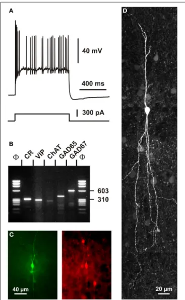
FIGURE 4 | Single cell RT-PCR analysis of a rat CR+ interneuron. (A)
Current-clamp recording obtained in response to a depolarizing current pulse (300 pA). Note the initial burst followed by irregularly discharged action potentials. (B) Single-cell RT-PCR analysis of the same neuron revealing co-expression of CR, VIP, ChAT, GAD65, and GAD67. (C) Comparison of the biocytin labeling of the recorded neuron (left panel) with the immunostaining of the slice with an antibody against CR (right panel). Note the immunoreactivity of the biocytin-labeled cell (scale bar 40μm, adapted fromCauli et al., 1997). (D) Intracellular biocytin labeling of another CR+ bipolar cell analyzed by singe cell RT-PCR. This neuron had a vertically oriented dendritic arborization. Pial surface is upward (scale bar, 20μm).
2000; Gallopin et al., 2006; Karagiannis et al., 2009; Lee et al.,
2010; Vucurovic et al., 2010
), contrasting sharply with the low
input resistance of fast spiking PV+ neurons (
Kawaguchi and
Kubota, 1993; Okaty et al., 2009; Battaglia et al., 2013
). This
property allows CR
+/VIP+ neurons to be substantially
depolar-ized by the small excitatory synaptic currents they receive from
thalamic inputs (
Lee et al., 2010
). The presence of a
promi-nent I
Hcurrent in SOM+ neurons underlying their
distinc-tive voltage sag induced by hyperpolarization partially explains
the slightly lower input resistance and the more depolarized
resting membrane potential of SOM
+ interneurons compared
with those of CR
+/VIP+ bipolar neurons (
Wang et al., 2004;
Halabisky et al., 2006; Ma et al., 2006; Xu et al., 2006; Karagiannis
et al., 2009
). Alike SOM+ interneurons, VIP+ cells have been
reported to exhibit the ability to discharge low-threshold spikes
driven by I
Tcalcium channels (
Kawaguchi and Kubota, 1996;
Cauli et al., 1997; Porter et al., 1999; Wang et al., 2004
). VIP
+
bipolar interneurons displayed action potentials of a duration
intermediate to those of fast spiking interneurons and
pyrami-dal cells (
Kawaguchi and Kubota, 1996; Cauli et al., 1997, 2000;
Karagiannis et al., 2009
). A complex repolarization phase of
their action potentials consisting of a fast after-hyperpolarization
(AHP), followed by an after-depolarization and a medium AHP
has been frequently observed in both VIP
+ and SOM+
interneu-rons (
Wang et al., 2004; Fanselow et al., 2008; Karagiannis
et al., 2009
), VIP+, but also SOM+ interneurons,
characteristi-cally displayed a pronounced frequency adaptation (
Kawaguchi
and Kubota, 1996; Cauli et al., 1997, 2000; Wang et al., 2004;
Halabisky et al., 2006; Ma et al., 2006; Xu et al., 2006
) which
can result in an irregular firing pattern (Figure 4) (
Cauli et al.,
1997, 2000; Wang et al., 2004
) when a slowly inactivating I
Dpotassium current is prominently present (
Porter et al., 1998
).
Similarly to SOM
+ interneurons, CR+ bipolar neurons exhibit
backpropagating action potentials accompanied by an
intracellu-lar Ca
2+increase (
Kaiser et al., 2001; Goldberg et al., 2003; Cho
et al., 2010
), which allows the release of neurotransmitters from
dendrites as reported for SOM+ interneurons (
Zilberter et al.,
1999
). Since the extent of action potential backpropagation is
modulated by the presence of I
Apotassium channels (
Goldberg
et al., 2003; Cho et al., 2010
), this suggests that the dendritic
release of CR+ bipolar interneurons (Figures 5, 6) can be finely
tuned.
In summary, CR
+ bipolar interneurons largely display the
electrophysiological features of adapting neurons (
Ascoli et al.,
2008
). Their distinctive intrinsic electrophysiological properties
indicate that they have specific integrative properties and
there-fore are likely to play specific roles in the physiology of cortical
circuits, e.g., state-dependently remove the blanket of inhibition
from principal neurons (
Karnani et al., 2014
).
SYNAPTIC INPUTS
CORTICAL
In general, our specific knowledge on connectivity is very
restricted. Probably the most precise data for local synaptic inputs
to CR
+ neurons come from paired recordings in layer II/III of the
primary somatosensory cortex in a CR-BAC-transgenic mouse
(
Caputi et al., 2009
). These authors reported that the two types
of neurons that they defined as the major cellular components
of the CR+ neuronal population receive different types of inputs
(and also form different types of output; see below). Bipolar
VIP
+/CR+ interneurons (BCR) receive functionally
depress-ing inputs from local pyramidal cells with a probability of
18.3%, from fast-spiking interneurons (29.7%), from multipolar
SOM+/CR+ interneurons (MCR; 41.1%) as well as facilitating
inputs from other BCR (31.8%) (Figure 5). By contrast, MCR
show facilitating inputs from pyramidal cells (17.4%) and from
BCR (76.4%) as well as depressing inputs from fast-spiking cells
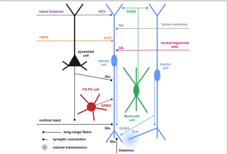
FIGURE 5 | Cortical and subcortical inputs of CR+ bipolar interneurons.
Schematic representation summarizing the different local cortical neurons (colored cells) and the long-range cortical and subcortical axon terminals (colored fibers) targeting CR+ bipolar interneurons (centered blue bipolar cell). Dendrites and axons are depicted by thick and thin lines, respectively. Anatomical synaptic connections are represented by triangles and putative volume transmission by spherical gradients. The local neuronal types targeting
bipolar CR+ and their respective neurotransmitters are schematized and color-coded; pyramidal cells (black, glutamate), fast spiking (FS)-PV neurons (red, GABA), Martinotti cells (green, GABA) other CR+ bipolar interneurons (blue, GABA and Enk [enkephalin]). Neurotransmitters of the long-range cortical and subcortical fibers are color-coded (Glu [glutamate; Black], 5-HT [serotonin, orange], ACh [acetylcholine, purple], NA [noradrenaline, gray], DA [dopamine, pink]) and their major origins indicated using the same color code.
(20%) and other MCR (9.8%). Interestingly, it appears that inputs
to CR+/VIP+ (BCR) neurons are mainly depressing (
Porter et al.,
1998; Rozov et al., 2001
), whereas all their outputs are facilitating
and, vice versa, for CR+/SOM+ (MCR) neurons inputs are often
facilitating whereas their outputs show a target cell type-specific
variability (
Caputi et al., 2009
). These data do not agree well with
paired recordings from rat barrel cortex, probably due to a
mix-ture of species differences and less defined populations of bipolar
vs. multipolar neurons in this study (
Reyes et al., 1998
).
Another rich source of observations describing the inputs to
layer II/III CR
+ neurons is provided by the glutamate uncaging
studies in different transgenic mouse lines (
Xu and Callaway,
2009
). They distinguished CR+ and CR- Martinotti cells and
(putatively CR+ and VIP+) bipolar cells. CR+ Martinotti cells
(as all other types of inhibitory neurons in their study) received
their strongest excitatory inputs within layer II/III whereas
bipo-lar cells possessed a second strong origin of inputs located in
layer IV.
Going beyond the local cortical circuits, Gonchar and
Burkhalter also described long-range cortical inputs to CR+
neu-rons (Figure 5) (
Gonchar and Burkhalter, 2003
). Here, tracer
injections to label connections between primary (V1) and
sec-ondary (V2) visual areas disclosed a circuit motif where CR
+
neurons (specifically in layer I) are involved in feedback inhibition
from V2 to V1, whereas few if any such long range connections
are found on layer II/III CR interneurons. In this layer, PV+
interneurons were the main recipients for feedback projections,
as they were for V1 to V2-directed feedforward projections.
CR
+ bipolar interneurons may integrate these glutamatergic
excitatory afferents by expressing ionotropic glutamate
recep-tors of the AMPA subtype exhibiting a low Ca
2+permeability
(
Porter et al., 1998; Rozov et al., 2001
) associated with high levels
of the GluR2 subunit (
Jonas et al., 1994
). They, however,
dis-tinctly exhibit a low occurrence of GluR3 subunits and mainly
express the flop variants of GluR subunits (
Lambolez et al., 1996;
Angulo et al., 1997; Porter et al., 1998; Cauli et al., 2000
). GluR5
and GluR6 are the main kainate receptor subunits expressed in
CR
+ bipolar neurons (
Porter et al., 1998; Cauli et al., 2000
).
Similarly to other cortical interneurons, CR
+ bipolar
interneu-rons express NR2A, B and D subunits of the NMDA receptor,
the latter being found at very low levels in pyramidal cells (
Flint
et al., 1997; Porter et al., 1998; Cauli et al., 2000
), indicating that
bipolar interneurons can express very slowly inactivating NMDA
receptors (
Vicini et al., 1998
). Similarly to SOM
+
interneu-rons, the activity of CR+ bipolar and can be also modulated
by the group I metabotropic glutamate receptors mGluR1 and
mGluR5 (
Baude et al., 1993; Kerner et al., 1997; Cauli et al.,
2000; van Hooft et al., 2000; Ferraguti et al., 2004
). The group III
metabotropic receptor mGluR7 expressed by CR
+/VIP+
bipo-lar neurons (
Cauli et al., 2000
) is presynaptically localized onto
synapses targeting SOM+ interneurons which suggests a
glu-tamatergic control of disinhibition (
Dalezios et al., 2002
). In
summary, CR
+/VIP+ bipolar neurons exhibit a cell-type-specific
expression pattern of glutamate receptors allowing them to
inte-grate glutamatergic inputs with distinct temporal and spatial
features (Figure 5).
In terms of their inhibitory inputs, alike SOM+ cells, CR+
bipolar interneurons sampled from layers II/III, IV, and V, which
was quite uncommon for most other cell types studied (
Xu and
Callaway, 2009
). In their study,
Gonchar and Burkhalter (1999)
corroborated with morphological methods that rat CR+ neurons
in layer II/III do innervate each other with numerous GABAergic
(symmetric) synapses on the soma as well as proximal and distal
dendrites. In addition, many symmetric and asymmetric synapses
that were not further identified by their specific origin could
be found on the CR
+ neurons. This is very similar to what we
have described for VIP+ interneurons in rat barrel cortex (
Staiger
et al., 2004
).
Since the expression of GABA receptor subunits by specific
cortical neuronal subtypes is still poorly documented (
Ruano
et al., 1997; Olah et al., 2009
) and given the diversity of these
subunits (
Laurie et al., 1992; Pirker et al., 2000
), the specific
GABA-A (but also GABA-B) receptor subunits of CR+ bipolar
interneurons remain largely undetermined.
Bipolar interneurons also possess receptors for intrinsic
cor-tical neuromodulators including opioids and endocannabinoids.
For instance, in the cerebral cortex
μ-opoid receptors are
mainly expressed by bipolar interneurons which also produce
enkephalin, its endogenous agonist (
Taki et al., 2000
). This
autocrine/paracrine
μ-opoid transmission has been shown to
restrict the inhibitory drive of CR
+ bipolar interneurons onto
pyramidal cells (
Férézou et al., 2007
) (Figure 5). Similarly, the
endocannabinoid transmission has been shown to exert a
long-lasting self-inhibition of putative SOM+ neurons (
Bacci et al.,
2004
) by activation of CB1 receptors, which are broadly expressed
by cortical neurons (
Bodor et al., 2005; Hill et al., 2007
). These
two neuromodulatory systems, recruited during sustained
fir-ing, provide activity-dependent negative feedback to restrict the
inhibitory actions of CR+ interneurons.
In summary, CR+ bipolar neurons possess a large integrative
capability for excitatory as well as inhibitory inputs across
dif-ferent layers of a cortical column. A lot remains to be done to
obtain a conclusive picture of the cortical input connectivity of
CR
+ bipolar neurons. However, already with the limited
infor-mation available, it appears that these GABAergic interneurons
do not only integrate excitatory and inhibitory inputs from local
origin but that they also process long-range inputs from within
the cortex and possibly subcortical sources (see next sections).
SUBCORTICAL INPUTS
Serotonergic
Due to the paucity of specific studies on subcortical targeting
of cortical bipolar CR
+ interneurons, inputs to the partially
overlapping populations of VIP+ as well as 5-HT3 R+
interneu-rons might be considered here, too (
Paspalas and Papadopoulos,
2001; Férézou et al., 2002; Cauli et al., 2004; Lee et al., 2010;
Vucurovic et al., 2010; Rudy et al., 2011
). The median raphe
nucleus was found to be the subcortical region that gives rise
to serotonergic input to CR+ interneurons in rat hippocampus
(
Acsady et al., 1993
). By using a bridge protein combined with
retrograde viral tracers, the paramedian raphe was also found to
project to a subpopulation of interneurons, which express ErbB4
in mouse primary somatosensory cortex (
Choi and Callaway,
2011
). Selectivity of the TVB-NRG1 bridge protein restricted the
transfection to the subgroups of interneurons immunopositive
for VIP and/or CR as well as ErbB4, including also small numbers
of PV
+/ErbB4 neurons but not SOM+ cells (
Choi et al., 2010; Xu
et al., 2010b
).
Although the expression of the ionotropic 5-HT3 R is
restricted to a subset of cortical interneurons, including CR+
bipolar neurons and neurogliaform cells (
Morales and Bloom,
1997; Férézou et al., 2002; Lee et al., 2010; Vucurovic et al.,
2010
), a subpopulation of hippocampal SOM
+ O-LM cells
orig-inating from the CGE was recently found to be 5-HT3 R
+
(
Chittajallu et al., 2013
). Cell-type-specific expression patterns
of metabotropic 5-HT receptors are by far less documented.
Nevertheless, 5-HT1a and 5-HT2a receptors are largely
co-expressed in pyramidal cells and in a minority of interneurons
positive for PV
+ or CB+ (
Aznar et al., 2003; Santana et al., 2004
),
suggesting that CR
+ bipolar interneurons barely express these
receptors. Similarly, 5HT-2c R is not prominently expressed in
CR+ neurons of the rat medial prefrontal cortex (
Liu et al., 2007
).
Given the reported segregation of 5-HT3 and 5-HT2a receptors in
the monkey cerebral cortex (
Jakab and Goldman-Rakic, 2000
), it
appears that 5-HT3 R is the major serotonin receptor expressed
by CR+ bipolar interneurons. These observations indicate that
serotonergic raphe neurons can rapidly activate CR+ bipolar
interneurons as shown in the cerebral cortex (
Férézou et al., 2002
)
(Figure 5).
Besides the paramedian raphe, additional origins of input
were detected (with a decreasing probability) in the thalamus,
secondary somatosensory, ipsilateral motor, retrosplenial, and
contralateral primary somatosensory cortex as well as the nucleus
basalis of Meynert (
Choi et al., 2010; Xu et al., 2010b
). Amongst
the detected brain regions, the thalamus and basal nucleus of
Meynert have been mostly investigated.
Thalamic
By anterograde tracing with PHA-L, neurons in the thalamus
(mainly ventral posterior nucleus and lateral geniculate nucleus)
were approved to form synaptic terminals onto the cortical VIP
+
population of inhibitory interneurons (
Staiger et al., 1996; Hajos
et al., 1997
). These anatomical observations were corroborated by
functional evidence showing that 5-HT3 R+ interneurons receive
monosynaptic thalamocortical inputs (
Lee et al., 2010
). By
con-trast, putative SOM
+ interneurons were found to barely receive
direct thalamic inputs (
Beierlein et al., 2000, 2003
).
Cholinergic
The projections from the basal nucleus of Meynert, where
most of the cholinergic neurons projecting to the neocortex are
located, are probably correlated to the nicotinic responsiveness
of VIP+/5-HT3 R+ bipolar interneurons (
Porter et al., 1999;
Férézou et al., 2002, 2007; Lee et al., 2010; Arroyo et al., 2012; Fu
et al., 2014
). CR+/VIP+ bipolar neurons express functional
high-affinity nicotinic receptors (
Porter et al., 1999; Férézou et al., 2002,
2007; Gulledge et al., 2006; Lee et al., 2010; Arroyo et al., 2012; Fu
et al., 2014
) composed of
α4β2 subunits and to a lesser extent
α5 subunit, a composition which is developmentally regulated
(
Winzer-Serhan and Leslie, 2005
). Alike SOM+ interneurons,
VIP
+ bipolar neurons are depolarized by muscarinic agonists
(
Kawaguchi, 1997; Fanselow et al., 2008
). Expression of m2
recep-tors has been reported in a minority CR
+ interneurons of the
rat entorhinal cortex (
Chaudhuri et al., 2005
) but the
subtype-specific identity of the muscarinic receptors inducing the
depo-larization of VIP
+ bipolar interneurons remains unknown. This
indicates that CR
+ interneurons integrate cholinergic afferences
from the basal forebrain (Figure 5) as recently shown for ChAT
+
bipolar interneurons using optogenetics (
Arroyo et al., 2012
),
which might be important for behavioral state-dependent control
of cortical circuits (
Pi et al., 2013
).
Noradrenergic
About 20% of VIP+ interneurons are contacted by noradrenergic
fibers (Figure 5), however, often with only a single
symmet-ric synapse (
Paspalas and Papadopoulos, 1998, 1999; Toussay
et al., 2013
). Despite the described expression of
α- and
β-adrenoreceptors in the cerebral cortex (
Nicholas et al., 1993a,b;
Pieribone et al., 1994; Scheinin et al., 1994; Venkatesan et al.,
1996
) and reported excitatory effects of
α-adrenoreceptors in
hippocampal and cortical interneurons (
Bergles et al., 1996;
Marek and Aghajanian, 1996; Kawaguchi and Shindou, 1998
),
the expression of adrenoreceptors in CR
+ interneurons has not
been specifically investigated. However, inhibitory effects of
α-adrenoreceptors have been observed in discrete subpopulations
of CCK+ and SOM+ interneurons (
Kawaguchi and Shindou,
1998
), whether or not this inhibitory effect of
α-adrenoreceptors
is existent in CR
+ bipolar interneurons remains unknown.
β1-and
β2-adrenoreceptors are not as frequently expressed in CR+
interneurons as compared to other types of interneurons (
Liu
et al., 2014
).
Dopaminergic
Dopaminergic terminals have been shown to target interneurons
(
Sesack et al., 1995b
), specifically CR+ interneurons in monkey
prefrontal cortex (
Sesack et al., 1995a
) (Figure 5). However, there
is no clear segregation in the expression profiles of dopamine
receptors, which are frequently observed in pyramidal cells and
interneurons of rodent and primate cortex with substantial
co-expression (
Vincent et al., 1993; Khan et al., 1998, 2000; Ciliax
et al., 2000; Wedzony et al., 2000; Rivera et al., 2008; Oda et al.,
2010
). Nevertheless, regarding the expression of D1-like
recep-tors, D1 receptors predominate in PV
+ interneurons of the
primate prefrontal cortex whereas it is D5 in CR
+ interneurons
(
Glausier et al., 2009
).
At the moment, all that can be stated with certainty is that
a lot of work is still needed to get a better idea how cortical
CR+ bipolar interneurons are influenced by subcortical sensory
or modulatory inputs (Figure 5), which receptors are involved
and how this modulation specifically affects sensory information
processing or more general brain-state activity.
TARGETS AND FUNCTIONS
CORTICAL CIRCUIT
Neuronal targets
Hippocampal
CR
+ interneurons are considered to be
interneuron-specific interneurons (
Klausberger and Somogyi,
2008
), which preferentially or even exclusively target other
interneurons, presumably on their dendritic shafts. In rat
hippocampus, post-embedding immunogold studies showed
that most of the targets of CR
+ and VIP+ boutons (which
might be derived from the same bipolar neurons co-localizing
CR and VIP) were GABAergic dendrites (
Acsady et al., 1996a;
Gulyas et al., 1996
). In the neocortex, the GABAergic dendrite
targeting property of CR+ or VIP+ interneurons is more
complex when taking cortical areas and layers into account. In
monkey and rat visual cortex, differential modes of innervation
by CR
+ interneurons exist in different layers. In layers I-III,
CR+ interneurons tended to synapse on dendritic shafts of
other not further specified GABAergic interneurons. However,
in layers IV-VI, GABA-negative spines belonging to pyramidal
neurons were primarily innervated (
Meskenaite, 1997; Gonchar
and Burkhalter, 1999
).
Paired recordings in layer II/III of the primary somatosensory
cortex of CR-BAC-transgenic mice (
Caputi et al., 2009
) gave an
idea of the functional output of these CR neurons. Bipolar CR+
(BCR) innervate pyramidal cells (11.6%), fast-spiking
interneu-rons (29.7%), other BCR (31.8%) and as already mentioned,
preferably MCR (76.4%). It is remarkable that the input with the
highest probability in this study represents indirect evidence for
the recently disclosed disinhibitory circuit from VIP neurons to
Martinotti cells (Figure 6), for which several direct (
Lee et al.,
2013; Pfeffer et al., 2013; Pi et al., 2013; Fu et al., 2014
) and
indi-rect (
Staiger et al., 2004; Gentet et al., 2012
) lines of evidence are
now available.
It has been shown in both, hippocampus and somatosensory
cortex of rodents, that calbindin-expressing (CB+)
interneu-rons are the targets of CR
+ or VIP+ interneurons (
Acsady
et al., 1996b; Gulyas et al., 1996; Staiger et al., 2004
). CB
+
interneurons form a heterogeneous population and can
coex-press other neuropeptides or other calcium binding proteins,
thus, it is reasonable to further differentiate CB+ interneurons
in order to distinguish the CR
+ or VIP+ outputs to specific
sets of interneurons. The most likely contacts are made with
FIGURE 6 | Neuronal and non-neuronal targets of CR+ bipolar cells.
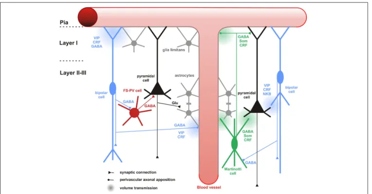
Schematic representation summarizing the different neuronal and non-neuronal targets of CR+ bipolar interneurons (blue neurons) and Martinotti cells (green neuron). Dendrites and axons are depicted by thick and thin lines, respectively. Anatomical synaptic connections are represented by triangles, axonal perivascular appositions by dots and putative volume transmission by spherical gradients. The cell types targeted by CR+ neurons are schematized and color-coded; FS-PV neurons (red), Martinotti cells (green), pyramidal cells (black), glial cells (gray).
Astro-glial coverage of blood vessels is illustrated by a gray-colored abluminal side. The specific action of the multiple neurotransmitters released by CR+ neurons is depicted by their location near the specific target and their origin color-coded (bipolar cell, blue; Martinotti cells, green). VIP, vasoactive intestinal polypeptide; CRF, corticotrophin releasing factor; NKB, neurokinin B; Som, somatostatin. The disinhibitory action of bipolar cells is represented by disynaptic circuits involving a bipolar interneuron, an intermediary interneuron (left: FS-PV cell; right: Som-Martinotti cell) and a pyramidal cell, respectively.
Martinotti cells which coexpress the markers CB and SOM
(
Kawaguchi and Kubota, 1996; Wang et al., 2004
). Supporting
this possible connection, recent functional studies described that
in mouse somatosensory, visual, auditory, and prefrontal
cor-tex, SOM
+ interneurons were preferentially innervated by local
VIP
+ interneurons (
Lee et al., 2013; Pfeffer et al., 2013; Pi et al.,
2013
). Because of the diversity of VIP+ and SOM+ neuronal
subgroups, it would be important to know what exact types in
terms of morphology contribute to this microcircuit. Some
indi-rect evidence already exists. Bipolar CR
+ interneurons (BCRs)
were immunopositive for VIP while multipolar CR
+
interneu-rons (MCRs) were not (
Caputi et al., 2009
) and subgroups of
SOM+ interneurons coexpressing CR with up to 95% or more
than 50% were found in mouse visual and somatosensory
cor-tex layers I–III, respectively (
Xu et al., 2006
). Therefore, it is
hypothesized that BCRs, which are presumably CR
+/VIP+
bipo-lar interneurons, target MCRs, which are probably CR+/SOM+
Martinotti interneurons.
Beside Martinotti interneurons, subgroups of basket cells such
as large basket cells and nest basket cells could be immunopositive
for CB, however, it is much more likely that these co-localize PV
and not SOM (
Wang et al., 2002
). Two anatomical studies showed
that VIP+ interneurons innervated PV+ interneurons, although
it is debated whether there exists a preferential targeting in terms
of putative PV+ subgroups (
Dávid et al., 2007; Hioki et al., 2013
).
Behavioral experiments on mouse auditory and prefrontal
cor-tex found that next to SOM
+ interneurons, PV+ interneurons
were the second major outputs of VIP
+ interneurons (
Pi et al.,
2013
) (Figure 6), lending further support to a functional role of
the above mentioned anatomical connections. In summary, the
CR+ bipolar interneurons seem to preferentially but not
exclu-sively target other GABAergic interneurons, the molecular and
anatomical properties of which need to be better analyzed.
Neuronal processing
The input-output connectivity pattern described above (Cortical
and Neuronal targets) implies that bipolar CR
+ interneurons do
participate in disinhibitory but also feedforward and feed-back
inhibitory circuits (
Isaacson and Scanziani, 2011
) of any
corti-cal layer, column and area, especially when considering their high
degree of colocalization with VIP (
Porter et al., 1998; Reyes et al.,
1998; Gonchar and Burkhalter, 2003; Caputi et al., 2009; Lee
et al., 2013; Pfeffer et al., 2013; Pi et al., 2013
). Whether the
con-tent of CR in these circuits is meaningful on its own, like it has
recently been suggested for another calcium-binding protein (i.e.,
parvalbumin in the hippocampus;
Donato et al., 2013
), is
cur-rently unknown (but see
Schwaller, 2014
for a recent review).
The best understood function is now the disinhibitory action
that is dependent on a unique circuit motif, i.e., targeting of
other inhibitory interneurons, mostly SOM- (or CB-)expressing
Martinotti cells. In the somatosensory system of mice,
Lee et al.
(2013)
found that a specific projection from the whisker motor
cortex excites VIP+ neurons and subsequently inhibits SOM+
neurons in the primary somatosensory cortex. This leads to a
disinhibition of the apical dendrites of the pyramidal cells
dur-ing exploratory behavior (Figure 6), probably actdur-ing as a
“gate-opener” for the paralemniscal pathway (
Gentet et al., 2012
). A
similar gate-opening mechanism might be functional in the
pri-mary auditory cortex, when animals learn to associate a tone
stimulus with an aversive context (
Pi et al., 2013
).
Peptidergic neuromodulation
The expression of numerous neuropeptides in CR
+ bipolar
interneurons (
Kubota et al., 1994, 2011; Kaneko et al., 1998; Taki
et al., 2000
), whose release requires a high level of neuronal
activ-ity (
Zupanc, 1996; Baraban and Tallent, 2004
), could also allow
an activity-dependent fine tuning of the cortical network. For
instance in the cat visual cortex, exogenously applied VIP had
little or no effect on recorded neurons in the absence of visual
stimulation, but enhanced their visual responses (
Murphy et al.,
1993; Fu et al., 2014
). Consistently, by activating VPAC1
recep-tors and cAMP/PKA signaling, VIP reduced the slow AHP current
and the tonic potassium current which regulates the excitability of
hippocampal and cortical pyramidal neurons (
Haug and Storm,
2000; Hu et al., 2011
). Interestingly, CRF whose CRF1 receptors
are also expressed by pyramidal neurons (
Gallopin et al., 2006
),
induced an even more pronounced increase in cAMP/PKA
signal-ing and modulation of potassium currents than VIP (
Haug and
Storm, 2000; Hu et al., 2011
). Presumably because of the rapid
desensitization of CRF1 receptors (
Hauger et al., 2000
), the action
of CRF rapidly declined.
CR
+ bipolar interneurons are also likely to enhance
gluta-matergic activity via NKB signaling (Figure 6). Indeed, NK-3
receptor, the most selective receptor for NKB (
Shigemoto et al.,
1990
), is expressed by layer V pyramidal cells (
Ding et al., 1996;
Shughrue et al., 1996; Gallopin et al., 2006
) and its activation
depolarizes them (
Stacey et al., 2002; Rekling, 2004; Gallopin
et al., 2006
).
In summary, to be operational, this complex peptidergic
neu-romodulation of the cortical circuit (Figure 6) requires a selective
enhancement of CR
+ bipolar neurons activity to achieve a
sub-stantial release of neuropeptides (
Zupanc, 1996; Baraban and
Tallent, 2004
). This prerequisite could be met during specific
brain states such as locomotion (
Fu et al., 2014
), which
asso-ciates an increased activity of basal forebrain cholinergic neurons
(
Lee et al., 2005
) and serotonergic neurons of the raphe (
Wu
et al., 2004
) leading to the concomitant activation of excitatory
receptors on CR
+ bipolar neurons (see Subcortical inputs).
GLIO-VASCULAR NETWORK