by
Bogdan I. Fedele§
B.S. Chemistry, B.S Biology (2002)
Massachusetts Institute of Technology, Cambridge, MA
SUBMITTED TO THE DEPARTMENT OF BIOLOGICAL ENGINEERING IN PARTIAL FULFILLMENT OF THE REQUIREMENTS
FOR THE DEGREE OF
DOCTOR OF PHILOSOPHY IN BIOLOGICAL ENGINEERING
AT THE
MASSACHUSETTS INSTITUTE OF TECHNOLOGY
JUNE 2009
@2009
Massachusetts Institute of Technology.
All rights reserved.
ARCHIVES
MASSACHUSETTS INSTITUa OF TECHNLOGYV
AUG 16 2(10
Ll PARI ES
The author hereby grants to MIT permission to reproduce and to distribute publicly paper and electronic copies of this thesis document in
whole or in part in any medium now known or hereafter created.
, ~-, / ~9 Signature of Author:
t 41
Certified by:
William and Betsy Leitch Professor
Department of Biological Engineering May 22, 2009
<1j John M. Essigmann
of Chemistry, Toxicology and Biological Engineering Thesis Supervisor
Accepted by:
V/ 'Alan J. Grodzinsky
Profer of Electrica chanic 1, and Biological Engineering Chairman, Departmenta mmittee for Graduate Students
THIS DOCTORAL THESIS HAS BEEN EXAMINED BY A COMMITTEE OF THE
DEPARTMENT OF BIOLOGICAL ENGINEERING AS FOLLOWS:
Leona D. Samson L(<--
--American Cancer Society Professor
Professor of Toxicology and Biological Engineering Director, Center for Environmental Health Sciences Chairman
John Martin Essigman_
William and Betsy Leitch Profe so of Chemistkioxicology and Biological Engineering Thesis Supervisor
,lA /9
Forrest White L/ - -L ' ( L/
-Mitsui Associate Professor of Biological Engineering
Robert G. Croy \
The Androgen Receptor Independent Mechanism of Toxicity of the
Novel Anti-tumor Agent 11
6-Dichloro
by
Bogdan I. Fedeles
Submitted to the Department of Biological Engineering in Partial Fulfillment of the Requirements for the Degree of Doctor of Philosophy in Biological Engineering
Abstract
Inspired by the toxicity mechanism of cisplatin in testicular cancer, a series of bi-functional genotoxicants has been designed that supplement their DNA damaging properties with the ability to interact with tumor specific proteins. One such compound, 11p -dichloro links an aniline mustard moiety to a steroid ligand for the androgen receptor (AR). In vitro and in vivo (cell cultures, mouse xenografts) studies highlighted the potent antitumor properties of the molecule, which can prevent growth of AR positive tumor xenografts in mice. However,
11
p-dichloro
also proved highly effective against many cancer lines that do not express the AR, more so than other nitrogen mustards commonly used in chemotherapy.To understand better the AR independent mechanism, the toxicity of 116 -dichloro was investigated in Saccharomyces cerevisiae, by interrogating the complete yeast single-gene deletion mutant library. Surprisingly, the screen revealed that the mutants most sensitive to
11p -dichloro are not the ones lacking genes involved in DNA repair, but rather mutants lacking genes involved in mitochondrial and ribosomal function. While some of the sensitive mutants are also sensitive to other DNA damaging agents, almost half of them are uniquely
sensitive to 11p -dichloro, suggesting a DNA-damage independent mechanism of action.
Based on the yeast findings, we tested mechanistic hypotheses in HeLa cells (an AR negative human cancer cell line), and discovered that 11p -dichloro induces a large amount of reactive oxygen species (ROS), accompanied by a significant depletion of the antioxidant pool and a perturbation of the mitochondrial inner membrane potential. We also discovered that co-treatment with antioxidants such as N-acetyl cysteine or vitamin E alleviates the toxicity of
11
p-dichloro,
suggesting that ROS play an important role in the mechanism of toxicity. In terms of transcriptional responses, previous observations were confirmed that 11p -dichloro upregulates expression of genes involved in the unfolded protein response (UPR) and sterol biosynthesis. Additional cellular responses consistent with these pathways were the striking increase in cytosolic calcium and significant changes in the size of the cells. Taken together, our results highlight the multifaceted toxicological profile of 11p -dichloro, indicating that besides DNA damage, the generation of ROS and induction of UPR may be the additional pathways responsible for the potent anticancer effects of this agent.Research on the scale required for doctoral work always involves significant human interaction and collaborative efforts. I am greatly indebted to all the people that have
influenced and shaped my doctoral career; their wisdom, advice and friendship have made all the difference, transforming these seven long years of PhD into an unforgettable life
experience.
My deepest gratitude goes to my advisor, John Essigmann, who has been a tremendous
source of inspiration, motivation and guidance. Ever since our first encounter at a student dinner, back in my college senior year, I have been fascinated by John's brilliant scientific ideas, his unstoppable enthusiasm and captivating story-telling ability. John has always been extremely supportive of all my overly-creative ideas and he encouraged me to explore paradigm-shifting approaches to research. I am very grateful for his laid-back mentoring style, which has been instrumental for allowing me to occasionally pursue interests outside the lab. Yet, I have always found John extremely available to consult on any matter, ranging from science to life and career advice. John, through his kind and selfless example, not only taught me how to become a better scientist, but he inspired me to become a better person. I have always been amazed by John's ability to gracefully handle a tightly packed research
schedule with his family duties and an entire dorm full of MIT undergraduates. For that, I consider John a true role model for me, and I hope to follow in his footsteps towards an exciting and fulfilling academic career.
I would like to thank my thesis committee members. Leona Samson has been another source
of inspiration for me. Her fascinating research and innovative approaches have been
instrumental for shaping my project. I am also very grateful to her for allowing me to work in her lab and learn all about yeast genomic phenotyping, and for her sharing of the yeast mutant library. Additionally, I would like to thank Leona for being the chair on my thesis committee; in this quality, she has played a pivotal role in focusing and guiding my research towards a fruitful completion. Her feedback, at every step of my graduate career has always been poignant and invaluable. I would like to thank Forest White for being a very supportive committee member. His insightful and goal-oriented comments have always been very helpful in structuring my work and focusing on the positive results of my research. I would also like to thank Bob Croy, the co-founder of the programmable chemotherapeutics project. Bob has helped me a great deal, both by personal example and by sharing his wealth of scientific knowledge with me. Bob has also been an excellent and avid critic of my work, helping me pose and search the answers for all the hard research questions, and thus helping me strive for scientific excellence. I am also grateful to all my thesis committee members for being accommodating and flexible with their schedules, which has allowed me to
successfully overcome crunch times that seemed virtually insurmountable.
Throughout my graduate career, all my scientific colleagues have been very helpful, friendly and supportive. Especially, the people in the Essigmann lab have often had a positive impact on my work and life. I would like to thank Nicole for being my very first Essigmann lab mentor, while I was still transitioning to grad school. Her neatness and attention to detail, doubled by a friendly, laid-back attitude have been attributes I have tried to emulate during
you for creating excellent compounds that kept the rest of the lab busy for years. I would like to thank Peter for always providing quick answers to my quick questions. I am very grateful to Shawn for having written a very well-documented thesis, which served as a model for my own. To Jim, I am thankful for always knowing the way to the nearest free food opportunity. To Charles, for being a cool wordsmith. To Lauren and Sarah, the most serious people in the lab, I am thankful for setting a high standard of dedication and work ethics, but also for organizing some excellent trips to Maine. I would like to thank Jeannette for proofreading some of my thesis and for being very supportive especially during my final grad days. I would also like to thank Jenn, Yuri, Francis, Will, Mayuree and Tang for always being very friendly. To Leslie, I am grateful for her help with proofreading and for her kind and friendly career insights and advice. I would like to thank Deyu for his excellent suggestions for my thesis defense talk. To Sreeja, I am grateful for all the help with the comet assays and other complicated protocols. Finally, I would like to thank Kyle for being a great year mate and friend. Our countless discussions about "figuring it out" yielded some of the most helpful ideas for my research. Kyle has also been extremely supportive and helped me through some trying emotional times with a positive outlook and sound advice.
I would like thank the people from other laboratories that I have collaborated with.
Especially, Tom Begley, Rebecca Fry and Rachel Ehrlich from the Samson lab, who have patiently taught me so many things about working with yeast, deserve an important acknowledgment. I would also like to thank Andrea from the Manalis lab, who helped me with the cell size measurements. I am also grateful to Michael Jie and Glenn Paradis for teaching me how to design and run flow cytometry experiments.
In my graduate career, I have been fortunate to have the opportunity to mentor several undergraduate students as they discover the joys and frustrations of experimental science. It has been a pleasure to teach them and it also helped me understand my work better. I would like to thank Angela, Kellie, Jessica, Alvin, Ryan and Daniel for being wonderful UROPs, very eager to learn new things, very dedicated and very receptive to my sometime eccentric ideas. The sheer amount of experimental data gathered throughout my project would not have been possible without your hard work. I have also learned a lot from you and had a great time working together.
I would like to extend a warm thank you to all my friends who have been helpful and
supportive all throughout my long grad school journey. The moments we shared at the occasional festive gatherings, on the town or around some intriguing board game have been immensely enjoyable and helped me keep my sanity after endless days of experiments. Many thanks go, in no particular order to: Daniel (Pylot), Stefan, Florin A., Florin C., Mihai B., Mihai & Simona, Theo & Smaranda, Simona T., Daniel N., Iuliu, Ioanid, Viorel and Florin
welcome oasis of diversion during the trying moments of grad school.
A special thank you goes to all my music teachers and coaches I have had at MIT. Music has
always been an integral part of my life and you helped me to continue to nurture it
throughout grad school. I would like to thank John Harbison, Randall Hodgkinson, Judith Gordon, Tim McFarland, Marcus Thompson and David Deveau. I would also like to thank all my music colleagues from all the chamber music groups I have been a part of, in
particular to : Way, Tracy, Joyce, Katherine, Katie, Sunny, Jodie, Sophie, Lanthe, Boris and Benjamin. Making music together has been a tremendous experience and a necessary respite from the lab grind.
Most importantly, I would like to extend the warmest thank you possible to my wonderful family. Many thanks go to my grandparents, who have always been supportive of me pursuing a scientific career. Sadly, none of them lived long enough to see me get my doctorate. My siblings, Flavia and Sorin and my sister-in-law Antonia, have also been extremely helpful and supportive. They have encouraged me every step of the way and I am very thankful for that. My parents deserve my most heartfelt gratitude; their unyielding love and dedication, their constant faith in my abilities and their unconditional support have made a world of difference to me. None of my accomplishments, including the present thesis would have been possible without their amazing optimism, care and emotional support.
Finally, my most special thank-yous go to the most important person in my life, the adorable
Abby Men. Meeting Abby has been a momentous event in my graduate career, because it
happened at a time of great emotional distress and seemingly insuperable difficulties in my work. She has been the miraculous catalyst that empowered me to find the ways to overcome the technical difficulties and persevere in my work towards a successful conclusion. I am forever indebted to her for her patience and understanding, for her amazing support and dedication, and for her love. Her efforts for easing my frustrating research toil have been unforgettable; after a long and laborious day of work, she would often surprise me with an exquisitely delicious, home-cooked dinner. I often feel that without her, my graduate school career would have been so much longer. I would never be able to thank her enough for the
joy and inspiration she brought into my life, for making me a better, more efficient person
A b stract ...
A cknow led gem ents... 7
T ab le of C ontents ... 11 L ist of A bbreviations ... 14 CHAPTER 1 In tro d u ctio n ... 17 1.1 C an cer ... 18 1.2 C hem otherapy ... 23
1.3 DNA-damaging agents as chemotherapeutics ... 26
1.4 Developing better chemotherapeutic agents ... 29
1.4.1 The inspiration - the mechanism of cisplatin in testicular cancer ... 29
1.4.2 The vision - engineering programmable cytotoxins... 31
1.4.3 The proof of principle - the phenyl-indole compounds... 32
1.4.4 E2-7a - optimizing a compound targeted towards ER ... 33
1.5 11
p-Dichloro
- an androgen receptor targeted compound ... 361.5.1 Physico-chemical properties ... 36
1.5.2 Toxicological properties ... 37
1.5.3 Investigating the mechanism of toxicity of 11
p-dichloro
... 401.6 F ig u re s... 4 2 1.7 R eferen ces ... 5 5 CHAPTER 2 Investigating the Mechanism of Toxicity of the Anticancer Agent 11p -Dichloro in Saccharomyces cerevisiae by Genomic Phenotyping... 61
2 .1 Intro du ction ... 62
2.2 Materials and Methods... 67
2 .2.1 R eagents... . . 67
2.2.2 Yeast cell lines and culture ... 67
2.2.3 Survival analysis ... 67
2.2.4 Library screening approach... 68
2.2.5 Statistical analysis... 69
2.3 Experimental Results ... 70
2.3.1 Validation of the spectrophotometric quantitation of cell growth... 70
2.3.2 Choosing and characterizing the control strains ... 71
2.3.3 Screening the complete single gene knockout mutant yeast library ... 72
2.3.4 The GO localization, processes and functions of mutants sensitive to 11p -d ich loro ... . . 74
2.3.5 Common mutants between most sensitive mutants to 11p -dichloro and most sensitive mutants to other DNA alkylating agents... 76
2.3.6 The GO localization, processes and functions of mutants uniquely sensitive to 11 -dich loro ... . . 77
2 .4 D iscu ssio n ... 7 8 2.5 T ables and Figures ... 82
3 .2 .1 R eag en ts ... 10 9
3.2.2 C ell lines and culture ... 109
3.2.3 Assays for cell growth, cell viability and cytotoxicity ... 109
3.2.4 Determination of intracellular ROS using flow cytometry... 110
3.2.5 Determination of the mitochondrial potential and reduced GSH ... 111
3.2.6 Determination of H202, lipid peroxidation and cellular NADPH... 111
3.2.7 Statistical analysis... 112
3.3 E xperim ental R esults ... 113
3.3.1 11
6-Dichloro
exhibits remarkable acute toxicity against HeLa cells in vitro, higher than simpler nitrogen mustards... 1133.3.2 11
5-Dichloro
exposure is accompanied by generation of ROS... 1143.3.3 Antioxidants attenuate 11p -dichloro toxicity ... 115
3.3.4 11
p-Dichloro
depletes cellular NADPH and the free thiol pool... 1153.3.5 Glutathione depletion sensitizes cells to 11p -dichloro ... 116
3.3.6 11 P-Dichloro treatment affects inner mitochondrial membrane polarization1 16 3.3.7 11
p-Dimethoxy
displays cellular responses similar to 11p -dichloro, but of low er m agn itu de... 1173 .4 D iscu ssio n ... 1 19 3 .5 F ig u re s... 12 8 3 .6 R eferen ces... 14 6 CHAPTER 4 The Anticancer Agent 11p -Dichloro Induces the Unfolded Protein Response in HeLa C e lls ... 1 5 1 4 .1 In tro du ction ... 152
4.2 M aterials and M ethods... 157
4 .2 .1 R eag en ts ... 157
4.2.2 C ell lines and culture ... 157
4.2.3 Quantifying changes in cell size ... 157
4.2.4 Determination of intracellular Ca+* using flow cytometry ... 158
4.2.5 Determination of transcript levels using RT-PCR ... 158
4.2.6 Determination of relative protein levels using Western blotting ... 159
4.2.7 Determination of cell viability in the presence of cell death inhibitors... 160
4.2.8 Determination of acidic cellular compartments using acridine orange staining 161 4.3 E xperim ental R esults ... 162
4.3.1 11
p-Dichloro
upregulates the expression of genes involved in the UPR response and sterol biosynthesis ... 1624.3.2 11 -Dichloro increases the levels of the SREBP protein ... 163
4.3.4 11fp-D ichloro causes an increase in cell size... 164
4.3.5 11
6-Dichloro
induces the proliferation of acidic compartments in the cell.. 1644.4 D iscussion... 166
4.5 Conclusions and Future D irections... 171
4.6 Tables & Figures... 180
4.7 References... 190
APPENDIX A The Com plete List of Y east M utants Sensitive to 11p -D ichloro... 197
A .1 D escription... 198
A .2 References ... 198
A .3 Table... 199
APPENDIX B Effects of the Medium Components on 11p -Dichloro Toxicity in Saccharomyces cerevisiae ... 269
B.1 Introduction ... 270
B.2 M aterials & M ethods... 270
B.3 Results and D iscussion... 271
B.3.1 The phenotypes of the control strains ... 271
B.3.2 The influence of the low ionic strength of the buffer... 271
B.3.3 The influence of glucose ... 272
B.3.4 The influence of divalent cations ... 273
B.4 Figures... 274
B.5 References ... 280
CU RRICU LU M V ITA E ... 281
4NQO AIF AMS ANT AO AR ATP BSO CFA CMFDA CM-H2DCFDA CML CTB DHE DHT DMSO DNA ER ER ERK ETC GO GPX GR GRID GSH GSSG HMG HMGCR 4-nitro-quinoline oxide apoptosis inducing factor accelerator mass spectrometry adenine nucleotide translocase acridine orange
androgen receptor adenosine triphosphate buthionine sulfoximine
colony forming assay
chloromethylfluorescein diacetate
5- (and 6-)-chloromethyl-2' ,7'-dichlorodihydrofluorescein diacetate chronic myelogenous leukemia
cell-titer blue dihydroethidine dihydrotestosterone dimehtylsulfoxide deoxyribonucleic acid
estrogen receptor (chapter 1) endoplasmic reticulum (chapter 4) extracellular-signal regulated kinase electron transport chain
gene ontology
glutathione peroxidase glutathione reductase
general repository for interaction datasets glutathione
glutathione dimer high mobility group
HPF hydroxyphenyl fluorescein
HPV human pappiloma virus
HRP horseradish peroxidase
hUBF human upstream binding factor
IP intra-peritoneal
JNK c-jun N-terminal kinase
LBD ligand binding domain
LDLR low density lipoprotein receptor
LFC log2 fold change
MAPK mitogen activated protein kinase
MDA malondiadehyde
MEK MAPK/ERK kinase
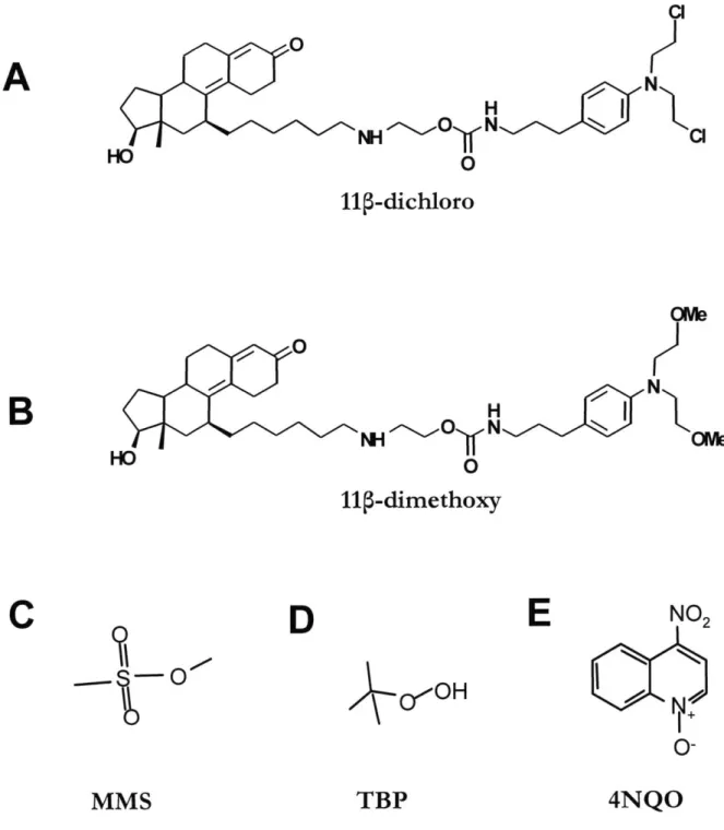
MMS methylmethane sulfonate
MnSOD manganese superoxide dismutase
MPT mitochodrial permeability transition
NAC N-acetyl L-cysteine
NADH nicotinamide adenine dinucleotide hydride
NADPH nicotinamide adenine dinucleotide phosphate hydride
NCI national cancer institute
PARP polyadenineribonucleotide polymerase
PBS phosphate buffered saline
PCR polymerase chain reaction
PDI protein disulfide isomerase
RNA ribonucleic acid
ROS reactive oxygen species
rRNA ribosomal RNA
SCAP SREBP cleavage activating protein
SERCA sarco-endoplasmic reticulum calcium ATPases
SQLE SREBP TBP TCA TH thioTEPA UPR
UV
WT YPD zVADfmk squalene epoxidasesterol responsive element-binding protein tert-butyl peroxide
tricarboxylic acid transhydrogenase
n,n',n"-triethylenethiophosphoramide unfolded protein response
ultra-violet wild-type
yeast-extract/peptone/dextrose
made in detection technologies and available therapies, we are still far from effective
treatments and cures for most malignancies, especially for advanced stage cancers. However, as our knowledge about the causes and biochemical mechanisms of cancer expands, we are approaching the critical mass of data necessary to map a path towards novel, paradigm-shifting treatments for cancers that are currently incurable.
Cancer constitutes a significant public health problem, accounting currently for about 25% of the total number of deaths in the US (Jemal et al., 2008), and for about 13% of the total
number of deaths worldwide (Boyle et al., 2008). The incidence of cancer in the Western world is staggering; it is estimated that one in two men and one in three women will develop a form of invasive cancer throughout their lifetime (Ries et al., 2009). Moreover, the
incidence of cancer is growing annually by about 1% worldwide. This growth is predicted
by some estimates to establish cancer as the leading cause of death worldwide as soon as
2010 (Boyle et al., 2008). In fact, in the US population aged 85 years or less, the death rate from cancer has already surpassed the death rate from cardiovascular diseases (Jemal et al.,
2008).
Cancer can affect people irrespective of their gender, race, locale's climate, micro-environment, or income, but it shows a strong dependence on age. On average, the lowest incidence of cancer occurs between the ages 20 to 40 years; the incidence increases about 10-fold for the ages 40 to 60 years and then another 2- to 3-10-fold for the ages 60 to 80 years. The cancer incidence then drops for people aged 80 years or more. Interestingly, children and teenagers (aged less than 20 years) have also an elevated risk of developing certain kinds of cancer (Jemal et al., 2008).
can occur in virtually every organ of the human body and in every type of tissue, and are usually classified by the location in which the primary tumor - the initial malignant
outgrowth, occurs. However, additional complexity is imparted by the extensive diversity at the cellular and molecular level; cancers occurring in the same kind of tissue may have very different genetic compositions and phenotypic manifestations, and thus exhibit variable and often unpredictable responses to treatments.
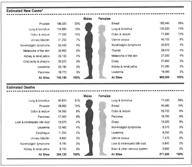
In 2008, in the US, the estimated top five most prevalent cancers have been cancers of prostate, lung, colon/rectum, urinary bladder and non-Hodgkin lymphoma for men, and cancers of breast, lung, colon/rectum, uterus and non-Hodgkin lymphoma for women (Figure
1.1). The most deadly cancers have been predicted to be the lung, prostate, colon/rectum,
pancreas and liver cancers for men, and lung, breast, colon/rectum cancer, pancreas and ovarian cancers for women (Figure 1.1).
A great body of work, which started in the 1980s, has firmly established that the molecular
basis for cancer is an accumulation of mutations in the cellular DNA (Hanahan and Weinberg, 2000). These genetic changes range from point mutations to large scale
chromosomal rearrangements, duplications and deletions. Often, changes also occur in the number and organization of the chromosomes, generating aneuploidy (Hede, 2005). Such genomic transformations alter the function of a number of genes involved in cell proliferation
- either activating genes that promote growth (oncogenes), or inactivating genes that suppress growth (tumor suppressor genes). Several such specific mutations are usually required for the cell to become cancerous, acquiring the six fundamental traits common to most malignancies (Hanahan et al., 2000) : unlimited replicative potential (immortality), self-sufficiency in growth stimuli, resistance to growth-inhibitory stimuli, avoidance of apoptosis, ability to trigger angiogenesis (blood vessel growth) and finally, invasiveness -the ability to metastasize (spread) to other parts of the body and generate secondary tumors. These traits are not acquired all at once, but rather one at time through selective pressures in the
micro-mutated cells acquires a second advantageous mutation, it proliferates even more. This cycle of mutation and selection goes on until cells acquire most of the cancer hallmarks; at that point, the growth of those cells becomes malignant. Unfortunately, cancer cells in the patient's body continue to evolve, becoming more invasive, faster growing and more
resistant to therapies. They metastasize and generate secondary tumors throughout the body, affecting the function of various organs. The loss of function in multiple systems due to secondary tumors is the cause of death in 90% of cancers (Sporn, 1997). This is why the earlier the cancer is detected, the higher the chance an effective therapy has for preventing the cancer from advancing to its deadly metastatic stage.
Understanding the molecular mechanisms responsible for the accumulation of the mutations that lead to cancer is essential for evaluating risk factors, designing better diagnostics and better treatments. In all living cells, there is a background mutation rate due to errors made
by polymerases when DNA is replicated. Taking into account all the proofreading and repair
mechanisms in the cell, mutation rates have been estimated to be between one in 109
replicated base pairs for somatic cells (Albertini et al., 1990) and one in 1011 replicated base pairs for stem cells (Cervantes et al., 2002). This rate translates into about one mutation for every somatic cell division and about one mutation for every four stem cell divisions. In addition to polymerase errors, environmental exposure to genotoxic agents can contribute to an even greater mutagenic load. Despite this barrage, most mutations that accumulate over a lifetime in our genomes will not lead to cancer. These benign mutations have been termed "passenger mutations." However, an occasional mutation that can contribute to cancer will occur - these are termed "driver mutations" (Greenman et al., 2007; Haber and Settleman,
2007). In general, the background and environmental rates of mutation are relatively low;
therefore, it is unlikely that a cell will accumulate the necessary driver mutations leading to cancer within a human lifetime (Loeb et al., 2008). To explain the epidemiological data on cancer incidence, it has been proposed that on their way to becoming cancerous, cells must
malignancy (Loeb et al., 2008). It has been mathematically proven that the mutator
phenotype hypothesis can satisfactorily explain the incidence of certain cancers (Beckman and Loeb, 2006). Additionally, a large number of clinical cancers exhibit a mutator
phenotype, which can be detected experimentally and used for diagnostic purposes.
Unfortunately, a mutator phenotype usually indicates a poor prognosis (Bielas et al., 2006; Loeb et al., 2008).
Genetic predispositions can significantly affect the probability of developing cancer. In light of the considerations above, inherited mutations in oncogenes or tumor suppressor genes shorten the time, sometimes dramatically, required for carcinogenesis, because fewer mutations are needed for cells to become cancerous (Pollard and Ratcliffe, 2009).
Additionally, mechanisms by which inherited mutations predispose the cells harboring them for acquiring dramatic mutator phenotypes and aneuploidy have also been documented (Hede, 2005). Recent efforts have focused on completely mapping many different cancers at genomic levels to understand better the genetic differences between cancerous and normal cells. More than 350 driver genes have been identified; interestingly 10% of these genes are already known to be associated with hereditary risk for cancer (Stratton et al., 2009). Among the genes most commonly mutated in cancers are the tumor suppressor gene TP53 (mutated in more than 50% of cancers) and the oncogene K-RAS (mutated in 20-30% of cancers). However, most mutated genes tend to be cancer specific; for example gliomas almost always have a mutation in K-RAS or another gene in the K-RAS pathway (Parsons et al., 2008), whereas many breast cancers show mutations in genes like BRCA1, BRCA2 and ERBB2, but very few show mutations in K-RAS (Bos, 1989).
There are numerous environmental risk factors that have been associated with increased incidence of cancer. These factors include smoking and tobacco use, exposure to sun light without UV protection, exposure to radiation (both cosmic and radioactive materials), obesity and high fat diet, exposure to chemical carcinogens and mutagens and various pathogenic microorganisms (including bacteria and viruses). Sometimes, the exposure to certain
pappiloma virus (HPV) infection has been associated with cervical cancer. Some
environmental agents have been shown to be mutagens - they can directly induce mutations in cellular DNA. Other agents, although not mutagenic, can still cause cancer by stimulating proliferation, hyperplasia or by causing chronic inflammation. Most of the time, it is the combination of these actions over a long period of time that leads to an increased risk of cancer.
Modem therapies for cancer are aimed at improving survival of the patient and managing the disease, given that in many cases, there is no cure. If the primary tumor is accessible, surgery is performed to remove the malignant tissue. This is, however, not an option for all
hematological cancers, such as leukemias and lymphomas. Given the risk of metastasis, even in the early stages of cancer, oncologists then prescribe courses of systemic treatments aimed at killing metastases and preventing the recurrence of the disease. Such treatments include radiation therapy, hormonal therapy, immunotherapy and chemotherapy, the best results usually being achieved by these approaches given in combination.
Significant progress has been made in last 30 years in improving the relative five year survival expectancy for all cancers, which has increased from 50% between 1975-1977 to about 66% between 1996-2003 (American Cancer Society, 2008). However, these numbers not only reflect the advancements in therapies, but also reflect better, earlier diagnoses and lifestyle changes in the US population (e.g., a reduction in smoking). The sobering reality is that for advanced cancers, the current available therapies prolong life, on average, only a few months more than the prevailing therapies did 30 years ago (American Cancer Society,
2008). Such statistics strongly underline the need for more research and development of
better, more effective therapies. The work presented here, aimed at investigating the mechanism of a novel synthetic agent with chemotherapeutic potential, is a small step towards fulfilling this need.
Chemotherapy - the use of chemical toxicants to kill cancer cells - is sometimes used alone, but in most cases it accompanies or follows other cancer treatment methods such as surgery, radiation therapy or hormonal therapy. Oncologists routinely prescribe chemotherapeutic regimens following surgery in an effort to eradicate local and distant micrometastases and to prevent the relapse. Due to its systemic nature, chemotherapy is among the very few
treatment choices for advanced, metastatic cancers.
All traditional chemotherapeutic agents act by preventing proliferating cells to divide further,
thereby inducing cell arrest, necrosis or apoptosis (Perry, 2001). Often, these compounds are termed cytotoxic, reflecting on their ability to kill cells. Most cancer cells are actively proliferating and thus, they would be preferentially targeted. However, other tissues in the human body. such as the bone marrow, the digestive tract epithelial lining and the hair follicles, have a high normal turnover rate; these cells will also be collaterally affected by chemotherapeutics. The toxicity to these normal tissues is responsible for the most common side-effects of chemotherapy: myelosupression (decrease in white blood cells), mucositis (inflammation of the digestive tract lining leading to nausea and loss of appetite) and alopecia (hair loss).
The most common target of chemotherapeutics is DNA and its associated processes. By interfering with the normal function of DNA, anti-neoplastic agents can slow down cell growth and trigger apoptosis (Hurley, 2002). As a function of their mechanism of toxicity, chemotherapeutic agents can be divided in several classes (Perry, 2001):
" Chromosomal apparatus inhibitors: agents that interfere with the chromosomal
microtubules assembly and disassembly during replication (e.g.,paclitaxel, vincristine)
* DNA antimetabolites: agents that interfere with the metabolic pathway required for the synthesis of new DNA. The common enzymes inhibited are folate synthase
" DNA polymerase inhibitors: agents that block the activity of DNA polymerases
inhibiting DNA replication (e.g., cytarabine)
" DNA topoisomerase inhibitors: agents that interfere with the unwinding activity of
the topoisomerases, thus blocking DNA replication and inducing single-strand and double-strand breaks in DNA (e.g., camtothecin, etoposide)
* DNA intercalating agents: agents that block DNA and RNA polymerases by
intercalating between the bases (e.g., doxorubicin, mitoxantrone). Doxorubicin is also a potent topoisomerase II inhibitor.
" DNA alkylating agents: agents that form covalent modifications (adducts) on the
bases in DNA (e.g., mechlorethamine, chlorambucil, cisplatin, carmustine, busulfan, thioTEPA, dacarbazine, mytomycin C)
Other classes of chemotherapeutics have been targeted at features of cancer cells other than
DNA. One common strategy is to inhibit the growth signals received by cancer cells and
slow down their growth. It has been observed that a large number of breast and prostate cancers depend, at least initially, on growth signals received from steroid hormones. About
50% of breast cancers overexpress the estrogen receptor (ER) and depend on it for growth
(Fem6 et al., 1990); therefore, anti-estrogens (aromatase inhibitors such as anastrozole, testolactone) and ER antagonists (tamoxifen) are successfully used as anticancer agents. In prostate cancer, the expression of the androgen receptor (AR) makes cells susceptible to anti-androgens (flutamide, bicalutamide). Unfortunately, although very effective initially, the signaling antagonists eventually fail when the cancers progress to a hormone refractory state.
Other strategies include targeting features that cancer cells acquire during their transformation and that are not found in normal cells. A classic example is the major translocation that occurs between the chromosomes 9 and 22 in chronic myelogenous leukemia (CML) and in other leukemias. The newly formed chromosome 22, named "Philadelphia chromosome," now contains a chimeric oncogene (BCR-Abl), formed by the fusion of the BCR gene from chromosome 22 and the gene for the tyrosine kinase Abl from
chromosome 9. The BCR-Abl kinase is constitutively active and plays an important role in driving carcinogenesis. Therefore, the BCR-Abl kinase constitutes an excellent target for chemotherapy. Antibody-based inhibitors against the BCR-Abl proteins (imatinib, market name Gleevec) have been notably successful in preventing the growth of cancer cells that express the chimeric oncoproteins. In clinical treatments, Gleevec has been shown to be more effective than previous chemotherapeutic regimens (O'Brien et al., 2003).
Another cancer-specific protein successfully identified and targeted with antibodies is ErbB2 (Her2), a pro-mitotic membrane tyrosine kinase amplified in about 15-30% of breast cancers, which was associated with reduced survival of breast cancer patients (Lohrisch and Piccart, 2001). Her2 overexpression was correlated with resistance to chemotherapy and hormonal therapy, and thus poor prognosis (Bange et al., 2001). Trastuzumab (Herceptin), the antibody against Her2, proved to be very effective at reducing the number of Her2 proteins on the cell surface, inhibiting cell growth and attracting an immune system response that killed the cancer cells (Sliwkowski et al., 1999). In combination with other chemotherapeutics, Herceptin regimens have been shown to improve median survival time of breast cancer patients by 25% compared with chemotherapy alone (Baselga, 2001).
The sad reality is that cancer cells also acquire resistance in almost all of the antibody based therapies, a phenomenon due to the large heterogeneity of the cells in a tumor (Jabbour et al.,
2009; Bedard et al., 2009). For this reason, even the very specific chemotherapeutics are
given in combination with broader spectrum, classical antineoplastic agents, in hopes of overcoming the development of therapy-refractory tumors. Unfortunately, in many cases, resistance to chemotherapy cannot be overcome, highlighting the dire need for development of better, more potent and more specific chemotherapeutics.
Among all chemotherapeutic agents, the DNA damaging agents have been in clinical use for the longest time. They also prove to be among the most versatile agents, being included in regimens for almost all types of cancers (Perry, 2001). Their versatility comes from the fact that although all alkylating drugs damage DNA, each agent has a slightly different reactivity and specificity.
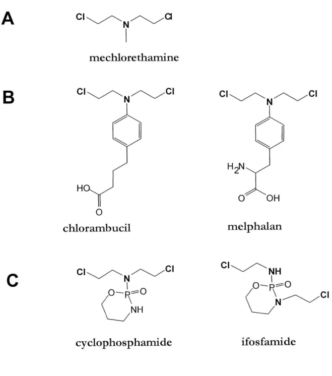
Among the DNA-damaging chemotherapeutics, nitrogen mustards constitute an important class. All nitrogen mustards are structurally derived from the parent compound,
mechlorethamine (HN2, Figure 1.2), an agent initially designed for chemical warfare. The presence of the electron pair on the nitrogen greatly increases the reactivity of the chloroethyl arms; an elimination-addition mechanism with the formation of an aziridine cation
intermediary is believed to be the mechanistic explanation for the remarkable reactivity of
HN2 (Colvin et al., 1976). The chemotherapeutic use of HN2 was suggested in 1943 when it
was observed that an accidental exposure to HN2 causes significant loss of white blood cells (Rappeneau et al., 2000). Although still in use today for certain aggressive leukemias, HN2 is an extremely toxic agent, causing massive non-specific damage to healthy tissues. Being a potent mutagen, HN2 can also induce secondary cancers (Povirk and Shuker, 1994).
A large number of nitrogen mustard derivatives have been synthesized in the last 30 years.
The chemical modifications were aimed at tempering the high reactivity of HN2 and at introducing specificity. The nitrogen mustards commonly used in clinical treatments maintain the two chloroethyl arms, but lower the nitrogen electron density by linking it to either a phenyl ring or to a more electropositive phosphorus (Figure 1.2). Although the reactive portion of each molecule (the warhead) is the same, the substituents on the mustard determine the reactivity, solubility and other biochemical properties, which then influence the toxicity and specificity of the agent.
Most cells are able to repair efficiently simple DNA alkylation damage; this is why nitrogen mustards are designed to be bifunctional, with one molecule of the drug alkylating the DNA
correctly (McHugh et al., 2001). Most mechanisms of repair involve multiple pathways such as nucleotide excision repair, homologous recombination and translesion repair. Given the complex mechanism required, the repair of crosslinks is likely to generate mutations and chromosomal aberrations (Dronkert and Kanaar, 2001). It is believed that the clinical efficacy of a nitrogen mustard is closely related to its ability to form DNA crosslinks.
Nevertheless, most normal cells with intact repair pathways can eventually repair crosslinks and thus tolerate crosslinking agents at reasonably low doses (Nojima et al., 2005). This observation suggests that nitrogen mustards would be most effective against cancer cells that lack (or have lost due to mutations) certain DNA repair abilities. It is believed that particular cancer cells exhibiting mutator phenotypes, have lost their ability to repair DNA faithfully and, hence, they are susceptible to toxicity induced by DNA-damage. Indeed, cells with impaired repair capabilities are orders of magnitude more sensitive to crosslinking agents (McHugh et al., 2001; Nojima et al., 2005; Trimmer and Essigmann, 1999).
Certain cancers develop resistance to DNA alkylating agents, either by upregulating efflux pumps that lower the effective intracellular drug concentration (Sharom, 2008), by
upregulating enzymes that can directly inactivate the reactive portion of the molecules (e.g., glutathione S-transferases) (O'Connor, 2007), or by a combination of the two mechanisms (Meijerman et al., 2008). Some malignancies, such as lung and prostate cancers, are
inherently resistant to alkylating agents because of their relatively slow growth and resistance to apoptosis. The long doubling times of these cancer cells allows more time for DNA repair, while the resistance to apoptosis prevents cell death in the wake of extended DNA damage. Hence, the effectiveness of DNA damaging agents in slow growing lung and prostate cancers is very limited.
The ability to form crosslinks is only part of the asset pool of an effective DNA alkylating drug. In the case of cisplatin (cis-diamminedichloroplatinum(II), (Figure 1.3 A), one of the most successful DNA-alkylating chemotherapeutics, the mechanism of toxicity has been
1.4.1 The inspiration - the mechanism of cisplatin in testicular cancer
Cisplatin and its analog carboplatin (Figure 1.3 A) are chemotherapeutic agents most
effective in the treatment of metastatic testicular cancer. These drugs are also used in various combinations to treat ovarian, head, neck, cervical, colon, lung, bladder and other cancers (Zamble and Lippard, 1995). In fact, the efficacy of cisplatin against testicular cancer is unprecedented; regimens including cisplatin achieve a cure rate of over 95% (Masters and K6berle, 2003; Ries et al., 2009), making cisplatin one of the most successful stories in chemotherapy.
It has been established that the cytotoxic properties of cisplatin are due to its ability to modify DNA covalently (Kartalou et al., 2001). Inside the cells, the two chloride ions are replaced with water molecules yielding a cationic aquated complex that is even more reactive towards DNA than the parental dichloro species, preferentially forming adducts at the N7 positions of guanine and N3 position of adenine (Bancroft et al., 1990). In vitro studies have shown that most of the adducts formed by cisplatin are in fact crosslinks: 65 % are
1,2-d(GpG) intrastrand crosslinks, 25% are 1,2-d(ApG) intrastrand crosslinks and 5-10% are 1 ,3-d(GpG) intrastrand crosslinks. Only a minor fraction of adducts are interstrand crosslinks
or monoadducts (Kartalou et al., 2001). A similar adduct profile is also observed in vivo, in cells isolated from cancer patients treated with cisplatin (Fichtinger-Schepman et al., 1987).
The ability of cisplatin to form a variety of DNA crosslinks is insufficient to explain its potent antitumor activity. The trans isomer (transplatin, trans-diamminedichloroplatinum(II)) (Figure 1.3 B) also forms a large number of DNA crosslinks, yet it is therapeutically
ineffective. It was shown that due to its geometry, the trans isomer forms a different pattern of crosslinks, mostly 1,3-d(GpC) intrastrand crosslinks and, unlike cisplatin, a great number of G-C interstrand crosslinks (Leng and Brabec, 1994). This result suggested that the cisplatin adducts were more than just crosslinks, but also substrates for some ulterior biochemical interactions that, potentially, interfered with the repair process. Indeed, the major adducts of cisplatin, 1,2-d(GpG) and 1,2-d(ApG), force the DNA into unusual
found in various non-histone components of chromatin. The HMG-domain proteins bind tightly to the DNA structures generated by the 1,2 crosslinks of cisplatin, but do not bind to the DNA structures generated by the 1,3 crosslinks, suggesting that these proteins might play a role in the mechanism of cisplatin toxicity. This observation could also explain why
transplatin does not display the same effectiveness (Pil and Lippard, 1992). The presence of HMG-domain proteins is thought to prevent the repair proteins from accessing the DNA crosslink, hence significantly extending the lifetime of the DNA adduct and its cytotoxic effect (Zamble et al., 1995). This mechanism has been labeled "repair shielding" (Figure 1.4
A).
Some of the HMG proteins have been shown to bind extremely tightly to the cisplatin
adducts. An important example is the human upstream binding factor (hUBF), a transcription factor involved in regulating transcription of ribosomal RNA (rRNA), which binds the 1,2 adducts of cisplatin with an affinity (Kd = 60 pM) very close to the affinity for its natural
response element in the rRNA promoter (Kd = 18 pM). This high affinity suggests that
increasing levels of cisplatin adducts will have a significant impact on the availability of
hUBF, effectively titrating hUBF away from its natural binding site (Treiber et al., 1994).
Disrupting the natural function of transcription factors such as hUBF may also be a mechanistic explanation for the toxicity of cisplatin. This mechanism has been labeled "transcription factor hijacking" (Figure 1.4 B).
A significant body of experimental work has demonstrated that both repair shielding and
transcription factor hijacking are possible players in the high effectiveness of cisplatin against testicular cancer cells. In vitro assays showed that the repair of cisplatin adducts is significantly inhibited in the presence of HMG domain proteins, such as HMG 1, tsHMG and
SRY (Huang et al., 1994; Zamble et al., 2002; Trimmer et al., 1998). Notably, the tsHMG and SRY proteins are commonly overexpressed in testicular cancers. Additionally, sensitivity
to cisplatin can be modulated in vivo by the levels of HMG-domain proteins. It was shown that mammalian cells overexpressing HMG 1 proteins are more sensitive to cisplatin, whereas cells knocked-out for IXR1, an HMG-domain gene, are 2-5 fold more resistant to cisplatin (McA'Nulty et al., 1996). Furthermore, the ability of cisplatin adducts to compete with hUBF has been shown in rRNA transcription assays, both in vitro (Zhai et al., 1998) and in vivo (Jordan and Carmo-Fonseca, 1998). Therefore, even though the complete mechanism of cisplatin in testicular cancer is thought to be even more complicated, involving the interplay of other cellular pathways (Cepeda et al., 2007), the repair shielding and transcription factor hijacking mechanisms are likely to be important contributors.
1.4.2 The vision
-
engineering programmable cytotoxins
Inspired by the mechanisms of cisplatin highlighted above, in the late 1990s, the Essigmann lab envisioned a design strategy for programmable cytotoxins: bifunctional agents comprised of a constant DNA alkylating moiety (a warhead) and a variable protein interacting moiety (ligand) that can be tailored to recognize cancer specific proteins (Essigmann et al., 2001). These compounds would alkylate DNA non-specifically, and in normal cells, these adducts would be readily repaired. However, in cancer cells expressing significant amounts of the target protein, the repair of the adducts would be impeded by the binding of the cancer specific protein to the molecule adducted to DNA. Furthermore, just like the cisplatin
adducts, the DNA adducts of the proposed compounds would compete with the natural ligand for the target protein, diverting it from its normal cellular function and leading to additional cytotoxicity.
The choice for the DNA alkylating moiety was an aniline mustard, derived from
chlorambucil. The reasoning behind this choice was the moderate toxicity of chlorambucil, the ease of incorporation into a synthetic scheme and bioavailability considerations (Rink et al., 1996). Another warhead derived from cisplatin was also considered; however,
incorporation of a platinum cation into a large organic molecule might impede the molecule's ability to cross the cellular membrane. Nevertheless, a compound with a cisplatin derived
For a protein interacting moiety, a ligand for the estrogen receptor (ER) was initially chosen. It has been known for decades that a significant number of breast and ovarian cancers
express high levels of the ER (Fern6 et al., 1990). Being a transcription factor, the ER localizes in the nucleus, where it would have increased chances of interacting with DNA adducts formed by the compound and would shield them from being readily repaired.
Additionally, given that each adduct represents a potential decoy binding site for the ER, the normal function of the steroid receptor will also be affected. Since receptor antagonism is a well established chemotherapeutic modality, an agent displaying an ER ligand could also squelch ER driven transcription in ER positive tumor cells, as well as block repair (Rink et al., 1996; Essigmann et al., 2001).
1.4.3 The proof of principle - the phenyl-indole compounds
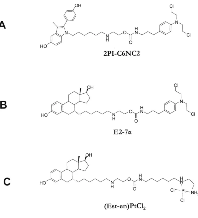
The first compound synthesized featured the 2-(4'-hydroxyphenyl)-3-methyl-5-hydroxy-indole (2PI) moiety as a ligand for ER and the chlorambucil-derived 4-(3-aminopropyl)-N,N-(2-chloroethyl)-aniline warhead, linked together by a 15 atom long alkyl chain functionalized with a secondary amine and a carbamate (Figure 1.5 A)(Rink et al., 1996). For control
purposes, another compound was synthesized using the 2-(4'-hydroxyphenyl)-3-methyl-indole moiety as the ligand. The subtle difference between the two ligands (one hydroxyl group) was introduced to drastically lower the affinity for the ER in the control compound. The lead compound was called 2PI-C6NC2 and the control compound was 2PI(OH)-C6NC2.
The linker design emerged from careful experimentation that balanced three distinct
requirements: 1) the linker should allow binding to the ER; 2) the linker should not introduce unnecessary hydrophobicity in the molecule, while also being synthetically accessible; 3) the linker should be stable when exposed to intracellular enzymes. The first requirement dictated the length of the hydrophobic chain attached to the ligand, while the third requirement dictated the use of the carbamate and the amine functional groups. The secondary amine also
negatively charged DNA backbone. Systematic variation of the length of each segment of the linker yielded the C6NC2 structure as optimal for ER binding (Figure 1.6).
The 2PI-C6NC2 compound was shown to have a significant binding affinity towards the ER
(7.1 relative binding affinity (RBA) compared to ER's natural ligand f-estradiol).
Additionally, 2PI-C6NC2 could alklyate DNA in vitro generating piperidine-labile N7 guanine adducts. Furthermore, a DNA oligonucleotide modified with 2PI-C6NC2 retained a modest affinity towards the ER (RBA = 0.5) (Rink et al., 1996). The in vivo results proved very encouraging. The toxicity of 2PI-C6NC2 was determined in two breast cancer cell lines:
MCF-7 which is ER positive, and MDA-MB-23 1, which is ER negative. These cell lines
allowed us to test whether the presence of ER contributed to differential toxicity. Indeed, the
MCF-7 cells were more sensitive to 2PI-C6NC2 than MDA-MB-23 1; as expected, the cell
lines were equally sensitive to agents that do not interact with the ER, such as chlorambucil, and the control compound 2PI(OH)-C6NC2, which has an ER affinity 70-fold less than the lead compound (Rink et al., 1996). This result strongly implicated the ER as a modulating factor in the toxicity of 2PI-C6NC2.
1.4.4 E2-7a - optimizing a compound targeted towards ER
Encouraged by the proof of principle highlighted by the 2PI-C6NC2 compound, a second generation of ER interacting compounds was designed, in which attempts were made to improve each aspect of the molecule: the ER affinity, the linker properties and the warhead.
To optimize the binding towards ER, a new compound was designed using
P-estradiol
as the ligand portion, while keeping the rest of the molecule identical to the first generation2PI-C6NC2 compound. The linker was attached at the 7a position of the steroid ring, in
accordance with the published studies of
P-estradiol
derivatives that maintain a high affinity for ER when substituted at the 7a position (Bowler et al., 1989; DaSilva and van Lier, 1990). Due to the new ligand, identical to the natural ER ligand, the new compound (named E2-7a,et al., 1996). It was also shown that E2-7a alkylates a DNA oligo in vitro, forming a
significant number of piperidine-labile adducts. These adducts could also bind the ER ligand binding domain (LBD), as demonstrated by a gel-shift assay;
p-estradiol
could compete away the shifts. Using the same two cell lines as before, it was shown that E2-7a toxicity isnoticeably enhanced by the presence of the ER, the ER positive MCF-7 cells being
significantly more sensitive than the ER negative MDA-MD-231 cells (Mitra et al., 2002).
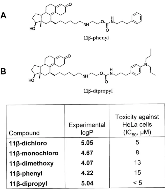
To optimize the linker properties, a series of compounds were synthesized that explored the type and number of functional groups present in the linker, while keeping constant the six carbon alkyl chain connecting to the estradiol moiety and the chlorambucil derived warhead (Sharma et al., 2004). The compounds made featured the following functional groups in the linker: one secondary amine group, two secondary amine groups, one amide group, two amide groups, one guanidine, one carbamate and one secondary amine and one guanidine Figure 1.7). For comparison, E2-7a compound, also included in Figure 1.7, features one secondary amine and one carbamate in its linker. The lengths of the linkers counted from the estradiol ring to the aniline ring varied between 11 and 16 atoms total. All of these
compounds were tested for their affinity to the ER, DNA reactivity and cytotoxicity in cell lines. Far from being intuitive, the results highlighted the subtle interplay between all parts of the molecule and the crucial importance of the linker. The presence of one or two secondary amine groups in the linker positively correlated with the compound's solubility, ability to react with DNA and toxicity (Sharma et al., 2004). While all compounds had good affinities for the ER (RBA between 9 and 46), the linker flexibility seemed to influence the ER binding the most. The more rigid linkers (such as those with the single amide, the carbamate alone and the guanidine alone) are likely limiting the number of conformations available for the linker, which limits the interactions of the molecule with the surface of the ER and thus might negatively affect binding (Sharma et al., 2004). The original compound (E2-7a) was found to have the best ER affinity (RBA = 45, in this assay), but the monoamine linker
compound was almost as good (RBA = 44). In terms of toxicity, only E2-7a, the monoamine and diamine linker compounds showed significantly higher toxicity than chlorambucil. These
the amine compounds. The investigation concluded that the parent compound had a linker that allowed the best compromise between ER affinity, DNA reactivity, and selective toxicity (Sharma et al., 2004).
Finally, a different warhead was explored in combination with the estradiol ligand and the linker featuring an amine and a carbamate (Kim et al., 2009). The new warhead was derived from cis-dichloroethylenediammineplatinum(II), by substitution on the carbon chain of the ethylenediammine (Figure 1.5 C). The resulting compound, [6-(2-amino-ethylamino)-hexyl]-carbamic acid 2-[6-(7a-estra-1,3,5,(10)-triene)-hexylamino]-ethyl ester platinum(II)
dichloride ((Est-en)PtCl2) (Figure 1.5 C) had all the intended properties: it had good affinity
for the ER, reacted with DNA, and was selectively toxic against ER positive cell lines. The ER affinity was measured to have an RBA of 28, which is somewhat less than the ER affinity of E2-7a. In terms of toxicity, the platinum compound was less toxic when compared with E2-7a; however, it had good selective toxicity for the ER positive cell lines, including the ER positive CAOV3 ovarian cancer cell line (Kim et al., 2009).
Taken together, all the optimization experiments confirmed E2-7a as the lead candidate in the class of ER targeted compounds, and highlighted the great flexibility of the
programmable cytotoxins design approach. The ER dependent mechanism of E2-7a is currently being investigated in several breast and ovarian cancer cell lines, in an effort to advance the compound in preclinical and eventually clinical trials (Gopal, 2009).
1.5.1 Physico-chemical properties
Given the success of the ER targeted compounds, a new class of compounds was proposed that would take advantage of another nuclear steroid receptor that is overexpressed in certain cancers: the androgen receptor (AR). The AR was shown to be highly expressed in many prostate cancers (Marcelli and Cunningham, 1999). Given the high incidence of prostate cancer - highest incidence and second highest rate of mortality in men (Jemal et al., 2008) (Figure 1.1) - the design of an effective agent targeting prostate cancer constitutes an important scientific challenge. The programmable cytotoxin paradigm seemed like a promising way to address this challenge.
The choice for an AR ligand was 17p-hydroxy-estraA4(5), 9(10)-3-one, a moiety inspired from the synthetic steroid RU-486, which possesses a significant affinity for the AR (Fuhrmann et al., 2000). Studies on RU-486 and related compounds indicated that the 11p position on the sterol ring system would be the least disruptive to AR binding, and thus, it was chosen as the place of attachment for the linker. Given that the ligand binding domain of AR has a high similarity to the ER ligand binding domain, the same linker found in the
E2-7a molecule was chosen, hoping that the properties of the C6NC2 linker that allowed ER
binding would also favor the AR binding. The complete compound (called 11 -dichloro , Figure 1.8 A) closely resembled E2-7a, except for the different steroid moiety and the attachment point on the steroid (Marquis et al., 2005). Furthermore, two related compounds were synthesized to test the contribution of DNA damage to the overall mechanism. One of the compounds, called 11
5-dimethoxy
(Figure 1.8 B) was identical to 11p -dichloro, except the chlorines on the chloroethyl arms of the mustard have been replaced by methoxy groups. Given that the methoxy group is a very poor leaving group, the reactivity of the mustard is1 IUPAC name: 2-(6-((8S, 11S,13S,14S,17S)- 1 7-hydroxy-
13-methyl-3-oxo-2,3,6,7,8,11,12,13,14,15,16,17 dodecahydro-1H-cyclopenta[a]phenanthren-1
replaced with a methoxy group. 11
p-monochloro
can still form DNA mono-adducts, yet it is unable to form DNA crosslinks. A comparison between 11 -dichloro and 11p -monochloro could indicate the importance of crosslinks in the mechanism of toxicity.The affinity for the AR was benchmarked against R1881, a synthetic androgen with one of the highest affinities for AR of all known ligands. Both 11p -dichloro and 11p -dimethoxy show good affinity for the AR (RBA = 11.3 and 18.3, respectively) when compared with the
RI 881 (Marquis et al., 2005). However, when compared with dihydrotestosterone (DHT), the best natural AR ligand, the RBA values would be 2- to 3-fold higher, because the synthetic androgen RI 881 has 2- to 3- times higher affinity for the AR than DHT (Zhao et al., 1999).
In vitro, 11p -dichloro displayed a good reactivity towards DNA oligonucleotides, generating
mostly piperidine-labile N7 guanine adducts, N3 adenine adducts and guanine-guanine crosslinks. Additionally, the adducts maintained a measurable, albeit lower affinity towards AR (RBA = 0.22 compared to R188 1). As expected, under the same conditions, 11p-dimethoxy was not able to modify DNA (Marquis et al., 2005).
1.5.2 Toxicological properties
The most impressive feature of 11p -dichloro is its potent toxicity against cancer cells in cell culture. The first experiments to test this property employed the AR positive prostate cancer LNCaP cell line. Treatment with 11p -dichloro at concentrations of 5 pIM and higher induced apoptosis in LNCaP cells, as suggested by the morphological changes (cells rounding up and detaching from the plate) and confirmed by apoptosis markers such as Annexin V staining, poly-ADP ribose polymerase (PARP) cleavage and DNA fragmentation (Marquis et al.,
2005). Interestingly, the cell cycle distribution of the LNCaP cells treated with 11p -dichloro
did not change, in spite of significant cell death suggested by the subGl population, indicating that 11p -dichloro toxicity may be cell cycle independent. However, the control compound, 11 -dimethoxy, showed very little toxicity and did not show any of the apoptosis
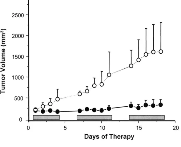
In vivo studies with 11 -dichloro produced even more exciting results. Nude mice bearing
LNCaP xenograft tumors were treated with 11p -dichloro or with vehicle alone, using a daily dose of 30 mg/kg in a 5 days/week, 7 week long regimen. By the last day, the treatment with 11p -dichloro showed an impressive 90% reduction in tumor growth compared with vehicle treated controls (Figure 1.9). More significantly, the overall toxicity to the treated mice was minimal, the treated animals having experienced a mean weight loss of only 9.7% (Marquis et al., 2005).
Encouraged by the promising effects of 11p -dichloro on LNCaP xenografts, the
biodistribution of the compound and the nature and quantity of DNA adducts formed was investigated in mice (Hillier et al., 2006). For these studies, a 14C labeled version of the molecule was prepared; small levels of radioactivity were then detected using the highly sensitive technique accelerator mass spectrometry (AMS). It was found that 11p -dichloro has a good biodistribution in the mouse and that it reaches the xenograft tumor in quantities sufficient to cause a significant number of DNA adducts. Interestingly, a much greater number of DNA adducts was detected in liver cells, although, at the therapeutically effective dose, none of the four commonly used hepatotoxicity markers were affected (Hillier et al.,
2006). This finding suggests that the level of DNA damage may not always correlate with
toxicity; other factors such as cell specific proteins and metabolism may play important roles. The result was consistent with the intended design mechanism, which predicted that 11 -dichloro adducts would be significantly more toxic in the AR positive LNCaP tumors, because of a repair shielding-like mechanism.
Unfortunately, recent experiments have seriously challenged the possibility of AR
modulating 11 -dichloro toxicity, at least in certain in vitro models. Initial data suggested the involvement of the AR consistent with a repair shielding mechanism, because the AR