Analysis of the Structural Changes Caused by Positive DNA
Supercoiling
by-Marita Christine Barth
B.S. Bioresource Research Oregon State University, 1998Submitted to the Division of Biological Engineering in partial fulfillment of the requirements for the degree of
Doctor of Philosophy in Macromolecular Biochemistry and Biophysics at the
MASSACHUSETTS INSTITUTE OF TECHNOLOGY February 2007
C 2007 Massachusetts Institute of Technology. All rights reserved.
Signature of Author:
Division of Biological Engineering January 12, 2007
Certified by:
Accepted by:
rMASSACK·KIEMEW IJf
OF TECHNOLOGYnNte
AUG 0 2 2007I
LIBRARIES
(I
Peter C. Dedon Professor of Toxicology and Biological Engineering Thesis Supervisor SLr/ % L". .,:
/1
Alan
Grodzinsky
Irof's r of Biological Engineering Chairman, Co ittee for Graduate StudentsThis doctoral thesis has been examined by a committee of the Division of Biological Engineering as follows:
Associate Professor Bevin P. Engelward
Chairman
Professor Peter C. Dedon
Supervisor
Professor John M. Essigmann
fl,(\
---Analysis of the Structural Changes Caused by Positive DNA
Supercoiling
by
Marita Christine Barth
Submitted to the Division of Biological Engineering on January 12, 2007 in partial fulfillment of the requirements for the degree of Doctor of Philosophy in Macromolecular
Biochemistry and Biophysics
ABSTRACT
The procession of helix-tracking enzymes along a DNA molecule results in the formation of supercoils in the DNA, with positive supercoiling (overwinding) generated ahead of the enzyme, and negative supercoiling (underwinding) in its wake. While the structural and physiological consequences of negative supercoiling have been well studied, technical challenges have prevented extensive examination of positively supercoiled DNA. Studies suggest that at sufficiently high levels of overwinding, DNA relieves strain by adopting an elongated structure, where the bases are positioned
extrahelically and the backbones occupy the center of the helix. This transition has only been identified, however, at a degree of supercoiling substantially higher than is
generated physiologically.
To examine the structural changes resulting from physiological levels of positive DNA supercoiling, I have developed a method for preparing highly purified positively supercoiled plasmid substrates. Based on a method previously developed in this laboratory, this allows for preparation of large quantities of very pure, highly positively supercoiled plasmid. It also expands on earlier methods by exploiting ionic strength to modulate the direction of supercoiling introduced, allowing preparation of either positively or negatively supercoiled substrates.
A combination of approaches has been used to elucidate changes to DNA
structure that result from physiological levels of positive supercoiling. Enzymatic probes for regions of single-stranded character are not reactive with positively supercoiled plasmid, indicating that stably unpaired regions are not present. Additionally, the effect of supercoiling on the activity of restriction enzymes has been examined. With the enzymes tested, no substantial differences in cleavage rates were observed with either positively or negatively supercoiled substrates. To examine structural changes at a wider range of superhelical densities, design and preparation was undertaken on 2-aminopurine-containing DNA substrates for use in fluorescence studies with a magnetic
micromanipulator. Technical limitations rendered these experiments infeasible with current instrumentation, but important insights were gained for future fluorescence-based micromanipulation experiments.
A destabilizing effect on the base pairs, however, can be seen using Raman difference spectroscopy, suggesting a subtle shift toward the more extreme extrahelical state. The Raman data suggest that structural adjustments due to positive supercoiling are small but significant, and in addition to the base-pairing effects, alterations are observed in phosphodiester torsion and the minor groove environment, as well as a slight shift in sugar pucker conformation to accommodate lengthening of the DNA backbone.
These results point to subtle changes in DNA structure caused by biologically relevant levels of positive superhelical tension and positive supercoiling. All of the changes are consistent with the mechanical effects of helical overwinding and suggest a model in which base pair destabilization in overwound DNA could affect the search mechanisms used by DNA repair enzymes and the binding of other proteins to DNA.
Thesis Supervisor: Peter C. Dedon
Acknowledgements
I would like to start by thanking my family, particularly my parents, Merritt and Jenny, my grandmother Mary, my brother Stephen and my aunt Marita Jo Broadus for their
support. They never failed to offer encouragement when I needed it, and I couldn't have done this without them.
The research experience I gained as an undergraduate was vital to my success in graduate school, and for that I wish to thank my undergraduate thesis advisor, Professor George S. Bailey, as well as Kate Mathews and Dr. Ulrich Harttig from his laboratory. They were
all amazingly kind and encouraging, and I appreciate their willingness to work with and trust an undergraduate in a research environment.
I have gained a lot from my interactions with all current and former members of the Dedon Lab, and I would like to extend my thanks to all of them. I would particularly like to thank Debra Dederich, who has been invaluable in preparation of substrates for this research, and whose dedication and hard work in the face of many technical challenges is tremendously appreciated. I would also like to thank Dr. C. Eric Elmquist, who
performed the MS analyses of chloroacetaldehyde-treated plasmid samples, and Dr. Michael DeMott who assisted greatly with the editing of this thesis. Dr. Koli Taghizadeh, Yelena Margolin, Dr. Min Dong, and Dr. Christiane Collins all gave important input into my research, and have been a pleasure to work with as well. The support staff for the Dedon Lab has been absolutely amazing, and I would like to thank them all. Marcia Weir, Kristine Marzilli and Dawn Erickson have all done much to make my life easier, and have my gratitude. Olga Parkin is in a class all her own, with an uncanny knack for solving any problem that might come up. Her assistance in all matters administrative, as well as her friendship, have meant a great deal to me.
I would also like to thank several of the grad students and post-docs whose friendship I have enjoyed through the years: Dr. Maxine Jonas, David Appleyard, Vasileios
Dendroulakis, Dr. Can Ozbal, Dr. Teresa Wright, Dr. Joe Newman, Dr. Maryann Timins, Dr. Elaine Chin, Dr. Janice Lansita, Dr. Jane Sohn, and J.P. Cosgrove.
This thesis project has involved multiple collaborations, which have significantly
enhanced both my education and the quality of the information obtained. I would like to thank Professor George Thomas at the University of Missouri at Kansas City for opening up his lab to me for the Raman spectroscopic studies of supercoiling. Within the Thomas lab, I wish to thank Professor James Benevides for sharing his expertise with me, and allowing me so much instrument time on my visits to Kansas City. Additionally, he and his family were wonderful hosts, and truly made me feel welcome during my stays there. For their collaboration on the 2-aminopurine studies, I wish to thank Professor Peter So and Dr. Serge Pelet for their input and experimental assistance.
I would like to thank my thesis committee, Professor Bevin Engelward, Professor John Essigmann and Dr. Richard Roberts for all of their support and input. They've been incredibly encouraging, and have been a wonderful source of challenging questions and intriguing ideas.
For help in more ways than I can list, I would like to thank Gavin McNett. His keen editorial eye and assistance with graphics have dramatically improved the quality of this thesis. Beyond that, his kindness, good nature, wit, phenomenal cooking skill, and (above all) patience have been vital to seeing me through the thesis writing process. I find it difficult to adequately express my gratitude and respect.
Finally, I would like to thank my thesis advisor, Professor Peter Dedon, for all his support and encouragement through the years. He has been incredibly generous and supportive, and I truly appreciate him sticking with me through what proved to be a very challenging and difficult project. His openness to new ideas and the freedom he has given me to explore different approaches to the problems I have faced have greatly enriched my education. I am truly grateful.
Biographical Note
Marita Christine Barth was born in 1975 in Dallas, Oregon, and graduated from Dallas High School in 1993. After a summer internship at the Laboratory Services Division of the Oregon Department of Agriculture, she enrolled at Oregon State University in
Corvallis, OR. Following a junior year abroad at Lincoln University in Canterbury, New Zealand, she returned to complete a thesis project, entitled "In vitro Mechanisms of Chlorophyllin Anticarcinogenesis Against Dibenzo[a, l]pyrene," in the laboratory of Professor George S. Bailey. She was awarded a Bachelor of Science in Bioresource Research with an option in Toxicology and a minor in Chemistry, in 1998. She subsequently enrolled at MIT, and during the course of her graduate studies has been awarded a National Defense Science and Engineering Graduate Fellowship, and a Poitras Pre-Doctoral Fellowship.
Table of Contents
A bstract ... ... 5 Acknowledgements ... 7 Biographical Note ... 9 List of Abbreviations ... 15 List of Figures ... 17 Chapter 1 - Introduction Packaging of DNA in Eukaryotic Cells ... 19Topological Domains and DNA Supercoiling ... 20
The Twin-Domain Model of DNA Supercoiling ... 21
Mathematical Descriptions of Supercoiling ... 23
Energetics of Supercoiling ... 26
Topoisomerases ... ... 27
Physiological Roles for Negative Supercoiling ... 30
Physiological Effects of Positive Supercoiling ... 34
Structural changes in negatively supercoiled DNA ... 35
Structural Changes in Positively Supercoiled DNA ... 37
Figures ... 41
Literature Citations ... 47
Chapter 2 - Preparation of Positively and Negatively Supercoiled Substrates Introduction ... 55
Materials and Methods ... 56
Results and Discussion ... 60
Conclusions ... 63
Figures ... ... 65
Literature Citations ... ... 68
Chapter 3 - Reactivity of Supercoiled DNA with Chemical and Enzymatic Probes of DNA Structure Introduction ... 71
Materials and Methods ... 73
Results ... 76
D iscussion ... 81
Figures ... 84
Literature Citations ... ... 89
Chapter 4 - Raman Spectroscopic Studies of Positive Supercoiling Introduction ... 93
Materials and Methods ... 95
Results ... 96
D iscussion ... 100
C onclusions ... 105
Figures ... 106
Literature Citations ... 112
Chapter 5 - Design and Optimization of Fluorescent DNA Substrates for Micromanipulator Studies Introduction ... 115
Materials and Methods ... 118
Results ... 120
D iscussion ... ... 125
Literature Citations ... 135
Chapter 6 - Effects of Supercoiling on Cleavage by Type II Restriction Enzymes Introduction ... 139
Materials and Methods ... 142
R esults ... 143 D iscussion ... 146 Figures ... 149 Literature Citations ... 153 Chapter 7 - Conclusions C onclusions ... 157 Literature Citations ... 164
Abbreviations
2AP 6MI A BCIP bp C CBE dA dAPTP dC ALk dG DIG DMS dNTP dT DTT EDTA EA EC G HPLC IPTG Lk Lko NBT NMR PCR SDS-PAGE Ua T TBE TIR Tris Tw Wr 2-Aminopurine 6-Methylisoxanthopterin Adenine 5-Bromo-4-chloro-3-indolyl phosphate Base pair(s) CytosineChicken blood extract 2'-Deoxyadenosine
2-Amino-2'-deoxyadenosine-5'-triphosphate 2'-Deoxycytidine
Linking number difference 2'-Deoxyguanosine Digoxygenin Dimethyl sulfate 2'-Deoxynucleotide triphosphate 2'-Deoxythymidine Dithiothreitol Ethylenediaminetetraacetic acid 1,N6-Ethenoadenine 3,N4-Ethenocytosine Guanine
High pressure liquid chromatography Isopropyl P-D-1 -thiogalactopyranoside Linking number
Linking number of a completely relaxed molecule p-Nitrotetrazolium blue
Nuclear magnetic resonance Polymerase chain reaction
Sodium dodecyl sulfate-polyacrylamide gel electrophoresis Superhelical density
Thymine
Tris-borate-EDTA Total internal reflection
2-Amino-2-(hydroxymethyl)- 1,3-propanediol Twist
List of Figures
Figure Figure Figure Figure Figure 1.1: 1.2: 1.3: 1.4: 1.5: Figure 1.6: Figure 2.1: Figure 2.2: Figure 2.3: Figure 3.1: Figure 3.2: Figure 3.3: Figure Figure Figure 3.4: 3.5: 4.1: Figure 4.2: Figure 4.3: Figure 4.4: Figure 4.5:Packaging of eukaryotic DNA into chromatin The twin-domain model of DNA supercoiling Twist and writhe parameters of DNA supercoiling Toroidal and plectonemic DNA writhe
Formation of a "chicken foot" structure at a stalled replication fork promoted by accumulated positive supercoiling
Two general types of cruciform extrusion at inverted repeat sequences of DNA
Reaction scheme for preparation of supercoiled plasmid substrates Optimization and analysis of prepared supercoiled substrates
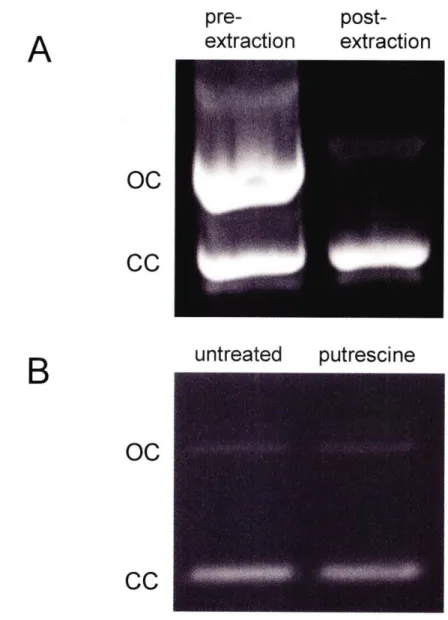
Purification of closed-circular plasmid preparation by extraction with acidic phenol
Kinetics of reaction of S1 nuclease with supercoiled and relaxed substrates Reaction of S I nuclease with supercoiled and relaxed substrates
Kinetics of reaction of nuclease BAL-31 with supercoiled and relaxed substrates
Reaction of chloroacetaldehyde with DNA bases
Reaction of chloroacetaldehyde with supercoiled and relaxed substrates Raman spectra of negatively supercoiled and relaxed pUC 18 plasmid DNA at 752 nm excitation
Raman spectra of negatively supercoiled and relaxed pUC 18 plasmid DNA at 532 nm excitation
Raman spectra of positively supercoiled and relaxed pUC 18 plasmid DNA at 752 nm excitation
Raman spectra of positively supercoiled and relaxed pUC 18 plasmid DNA at 532 nm excitation
Raman spectra of negatively supercoiled and relaxed pUC 18 plasmid DNA in D20
Figure 4.6: Figure 5.1: Figure 5.2: Figure 5.3: Figure 5.4: Figure 5.5: Figure Figure Figure Figure 6.1: 6.2: 6.3: 6.4:
Raman spectra of positively supercoiled and relaxed pUC 18 plasmid DNA in D20
Hydrogen bonding schemes of the canonical Watson-Crick adenine-thymine base pair, the fluorescent base analog 2-aminopurine paired to thymine, and a proposed protonated form of 2AP that could pair with cytosine
Preparation of components of micromanipulator substrate Assembly scheme for micromanipulator substrate
Fluorescence lifetime of free and incorporated 2-aminopurine Comparison of original design of micromanipulator substrate with optimized design
Possible reaction modes of closed circular DNA with restriction enzymes Kinetics of reaction of NarI with supercoiled and relaxed substrates Kinetics of reaction of Ehel with supercoiled and relaxed substrates Kinetics of reaction of EcoRI with supercoiled and relaxed substrates
Chapter
1
- Introduction
Packaging of DNA in Eukaryotic Cells
The extensive length of genomes makes it necessary for both prokaryotic and eukaryotic cells to compact their DNA. The human genome, for example, consists of -3xl09 bp, and codes for an estimated 20,000-25,000 genes (1). For a single human diploid cell, this amount of DNA laid end-to-end would result in a length of more than two meters. Since the nucleus that must contain this DNA is a mere -10-5 m in diameter, a significant degree of compaction is necessary.
While complete compaction of the genome might be ideal for storage, as well as for cellular events like chromosomal segregation, other cellular processes periodically require at least some of the DNA to be unpackaged. RNA polymerases need to process along the DNA to function, so any compaction would need to be removed from
transcriptionally active genes before transcription ensues. This means that in certain stages of the cell cycle the DNA needs to be partially compacted and partially unpacked. To further complicate the situation, cells need the ability to rapidly up- and down-regulate genes in response to stimuli, so compaction must be both dynamic and tightly regulated. All of this must be accomplished in a fashion that minimizes tangling and knotting of the DNA molecules.
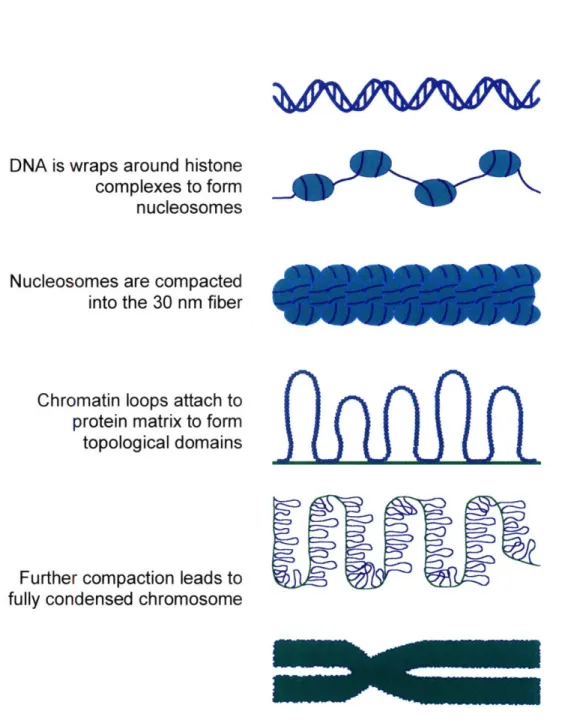
The eukaryotic solution to the problem is to store the DNA as chromatin, which utilizes protein-DNA and protein-protein interactions to package the DNA in increasing levels of compaction (reviewed in Ref. 2). First, the naked DNA is wrapped into
positively charged histone proteins in a left-handed toroid. The nucleosomes are separated from one another by 50-100 bp segments of linker DNA, giving extended nucleosomal arrays the appearance of "beads on a string" when viewed in electron micrographs. This nucleosomal array, also known as the 10 nm fiber, is further
compacted into a secondary structure known as the 30 nm fiber, although debate persists as to whether this takes a solenoidal (3,4), or irregularly zig-zagging "slinky-like" form
(5,6). Further compaction of chromatin into a tertiary structure requires long-range interactions of the chromatin fiber, and it is affected by a number of different proteins in a process that is still not well understood (Fig. 1.1).
Topological Domains and DNA Supercoiling
One important consequence of the compaction into chromatin is the division of DNA molecules into topological domains (7). The DNA in chromatin is anchored to the nuclear matrix at intervals, in such a fashion that it cannot freely rotate. The DNA between any two of these points is known as a topological domain. In an unconstrained linear DNA molecule, any change in twist that is introduced will diffuse through the molecule and will gradually dissipate, as one strand of the DNA rotates relative to the other. When such free rotation is limited, as happens within a topological domain, any added twist will be retained by the DNA, since it cannot diffuse past the boundaries of the domain. Topological domains vary dramatically in size. In human cells, the most recent estimate ranges from <95,000 to >150,000 bp (8), although other estimates offer a broader distribution from 1,000 to 300,000 bp for eukaryotic genomes (9-11). If an
average of 100,000 bp/domain is assumed, this would suggest that a diploid human cell has -60,000 topological domains.
Supercoiling in DNA occurs when the DNA is either underwound (negative supercoiling) or overwound (positive supercoiling) relative to the B-form helix.
Constrained toroidal supercoiling, in the form of DNA wrapped around histone cores, is present in cells independent of any topological domains. This supercoiling is trapped by the binding of the proteins to the DNA, and as such cannot diffuse out. Because DNA wraps around histones in a left-handed fashion, the supercoiling that is constrained is negative.
The partitioning of cellular DNA into topological domains does allow the retention of unconstrained supercoiling. This type of supercoiling is free to diffuse through the molecule, within the limits set by the boundaries of the topological domain. This normally occurs when the strands of DNA are separated during various cellular processes. The earliest detailed description of this phenomenon is the twin-domain model of transcription proposed by Liu and Wang (12).
The Twin-Domain Model of DNA Supercoiling
In the twin-domain model, Liu and Wang account for the consequences of the requirement for the RNA polymerase and the DNA template to rotate relative to each other during the transcription process (12). In theory, either molecule could rotate, however evidence indicates that polymerases are anchored to the nuclear matrix (13,14). Therefore, for transcription to occur, the DNA must rotate relative to the polymerase. As this happens, negative supercoiling is generated behind the complex, while positive
supercoiling is generated ahead of it. (Fig. 1.2) This supercoiling can diffuse rotationally along the helix, but cannot diffuse past the limits of the topological domain.
According to this model, the degree of supercoiling present in a topological domain is determined by the orientation of transcribed genes, by how transcriptionally active these genes are, and by the activity of different topoisomerases within the cell. If two transcribed genes are oriented in the same direction, the supercoils will eventually cancel each other in the region between the genes, as the positive supercoils generated ahead of the one gene are met by the negative supercoils generated behind the other. If the genes are oriented in opposite directions, significant supercoiling of either sign can be generated between the two genes, as it will be accumulating from both directions.
Topoisomerases can affect the degree of supercoiling within a topological domain by selectively adding or removing supercoils. If a topoisomerase that is removing supercoiling of one sign is more active than the topoisomerase removing the other, an imbalance will develop in the net amount of supercoiling. Topoisomerases will be discussed in greater detail later in this chapter.
Evidence in support of the twin-domain model has been found in vitro (15,16), in prokaryotes (17,18), and in eukaryotes (19,20), illustrating its importance as a widespread biological phenomenon. The phenomenon also isn't limited to transcription. While the original model described transcriptionally-induced supercoiling, any protein that forces rotation of the DNA template can generate supercoiling in a similar fashion. This
includes helicases such as the E. coli nucleotide excision repair complex UvrAB (21) and the SV40 large tumor antigen (22), and translocating enzymes such as the type I
Mathematical Descriptions of Supercoiling
Before delving into the structural and physiological consequences of supercoiling, it is useful to consider the mathematical conventions used to describe it. In most
respects, a topological domain is analogous to a closed-circular DNA molecule, so several of the same concepts apply.
DNA has a double helical structure comprised of two interwound strands. The linking number of the DNA, Lk, is the number of times the strands are intertwined for a given molecule. For B-form DNA, the linking number of a completely relaxed molecule, Lko, can be obtained by dividing the number of base pairs, n, by the helical repeat. In physiological solution conditions, the helical repeat is -10.4 bp/turn (24), so:
Lko
=
n/10.5
After torsion has been introduced into the molecule, Lk will change by 1 for every complete turn of the helix added (or removed). The linking number difference, ALk, is equal to the new linking number minus the relaxed linking number:
ALk= Lk- Lko
In a closed-circular molecule, both strands of the DNA are unbroken, which means that Lk must be an integer value. From the definition of Lko, however, we can see that it is not necessarily an integer value. Thus, depending on the precise length of the DNA,
some closed-circular molecules of DNA cannot reach a completely relaxed state. It also means that ALk is not necessarily an integer value. Because changes in ALk for an intact molecule can only occur by breaking one or more strands of the DNA and then
It is also important to note that under consistent solution conditions, localized supercoiling can occur in an intact topological domain, but the net Lk for that domain cannot change without breakage of at least one strand of the DNA followed by re-ligation. This means that if one portion of an intact molecule is subjected to a localized positive linking number change, a compensatory negative supercoiling of equivalent Lk value must occur elsewhere in the domain.
Two closed circular molecules differing only in ALk are referred to as topoisomers. Populations of topoisomers can be resolved using agarose gel
electrophoresis, as an increase in I ALkJ will result in increased mobility of a plasmid on a gel (25).
Changes in the linking number describe differences between topoisomers of the same molecule, but give no information about the length of DNA that is accommodating the introduced torsion. A ALk of +1 would put considerably more strain on a 100 bp molecule than it would on a 1,000 bp molecule. To account for the length of a molecule when describing supercoiling, the superhelical density, o, is used:
a = ALLk/Lko
Superhelical density values allow for comparison of the degree of supercoiling among molecules of different sizes. DNA isolated from most cells is negatively supercoiled; in E. coli the superhelical density will generally vary from o Z -0.03 to -0.09, depending on growth conditions (26-28).
Since the linking number represents the number of times the two strands of the DNA are intertwined, it is tempting to think of changes in Lk as simple changes in the twist (or helical repeat) of the molecule. However, this is oversimplified. By way of
analogy, consider an electrical cord that has one end held stationary while the other end is turned. As the number of turns increases and twists along the length accumulate, the cord
will eventually begin to fold upon itself. DNA behaves in a similar fashion; Lk is partitioned between two components - twist and writhe:
Lk= Tw + Wr
The twist value represents the number of times the strands wrap around the helical axis, while writhe is the crossing of the helical axis over itself (Fig. 1.3). The double helical structure of DNA means that twist is always present, while writhe will only occur when Lk • Lko. Therefore:
ALk = ATw + Wr
Because of this relationship between twist and writhe, any change in one will necessarily affect the other. Partitioning between the two can therefore be changed by any factor that affects either component. A number of factors have been identified that can shift this distribution.
A change in twist effectively means a change in the helical repeat of the DNA. With underwinding, the helical repeat will be >10.4 bp/turn, while with overwinding it will be <10.4 bp/turn. Intercalating agents are one example of a factor that will change the twist component of DNA. They act by introducing localized unwinding of the DNA
at the site of intercalation, which requires a compensatory change in the writhe
component of the DNA. Depending on the degree of intercalator binding, the writhe may actually have to be of the opposite sign, with a positive writhe offsetting an excess of negative twist.
Writhe exists in two forms, toroidal and plectonemic. Toroids form when the DNA is wrapped around a central axis such as a histone complex. Unconstrained
supercoiling, such as occurs in the absence of toroidal wrapping in nucleosomes, takes a plectonemic form (29,30) (Fig. 1.4). Salt conditions have a significant effect on
plectonemic writhing in DNA. The highly negatively charged backbone of DNA makes it difficult for different segments of the molecule to come into close proximity during the crossing-over that occurs in plectonemic writhing. Cations in the salt solution help to neutralize this charge repulsion, reducing the effective diameter of the DNA and allowing it to interwind more tightly (31). At physiological salt conditions, writhe has been
estimated to account for approximately two-thirds to three-quarters of the linking number difference (29,30,32).
Energetics of Supercoiling
A DNA molecule is considered to be supercoiled if it has a ALk # 0 and there is a considerable free energy cost associated with supercoiling. The free energy of
supercoiling (AG,,) has been empirically determined by measuring the binding affinities of intercalating dyes (33), and examining distribution of topoisomers (34,35). It has also been estimated computationally, using Monte Carlo simulations (32). The results are in
general agreement (for a comparison, see ref. (32); the dependence of free energy on the degree of supercoiling is quadratic:
AGsc=KRT(ALk)2/N
Where R is the gas constant, T is the absolute temperature, N is the number of base pairs, and K is a length- and ionic strength-dependent constant. As ionic strength increases, K
decreases, resulting in a reduction in the free energy of supercoiling (35). The length dependence of K is more significant for shorter segments of DNA; above -2000 bp K is inversely proportional to N, and the value of NK becomes length independent (36).
The enthalpy (AH) and entropy (AS) of supercoiling have been estimated computationally by Monte Carle simulations (37), and also measured experimentally using microcalorimetry (38), Gibbs-Helmholtz (39), and van't Hoff (40) methods.
Experimental methods show the enthalpy of supercoiling to be positive, and substantially larger than the free energy. Given this, and the relationship:
AG = AH - (TAS)
We can conclude that the entropy of supercoiling must also be large and positive. This is in direct contradiction to the computationally predicted values, which calculate a negative entropy for supercoiling. Intuitively, a negative entropy makes sense: The limitations in the conformations which the DNA can adopt upon supercoiling would result in a negative entropy for supercoiling. This is not experimentally borne out, however, so the small decrease in configurational entropy must be more than compensated for by a much larger positive entropy component. This has been hypothesized to be due to the disruption of bound ions and water molecules resulting from the twisting and bending motions of the
DNA upon supercoiling (32), as well as changes in local interactions between base pairs
and between base pairs and solvent (39).
Topoisomerases
Topoisomerases are the enzymes responsible for regulating and maintaining levels of supercoiling within a cell. They can be classified into two major types, I and II.
Type I enzymes function via a swivel mechanism, cleaving one strand of the DNA and either allowing or driving rotation prior to re-sealing the cleaved strand (41,42). Type II topoisomerases act via a strand passage mechanism: Both strands of the DNA are cleaved
and held open by the enzyme, like a gate, while another segment of the molecule is passed through the opening (43). The mechanisms are directly related to the degree to which the topoisomerases change the linking number of the molecule. Type I enzymes can alter ALk in single integer increments, while Type II always change the linking number in multiples of two.
Both major types of topoisomerase are further split into two sub-families, A and B. Type lA enzymes pass the intact strand through the gap created by the single strand cleavage prior to religation, changing the linking number in steps of __l (44). While most
lA enzymes require only a metal as a cofactor, reverse gyrase is a unique type lA that can introduce positive supercoils in an ATP-dependent fashion (45,46). Type 1B enzymes, while also cleaving a single strand, are thought to allow rotation around the intact strand, changing the linking number by an average of as much as ±5 in a single step (47).
In most known cases, Type I topoisomerases act to remove superhelical tension from the molecule. In large molecules, however, thermal fluctuations in structure can occur that result in a molecule being apparently relaxed at the site of topoisomerase action while there is still some supercoiling present in the molecule. Because of this, whenever a Type I topoisomerase acts to relax a population of supercoiled molecules, a distribution of topoisomers, rather than an entirely relaxed population, will result. The
pattern of topoisomer distribution is predictable and follows a Boltzmann distribution, with the median at the most relaxed possible state for the given conditions (34).
Type II topoisomerases utilize ATP to drive their reaction, and depending on the specific enzyme, can either relax DNA or introduce supercoils. Types IIA and IIB both exhibit similar mechanisms, with the primary difference between the two sub-families being global structural features. These enzymes change the superhelicity of a molecule using a sign inversion mechanism: At the site of a DNA crossover, when both strands of a molecule are cleaved and another segment of the molecule is passed through, it reverses the handedness of the crossover (e.g changing a positive writhe to a negative writhe), resulting in a change in ALk of ±2 (48). Because of the double-stranded nature of the break induced by Type II topoisomerases, under certain conditions they can
catenate/decatenate and knot/unknot DNA in addition to their function in modulating superhelicity (49,50).
While the mechanism and degree of supercoiling change is defined by the
subfamily to which a topoisomerase belongs, the directionality of the supercoiling that is removed (or introduced) varies with the enzyme. Some topoisomerases will only act on supercoils of a specific handedness, while others will remove supercoils of either sign. The balance of activities of these different topoisomerase species controls the overall level of unconstrained supercoiling within a cell.
One well-studied example of this topoisomerase balance is in E. coli, where the level of supercoiling is tightly homeostatically regulated (51,52). It is primarily
controlled by two enzymes: The type IA enzyme topoisomerase I is responsible for removal of negative supercoils (53), and the type II enzyme gyrase selectively removes
positive supercoils and introduces negative ones (54,55). Increasing the level of negative supercoiling results in an up-regulation of topoisomerase I expression and a down-regulation of gyrase (56); reducing the level of supercoiling has the opposite effect (51). Perturbation of the activity of either of these enzymes through gene alteration (53) or use of chemical inhibitors (57) will change the overall level of supercoiling in the cells. Until recently, a deletion of the topA gene coding for topoisomerase I was thought to be lethal without compensatory gyrase mutations (58). Strains have now been created where this is not the case, although deletion of topoisomerase III in addition to topA will result in non-viable cells (59).
Topological controls are also present in eukaryotic cells (19,60,61), suggesting that this phenomenon is of broad biological relevance. Taken together, this evidence
suggests that tight control of supercoiling is important for cellular survival, and as such, superhelicity must play a vital physiological role.
Physiological Roles for Negative Supercoiling
The importance of supercoiling implied by these tight controls bears out, with many cellular processes are affected by supercoiling. Physiological effects can arise from the alteration in either the twist or the writhe of the molecule. Effects due to twist
can result from the change in the helical repeat, transitioning to alternative secondary structures, and torsional tension on the helix. Writhe is required for compaction,
facilitates looping and bending, and can bring distant elements on a single molecule into close proximity. One area that has been well studied in regards to supercoiling is gene
expression; the supercoiling state of the molecule can affect gene expression via a number of different mechanisms.
One way in which negative supercoiling can affect gene expression is by assisting in open complex formation in the DNA (62). When the RNA polymerase complex
initiates transcription, it first binds to the -35 and -10 elements in the promoter, forming a closed complex (63). This is followed by nucleation, where DNA in the -10 region is unwound. The open region of the DNA is then extended to the start site of transcription,
allowing the polymerase complex to initiate transcription (reviewed in (64). Denaturation of DNA is energetically unfavorable, and creation of an open region of DNA by a protein
carries a significant energetic cost. When the DNA is negatively supercoiled, this energetic cost can be "paid" by the supercoiling-associated free energy trapped in the molecule, concentrating the negative supercoiling into the denatured region and as a result relieving the superhelical strain on the remainder of the molecule (65).
While a high degree of negative supercoiling is useful for stabilizing denatured regions of DNA, other mechanisms that control gene expression are more precise in the degree of superhelical density required to exert an effect. Increasing the degree of
negative supercoiling will not always increase the level of expression of a particular gene. Early evidence of this was found by Stirdivant and co-workers, who examined two adjacent genes from the maize chloroplast chromosome and discovered that they were optimally expressed at different levels of supercoiling in vitro (66). Interestingly, while one gene exhibited an increase in expression followed by a plateau as negative
the same conditions. This suggests that multiple mechanisms are responsible for the supercoiling-based changes in expression levels.
Steck and co-workers did a broader study in vivo and discovered that the optimal level of supercoiling for gene expression in E. coli could generally be correlated to the length of the spacer DNA between the -10 and -35 hexameric elements in the promoter region of the genes (67). Genes with a 17 bp spacer were optimally expressed at normal levels of supercoiling. For genes with a shorter spacer, a reduction in negative
supercoiling increased expression, and for those with a long spacer, increased levels of negative supercoiling increased expression.
The spacer phenomenon can be explained in terms of the relative rotational orientation of the -10 and -35 elements on the molecule (68). The RNA polymerase complex binds to both elements during initiation, and as such the rotational orientation of the elements is an important determinant of binding. If 17 bp is an optimal spacer for the elements to be in a given orientation at physiological levels of supercoiling, then a longer spacer would need an increased level of negative supercoiling to have the same number of helical turns (and hence the same orientation) in between the two elements. The reverse is true for a shorter spacer.
The writhe component of DNA supercoiling can also play a role in gene
expression. One notable example of how it may do so involves the ability of prokaryotic enhancers to affect transcription when they are located at a substantial distance from the promoter. This works by increasing the probability that the enhancer will be brought
close enough to the promoter for the two to interact. In a molecule with no writhe, any effort to bring the two elements together is essentially a search in three-dimensional
space. Normal fluctuations in the molecule may bring the two elements into proximity, but since the chances of the two elements meeting in three-dimensional space are low, these events are infrequent. When the molecule is crossed over itself, as in writhe, the search becomes two-dimensional; the molecule can move along itself in a slithering fashion (reptation), and in this manner dramatically increase the odds of bringing two distant sites into close proximity. A Monte Carlo simulation estimated that for sites separated by 3 kb, the probability of the two sites coming into contact is two orders of magnitude higher when the DNA is subjected to physiological levels of supercoiling than when it is relaxed (37). Experiments in vitro with the glnAP2 promoter and the NtrC-dependent enhancer from E. coli confirm this prediction: in a molecule where the promoter and enhancer are separated by 2500 bp, effective communication between the two elements is increased 50-fold when the molecule is negatively supercoiled relative to when it is relaxed (69). This observed difference was significantly reduced when the promoter and enhancer were located closer together on the molecule.
Control of gene expression by negative supercoiling is not limited to these mechanisms, nor are the physiological effects of negative supercoiling limited to control of gene expression. In addition to affecting the initiation of transcription, negative supercoiling has been demonstrated to play a role in transcription elongation, as well (70). In other cellular processes, the twist and the writhe components of supercoiling have been implicated in the binding of histone proteins to DNA (71), replication (72), site-specific recombination (73), and transposition (74).
Physiological Effects of Positive Supercoiling
The discussion thus far has been limited to the effects of negative supercoiling. However, evidence suggests that positive supercoiling has important physiological consequences as well. These have not been as well elucidated; plasmid DNA isolated from eubacteria under normal conditions is negatively supercoiled, making it a much more easily obtained substrate than positively supercoiled plasmid, which must be prepared artificially. Additionally, because DNA isolated from most organisms is negatively supercoiled, it is a more obvious candidate for study. Still, the twin-domain model predicts the existence of at least transient positive supercoiling in cellular DNA, and this presence has been verified experimentally (17,19,75). Also, several proteins have been identified that can introduce either free or constrained positive supercoiling into DNA independent of a helix-tracking process, including the archaebacterial topoisomerase reverse gyrase (46), and the ubiquitous human chromatin-associated protein DEK (76). This suggests an important role for positive supercoiling in the
functioning of the cell.
With the enhancing effect of negative supercoiling on transcription and replication, an obvious function of positive supercoiling would be to act as a negative regulator of these processes. Any process that requires unwinding of the helix will face a growing energy barrier to activation as the degree of positive supercoiling increases. Positive supercoiling has been demonstrated to inhibit transcription in vitro at levels as low as a = +0.03, potentially acting by interfering with initiation and elongation steps (77). Histone complexes bind with less affinity to positively supercoiled DNA (71,78), leading to the theory that waves of positive supercoiling generated ahead of
helix-tracking enzymes can help to drive nucleosomes off the DNA, making it more accessible (71,78). Accumulated positive supercoiling can force regression of a stalled replication fork to form a "chickenfoot" structure analogous to a Holliday junction (Fig. 1.5) (79). Most recently, evidence has been found that positive supercoiling significantly enhances the rate of telomere resolution in the linear chromosomes of the Lyme disease spirochete Borrelia burgdorferi (80). The diversity of processes that appear to be affected by positive supercoiling suggests a broad biological role, but considerably more information is necessary to get a more complete picture of its effects within a cell.
Structural changes in negatively supercoiled DNA
Since plasmid DNA isolated from eubacteria under standard growth conditions is negatively supercoiled, a ready substrate is available for researchers to use in examining the effects of helical unwinding on the structure of DNA, and the phenomenon is well-studied. Negative supercoiling increases the susceptibility of DNA to single-strand specific nucleases (81-83), a result that has been attributed to stabilization of regions of denatured base pairs within the DNA. The nature of negative supercoiling lends itself to denaturation; the two strands of the DNA molecule become less intertwined, making disruption of base pairing more likely as the level of underwinding increases. This phenomenon has been treated theoretically by Benham, who predicted that stably denatured regions would occur at physiologically expected levels of untwisting (equivalent to c• -0.05) (65). As the level of negative supercoiling increases, so too does the likelihood of forming a denatured region.
In some cases, certain sequences of DNA can undergo more substantial transitions to alternative secondary structures as a result of negative supercoiling. These alternative structures occur in much the same way as stably denatured regions, with the free energy that is stored in the molecule as supercoiling acting to stabilize otherwise unstable conformations. This concentration of free energy into a relatively small region of the DNA molecule reduces the superhelical strain in the rest of the topological domain. One example of this phenomenon is cruciform extrusion, which occurs at sites of inverted repeats in the DNA.
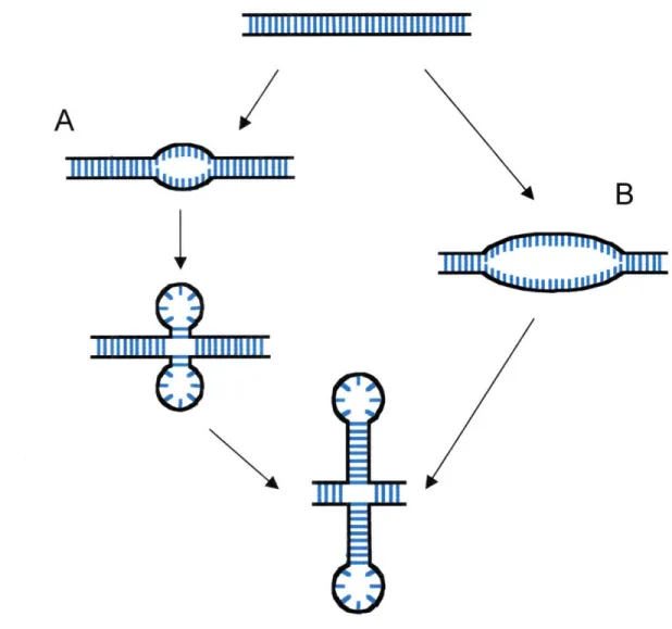
Cruciforms are branched DNA structures in which an inverted repeat, the halves of which are usually separated by a short segment of non-palindromic sequence, extrudes from the helix to form two opposing stem-loop structures (84). Cruciform extrusion can happen by one of two processes. In C-type extrusions, the formation of the cruciform is preceded by the stable melting of the palindromic region of DNA, while S-type
extrusions are more complex and involve the formation of partially extruded
intermediates that develop into cruciforms through a branch migration process (Fig. 1.6). The mechanism of extrusion is dictated by the sequence context of the inverted repeat; the sequences flanking the ColE 1 inverted repeat, a prototype for C-type extrusion, are A-T rich and more likely to be prone to denaturation. Consistent with the requirement for
large-scale melting, C-type cruciforms extrude preferentially under low salt, and have a high activation energy. S-type cruciforms, which are much more common, require salt of
at least 50-60 mM concentration and have a lower activation energy for extrusion (85,86).
There is still some debate over the physiological role of cruciforms. Some studies have suggested that such structures can affect gene expression, forming above a certain negative superhelical density and acting as a block to the progression of the polymerase complex (77). The structure has also been demonstrated to be a specific binding site for regulatory proteins. In one instance, a combination of cruciform extrusion and the
interaction of the cruciform with the RuvA protein from E. coli appears to act as a topological switch, regulating processes that require interaction of cis-acting DNA
elements by controlling the reptation of supercoiled molecules (87).
Besides denatured regions and cruciforms, negative supercoiling can assist with the formation of other alternative structures as well. These include left-handed Z-DNA
(88) and triplex H-DNA (89), both of which have their own unique sequence and supercoiling requirements, and their own predicted biological function(s).
Structural Changes in Positively Supercoiled DNA
As is the case with physiological effects, much is unknown about the structural consequences of positive supercoiling in DNA. Again, since positively supercoiled substrates are much more difficult to obtain than the negatively supercoiled form, they have not been studied in as much depth. Most studies are limited by a low level of positive superhelical density in the substrates, or by a reliance on the presence of small molecules bound to the DNA helix to generate positive supercoiling. Much of the current information about structural consequences of positive supercoiling comes from
computational studies, and suffers from a lack of supporting experimental evidence. Understanding the structural changes that are induced by positive supercoiling is vital to
further understanding the mechanisms of the physiological effects already hypothesized, as well as predicting effects that have not yet been identified.
An important insight into structural changes caused by positive supercoiling came from Allemand et al. (90). This group used single-molecule biomechanical experiments to look at global structural changes in DNA at varying levels of introduced torsion. A long (17,000 bp) piece of DNA was attached at one end to a solid support, and on the other to a paramagnetic bead. Magnets were used to rotate the bead, introducing precise amounts of either positive or negative torsion into the molecule. The magnets were also used to stretch the DNA, preventing writhe, and therefore forcing the entire ALk to be partitioned into twist. Both the length of the DNA and the force being used to pull on the bead can be measured using this system. To monitor changes in DNA structure resulting from introduced torsion, both length vs. force at a constant o, or length vs. oy at a constant force were measured.
In the case of underwound DNA, the molecule underwent denaturation as a means of relieving the overall torque (90). This phenomenon has been previously established through modeling (65), experiments with negatively supercoiled plasmids (39,91), and micromanipulator studies (92). With overwinding of the DNA, a different structural transition occurs, albeit at a higher level of force (3 pN vs. 0.3 pN for underwinding). Modeling of the data led to a proposed structure with a helical repeat of 2.6 bp/turn where the DNA is significantly (-75%) longer than B-DNA, and the phosphate backbones occupy the center of the helix, with extruded bases along the outside. Similarities to the DNA structure originally proposed by Linus Pauling (93) led to the structure being termed "P-DNA" (90). The solvent-exposed positioning of the bases was confirmed by
reaction of the twisted molecules with glyoxal, a chemical agent that reacts selectively with unpaired bases (94).
In both under- and overwinding, the structural change occurs as a phase transition, where above a certain critical force (and associated critical torque) writhing becomes energetically unfavorable (90). At this point, the molecule adopts regions of the alternative structure to alleviate strain. Just as in the case of cruciform structure
discussed previously, a small region of alternative structure can reduce the strain on the rest of the molecule considerably; in the case of overwinding, adopting the new structure reduces the twist in the rest of the molecule by three turns for every 10.4 bp converted.
While evidence for a non-base paired structure is compelling, much of the study was completed under conditions that would not be expected physiologically.
Experiments were carried out at low salt (10 mM phosphate), at which the helix is less stable than at physiological ionic strength; experiments in higher salt showed that greater thresholds were necessary to observe the same structural transitions. Additionally, the entire linking number difference was confined to twist, a partitioning that would not be expected in unrestrained supercoiling. Conditions resulting in this phase transition may be transiently generated immediately ahead of a processing helix tracking enzyme, but are less likely to be sustained in other circumstances.
At the same time, it may be possible for some degree of base unpairing to be present at lower levels of torsional strain. In negatively supercoiled plasmid, single strand specific nuclease sensitivity can be detected at values as low as c = -0.02 (82), despite the denaturation phase transition requiring significant pulling force to occur at a c = -0.015. Could some regions of P-DNA-like structure develop in DNA that is
overtwisted to a lesser extent than the critical force that was identified in the micromanipulator experiments?
There is some experimental evidence to suggest this may be possible. Nuclease BAL-31 reacts with DNA when enough of the intercalating agent ethidium bromide has bound to give an estimated superhelical density of Ya +0.15 (82). A long AT(,n) tract
becomes sensitive to the unpaired-base specific agent osmium tetraoxide when positive supercoiling is induced by actinomycin D, a CpG binding agent (95). While these results are intriguing, both studies suffer from a major flaw: reliance on a bound molecule to introduce positive supercoiling prevents a realistic assessment of how positively supercoiled DNA behaves in the absence of a bound ligand.
The ability to prepare positively supercoiled substrates with a high superhelical density in the absence of extraneous ligands is crucial for further understanding of the
structural consequences of positive supercoiling in DNA. In experiments presented in the succeeding chapters, I describe modifications to such a method that was previously developed in this laboratory that improve the consistency and purity of prepared positively supercoiled substrates. Using these substrates, I have performed chemical,
enzymatic, biomechanical, and spectroscopic studies to characterize structural changes that occur with positive supercoiling. Taken together, these studies should provide an
important guide for future research into the structural and physiological consequences of positive supercoiling in DNA.
DNA is wraps around histone
complexes to form
nucleosomes
Nucleosomes
are compacted
into the 30 nm fiber
Chromatin loops attach to
protein matrix to form
topological domains
Further compaction leads to
fully condensed chromosome
Figure 1.1. Packaging of eukaryotic DNA into chromatin. DNA is packaged in successive stages of compaction, going from naked DNA through nucleosomes and the 30 nm fiber to chromatin loops, and finally a completely condensed chromosome.
Ail
Negative Supercoiling
(underwinding)
> 10.4
bp/turn
Positive Supercoiling
(overwinding)
<
10.4 bp/turn
je
helix-tracking enzyme
as RNA polymerase)
Figure 1.2. The twin-domain model of DNA supercoiling. As a helix-tracking enzyme moves along a DNA molecule, the two must rotate relative to each other. If the enzyme is prevented from rotating, the DNA molecule itself must rotate, causing a change in the helical repeat of the DNA (supercoiling). Positive supercoiling (overwinding) will be generated ahead of the enzyme, while negative supercoling (underwinding) will be generated behind it. If the ends of the DNA are constrained in such a fashion that they cannot freely rotate, this supercoiling will be retained in the DNA.
underwinding
overwinding
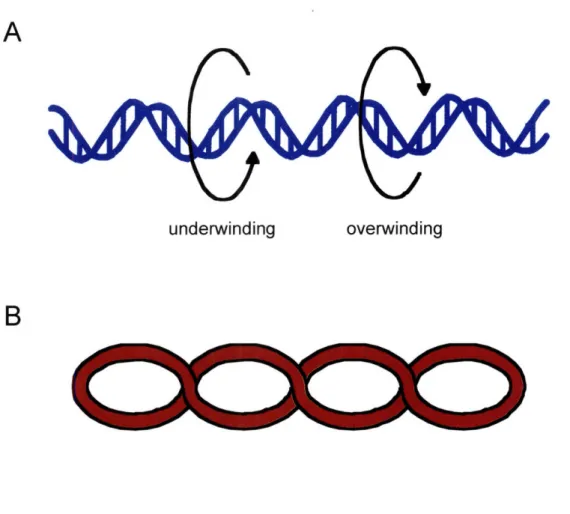
B
Figure 1.3. Twist and writhe parameters of DNA supercoiling. A) Twist refers to the number of times the strands wrap around the helical axis. Supercoiled DNA can be either under- or over-twisted relative to relaxed B-DNA B) Writhe is the crossing of the DNA helical axis over itself. The direction of cross-over is determined by the direction of suDercoiline.
A
B
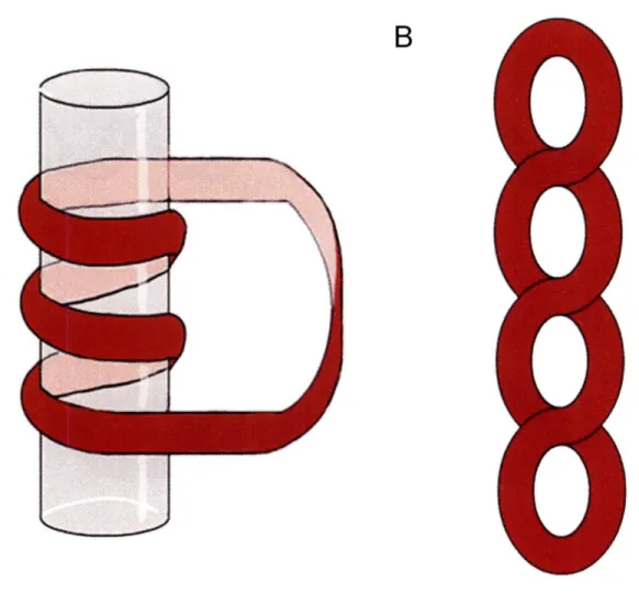
K~)
Figure 1.4. Toroidal and plectonemic DNA writhe. A) Toroidal writhe occurs when the DNA wraps around a central axis, such as a protein. B) Plectonemic writhe is unconstrained and occurs in the absence of toroidal wrapping.
A
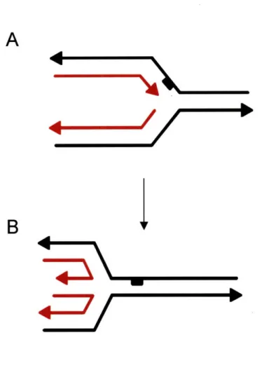
Figure 1.5: Formation of a "chicken foot" structure at a stalled replication fork promoted by accumulated positive supercoiling. A: A lesion on the leading strand causes replication to
stall. Arrowheads indicate the 3' ends of the strands; parent strands are depicted in black while daughter strands are in red. B: A regression of the stalled replication fork promoted by accumulated positive supercoils ahead of the fork. The daughter strands anneal to each other due to this regression, forming a "toe" in the "chicken foot". Figure adapted from reference
A
Figure 1.6: Two general types of cruciform extrusion at inverted repeat sequences in DNA. A. S-type extrusion: Extrusion is initiated by formation of a small denatured bubble that develops into opposing hairpin loops. Branch migration leads to a fully extruded cruciform structure. B. C-type extrusion: under low salt conditions a large denatured bubble forms. This denatured region form the full cruciform directly. Negative supercoiling is necessary for either type of extrusion; the type a particular sequence undergoes is sequence dependent.
1111111 lllilllllIl1illnllilnlrir
LITERATURE CITATIONS
1. (2004) Finishing the euchromatic sequence of the human genome. Nature, 431, 931-945.
2. Hansen, J.C. (2002) Conformational dynamics of the chromatin fiber in solution: determinants, mechanisms, and functions. Annu Rev Biophys Biomol Struct, 31, 361-392.
3. Finch, J.T. and Klug, A. (1976) Solenoidal model for superstructure in chromatin. Proc ANatl Acad Sci US A, 73, 1897-1901.
4. Robinson, P.J., Fairall, L., Huynh, V.A. and Rhodes, D. (2006) EM measurements define the dimensions of the "30-nm" chromatin fiber: evidence for a compact, interdigitated structure. Proc Natl Acad Sci USA, 103, 6506-6511.
5. Woodcock, C.L., Grigoryev, S.A., Horowitz, R.A. and Whitaker, N. (1993) A chromatin folding model that incorporates linker variability generates fibers resembling the native structures. Proc Natl Acad Sci U S A, 90, 9021-9025. 6. Dorigo, B., Schalch, T., Kulangara, A., Duda, S., Schroeder, R.R. and Richmond,
T.J. (2004) Nucleosome arrays reveal the two-start organization of the chromatin fiber. Science, 306, 1571-1573.
7. Cook, P.R. and Brazell, I.A. (1975) Supercoils in human DNA. J Cell Sci, 19, 261-279.
8. Kramer, P.R. and Sinden, R.R. (1997) Measurement of unrestrained negative supercoiling and topological domain size in living human cells. Biochemistry, 36, 3151-3158.
9. Benyajati, C. and Worcel, A. (1976) Isolation, characterization, and structure of the folded interphase genome of Drosophila melanogaster. Cell, 9, 393-407. 10. Hofmann, J.F., Laroche, T., Brand, A.H. and Gasser, S.M. (1989) RAP-1 factor is
necessary for DNA loop formation in vitro at the silent mating type locus HML. Cell, 57, 725-737.
11. Jackson, D.A., Dickinson, P. and Cook, P.R. (1990) The size of chromatin loops in HeLa cells. Embo J, 9, 567-571.
12. Liu, L.F. and Wang, J.C. (1987) Supercoiling of the DNA template during transcription. Proc Natl A cad Sci USA, 84, 7024-7027.
13. Jackson, D.A., Hassan, A.B., Errington, R.J. and Cook, P.R. (1993) Visualization of focal sites of transcription within human nuclei. Embo J, 12, 1059-1065.
14. Hozak, P., Hassan, A.B., Jackson, D.A. and Cook, P.R. (1993) Visualization of replication factories attached to nucleoskeleton. Cell, 73, 361-373.
15. Tsao, Y.P., Wu, H.Y. and Liu, L.F. (1989) Transcription-driven supercoiling of DNA: direct biochemical evidence from in vitro studies. Cell, 56, 111-118. 16. Leng, F. and McMacken, R. (2002) Potent stimulation of transcription-coupled
DNA supercoiling by sequence-specific DNA-binding proteins. Proc Natl Acad Sci USA, 99, 9139-9144.
17. Wu, H.Y., Shyy, S.H., Wang, J.C. and Liu, L.F. (1988) Transcription generates positively and negatively supercoiled domains in the template. Cell, 53, 433-440. 18. Figueroa, N. and Bossi, L. (1988) Transcription induces gyration of the DNA
template in Escherichia coli. Proc Natl Acad Sci USA, 85, 9416-9420.
19. Giaever, G.N. and Wang, J.C. (1988) Supercoiling of intracellular DNA can occur in eukaryotic cells. Cell, 55, 849-856.
20. Brill, S.J. and Sternglanz, R. (1988) Transcription-dependent DNA supercoiling in yeast DNA topoisomerase mutants. Cell, 54, 403-411.
21. Koo, H.S., Claassen, L., Grossman, L. and Liu, L.F. (1991) ATP-dependent partitioning of the DNA template into supercoiled domains by Escherichia coli UvrAB. Proc Natl Acad Sci US A, 88, 1212-1216.
22. Yang, L., Jessee, C.B., Lau, K., Zhang, H. and Liu, L.F. (1989) Template supercoiling during ATP-dependent DNA helix tracking: studies with simian virus 40 large tumor antigen. Proc Natl Acad Sci US A, 86, 6121-6125. 23. Janscak, P. and Bickle, T.A. (2000) DNA supercoiling during ATP-dependent
DNA translocation by the type I restriction enzyme EcoAI. JMol Biol, 295, 1089-1099.
24. Wang, J.C. (1979) Helical repeat of DNA in solution. Proc Natl Acad Sci USA, 76, 200-203.
25. Keller, W. and Wendel, I. (1975) Stepwise relaxation of supercoiled SV40 DNA. Cold Spring Harb Symp Quant Biol, 39 Pt 1, 199-208.
26. McClellan, J.A., Boublikova, P., Palecek, E. and Lilley, D.M. (1990) Superhelical torsion in cellular DNA responds directly to environmental and genetic factors. Proc Natl Acad Sci US A, 87, 8373-8377.
27. Kusano, S., Ding, Q., Fujita, N. and Ishihama, A. (1996) Promoter selectivity of Escherichia coli RNA polymerase E sigma 70 and E sigma 38 holoenzymes. Effect of DNA supercoiling. JBiol Chem, 271, 1998-2004.
28. Higgins, C.F., Dorman, C.J., Stirling, D.A., Waddell, L., Booth, I.R., May, G. and Bremer, E. (1988) A physiological role for DNA supercoiling in the osmotic regulation of gene expression in S. typhimurium and E. coli. Cell, 52, 569-584. 29. Adrian, M., ten Heggeler-Bordier, B., Wahli, W., Stasiak, A.Z., Stasiak, A. and
Dubochet, J. (1990) Direct visualization of supercoiled DNA molecules in solution. Embo J, 9, 4551-4554.
30. Boles, T.C., White, J.H. and Cozzarelli, N.R. (1990) Structure of plectonemically supercoiled DNA. JMol Biol, 213, 931-951.
31. Bednar, J., Furrer, P., Stasiak, A., Dubochet, J., Egelman, E.H. and Bates, A.D. (1994) The twist, writhe and overall shape of supercoiled DNA change during counterion-induced transition from a loosely to a tightly interwound superhelix. Possible implications for DNA structure in vivo. JMol Biol, 235, 825-847. 32. Vologodskii, A.V. and Cozzarelli, N.R. (1994) Conformational and
thermodynamic properties of supercoiled DNA. Annu Rev Biophys Biomol Struct, 23, 609-643.
33. Bauer, W. and Vinograd, J. (1970) Interaction of closed circular DNA with intercalative dyes. II. The free energy of superhelix formation in SV40 DNA. J Mol Biol, 47, 419-435.
34. Pulleyblank, D.E., Shure, M., Tang, D., Vinograd, J. and Vosberg, H.P. (1975) Action of nicking-closing enzyme on supercoiled and nonsupercoiled closed circular DNA: formation of a Boltzmann distribution of topological isomers. Proc Natl Acad Sci USA, 72, 4280-4284.
35. Rybenkov, V.V., Vologodskii, A.V. and Cozzarelli, N.R. (1997) The effect of ionic conditions on DNA helical repeat, effective diameter and free energy of supercoiling. Nucleic Acids Res, 25, 1412-1418.
36. Horowitz, D.S. and Wang, J.C. (1984) Torsional rigidity of DNA and length dependence of the free energy of DNA supercoiling. JMol Biol, 173, 75-91. 37. Vologodskii, A.V., Levene, S.D., Klenin, K.V., Frank-Kamenetskii, M. and
Cozzarelli, N.R. (1992) Conformational and thermodynamic properties of supercoiled DNA. J Mol Biol, 227, 1224-1243.
38. Seidl, A. and Hinz, H.J. (1984) The free energy of DNA supercoiling is enthalpy-determined. Proc Natl Acad Sci US A, 81, 1312-1316.
39. Bauer, W.R. and Benham, C.J. (1993) The free energy, enthalpy and entropy of native and of partially denatured closed circular DNA. JMol Biol, 234,