Analysis of Cell Cycle
Surveillance Mechanisms in Meiosis
byAndreas Hochwagen
Magister rerum naturalis, Chemistry University of Vienna, Austria, 2000
Submitted to the Department of Biology in partial fulfillment of the requirements for the degree of
Doctor of Philosophy in Biology at the Massachusetts Institute of Technology
January 2006
© 2006 Massachusetts Institute of Technology. All rights reserved.
Signature of the Author:___________________________________________________________ Department of Biology January 14, 2006 Certified by:_______________________________________________________________ Angelika Amon Associate Professor of Biology Thesis Supervisor Accepted
by:_______________________________________________________________ Steve P. Bell Professor of Biology Chair, Committee for Graduate Students
Summary
Numerous DNA double-strand breaks (DSBs) are introduced into the genome in the course of meiotic recombination. This poses a significant hazard to the genomic integrity of the cell. Studies in a number of organisms have unveiled the existence of surveillance mechanisms or checkpoints that couple DNA repair and microtubule integrity to meiotic cell cycle progression. Through their action, aberrant meiocytes are delayed in their meiotic progression to facilitate repair of meiotic DSBs, or are culled through programmed cell death, thereby protecting the germline from aneuploidies that could lead to spontaneous abortions, birth defects and cancer predisposition in the offspring. Two such surveillance mechanisms are analyzed in this thesis. The first is the meiotic recombination checkpoint, which delays meiotic cells in G2/prophase if recombination intermediates remain unrepaired. The extent of the delay is modulated by protein phosphatase 1 (PP1), whose activity allows cells to overcome the checkpoint dependent delay in a process called adaptation. In this work, experiments in the budding yeast Saccharomyces cerevisiae are described that show that premature adaptation is prevented by the FK506-binding protein Fpr3, which associates with and counteracts PP1 in vivo. The checkpoint activity of Fpr3 can be inhibited by the small molecule inhibitor rapamycin and requires the proline isomerase domain of Fpr3, but not its catalytic activity. The second surveillance mechanism analyzed here is a spindle checkpoint independent arrest response of meiotic cells to microtubule perturbation. This arrest is caused by down-regulation of the meiotic transcriptional program and occurs at one of two possible stages, in meiotic G1 prior to entry into the meiotic program, or in meiotic G2/prophase after pre-meiotic DNA replication. Both mechanisms described in this work may be conserved in other organisms, including mammals. The findings presented herein are incorporated into a general model of the surveillance mechanisms of meiotic recombination.
Table of Contents
Summary... 3 Table of Contents... 5 Chapter 1: Introduction. ... 9
The Significance of Meiosis 10
Meiosis – An Overview 11
Meiotic Entry 12
Premeiotic DNA Replication 14
Meiotic Recombination 15
The Meiotic Divisions 19
Meiotic Surveillance Mechanisms 20
The Double-Strand Break Checkpoint 21
Checkpoints Monitoring DSB Repair 24
The Meiotic DNA Damage Checkpoint 24
The rad50S Checkpoint 26
The Recombination Checkpoint 30
The zip1 Checkpoint 37
A Synapsis Checkpoint? 38
Signal Integration 38
Checkpoint Targets 39
Cell cycle progression: 39
DSB repair: 42
Apoptosis: 42
Development: 43
Adaptation 44
The Meiosis I Spindle Checkpoint 46
Why delay in G2/prophase? 50
Conclusion 51
References 52
Chapter 2: The FK506 and Rapamycin Binding Protein Fpr3 Counteracts the Protein Phosphatase 1 to Maintain Recombination Checkpoint Activity.
... 69
Summary 70
Introduction 71
Results 74
FPR3 is required for continued checkpoint arrest. 74
DSBs form normally and persist in dmc1D fpr3D cells. 80
FPR3 is a checkpoint factor. 83
Fpr3 spreads from the nucleolus into the nucleoplasm during meiosis. 90
Fpr3 associates with and anchors Glc7/PP1 in the nucleolus. 97
Fpr3 antagonizes Glc7 function. 99
The PPIase domain of FPR3 is necessary for complex formation with Glc7. 101 The proline isomerase domain of FPR3 is necessary for FPR3’s checkpoint
function. 103
Discussion 107
Fpr3 is a novel component of the recombination checkpoint. 107 What is the function of Fpr3 in the recombination checkpoint? 108 The PPIase domain of Fpr3 is required for its checkpoint function. 110 Is the checkpoint function of Fpr3 shared by other FKBPs? 111
Acknowledgements 113
Materials and Methods 114
References 123
Chapter 3: A Novel Response to Microtubule Perturbation in Meiosis...131
Summary 132
Introduction 133
Results 137
Benomyl reversibly arrests cells during meiosis. 137 Benomyl treatment prevents Clb-CDK accumulation in meiotic cells. 139 Chromosome pairing is defective in the presence of high levels of benomyl. 141 Benomyl treatment causes cells to arrest in G1 or G2/prophase. 144 High levels of benomyl interfere with gene expression during meiosis. 147 The down-regulation of meiotic transcripts depends at least in part on
microtubule depolymerization. 148
Benomyl causes a global change in meiotic gene expression. 150
Cold shock causes similar effects as benomyl. 158
The G2/prophase arrest caused by benomyl treatment is independent of known
meiotic checkpoints. 161
Discussion 165
A novel response to microtubule perturbation. 165
Benomyl treatment causes as cell cycle arrest in G1 and G2/prophase. 168 Why does microtubule perturbation cause a G1 or G2/prophase arrest during
the meiotic cell cycle? 170
Acknowledgements 174
Materials and Methods 175
References 184
Chapter 4: Conclusions and Future Prospects...191
Conclusion 192
Searching for Recombination Checkpoint Components. 192
Defining a Checkpoint Role. 194
Environmental Effects on Meiotic Progression. 202
Conservation and significance. 204
References 207
Appendix A: Control of Meiotic Double Strand Break Formation by Cyclin Dependent Kinases. ... 213
Introduction 214
Preliminary Results and Discussion 216
Search for CDK substrates required for DSB formation. 219
References 224
Acknowledgements ... 227 Curriculum vitae ... 229
Chapter 1
Introduction.
Part of this chapter has been submitted to Current Biology for publication. Hochwagen, A., and Amon, A. (). Checking your breaks: surveillance
The Significance of Meiosis
The incidence of chromosomal abnormalities in human embryos is dramatically high; according to recent estimates the fraction of fertilized human oocytes that contain the wrong number of chromosomes lies between 10 and 30 per cent (Hassold and Hunt, 2001). This exceeds the error rate of most other sexually reproducing organisms by several orders of magnitude. The consequences are a prevalence of spontaneous abortions and genetic diseases such as Down’s syndrome, the most common form of mental retardation. What underlies this high error rate remains obscure, but many chromosomal abnormalities can be traced to errors that occurred during meiosis (Hassold and Hunt, 2001), the cell division program leading to the formation of sperm and egg. Meiosis is a still poorly understood process that is exceedingly difficult to study in humans for many reasons, not the least of which is that in human females meiosis can take decades to complete. However, in recent years research using model organisms has helped elucidate many of the basic meiotic mechanisms at the molecular level. The observation that many meiotic factors are conserved through evolution suggests that similar mechanisms are also operative in humans.
One major finding that resulted from the study of meiosis was the discovery of a number of meiotic surveillance mechanisms, or checkpoints. These mechanisms monitor cellular and in particular chromosomal integrity as the cells progress through meiosis, allow for the coordination of distinct meiotic processes, and arrest or eliminate cells when things go awry. The present work will focus on the
control and molecular basis of several such surveillance mechanisms using the budding yeast Saccharomyces cerevisiae as a model organism.
Meiosis – An Overview
The characteristic division pattern of meiosis consists of two divisions without intervening DNA replication, during which first homologous chromosomes (meiosis I) and then sister chromatids (meiosis II) are segregated away from each other. As a consequence, gametes are produced that contain half the genome complement number of the adult organism. Ploidy is doubled again when two gametes fuse during fertilization. Thus, meiosis is the essential counterpart of fertilization, allowing ploidy to be held constant from one generation to the next. However, this unusual division pattern also introduces a number of constraints that do not exist when a chromosomes separate during mitosis (Marston and Amon, 2004; Petronczki et al., 2003). First, homologous chromosomes differ fundamentally from sister chromatids, because unlike sister chromatids, which are held together by cohesin complexes, homologous chromosomes are not a priori linked to each other. Thus, for correct alignment of chromosomes during metaphase I, connections between homologs need to be established, which occurs in the course of a process of controlled DNA breakage and homolog-directed repair, called meiotic recombination. Second, the connections between chromosomes need to be lost in a stepwise manner, such that homologs separate first, whereas sister chromatids remain connected until meiosis II. Finally, the
kinetochores of sister chromatids need to be co-oriented during the first division, to ensure that they are segregated to the same spindle pole during meiosis I.
Meiotic Entry
Since the meiotic products are often highly specialized cell types – sperm and egg in higher eukaryotes, highly stress-resistant spores in the yeasts – the decision to enter the meiotic cell cycle rather than to continue proliferation is strictly regulated and depends predominantly on external stimuli. This observation is true for higher eukaryotes, where germ cells require (as yet unidentified) signals from the surrounding somatic tissues of the gonads to enter the meiotic program (Bullejos and Koopman, 2004; Pepper et al., 2003; Zhao and Garbers, 2002). It is also true for the single-celled yeasts, for which the major signal for entry into meiosis is nutrient limitation (Figure 1). In budding yeast, starvation signals are integrated at the promoters of IME1, a transcription factor that controls the expression of early meiotic genes, and IME2, a protein kinase that controls both the initiation of premeiotic DNA replication and exit from meiotic G2/prophase (Honigberg and Purnapatre, 2003). Since budding yeast can grow in both haploid and diploid form, they use a/a mating type heterozygosity as a means to ensure that only diploid cells enter meiosis. In haploid cells, the transcriptional repressor Rme1 prevents entry into the meiotic program under nutrient limiting conditions by inhibiting IME1 expression. By contrast, the combined activity of the mating-type specific transcription factors a1 and alpha2 represses the expression of RME1
in diploid cells, thus allowing IME1 expression and entry into the meiotic program (Covitz et al., 1991).
Figure 1: Induction of the meiotic program.
In diploid cells, expression of the transcriptional repressor Rme1 is repressed by the mating type specific transcription factors a1 and alpha2, permitting the nutrient limitation dependent expression of the transcription factor Ime1. The expression of the kinase Ime2 depends on both nutrient limitation and Ime1. Both Ime2 and Ime1-dependent early gene expression are necessary for the induction of premeiotic DNA replication and subsequent meiotic events.
Meiotic entry and progression are controlled in part by successive waves of transcription that coordinate the stage-specific expression of meiotic genes (Chu et al., 1998; Primig et al., 2000). Ime1 controls the first wave of meiotic gene expression, which includes predominantly genes required for premeiotic DNA replication, such as IME2, MUM2, CLB5 and CLB6 and meiotic recombination such as SPO11, RED1, ZIP1, and DMC1 (Chu et al., 1998; Primig et al., 2000;
Smith et al., 1990). In a two-step process this leads to the activation of S phase cyclin dependent kinases (CDKs) complexes, first through the induction of the activating cyclin subunits CLB5 and CLB6, and second through activation of Ime2, which promotes the degradation of the CDK inhibitor Sic1 (Dirick et al., 1998). Active Clb5-CDK and Clb6-CDK complexes then trigger the initiation of premeiotic DNA replication (Benjamin et al., 2003; Dirick et al., 1998; Stuart and Wittenberg, 1998).
Premeiotic DNA Replication
Premeiotic DNA replication appears to initiate largely at the same origins of replication that are used during mitotic proliferation (Collins and Newlon, 1994). Moreover, meiotic and mitotic origin firing, at least in the yeasts, requires many of the same proteins, including the origin recognition complex (ORC), the putative replicative helicase Mcm2-7, and the helicase loading factor Cdc6 (Hochwagen et al., 2005a; Lindner et al., 2002; Murakami and Nurse, 2001; Ofir et al., 2004). One meiosis-specific replication factor, Mum2, has been identified in budding yeast, but its role in premeiotic DNA replication is not understood (Davis et al., 2001). Nevertheless, origin firing differs somewhat between mitotic and meiotic cell cycles. In both cases, the activity of S phase Clb5/6-CDKs is necessary for firing; however, while B-type (Clb1-4)-CDKs can substitute for a lack of Clb5/6-CDKs in mitosis (Kuhne and Linder, 1993; Schwob and Nasmyth, 1993), this does not occur in meiosis (Dirick et al., 1998; Stuart and Wittenberg, 1998). This may be because the major mitotic B-type cyclin CLB2 is not
expressed in meiosis, and CLB1, CLB3, and CLB4 are not induced until after cells leave the extended meiotic G2/prophase phase (Dahmann and Futcher, 1995; Grandin and Reed, 1993). An additional difference between mitotic and meiotic DNA replication may be the requirement for the kinase complex Cdc7/Dbf4. While absolutely required for the firing of origins of replication in mitosis, CDC7 appeared to be dispensable for premeiotic DNA replication (Hollingsworth and Sclafani, 1993; Schild and Byers, 1978). However, this apparent lack of requirement may also have been due to leakiness of the temperature-sensitive
CDC7 allele used in those studies. The cohesin complexes connecting sister
chromatids after premeiotic DNA replication also differ from their mitotic counterparts. In particular, the cleavable subunit of cohesin, Scc1/Mcd1, is replaced by Rec8 in meiotic cohesin complexes. This replacement is necessary both for correct meiotic recombination and for the stepwise loss of cohesion during the subsequent meiotic divisions (Klein et al., 1999; Toth et al., 2000).
Meiotic Recombination
Sister chromatids never exist independently of each other. From the moment they are produced during premeiotic DNA replication to the moment they are separated during meiosis II, they are always linked by sister chromatid cohesion. In contrast, homologous chromosomes are not connected to each other as cells enter the meiotic program. It is during the lengthy meiotic G2 phase that meiotic recombination establishes the links between homologs (Figure 2). Throughout this work we will use the term “G2/prophase” instead of the commonly used meiotic
“prophase”. Prophase is a cytologically defined stage, during which chromosome morphology becomes apparent. However, on a molecular level, CDK activity is low during meiotic “prophase”, a feature that, in mitotic cells, indicates that the cells are in G2 phase rather than in prophase (Dahmann and Futcher, 1995; Grandin and Reed, 1993). To account for this difference, we will refer to the period of low CDK activity that follows premeiotic DNA replication, as G2/prophase.
(A) Meiotic chromosome segregation: Following loss of cohesins at chromosome arms during meiosis I, homologous chromosomes segregate to opposite poles. Subsequently, sister chromatids segregate to opposite poles during meiosis II. (B) Meiotic recombination. DSBs can be processed to result in two types of recombination products, crossovers (COs) where flanking sequences are exchanged, and non-crossovers (NCOs) where flanking sequences are in the parental configuration. Unlike COs produced by the major ZIP1 dependent pathway (solid arrows), COs produced by the less active MMS4-dependent pathway (dashed arrows) may not be formed via a double Holliday junction intermediate and do not exhibit interference – i.e. COs produced by the latter pathway are randomly distributed (Argueso et al., 2004; Hollingsworth and Brill, 2004). Recombination factors, whose inactivation results in a checkpoint response, are indicated next to the stage of recombination for which they are required. Adapted from (Bishop and Zickler, 2004).
Concomitant with premeiotic DNA replication, a number of protein complexes assemble onto chromosomes, including the recombination factors Hop2 and Mnd1, and the meiotic chromosome structure components Red1 and Hop1 (Blat et al., 2002; Smith and Roeder, 1997; Tsubouchi and Roeder, 2002; Zierhut et al., 2004). Within this context, the topoisomerase-like enzyme Spo11 introduces double strand breaks (DSBs) into the DNA (Keeney, 2001). Break formation by Spo11 requires a large set of accessory factors and the activity of Clb5/6-CDK (Arora et al., 2004; Hochwagen et al., 2005a; Kee et al., 2004; Keeney, 2001; Pecina et al., 2002; Prieler et al., 2005). Following DSB formation, Spo11 is nucleolytically cleaved off the break ends in a manner dependent on the Mre11/Rad50/Xrs2 complex, and DSBs are resected in the 5’ to 3’ direction to
expose 3’ single-stranded overhangs (Alani et al., 1990; Neale et al., 2005) (Figure 2B). Single-stranded DNA (ssDNA) is incorporated into nucleoprotein filaments containing among other proteins the RecA-like strand invasion factors Rad51 and Dmc1 (Bishop, 1994). These filaments then engage in the search for homologous repair templates with a strong bias towards the homologous chromosomes rather than the sister chromatid (Schwacha and Kleckner, 1997). Template selection also requires factors such as the chromosome-associated kinase Mek1 that block the sister chromatid as a possible repair template (Niu et al., 2005; Wan et al., 2004). As DSBs are processed, a proteinaceous structure, the synaptonemal complex (SC), forms along meiotic chromosomes in many organisms (Page and Hawley, 2004; Zickler and Kleckner, 1999). Typically, the SC assembles around pairs of homologous chromosomes. However, in some mutant situations, such as yeast hop2 mutants and Msh5-/- mice, synapsis can also occur between chromosomes that are not homologous (Leu et al., 1998; Mahadevaiah et al., 2001). Components of the SC, notably budding yeast Zip1, Zip2, and Zip3 proteins, as well as Mer3 helicase and the Msh4/Msh5 complex are required to ensure that recombination intermediates stably invade the homologous chromosomes and mature into crossovers (Borner et al., 2004; Kleckner et al., 2004). Crossover formation is the crucial step in the establishment of physical links between homologous chromosomes, which are manifested cytologically as chiasmata (Figure 2A).
The Meiotic Divisions
Once meiotic recombination is complete, cells prepare for the first meiotic division by inducing a new wave of gene expression that depends on the transcription factor Ndt80 (Chu and Herskowitz, 1998; Hepworth et al., 1998). Expression of the B-type cyclins Clb1, Clb3, and Clb4 leads to a dramatic increase in mitotic CDK activity and activates the formation of the meiosis I spindle (Dahmann and Futcher, 1995; Grandin and Reed, 1993). At the same time the kinetochores (the chromosomal microtubule attachment sites) of sister chromatids become modified by the monopolin complex, which relocalizes from the nucleolus to the kinetochores as cell exit meiotic G2/prophase (Rabitsch et al., 2003). The monopolin complex, together with the Polo kinase Cdc5 and the meiotic protein Spo13 is required for co-orientation of sister kinetochores during meiosis I, possibly by preventing microtubule attachment to one of the two sister kinetochores (Lee and Amon, 2003; Lee et al., 2004; Rabitsch et al., 2003; Toth et al., 2000; Watanabe, 2004; Winey et al., 2005). Once chromosomes are correctly attached to the meiosis I spindle, cohesin complexes are cleaved along chromosome arms by the protease Separase, leading to segregation of homologous chromosomes (Buonomo et al., 2000). In contrast, centromeric cohesin is protected from cleavage during meiosis I in a manner dependent on the kinetochore-associated protein MEI-S332/shugoshin (Kerrebrock et al., 1995; Kitajima et al., 2004; Lee et al., 2005; Marston et al., 2004; Watanabe, 2005). Only after homologs have separated to opposite poles of the spindle, do the monopolin complex and shugoshin leave the centromeric and pericentromeric
regions of sister chromatids and thus allow sister separation during meiosis II (Figure 2A). Therefore, a precise sequence of events is required for the establishment of the meiotic chromosome segregation pattern, starting with premeiotic DNA replication and ending only after sister chromatid cohesion has been lost at second meiotic division. This extraordinary choreography of events is in part controlled by the action of surveillance mechanisms, or checkpoints. The second part of this introduction will attempt to summarize our current understanding of these mechanisms.
Meiotic Surveillance Mechanisms
The sequence of events surrounding meiotic recombination is highly stereotyped. For example, DSB formation always occurs after DNA replication, cells exit from meiotic G2/prophase only after all DSBs have been repaired, and anaphase I is only initiated once chromosomes are correctly aligned on the metaphase I spindle. Research conducted in the past decade has uncovered some of the coupling mechanisms, so-called checkpoints, responsible for this temporal coordination between meiotic recombination and cell cycle progression.
Throughout this work we will use the term “checkpoint” to describe a mechanism that couples two events, which would occur in an uncoordinated manner in the absence of this mechanism. In this manner, a checkpoint comprises the following components: a signal (1), which is detected by signal sensors (2), which in turn activate signal transduction pathways (3) that translate the signal into an output by
modifying checkpoint targets (4). On the molecular level different checkpoints can share sensors and signal transduction pathways, and can impinge on the same targets. Here, we define checkpoints as distinct, if they differ in the signal and at least one of the above components.
The Double-Strand Break Checkpoint
DSB formation is coupled to the completion of premeiotic DNA replication, presumably to prevent aberrant replication across unrepaired DSBs or double Holliday junctions. Recent work in S. cerevisiae and S. pombe suggests that meiotic cells monitor the progression of the replication fork and permit DSB formation only once the replication fork has passed. If replication forks are stalled early in S phase by using mutations in ribonucleotide reductase (RNR) or the RNR inhibitor hydroxyurea (Borde et al., 2000; Tonami et al., 2005), DSBs are not formed. Furthermore, Borde and colleagues showed that the coupling of DSB formation to DNA replication is a local chromosomal phenomenon. A delay in replication on one arm of chromosome III selectively delayed DSB formation on that arm without influencing the kinetics of DSB formation on other chromosomes or even on the other (normally replicating) arm of chromosome III (Borde et al., 2000).
Interestingly, the mechanisms that ensure this coupling are only active once DNA replication has been initiated. If the firing of origins of replication is prevented, for example by inactivating the S. cerevisiae pre-replication complex component
CDC6 (Hochwagen et al., 2005a) or its S. pombe homologue CDC18 (Murakami
and Nurse, 2001), cells form almost wild-type levels of DSBs, and after a delay repair these DSBs. Thus, the mechanism blocking premature meiotic DSB formation may require the presence of replication forks. This notion is highly reminiscent of the S phase and DNA damage checkpoint controls that couple mitosis to DNA replication. Cells preparing for mitosis are able to detect active and/or stalled replication forks and delay entry into mitosis accordingly. On the other hand if DNA replication is never initiated, cells initiate mitosis with unreplicated chromosomes (Kelly et al., 1993; Piatti et al., 1995; Tercero et al., 2003; Toyn et al., 1995; Whittaker et al., 2000) presumably due to the absence of a signal that engages the S phase and/or DNA damage checkpoints.
The possibility of a checkpoint sensing the presence of replication forks and delaying meiotic DSB formation is supported by the finding that inactivation of
RAD3 (Atr, Table 2) allows meiotic cells to form DSBs in the presence of stalled
forks in S. pombe (Figure 3). A number of other DNA damage checkpoint components, including Rad1, Rad9, Rad17, Rad26, Hus1 and Cds1, are also required for this double-strand break checkpoint (Tonami et al., 2005). In S .
cerevisiae, Atr (MEC1) does not appear to be required for the meiotic DSB block
in response to stalled replication forks (Borde et al., 2000). However, we speculate that a checkpoint similar to the S. pombe double-strand break checkpoint, maybe dependent on redundant activities of both Mec1 and Tel1 (a checkpoint kinase closely related to Mec1), also exists in S. cerevisiae. Such a
checkpoint would presumably produce a global inhibitory signal preventing DSB formation once premeiotic DNA replication has been initiated (Figure 3). That block would then be inactivated locally by the passing replication fork, maybe by producing chromatin states permissive to DSB formation (Murakami et al., 2003).
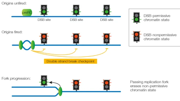
Figure 3. The double strand break checkpoint.
Prior to the initiation of DNA replication (pre-RC – pre-replicative complex), potential sites of DSB formation are permissive for DSB formation (indicated by green traffic lights). Once origins of replication have fired a global signal prevents DSB formation at all potential sites (indicated by red traffic lights). Passage of the replication fork (indicated as green oval) erases the checkpoint signal and resets potential sites of DSB formation to the permissive state.
Checkpoints Monitoring DSB Repair
Once DSBs are introduced, entry into meiosis I is delayed until the completion of meiotic DSB repair. This coupling mechanism becomes apparent in mutants defective in DSB repair. If recombination intermediates persist, meiotic cells arrest or undergo programmed cell death. Such a checkpoint response can be observed in many organisms, including budding yeast, S. pombe, C. elegans,
Drosophila, and mouse (Barlow et al., 1997; Gartner et al., 2000; Ghabrial and
Schupbach, 1999; Roeder and Bailis, 2000; Shimada et al., 2002). However, over the past years evidence has accumulated indicating that the response to DSB repair defects is far from homogeneous. First, both the exact arrest point and the duration of the delay frequently vary depending on the nature of the defect. This could be explained by different severities of the respective defects, and hence quantitative differences in the signaling of a single checkpoint. However, increasingly, checkpoint proteins are being identified that are only required for the response to a particular type of repair defect and are dispensable for others. Thus, it appears that the recombination checkpoint or pachytene checkpoint needs to be thought of as a set of distinct pathways. Below, we attempt to define these checkpoint pathways, notably the meiotic DNA damage checkpoint, the rad50S checkpoint, the recombination checkpoint, and the zip1 checkpoint.
The Meiotic DNA Damage Checkpoint
Broken DNA ends and in particular the resulting ssDNA (coated with the ssDNA-binding protein RPA) activate the DNA damage checkpoint during the mitotic
cell cycle (Garvik et al., 1995; Lydall, 2003; Zou and Elledge, 2003). Evidence that the DNA damage checkpoint is also active prior to the meiotic division comes from the study of budding yeast cdc13 mutants. At the restrictive temperature, temperature-sensitive cdc13 mutants accumulate large amounts of ssDNA at the telomeres (Garvik et al., 1995). During the mitotic division, this triggers the DNA damage checkpoint and leads to a cell cycle arrest at metaphase depending on the DNA damage checkpoint factor Rad9 (Lydall, 2003). Inactivation of CDC13 prior to the meiotic divisions leads to an arrest in G2/prophase that also depends on Rad9 (Weber and Byers, 1992). Rad9 is an adaptor protein in the checkpoint kinase cascade that allows the checkpoint kinase Mec1 to phosphorylate and activate the protein kinase Rad53 (Rouse and Jackson, 2002) (Figure 4). Inactivation of the RecQ family helicase Sgs1 likely also triggers the meiotic DNA damage checkpoint. sgs1 mutants exhibit chromosome instability and a delay in meiotic G2/prophase even in the absence of Spo11-induced DSBs, suggesting a general defect in DNA metabolism that is sensed by the checkpoint. The delay of sgs1 mutants depends on the DNA damage checkpoint components Rad24, Ddc1, and Mec3 (Figure 4A) (Rockmill et al., 2003). Rad24 recognizes ssDNA independently of Mec1, and loads a PCNA-like clamp consisting of Rad17, Ddc1, and Mec3 onto broken ends, which is required for full activation of Mec1 (de la Torre-Ruiz et al., 1998; Rouse and Jackson, 2002) (Figure 4B). These five factors (Mec1, Rad24, Rad17, Ddc1, and Mec3) are central DNA damage sensors common to all meiotic (and mitotic) checkpoints (see below). What distinguishes the meiotic DNA damage checkpoint from the
recombination checkpoint and the zip1 checkpoint is a functional requirement for Rad9 (and presumably Rad53), which is not needed for the latter two checkpoints (Lydall et al., 1996; Roeder and Bailis, 2000). Furthermore, the chromosome structure proteins Red1 and Mek1, which play an important role in the rad50S checkpoint, recombination checkpoint, and zip1 checkpoint, are dispensable for the meiotic DNA damage checkpoint (Rockmill et al., 2003; Xu et al., 1997) (Figure 4). Thus, even though both meiotic recombination and DNA damage (or stalled replication forks) lead to the formation of DSBs, which are repaired through ssDNA intermediates, different surveillance mechanisms are responsible for detecting these DNA lesions and halting cell cycle progression.
A role for the meiotic DNA damage checkpoint in detecting non-recombination induced DNA lesions appears to be conserved across species. Radiation-induced DNA damage triggers programmed cell death in mouse spermatocytes as well as in oocytes of C. elegans hermaphrodites. In both cases, germ cell apoptosis depends on p53, a key regulator of DNA damage dependent apoptosis in mitotic cells (Gartner et al., 2000; Odorisio et al., 1998), suggesting that a meiotic DNA damage checkpoint is also active in mice and worms.
The rad50S Checkpoint
Unlike the DNA damage checkpoint described above, the checkpoints described in the following sections appear to respond to particular meiosis-specific recombination intermediates. rad50S-like mutations, a set of non-null alleles of
RAD50, as well as null mutations in SAE2/COM1, result in a repair defect early
during recombination (refer to Table 1 in which the mutants are grouped according to which checkpoint they activate). In these mutants, Spo11 remains covalently attached to the ends of DSBs and breaks are not resected (Alani et al., 1990). rad50S-like mutants delay in G2/prophase for several hours. (Note however, that eventually these cells enter meiosis despite the persistence of breaks, which may be a form of adaptation, see below). A rad50S checkpoint may also be active in mice. Although spermatocytes of Rad50S/S mice do not enter a permanent meiotic block, they exhibit increased apoptosis resulting in testes that are progressively depleted of mature spermatocytes (Bender et al., 2002).
Table 1: Speculative classification of budding yeast mutants exhibiting a checkpoint dependent G2/prophase delay1.
Meiotic DNA damage checkpoint
rad50S checkpoint Recombination checkpoint
zip1 checkpoint
cdc13 rad50S dmc1 mms4
sgs1 com1/sae2 sae3 zmm mutants2 (23°C
rad51? mei5 and 33°C)
mnd1 hop2 rec8
zmm mutants2 (33°C)
1 It is possible that some of the indicated mutants activate more than one checkpoint. 2 zmm mutants are zip1, zip2, zip3, mer3, and msh5 (Borner et al., 2004)
Like all other checkpoints, the rad50S checkpoint requires the DNA damage sensors Mec1 and Rad24 (Usui et al., 2001). However, as ssDNA does not appear to be exposed in rad50S-like mutants, it is unclear how these proteins recognize
the recombination intermediates. Based on the observations that neither rad50S mutants lacking the protein kinase TEL1 nor mre11-58 mutants (which also accumulate Spo11-linked DSBs) exhibit a delay, it has been suggested that the Mre11/Rad50/Xrs2 (MRX) complex and Tel1 are the primary sensors of protein-linked DSBs (Usui et al., 2001). Consistent with this, Tel1 and the MRX complex appear to be exclusively required for the rad50S checkpoint (Usui et al., 2001).
The checkpoint signal is then relayed through Rad9 (and presumably Rad53), which is similar to the meiotic DNA damage checkpoint but further distinguishes the rad50S checkpoint from the recombination checkpoint and the zip1 checkpoint. Unlike the meiotic DNA damage checkpoint, however, the chromosomal structure proteins Mek1, Red1 and Hop1 are also required for
rad50S checkpoint function (Usui et al., 2001; Woltering et al., 2000; Xu et al.,
1997) (Figure 4B). Mek1 is a meiosis-specific paralog of the protein kinase Rad53 that also exhibits Mec1-dependent phosphorylation (Bailis and Roeder, 2000). It is possible that Mek1 substitutes for some of Rad53’s functions in the context of meiotic recombination intermediates. In this context it is interesting to note, that Red1 also undergoes Mec1-dependent phosphorylation, and is required for the phosphorylation of Mek1 (Bailis and Roeder, 2000). Furthermore, Mek1 binds to phosphorylated Red1 with its phospho-specific FHA domain (Wan et al., 2004). In this way, Red1 may act as an adaptor between Mec1 and Mek1, similar to role of Rad9 in the activation of Rad53. Rad53 is recruited to phosphorylated Rad9 through its FHA domain, which allows Mec1 to phosphorylate Rad53 in the
mitotic DNA damage checkpoint (Rouse and Jackson, 2002; Sweeney et al., 2005).
Figure 4. Surveillance of recombinatorial repair in budding yeast.
(A) Meiotic DNA damage checkpoint, (B) rad50S checkpoint, (C) Recombination checkpoint, (D) zip1 checkpoint. The potential signal activating each checkpoint is depicted at the top. The proteins comprising each checkpoint pathway are listed below. Shared components are indicated by boxes overlapping the corresponding checkpoints. Components that have been demonstrated to act in a particular checkpoint are depicted in color; predicted checkpoint components are depicted in grey. P indicates phosphorylation.
Interestingly, aside from their checkpoint function, Mek1, Red1 and Hop1 are also directly involved in the control of repair template choice and serve as
structural components of the SC (Bailis and Roeder, 1998; Niu et al., 2005; Schwacha and Kleckner, 1997; Wan et al., 2004; Woltering et al., 2000). This raises the possibility that the meiotic chromosomal context is important for sensing unprocessed DSBs and/or relaying the checkpoint signal. Which aspect of chromosome structure, if any, is important for checkpoint signaling is an important question to be addressed.
The Recombination Checkpoint
The recombination checkpoint has been investigated mostly in cells lacking factors required for the initial strand invasion step of meiotic recombination, such as DMC1, HOP2, and others (Figure 2B, Table 1), which, unlike rad50S-like mutants, are competent to remove Spo11 from the ends of DSBs. However, because of a failure to engage in interhomolog repair, these mutants accumulate large amounts of hyperresected DSBs and exhibit a delay in G2/prophase that is substantially more pronounced than that caused by activation of the rad50S checkpoint (Bishop et al., 1992; Gerton and DeRisi, 2002; Leu et al., 1998).
The hyperresection of DSBs observed in homology search mutants leads to large amounts of Rad51-coated ssDNA and it has been suggested that the Rad51 nucleoprotein filament may constitute a signal recognized by the recombination checkpoint (Lydall et al., 1996; Shinohara et al., 1997). Consistent with this interpretation, a rad50S mutation (which prevents formation of the Rad51 filament) strongly reduces the delay of dmc1 mutants (Bishop et al., 1992).
Furthermore, in the absence of Rad51, the delay of dmc1 mutants is substantially reduced (Shinohara et al., 1997). Indeed, rad51 mutants which also accumulate large amounts of ssDNA, exhibit only a modest (and presumably meiotic DNA damage checkpoint dependent) delay in meiotic G2/prophase (Shinohara et al., 1997)(unpublished observations), supporting a signaling role for the Rad51 filament. Similar arguments can also be made for the Dmc1 nucleoprotein filament. hop2 and mnd1 mutants, which accumulate both Rad51 and Dmc1 filaments exhibit a more pronounced cell cycle arrest than dmc1 mutants (Leu et al., 1998). In support of the notion that Dmc1 and Rad51 filaments constitute additive signals, lack of DMC1 reduces the G2/prophase delay of hop2 mutants to the level of dmc1 single mutants (Tsubouchi and Roeder, 2003). On the other hand, however, lack of RAD51 does not alleviate the arrest of hop2 mutants (Tsubouchi and Roeder, 2003). It is, therefore, also possible that the absence of
RAD51 and DMC1 prevents the recombination intermediates of dmc1 and hop2
mutants, respectively, from being processed into structures that are detected by the checkpoint.
The recombination checkpoint shares components with the rad50S checkpoint and the DNA damage checkpoint, including Mec1, Rad24, Rad17, Mec3 and Ddc1 (Hong and Roeder, 2002; Lydall et al., 1996; Roeder and Bailis, 2000) (Figure 4C). In their absence, dmc1 mutants do not experience a G2/prophase delay and initiate the first meiotic division despite a large number of unrepaired DSBs (Grushcow et al., 1999; Hong and Roeder, 2002; Lydall et al., 1996). In
contrast to the rad50S checkpoint and the meiotic DNA damage checkpoint, however, neither Rad9 nor Tel1 play a role in the recombination checkpoint (Lydall et al., 1996; Usui et al., 2001).
Similar to the rad50S checkpoint, a macromolecular assembly of the meiotic chromosomal proteins Hop1, Red1 and Mek1 is thought to provide a framework for the activation of the recombination checkpoint (Bailis and Roeder, 1998; Bailis et al., 2000; Hollingsworth and Ponte, 1997; Woltering et al., 2000; Xu et al., 1997). Their correct localization to chromosomes appears to depend in part on the histone methyltransferase Dot1 (San-Segundo and Roeder, 2000). The recombination checkpoint response of dmc1 mutants is completely eliminated in cells lacking HOP1, RED1, or MEK1 and much reduced in the absence of DOT1 (Hochwagen et al., 2005a; San-Segundo and Roeder, 2000; Xu et al., 1997). The kinase activity of Mek1 is necessary to maintain the arrest of dmc1 mutants (Bailis and Roeder, 1998; de los Santos and Hollingsworth, 1999; Wan et al., 2004) and both Ddc1 and Red1 exhibit Mek1-dependent phosphorylation (Bailis and Roeder, 1998; de los Santos and Hollingsworth, 1999; Hong and Roeder, 2002). However, recent experiments using kinase-specific ATP-analogues indicate that Mek1-dependent phosphorylation of Red1 is not direct (Wan et al., 2004). It is possible that Mek1 serves to hyperactivate Mec1 by phosphorylating Ddc1, thereby leading to further phosphorylation of Red1 (Figure 4C).
The recombination checkpoint is widely conserved. Mice lacking Dmc1, Hop2, or
Msh5 (and a growing list of other factors) experience a block in gametogenesis
followed by widespread apoptosis of germ cells (de Rooij and de Boer, 2003). Also, inactivation of Spo11 or Mei1 (another factor likely required for DSB formation) in a Dmc1-/- or Msh5-/- mutant background result in the bypass of the cell cycle arrest (Barchi et al., 2005; Di Giacomo et al., 2005; Reinholdt and Schimenti, 2005), suggesting that, in mice as in yeast, a checkpoint detects DSBs and/or subsequent repair intermediates. To date, however, no components of the mouse recombination checkpoint have been identified. Atm-/-mutants, which show a profound defect in the somatic DNA damage checkpoint, exhibit a meiotic arrest very similar to Dmc1-/- mutants (Barlow et al., 1996; Xu et al., 1996). This suggests that Atm has a direct role in DSB repair. Atm may still be involved in the checkpoint, but given that Atm-/- mutants arrest, other aspects of the checkpoint are clearly intact. The analysis of another likely checkpoint component, Atr, has been precluded by the fact that loss of Atr is embryonic lethal (Brown and Baltimore, 2000; de Klein et al., 2000). Nevertheless cytological evidence is consistent with a role for Atr in the recombination checkpoint (Keegan et al., 1996; Moens et al., 1999). A number of other somatic checkpoint factors have been implicated in the recombination checkpoint based on cytological data, including TopBP1 (Barchi et al., 2005; Perera et al., 2004) and Rad1 (Freire et al., 1998).
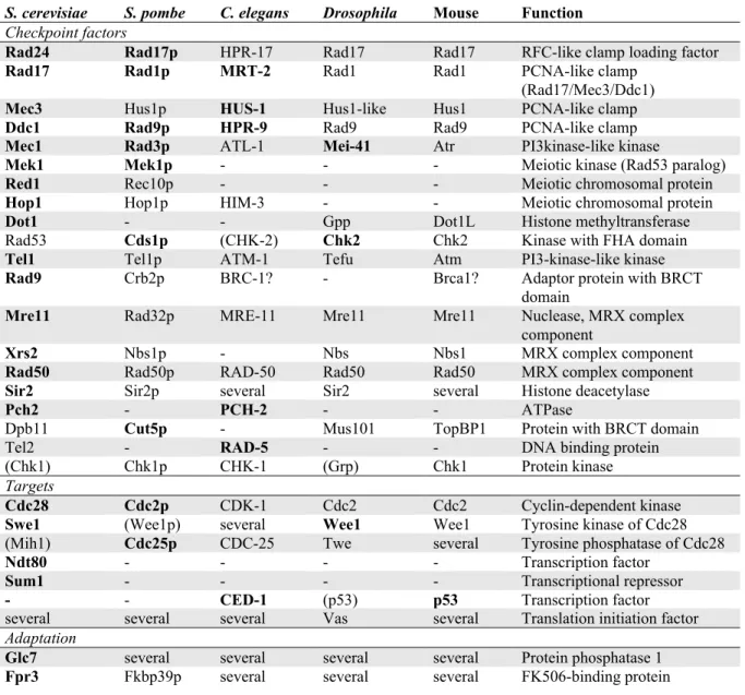
Table 2. Meiotic checkpoint proteins and their homologues.
Protein names in bold indicate factors whose meiotic checkpoint role has been demonstrated experimentally. Names in brackets indicate factors for which experiments did not identify a meiotic checkpoint role.
S. cerevisiae S. pombe C. elegans Drosophila Mouse Function
Checkpoint factors
Rad24 Rad17p HPR-17 Rad17 Rad17 RFC-like clamp loading factor
Rad17 Rad1p MRT-2 Rad1 Rad1 PCNA-like clamp (Rad17/Mec3/Ddc1)
Mec3 Hus1p HUS-1 Hus1-like Hus1 PCNA-like clamp
Ddc1 Rad9p HPR-9 Rad9 Rad9 PCNA-like clamp
Mec1 Rad3p ATL-1 Mei-41 Atr PI3kinase-like kinase
Mek1 Mek1p - - - Meiotic kinase (Rad53 paralog)
Red1 Rec10p - - - Meiotic chromosomal protein
Hop1 Hop1p HIM-3 - - Meiotic chromosomal protein
Dot1 - - Gpp Dot1L Histone methyltransferase
Rad53 Cds1p (CHK-2) Chk2 Chk2 Kinase with FHA domain
Tel1 Tel1p ATM-1 Tefu Atm PI3-kinase-like kinase
Rad9 Crb2p BRC-1? - Brca1? Adaptor protein with BRCT
domain
Mre11 Rad32p MRE-11 Mre11 Mre11 Nuclease, MRX complex component
Xrs2 Nbs1p - Nbs Nbs1 MRX complex component
Rad50 Rad50p RAD-50 Rad50 Rad50 MRX complex component
Sir2 Sir2p several Sir2 several Histone deacetylase
Pch2 - PCH-2 - - ATPase
Dpb11 Cut5p - Mus101 TopBP1 Protein with BRCT domain
Tel2 - RAD-5 - - DNA binding protein
(Chk1) Chk1p CHK-1 (Grp) Chk1 Protein kinase
Targets
Cdc28 Cdc2p CDK-1 Cdc2 Cdc2 Cyclin-dependent kinase
Swe1 (Wee1p) several Wee1 Wee1 Tyrosine kinase of Cdc28 (Mih1) Cdc25p CDC-25 Twe several Tyrosine phosphatase of Cdc28
Ndt80 - - - - Transcription factor
Sum1 - - - - Transcriptional repressor
- - CED-1 (p53) p53 Transcription factor several several several Vas several Translation initiation factor
Adaptation
Glc7 several several several several Protein phosphatase 1
Fpr3 Fkbp39p several several several FK506-binding protein RFC – replication factor C, PCNA – proliferating cell nuclear antigen, PI3kinase – 3-phospho inositol kinase, FHA domain – forkhead associated domain, BRCT domain – Brca1 carboxy terminal domain, MRX complex – Mre11/Rad50/Xrs2 complex, Gpp – Grappa, Tefu – Telomere fusion, Grp – Grapes, Twe – Twine, Vas - Vasa
As observed in mouse gametogenesis, cells with meiotic DSB repair defects are removed by apoptosis in the female germline of C. elegans hermaphrodites (Colaiacovo et al., 2003; Gartner et al., 2000). The damage-dependent programmed cell death is induced in the pachytene stage of meiotic G2/prophase and requires the checkpoint factors MRT-2, HUS-1, HPR-9 and RAD-5 (Gartner et al., 2000; Stergiou and Hengartner, 2004)(Table 2). MRT-2, HUS-1, and HPR-9 likely act as a complex in parallel to RAD-5 (Hofmann et al., 2002). It is unclear whether the checkpoint kinase CHK-2 has a role in the worm recombination checkpoint. A mutation in chk-2 does prevent apoptosis in oocytes lacking rad-51. However, this may be due to a defect in DSB formation rather than inactivation of the checkpoint (Alpi et al., 2003; MacQueen and Villeneuve, 2001). Not all worm repair mutants trigger checkpoint dependent apoptosis. No programmed cell death is elicited in oocytes lacking the SC components him-3 or
rec-8, despite defects in synapsis and an accumulation of RAD-51 foci (a
cytological marker for unrepaired DSBs) (Alpi et al., 2003). Given that him-3 is related to HOP1 (Table 2), this may also indicate a checkpoint role for HIM-3.
The recombination checkpoint of S. pombe has long eluded detection, because most S. pombe repair mutants do not exhibit dramatic cell cycle delays, and even mutants completely deficient in DSB repair progress through meiosis (Catlett and Forsburg, 2003). Careful analysis of meiotic cell cycle kinetics, however, indicated that repair-deficient meu13 (hop2) mutants delay entry into meiosis I by approximately 30 minutes (Perez-Hidalgo et al., 2003; Shimada et al., 2002). The
meu13 delay depends on the formation of DSBs and requires a set of conserved
checkpoint factors, including Rad17, Rad9, Rad1, Rad3, Mek1, Cds1, and Cut5 (Perera et al., 2004; Perez-Hidalgo et al., 2003; Shimada et al., 2002), most of which are also involved in the recombination checkpoint in other organisms (Table 2).
Evidence for a recombination checkpoint in Drosophila oocytes comes from the analysis of spnA, spnB, spnD, and okra mutations, which disrupt several Rad51-like factors (Abdu et al., 2003; Ghabrial and Schupbach, 1999; Staeva-Vieira et al., 2003). These mutants exhibit defects in the formation of the karyosome, a chromosome structure specific for meiotic G2/prophase. Furthermore, the subsequent patterning of the eggshell is abnormal in these mutants due to a failure to accumulate wild-type levels of the patterning protein Gurken (Ghabrial et al., 1998). Both defects are suppressed in mutants of the Spo11 homolog Mei-W68, suggesting that they result from a defect in DSB repair. Furthermore, both karyosome and egg patterning defects depend on the checkpoint factors Mei-41 and Chk2, and Chk2 is phosphorylated in a Mei-41-dependent manner in spnB,
spnD and okra mutants (Abdu et al., 2002; Ghabrial and Schupbach, 1999).
Several other DNA damage checkpoint factors including the Chk1-homolog
grapes, and the Mei-41 interacting factor Mus304 are likely not involved in the
The zip1 Checkpoint
The stable invasion of the homolog by a subset of DSBs that will later be repaired as crossovers requires the SC components Zip1, Zip2, and Zip3 as well as a set of other recombination factors (Figure 2B). In their absence, cells undergo a temperature-dependent delay in G2/prophase (Agarwal and Roeder, 2000; Borner et al., 2004; Chua and Roeder, 1998; Sym et al., 1993). The best-analyzed checkpoint response is the delay of zip1 mutants, which requires Rad24, Rad17, Ddc1, Mec3, and Mec1, as well as Red1, Hop1 and Mek1 (Roeder and Bailis, 2000). The nature of the checkpoint signal in these mutants is unclear. However in contrast to the other checkpoints, lesion detection requires the ATPase Pch2 (San-Segundo and Roeder, 1999). Pch2 is specifically required for the zip1 checkpoint, because inactivation of PCH2 eliminates the cell cycle delay of zip1,
zip2, and mms4 mutants (de los Santos et al., 2001; San-Segundo and Roeder,
1999), but does not impair the arrest of hop2, mnd1 and sgs1 mutants (Rockmill et al., 2003; Roeder and Bailis, 2000; Zierhut et al., 2004). The bypass of the dmc1 in the absence of PCH2 appears to depend on the strain background (Hochwagen et al., 2005a; San-Segundo and Roeder, 1999). Pch2 localizes to the nucleolus, which appears to be important for Pch2 function and depends both on Dot1 and the histone deacetylase Sir2 (San-Segundo and Roeder, 1999; San-Segundo and Roeder, 2000). The exact checkpoint role of Pch2 in the zip1 checkpoint is, however, unclear. Pch2 may be involved in the production or accumulation of the recombination intermediate detected by the zip1 checkpoint, because PCH2 also
plays a direct role in recombination (V. Börner, personal communication) (Hochwagen et al., 2005a).
A Synapsis Checkpoint?
Not all meiotic checkpoints respond to DSB-derived recombination intermediates. Some mutant mice, such as Spo11-/- or Mei1-/- mice, also exhibit meiotic blocks in the absence of DSBs (Baudat et al., 2000; Libby et al., 2002; Romanienko and Camerini-Otero, 2000), suggesting that some aspect of synapsis or lack thereof may constitute another checkpoint signal. The block in Spo11-/- and Mei1 -/-spermatogenesis and oogenesis occurs at a later stage than the block in Dmc1-/- or
Msh5-/- meiocytes (Ashley et al., 2004a; Barchi et al., 2005; Di Giacomo et al., 2005; Reinholdt and Schimenti, 2005), supporting the notion that the defects of
Spo11-/- and Mei1-/- germ cells are potentially detected by a distinct checkpoint. Recently, PCH-2 has been implicated in the C. elegans checkpoint response to unsynapsed chromosomes (cited in (McDougall et al., 2005)). In budding yeast, absence of DSBs does not cause a checkpoint response, and may in fact accelerate meiotic progression (Malone et al., 2004), suggesting that the absence of synapsis is not detected in this organism.
Signal Integration
Some recombination mutants may activate more than one checkpoint. A striking example of additive checkpoint activation comes from the analysis of zmm mutants, a class of mutants in budding yeast that includes zip1, zip2, zip3, mer3,
and msh5. These mutants are proficient in strand invasion at 23°C, albeit with a delay, but fail to form single-end invasion intermediates at 33°C. Concomitantly,
zmm mutants only delay in G2/prophase at 23°C but completely arrest at 33°C
(Borner et al., 2004), presumably because the failure to process Rad51 and Dmc1 filaments at 33°C activates the recombination checkpoint in addition to the zip1 checkpoint. Given that several of the checkpoint components (e.g. Mec1) are shared between different checkpoints, we speculate that these factors may serve as signal integrators that translate the inputs of the various checkpoints into a corresponding cell cycle delay.
Checkpoint Targets
The activated checkpoint factors transmit their signal to downstream targets that control cell cycle progression, DNA repair, programmed cell death and, in some cases, development. Most studies concerning checkpoint targets have been conducted in the context of the recombination checkpoint and the zip1 checkpoint. Whether the different checkpoints activate distinct targets has thus far not been investigated.
Cell cycle progression:
The major cell cycle targets of the recombination checkpoint and zip1 checkpoint are cyclin-dependent kinases, protein kinases composed of a catalytic kinase subunit (CDK) and a regulatory cyclin subunit. CDKs, when associated with cyclin A or B in higher eukaryotes, or Clb1, 3, or 4 in budding yeast, drive cells
into meiosis (reviewed in (Marston and Amon, 2004)). Two mechanisms keep CDKs inactive in response to checkpoint activation. First, CDK is inhibited in a checkpoint-dependent manner by the dual specificity protein kinase Wee1, which phosphorylates CDK on a crucial threonine and tyrosine (T14, Y15). In budding yeast hop2 mutants, Swe1 (budding yeast Wee1, Table 2) is hyperphosphorylated and stabilized, and inactivation of SWE1 allows the partial bypass of checkpoint dependent delay (Leu and Roeder, 1999; Pak and Segall, 2002). In Drosophila
spnB (rad51-like) mutants, Wee1 is modified in a Chk2-dependent manner (Abdu
et al., 2002) indicating that similar to budding yeast, cell cycle arrest also occurs by modulating Wee1 activity. The S. pombe recombination checkpoint, on the other hand, does not regulate CDKs throughWee1. Rather, CDKs remain phosphorylated on Y15 during the hop2 delay, because of Mek1-dependent inhibition of Cdc25, a CDK-Y15 phosphatase (Perez-Hidalgo et al., 2003; Shimada et al., 2002). In contrast, Cdc25 (Mih1) does not play a checkpoint role in S. cerevisiae (Leu and Roeder, 1999). Nevertheless, this suggests that CDK is a conserved meiotic checkpoint target.
In budding yeast, checkpoints activation also keeps the transcript (and protein) levels of the B type cyclins low (Chu and Herskowitz, 1998; Hepworth et al., 1998). The promoters of meiotically expressed B type cyclins contain a short DNA element called the middle sporulation element (MSE), which is found in many other so-called “middle genes” whose expression is induced once cells exit from meiotic G2/prophase and enter meiosis I. Meiotic cyclin expression is
controlled by two transcription factors, Ndt80 and Sum1. Ndt80 is a transcriptional activator that binds to the MSE and induces middle gene expression (Chu and Herskowitz, 1998; Hepworth et al., 1998). Sum1 is a transcriptional repressor that recognizes a DNA element that overlaps with the MSE, and thereby competes with Ndt80 for MSE binding at a subset of middle genes (Lindgren et al., 2000; Pierce et al., 2003; Xie et al., 1999). Both Ndt80 and Sum1 are under checkpoint control. NDT80 expression levels are kept low and overexpression of NDT80 allows a partial bypass of the dmc1 G2/prophase delay (Pak and Segall, 2002; Tung et al., 2000). Furthermore, the extensive phosphorylation of Ndt80 is reduced, albeit not eliminated, in a checkpoint-dependent manner in dmc1 or zip1 cells (Hepworth et al., 1998; Shubassi et al., 2003; Tung et al., 2000). Ndt80 phosphorylation has been shown to depend in part on the meiotic kinase Ime2 and the Polo kinase Cdc5 (Benjamin et al., 2003; Clyne et al., 2003), but it is unclear whether these kinases are involved the checkpoint-dependent phosphorylation changes of Ndt80. Sum1, on the other hand, appears to be regulated at the level of protein stability. The level of Sum1 protein transiently drops as meiotic cells progress from G2/prophase into meiosis I, despite increasing levels of SUM1 mRNA (Lindgren et al., 2000). Moreover, Sum1 protein remains at high levels while cells are delayed in G2/prophase by the checkpoint, and SUM1 is required for the checkpoint arrest of dmc1 mutants (Lindgren et al., 2000; Pak and Segall, 2002). The checkpoint factors controlling both Ndt80 phosphorylation and the drop in Sum1 protein levels remain to be identified.
DSB repair:
At least in budding yeast, the meiotic checkpoints also induce DSB repair. For example, Rfa2, a subunit of the ssDNA-binding protein complex RPA is hyperphosphorylated in dmc1 mutants. This phosphorylation is dependent on
MEC1 and DSBs, and is thought to be required for DSB repair (Bartrand et al.,
2005; Brush et al., 2001). Furthermore, Rad24 interacts with the repair protein Rad57 specifically during meiosis, suggesting another link between checkpoint surveillance and repair (Hong and Roeder, 2002).
Apoptosis:
In multicellular organisms, programmed cell death frequently eliminates repair defective meiocytes, and the apoptotic machinery appears to be an important checkpoint target in both mouse and C. elegans. Reports differ as to whether p53 is required for the induction of apoptosis in mouse repair mutants. The finding that inactivation of p53 (or the CDK inhibitor p21Cip1) allows Atm-/- mutant spermatocytes to partially overcome the G2/prophase arrest (Barlow et al., 1997) has been confirmed by some albeit not all subsequent reports (Ashley et al., 2004b; Scherthan et al., 2000). p53-independent apoptosis has been observed in spermatocytes harboring certain chromosomal translocations (Odorisio et al., 1998), while both p53-dependent and p53-independent apoptosis of spermatocytes occurs in several other mouse meiotic mutants (Salazar et al., 2005; Xu et al., 1996). These findings suggest that only a subset of the checkpoint
pathways that are active during mouse spermatogenesis trigger p53-dependent apoptosis.
In C. elegans, apoptosis of rad-51 mutant oocytes is induced through the action of the p53-homolog ced-1 (Alpi et al., 2003). Prior to pachytene stage of meiotic G2/prophase, translation of ced-1/p53 mRNA is inhibited by the RNA-binding protein GLD-1 (Schumacher et al., 2005). GLD-1 levels drop during pachytene leading to an increase in CED-1 protein levels in pachytene oocytes, which in
rad-51 mutant oocytes allows the recombination checkpoint signal to be
translated into a proapoptotic signal (Schumacher et al., 2005).
Development:
In budding yeast, checkpoint activation inhibits spore development concomitantly with cell cycle progression as a consequence of the inhibition of the transcription factor Ndt80, which controls the expression of genes required for both processes (Chu and Herskowitz, 1998; Hepworth et al., 1998). Curiously, the Drosophila recombination checkpoint affects the patterning of the embryo. The recombination defective spn mutants exhibit defects in karyosome formation and Gurken accumulation similar to that of mutants lacking the translation initiation factor vasa. However, unlike the spn mutants, the vasa mutant phenotype is not
mei-41 dependent, suggesting that Vasa acts downstream of the Drosophila
Chk2-dependent manner in spnB mutants (Abdu et al., 2002; Ghabrial and Schupbach, 1999).
Adaptation
The meiotic checkpoints, at least in budding yeast, appear to be less responsive to DNA damage than the mitotic DNA damage checkpoint. In mitotic cells, a single irreparable DSB can trigger an extended checkpoint delay (Lee et al., 2000; Sandell and Zakian, 1993). Meiotic cells, on the other hand, are able to progress through meiosis and form spores even if a DSB remains unrepaired (Malkova et al., 1996). Moreover, despite the large number of DSBs typically introduced during meiosis – more than 200 recombination events are estimated to occur per meiosis – the cell cycle block of many repair mutants is transient. As indicated above, exogenous DNA damage may be sensed differently than recombination intermediates, which may partially explain the less dramatic response. An additional, non-exclusive possibility is that meiotic cells adapt more easily to damage than mitotic cells. Adaptation is known to occur in mitotic cells and allows cells with very limited DNA damage to overcome the checkpoint-dependent block and progress through the cell cycle (Lee et al., 2000; Lupardus and Cimprich, 2004).
Adaptation has also been demonstrated for the recombination checkpoint in budding yeast. A factor likely involved in this process is protein phosphatase 1 (PP1). Overexpression of the catalytic subunit of PP1, GLC7 shortens the
G2/prophase delay of many meiotic repair mutants and can alleviate the arrest caused by constitutively active MEK1 (Bailis and Roeder, 2000; Hochwagen et al., 2005a). Glc7, associates with a variety of targeting factors that provide substrate specificity. In one strain background, inactivation of GIP1, a meiosis-specific substrate-targeting factor of Glc7, causes a block in meiotic G2/prophase (Bailis and Roeder, 2000). However, gip1 mutants do not arrest in other strain backgrounds (Tachikawa et al., 2001), suggesting that there may be other specificity factors acting redundantly to Gip1. Glc7 is inhibited by the FK506-binding protein Fpr3 (Hochwagen et al., 2005a). Fpr3 interacts with Glc7 through its proline isomerase (PPIase) domain and the PPIase domain of Fpr3, though not its catalytic activity, is required to prevent premature adaptation. Inactivation of Fpr3 using the small-molecule inhibitor rapamycin and Fpr3 PPIase mutants cause a reduced checkpoint delay in many repair mutants, similar to the overexpression of GLC7. Furthermore, co-overexpression of GLC7 and FPR3 re-establishes the checkpoint delay (Hochwagen et al., 2005a). Thus, adaptation to persistent recombination intermediates depends on the modulation of PP1 activity. Glc7 may allow adaptation by dephosphorylating Red1 or Red1-dependent targets. Indeed, Glc7 interacts with Red1, and Red1 can be dephosphorylated by Glc7 in vitro (Bailis and Roeder, 2000; Tu et al., 1996). Moreover, a mutant of Glc7 that fails to interact with Red1 (glc7-T152K) exhibits a DSB-dependent cell cycle arrest in meiotic G2/prophase that is bypassed by the inactivation of RED1 (Bailis and Roeder, 2000).
In summary, meiotic recombination appears to be monitored by a battery of surveillance mechanisms. Moreover, many of the checkpoint factors involved in this surveillance are members of multiple checkpoint signaling cascades, and all pathways ultimately lead to cell cycle arrest in meiotic G2/prophase and/or apoptosis. Whether this means that the signals converge into one single pathway or whether the checkpoint components required for more than one checkpoint are assembled into distinct signaling modules by checkpoint specific factors remains to be determined.
The Meiosis I Spindle Checkpoint
Once cells have successfully completed meiotic recombination, the homologs need to be aligned correctly on the metaphase I spindle, such that (1) microtubules are attached to all (unmasked) kinetochores, and (2) the kinetochores of homologs face opposite poles of the spindle. In mitosis, correct bipolar attachment is sensed by the spindle checkpoint, which prevents anaphase onset as long as chromosomes are not properly attached to the spindle (Lew and Burke, 2003; Taylor et al., 2004). For anaphase to occur, a multi-subunit ubiquitin ligase, the anaphase promoting complex or cyclosome (APC/C) needs to target the separase inhibitor securin for degradation, thereby triggering the separase dependent destruction of sister chromatid cohesion (Peters, 2002). Kinetochores that are not bound to microtubules cause the sequestration of the APC/C adaptor protein Cdc20, a specificity factor required for securin destruction. Current models postulate that if a kinetochore is unattached, the kinetochore-associated
checkpoint factors Mad2 and Mad1 associate with that kinetochore. Once kinetochore-associated, they form a template that allows soluble Mad2 proteins to undergo a conformational switch, which binds and entraps Cdc20. Mad2-Cdc20 complexes may then act as templates themselves to further amplify the signal (Nasmyth, 2005). The checkpoint kinase Bub1 is required for the kinetochore association of Mad1 and Mad2 and also activates a second pathway of Cdc20 inhibition involving the checkpoint factor Bub3 and the Cdc20 binding factor Mad3 (Lew and Burke, 2003).
Both microtubule attachment and the tension that is created when the kinetochores of two connected chromosomes (sister chromatids in mitosis and meiosis II, homologs in meiosis I) are attached to opposite spindle poles appear to be monitored by the checkpoint. It is not clear, however, to what extent lack of microtubule attachment and lack of tension constitute separate signals (Lew and Burke, 2003). The Aurora kinase Ipl1 constitutes part of a tension sensitive microtubule severing activity required for the biorientation of kinetochores (Dewar et al., 2004; Tanaka et al., 2002). Thus, in the absence of tension, microtubules become detached from kinetochore, which may be enough to activate the spindle checkpoint.
The inactivation of spindle checkpoint components causes lethality in many higher eukaryotes but has little effect on the viability of proliferating yeast cells (Taylor et al., 2004). In contrast, deletion of MAD1 or MAD2 (but not MAD3)
significantly affects the fidelity of meiosis I chromosome segregation in budding yeast, suggesting that spindle checkpoint function is important for the correct alignment of homologs on the metaphase I spindle (Cheslock et al., 2005; Shonn et al., 2000; Shonn et al., 2003). Likewise, reduced Mad2 function also causes meiosis I chromosome missegregation during mouse oogenesis (Homer et al., 2005). It is thought that Mad1 and Mad2 delay the onset of anaphase I in response to incorrectly attached or unattached chromosomes, because artificial delay of anaphase I onset partially restores correct meiosis I chromosome segregation to
mad1 and mad2 mutants (Homer et al., 2005; Shonn et al., 2000; Shonn et al.,
2003).
Interestingly, even though loss of Mad3 does not affect the fidelity of meiotic chromosome segregation, both Mad2 and Mad3 are required to prevent securin destruction and thus anaphase I entry in spo11 mutants, which cannot establish tension between homologs due to the absence of chiasmata (Shonn et al., 2003). The mechanism by which Mad2 delays anaphase entry likely differs from the role of Mad3, because unlike Mad2, Mad3 delays cells in already in meiotic G2/prophase irrespective of whether Spo11 is active (Cheslock et al., 2005; Shonn et al., 2003). Furthermore, Mad3 appears to be specifically required for chromosomes that have failed to recombine. In budding yeast wild-type cells, non-exchange chromosomes only missegregate in about 7% of cases, because a recombination-independent distributive system can non-specifically pair centromeres that have failed to align during meiotic recombination (Kemp et al.,
2004; Stewart and Dawson, 2004; Tsubouchi and Roeder, 2005). Inactivation of Mad1 and Mad2 causes missegregation of non-exchange chromosomes to increase to about 25%. Loss of Mad3, on the other hand causes completely randomized segregation (Cheslock et al., 2005). Thus, the spindle checkpoint components appear to act in several pathways to ensure correct meiosis I chromosome segregation.
In mitosis, the spindle checkpoint prevents anaphase initiation if microtubules are chemically destabilized using microtubule poisons, such as benomyl or nocodazole (Lew and Burke, 2003; Taylor et al., 2004). A similar Mad2-dependent metaphase I arrest in response to nocodazole treatment is also observed in mouse oocytes (Wassmann et al., 2003). On the other hand, mouse spermatocytes treated with colcemid, another microtubule poison, already arrest in meiotic G2/prophase (Tepperberg et al., 1997; Tepperberg et al., 1999). In budding yeast, it has been suggested that microtubule destabilization using benomyl causes an arrest in metaphase I (Shonn et al., 2000; Shonn et al., 2003). However, analysis of the exact arrest point of benomyl-treated yeast cells indicated that microtubule perturbation triggers a reversible arrest in meiotic G2/prophase rather than metaphase I. A similar arrest is also observed, if microtubules are destabilized by low temperatures (Hochwagen et al., 2005b). Unlike the spindle checkpoint response that occurs in mitosis, the meiotic G2/prophase arrest was caused by a dramatic downregulation of the meiotic transcriptional program and appears to be independent of Mad2 (Hochwagen et