Chromosoma (2004) 113: 113–125 DOI 10.1007/s00412-004-0303-7
R E V I E W
Magali Toueille . Ulrich Hübscher
Regulation of the DNA replication fork: a way to fight genomic
instability
Received: 17 May 2004 / Revised: 15 June 2004 / Accepted: 17 June 2004 / Published online: 6 August 2004 # Springer-Verlag 2004
Abstract DNA replication is a complex mechanism that functions due to the coordinated interplay of many factors. In the last few years, numerous studies have suggested that DNA replication factors are closely implicated in several DNA transaction events that maintain the integrity of the genome. Therefore, DNA replication fork factors have to be considered as part of a general process that aims to protect and replicate the genome in order to allow correct functioning of a cell and its eventual daughter cells. This is illustrated by the numerous factors that have a well-defined function at the DNA replication fork, but also play crucial roles in different DNA repair pathways such as base excision repair, nucleotide excision repair, double-strand break repair, and mismatch repair. Moreover, several of the replisome proteins have also been shown to be essential in sensing and transducing DNA damages through the checkpoint cascade pathways, including the recently characterised alternative clamps and clamp-loaders. In this review we present DNA replication factors that are involved in different DNA transaction and checkpoint regulation pathways, with emphasis on the link between DNA replication and maintenance of genomic stability.
Introduction
DNA carries all the genetic information necessary for a cell to grow and develop normally. This information has to be faithfully replicated before cell division, in order to be transmitted to the daughter cells. A large protein complex, called the replisome, is responsible for DNA replication (Waga and Stillman 1998). One crucial step in the DNA
replication process is to replicate the correct genetic information. However, DNA is a chemically reactive molecule, which is a target for many internal or external DNA-damaging agents (Lindahl and Wood1999). Occur-rence of DNA damage leads subsequently to an altered genomic sequence. Several molecular mechanisms exist to fight genomic instability generated by these agents: cell cycle checkpoints and several DNA repair pathways. Cell cycle checkpoints are sophisticated cascades that result in slowing down cell cycle progression and DNA replication, in order to prevent the cell from continuing to replicate damaged DNA (Melo and Toczyski 2002). Lesions are processed by multiprotein complexes specialised in different DNA repair pathways, such as base excision repair (BER), nucleotide excision repair (NER), double-strand break repair (DSBR), and mismatch repair (MMR) (Hoeijmakers 2001). Coordinated functioning of these various processes is indispensable (Stucki et al.2001a,b), implicating tight regulation of the replicative machinery in synchronization with DNA repair and checkpoint mechan-isms.
In this review, we first describe the DNA replisome, and then focus on its regulation in view of the different pathways involved in maintenance of genomic stability. The DNA replication fork is, however, also regulated at several other levels such as the control of replication origins (recently reviewed in Kearsey and Cotterill2003) and its linkage to other cellular mechanisms such as the cell cycle, in which cyclin dependent kinases (CDKs) and cyclins phosphorylate DNA replication factors (recently reviewed in Henneke et al. 2003).
The DNA replisome
DNA replication is a complex mechanism that necessitates the coordinated interplay of dozens of different proteins (Waga and Stillman 1998). Due to the antiparallel nature of DNA and the 3′→5′ polymerisation direction of all DNA polymerases known, one strand, called the leading strand, is synthesised continuously, and the other, called Communicated by E.A. Nigg
M. Toueille . U. Hübscher (*)
Institute of Veterinary Biochemistry and Molecular Biology, University of Zürich-Irchel,
Winterthurerstrasse 190, 8057 Zurich, Switzerland e-mail: [email protected]
the lagging strand, discontinuously in short pieces of about 200 bases. These pieces are called Okazaki fragments. The successful processing of the 20 million Okazaki fragments at the lagging strand requires at least 23 polypeptides (Hubscher and Seo 2001). The complex replication machinery is assembled at the so-called replication forks, which are gathered in “nuclear replication factories” (Hozak et al.1993). The molecular bases of the replisome were established several years ago thanks to the SV40 in vitro model system, in which nuclear extracts of different origins (Stillman 1994; Waga and Stillman 1994) were used to define the factors involved. Later, functional replisomes have been reconstituted with purified proteins, allowing more accurate depiction of the mechanisms involved in DNA replication. The replisomal proteins can be divided into two main categories: the polymerases and the “accessory” proteins (see Table 1). The polymerases catalyse DNA polymerisation, whereas the accessory proteins are indispensable for assembly and functioning of the replisome at the DNA replication fork. All these polypeptides are spatially arranged and temporally co-ordinated in order to achieve complete bidirectional replication of the replicon (see Maga and Hubscher
1996; Waga and Stillman 1998, for DNA replication models).
The replisomal proteins
Three major polymerases are responsible for DNA synthesis during the progression of the DNA replication fork. First, pol α/primase, a four subunit enzyme, is involved in the initial steps of DNA synthesis. The primase subunits synthesize a short RNA oligonucleotide that can be subsequently elongated by a short stretch of DNA synthesized by pol α (Mizuno et al.1999). Second, the elongation step of DNA synthesis is performed by two polymerases,δ and ε, after a DNA polymerase switch has occurred. Polymeraseε is a four subunit enzyme of which the large catalytic subunit contains DNA polymerase and intrinsic 3′→5′ exonuclease activities. Furthermore, one of the small subunits (p80) could be involved in formation of polymerase dimers. Multiple evidence suggests that polε might be a“regulatory” DNA polymerase inside the DNA replication fork (see below and Araki et al.1995; Feng and
D’Urso 2001; Takayama et al. 2003). Polymerase δ is a four subunit enzyme of which the catalytic subunit possesses an intrinsic 3′→5′ exonuclease proofreading activity. Its polymerase activity can be stimulated by the processivity factor proliferating cell nuclear antigen (PCNA). Among the many accessory proteins, PCNA appears to be a key player. It not only acts as a processivity factor or as a clamp for polδ, but it also plays a role in coordinating the complex network of proteins interacting at the replication fork (Yuzhakov et al. 1999; Maga et al.
2000). Moreover, in view of its numerous interacting partners, PCNA may be one of the proteins linking DNA replication to other cellular pathways (recently reviewed in Maga and Hubscher2003). Due to its thoroidal structure, PCNA forms a homotrimeric ring (three 30 kDa subunits) that encircles DNA, which confers on it a central position in the replisome (Krishna et al.1994). PCNA is not able to load autonomously on DNA, but needs a so-called “clamp-loader”. The five subunit replication factor C (RF-C) is able to open the PCNA ring and to load it onto DNA in an ATP-dependent process. In addition to its clamp-loader role, it was recently shown that the RF-C protein plays a role in polymerase switching from polα/ primase to polδ (Maga et al.2000). Additionally, the four subunit pol ε is also thought to be involved in Okazaki fragment processing, even if its activity appears dispen-sable in this process.
Another important component of the DNA replication machinery is the single-strand binding protein, called replication protein A (RP-A). It covers and protects single-strand DNA during replication fork progression. RP-A is involved in the regulation of several steps of DNA replication. First, it has been shown that RP-A acts as a “fidelity clamp” for pol α/primase (Maga et al. 2001a,b). Second it can modulate the strand displacement synthesis of polδ (Maga et al.2001a,b) and third it can regulate the sequential action of the two nucleases flap endonuclease 1 (Fen1) and Dna2, both involved in Okazaki fragment processing (Bae et al.2001).
Fen1 and Dna2 are endonucleases that specifically cut flap structures, Fen1 being specialised to cleave DNA and to some extent also RNA containing flaps and Dna2, RNA containing flaps only. These activities are required to remove RNA primers synthesised by primase (Budd and Campbell 1997; Waga and Stillman 1998). Finally, the
Table 1 Proteins and their functions at the replication fork. (PCNA, proliferating cell nuclear antigen; Pol, polymer-ase; RF-C, replication factor C; RP-A, replication protein A)
Protein Function
DNA helicase Opens the DNA at the replication fork
Polα/primase RNA–DNA primer synthesis at the origin and at the lagging strand Polδ/pol ε Elongation of primers at the leading and lagging strands
PCNA Processivity factor for polδ and pol ε RF-C Loading of PCNA, polymerase switching
RP-A Protection of single-strand DNA, fidelity clamp for polα/primase Fen1 Cleavage of RNA–DNA flap structures at the lagging strand Dna2 Cleavage of RNA flap structures at the lagging strand DNA ligase I Ligation of DNA pieces
single subunit DNA ligase I is able to seal the nicks on the DNA (Ayyagari et al. 2003; Jin et al. 2003).
Additionally, a very important set of proteins is involved in the pre-initiation and initiation steps of replication. These include Dpb11/Cut5/TopBP1, Cdc45 and the recently discovered tetrameric ring-shaped GINS complex. Dpb11/Cut5/TopBP1 has been found to be associated with polε and the recruitment of both proteins to DNA is interdependent, which provides additional evidence for a regulatory role for polε in DNA replication (Araki et al.1995; Feng and D’Urso2001; Takayama et al.
2003). All these pre-initiation proteins are indispensable for transition from pre-initiation to initiation complex and their binding to replication origins has been shown to be mutually dependent (Kubota et al. 2003; Takayama et al.
2003). The replication helicase Mcm is a factor indis-pensable for melting of the DNA double helix in the pre-initiation step of DNA replication. This hexameric helicase is formed by the stoichiometric assembly of six Mcm monomers, Mcm-2 to Mcm-7, and exhibits a molecular weight of about 600 kDa (Thommes et al. 1997). The Mcm complex exerts helicase and ATPase activities that have been proposed to result from the coordinated function of the regulatory Mcm-2-3-5 and the catalytic Mcm-4-6-7 complexes (Schwacha and Bell2001).
Dynamics of the DNA replication fork
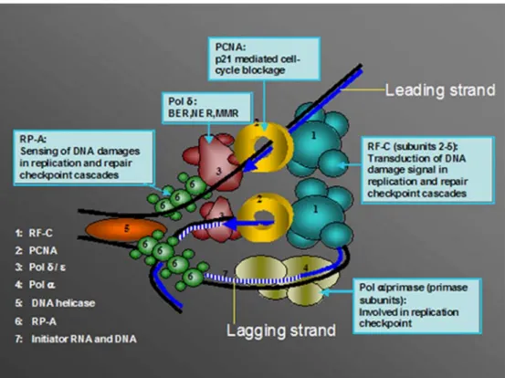
The DNA replication fork is a complex structure in which all the components described above function in a spatially and temporally coordinated manner, allowing the simulta-neous replication of both DNA strands. It has been postulated that, at the replisome, DNA forms a loop and dimerisation of two polymerases occurs via a putative dimerisation factor (Fig.1). Prior to formation of the DNA replication fork, a pre-initiation step is required. Replica-tion origins are recognized by a complex called “origin recognition complex”, which binds the Cdc6 and Cdt1 proteins (Liang et al. 1995; Mizushima et al. 2000). The latter are subsequently involved in recruitment of the Mcm helicase, which results in the formation of an initiation complex that is able to open the DNA structure thanks to the Mcm DNA helicase activity (Liang et al. 1995; Donovan et al. 1997; Tanaka et al. 1997; Kearsey et al.
2000; Maiorano et al. 2000). One model proposes that phosphorylation of the Mcm complex by the Dbf4-dependent kinase leads to an allosteric change in the complex that allows interaction with the Cdc45 initiation protein (Hardy et al.1997; Owens et al.1997; Jiang et al.
1999; Forsburg 2004). However, CDK activity also appears to be necessary for Cdc45 recruitment, and this may be mediated by the Dpb11/Cut5/TopBP1 protein, which is part of a CDK-regulated complex (Kamimura et al. 1998; Wang and Elledge 1999; Hashimoto and Takisawa 2003). In this process, the recently identified GINS complex appears to mediate between the two above-mentioned proteins in order to facilitate proper association between origins and DNA polymerases at the initiation of
DNA replication (Takayama et al. 2003). Subsequently, the DNA helix is unwound under the action of DNA topoisomerase I (Tsurimoto and Stillman 1989). Starting from this point, the DNA replication fork is formed by recruitment of the diverse DNA replication factors. The single-strand DNA is immediately covered by RP-A, which also participates in unwinding of DNA. Polymerase α/primase is then recruited thanks to its interaction with RP-A and Cdc45 (Mimura and Takisawa1998; Uchiyama et al. 2001; Pollok et al.2003), and initiates synthesis by first synthesising an RNA fragment of about ten bases, prolonged by a 30 nucleotide DNA fragment. This initiation step and the following occur during synthesis of each Okazaki fragment on the lagging strand, and only once for the leading strand (Hubscher et al. 2002). After the initiation step, an ATP-dependent polymerase switch from pol α to pol δ and ε occurs, and for this ATP hydrolysis by RF-C is required (Maga et al.2000; Mossi et al.2000). PCNA is simultaneously loaded by RF-C at the primer end, and polδ bound to PCNA is able to synthesise long stretches of DNA. When polδ encounters the 5′ end of the previous Okazaki fragment, it is able to perform strand displacement DNA synthesis (Podust and Hubscher
1993). The flap structure thus generated is eventually a substrate for combined cleavage by the Dna2 and Fen1 endonucleases, in which the single-strand binding protein RP-A, PCNA and RF-C can act as modulators (Mossi et al.2000). The resulting ligatable nick is finally sealed by DNA Ligase I.
Link between DNA replication and DNA repair
Upon formation of DNA lesions by DNA-damaging agents, several DNA repair machineries can be triggered, depending on the type of DNA damage. The lesions are processed by multiprotein complexes specialising in
Fig. 1 Dynamics of the DNA replication fork. DNA forms a loop and dimerisation of the two polymerases via a putative dimerisation factor is proposed, thus rendering the same directionality for the leading and the lagging DNA strands.1 Replication factor C (RF-C), 2 proliferating cell nuclear antigen (PCNA), 3 DNA polymerase δ (pol δ), 4 DNA polymerase α (pol α), 5 DNA helicase, 6 replication protein A (RP-A), 7 initiator RNA and DNA. For more details see text
different DNA repair pathways such as BER, NER, DSBR, or MMR (recently reviewed in Christmann et al.
2003). Several of the DNA replication factors mentioned above have been shown to be directly or indirectly implicated in various DNA repair events, either by participating actively in certain DNA repair processes, or by interacting with DNA repair proteins (Table 2). This has led to the conclusion that a dual role in these two DNA metabolic pathways could involve the same proteins, which, however, might be regulated differently (e.g. by post-translational modifications).
PCNA and the replicative polymerases
The most striking example of a dual role in replication and repair is the DNA clamp PCNA. First, all the above-mentioned repair pathways involve a DNA resynthesis step that can be carried out by pol δ or pol ε. Therefore, polδ, pol ε and PCNA are essential factors both in DNA replication and repair. However, considering the many interacting partners discovered for PCNA, its role in DNA repair goes far beyond being a simple processivity clamp (Maga and Hubscher 2003).
Several lines of evidence support the implication of PCNA in BER. First, PCNA has been shown to interact with the uracil DNA glycosylase UNG and with the apurinic endonucleases Apn and Apn2, suggesting a role for PCNA in the excision step (Dianova et al. 2001; Krokan et al. 2001; Unk et al. 2002). It has also been shown that PCNA interacts in vivo and in vitro with polβ, the main BER polymerase (Kedar et al.2002). Moreover, the long-patch BER pathway has been reconstituted and studied in vitro, and showed a requirement for PCNA in the DNA synthesis step achieved by pol δ and/or pol ε (Klungland and Lindahl1997; Stucki et al.1998; Pascucci et al. 1999).
Furthermore, PCNA is necessary in the reconstituted NER system in vitro, probably due to its processivity effect on polδ. However, an interaction between the NER endonuclease XP-G and PCNA has been detected, which
together likely stabilise the NER complex (Gary et al.
1997). A requirement of the XP-A protein for localisation of PCNA at the sites of DNA damage upon UV irradiation also suggested a role for PCNA in the lesion recognition step of NER (Aboussekhra and Wood 1995; Miura and Sasaki 1996).
An interaction between PCNA and the MMR com-plexes MSH2–MSH6 and MSH2–MSH3 has been dis-covered (Clark et al. 2000; Flores-Rozas et al. 2000; Kleczkowska et al. 2001). PCNA interaction leads to increased mispair binding specificity of the MSH2–MSH6 complex. Recent data suggested a model of mispair recognition in which MSH2–MSH6 binds to PCNA on the newly synthesised DNA and is then transferred to the mispair in an ATP-dependent manner (Lau et al. 2002), suggesting an important role of PCNA in mismatch recognition. In vitro studies also suggested that PCNA is involved in the recognition step of interstrand cross-link repair in coordination with MMR proteins (Zhang et al.
2003).
Finally, PCNA, as well as pol δ, may be involved in recombinational repair of double-strand breaks. This is possibly due to their interaction with the Werner DNA helicase (Lebel et al.1999; Kamath-Loeb et al.2000). A localisation study of green fluorescent protein (GFP)-labelled PCNA in Schizosaccharomyces pombe showed that PCNA is recruited in so-called“repair factories” after exposure to ionising radiation, thus suggesting a role for PCNA in the early steps of homologous recombination (Meister et al.2003).
RP-A, the single-strand binding protein
Studies of DNA repair mechanisms have shown that RP-A plays a major role in coordinating DNA repair mechan-isms and is therefore more than a simple “protecting” protein for single-strand DNA. Indeed, single-strand DNA is the most commonly generated structure found after DNA damage or during DNA repair. Therefore, binding of RP-A to single-strand DNA brings it to an excellent
Table 2 Roles of DNA replica-tion proteins in DNA repair. (BER, base excision repair; DSBR, double-strand break re-pair; MMR, mismatch rere-pair; NER, nucleotide excision repair; PCNA, proliferating cell nuclear antigen; Pol, polymerase; RF-C, replication factor C; RP-A, rep-lication protein A)
Protein Role in DNA repair
Polδ/polε Gap filling DNA synthesis in BER, NER, DSBR, MMR PCNA Processivity factor for polδ
Excision step in long patch BER Stabilization of the NER complex Mismatch recognition in MMR
RF-C Loading of PCNA
RP-A Recognition of DNA damage in NER
Stabilisation of the DSBR complex
Modulation of the MutSα and MutLα proteins in MMR Fen1 Cleavage of DNA flap structures in BER
Role in DSBR
Role in UV damage excision repair pathway (UVER) DNA ligase I Ligation of cleaved products in NER
strategic position for modulation of DNA metabolic processes.
Several studies have suggested that RP-A, in complex with the NER factor XP-A, might be responsible for the initial recognition of DNA damage, hence initiating the NER pathway (Wakasugi and Sancar 1999; Reardon and Sancar 2002). However, recently, a different model in which RP-A acts in a later step of nucleotide excision was proposed, since RP-A was found to be recruited to the already formed NER complex by interaction with XP-A, allowing stabilisation of XP-A and XP-G in the repair complex (Riedl et al.2003). After the incision step, RP-A remains bound to the incised DNA, in contrast to other NER factors. This, in turn, allows the recruitment of RF-C and PCNA, which are then required together with pol δ and/or pol ε to carry out DNA resynthesis (Riedl et al.
2003).
During DSBR, RP-A is involved in homologous recombination. After the exonucleolytic resection step, Rad52 binds to the single-strand DNA, and interacts with RP-A and Rad51. Rad51 catalyses the strand exchange under stimulation by RP-A. Indeed, RP-A is proposed to bind the displaced DNA strand during this process, thus stabilizing the rad51-mediated DNA pairing (Eggler et al.
2002; Wang and Haber2004).
Recently, evidence was provided that RP-A participates in MMR (Genschel and Modrich 2003). These authors showed that RP-A covers the single-strand DNA gener-ated by the action of the 5′→3′ exonuclease 1 that catalyses the cleavage of the mismatched base pair. The presence of RP-A subsequently results in the displacement of MutSα and exonuclease 1, thus supressing the exonu-clease activity of the latter on DNA lacking a mismatch. After removal of the mismatch, MutSα and MutLα suppress the exonuclease 1 activity on DNA containing an RP-A filled gap (Genschel and Modrich 2003). Finally, similarly to PCNA, RP-A has also been shown to be indispensable in both the incision and the DNA synthesis step of interstrand cross-link repair (Zhang et al.2003)
Fen1, a novel tumour suppressor
A role of Fen1 in BER was suggested by genetic experiments in Saccharomyces cerevisiae in which RAD27 mutants (RAD27 is the S. cerevisiae homologue of Fen1) showed high sensitivity to methylmethane sulphonate, a DNA alkylating agent (Reagan et al.
1995). Moreover, Fen1 is an essential factor in the in vitro reconstituted PCNA-dependent long-patch BER (Klungland and Lindahl 1997; Pascucci et al. 1999; Dianova et al. 2001). In this pol β-independent BER pathway, Fen1 is able to excise the 5′-dRP group, together with adjacent 3′ nucleotides as part of an oligonucleotide (Prasad et al. 2000).
As a result of deletion mutant analysis, Wu et al. proposed an involvement of Fen1 in NHEJ processing of DSBs. This work showed that only the endonucleolytic activity of Fen1 would be involved in such a mechanism,
predicted to proceed by means of 5′-flap intermediates (Yonemasu et al.1997). Similarly to PCNA, Fen1 has also been shown to interact physically with the Werner protein (Brosh et al. 2002), providing additional evidence for its involvement in DSB repair.
Finally, inS. pombe, which possesses a specialised UV damage excision repair (UVER) pathway, Fen1 has been shown to participate in removal of the lesion in coordi-nation with the UV damage endonuclease (Yonemasu et al.1997). Apparently, only the 5′→3′ exonuclease activity is required in this pathway, leaving a gap that could be filled by polδ (Alleva and Doetsch 2000).
DNA ligase I
DNA ligase I, due to its ability to seal single-strand nicks in DNA, is involved in the resynthesis step of several DNA repair pathways such as the global genomic repair branch of NER and long-patch BER (Christmann et al.
2003). Moreover, DNA ligase I was suggested to regulate the patch size of long or short patches in BER (Pascucci et al.1999).
How to fight against genomic instability during replication fork progression?
Processing of Okazaki fragments
Processing of Okazaki fragments is a crucial step in the maintenance of genomic stability in cells. Fen1 appears to be a key player in this process. Indeed, yeasts defective for Rad 27, even if viable, exhibit numerous defects in DNA replication and repair (Tishkoff et al. 1997). Deletion of Fen1 in strains lacking polδ or Dna2 is lethal. These data suggest that proteins acting in Okazaki fragment proces-sing can promote mechanisms to overcome the failure in this processing resulting from a Fen1 defect (Budd and Campbell 1997; Kokoska et al. 1998). However, this might be inaccurate, leading to genomic instability. Moreover, human diseases such as myotonic dystrophy, Huntington’s disease, ataxias and fragile X syndrome may in some cases be due to aberrant flap removal by Fen1 during DNA replication, leading to DNA triplet repeat expansion. Such sequences are able to form secondary structures that cannot be properly processed by Fen1, which, in turn, leads to expansion of DNA triplets (Spiro et al. 1999; Henricksen et al.2002).
In addition, Fen1 has been shown to be acetylated in vitro by p300 and in vivo upon UV treatment (Hasan et al.
2001). Acetylation in vitro led to reduced activity of Fen1, probably due to acetylation of its DNA binding domain located in its C-terminal part (Stucki et al.2001a,b). This post-translational regulation is proposed to prevent pre-mature Okazaki fragment processing, thus allowing full removal of the DNA primer synthesised by the inaccurate polα.
Quality control of DNA by DNA polymerases and proofreading exonucleases
Translesion DNA synthesis
In the past few years, a growing number of polymerases have been discovered, resulting in the 19 eukaryotic polymerases known today (see Hubscher et al. 2002 for review). Most of the newly discovered polymerases are translesion polymerases or terminal deoxynucleotidyl transferases belonging to the pol Y or X family. The translesion polymerases are thought to play a crucial role in the S-phase of the cell cycle. Indeed, when replicative polymerases encounter a lesion during DNA replication, the replication fork is stalled. To resume the stalled fork, DNA replication by polymerases capable of bypass synthesis is needed. Several translesion polymerases such as pol η, pol ι, pol κ and pol λ have been shown to interact with PCNA. This interaction leads to an increase in lesion bypass by the translesion polymerases (Haracska et al. 2001a,b,c, 2002; Maga et al. 2002). Moreover, polκ and ι have been shown to accumulate at stalled DNA replication forks in vivo (Kannouche et al.
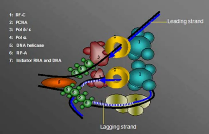
2002). These observations led to the conclusion that PCNA, thanks to its interaction with pol δ, could be located at the point of polymerase stalling, and would play a role as a recruiting platform, allowing the switch from replicative to translesion polymerases required to resume the replication fork (Fig. 2).
Recent studies in S. cerevisiae shed light on an additional level of regulation of bypass synthesis by
PCNA. PCNA has been shown to be competitively ubiquitinated by the RAD 6 dependent pathway or SUMOylated in association with DNA replication (Hoege et al. 2002). Stelter and Ulrich showed that DNA synthesis by the translesion pol ζ is differentially affected by mono-ubiquitin and SUMO modification of PCNA in yeast. Ubiquitination was shown to be required for damage-induced activity of polζ, whereas SUMOyla-tion seemed to be involved only during S-phase, in damage-independent synthesis, which may occur to resume fork stalling caused by refractory DNA structures (Stelter and Ulrich 2003; Haracska et al. 2004). It was proposed that this effect might be triggered by dissociation of PCNA from the replicative polymerases upon SUMOy-lation.
Furthermore, data from the fission yeast S. pombe demonstrated a role of the alternative clamp/clamp-loader pair, 9–1–1/Rad17–RF-C2–5(for details see“Transducing the damage signal via a checkpoint clamp–clamp-loader pair”), in recruiting the translesion polymerase DinB/pol κ
to chromatin upon DNA replication perturbation (Kai and Wang 2003). This finding, in agreement with models in which the 9–1–1 complex stabilises stalled DNA replica-tion fork structures (Bermudez et al. 2003; Ellison and Stillman2003; Majka and Burgers2003; Zou et al.2003), suggests an alternative pathway for recruiting translesion polymerases at the sites of fork stalling. The signals leading to differential selection of these pathways remain to be assessed.
Fig. 2 Switch from the replicative to the translesion DNA polymerase. During DNA replication, the DNA replication machin-ery [schematically drawn as the polδ/ε holoenzyme (pol δ/ε, RF-C, andPCNA)], eventually meets lesions on the DNA (represented with black symbols). Polymerases δ and ε are unable to traverse DNA lesions and their arrest causes a block of the replication fork. We propose the existence of subnuclear compartments or foci where
replicative or translesion (TLS) polymerases are stored. Upon stalling of the fork, checkpoints are activated, leading to the recruitment of specific factors at the lesion through as yet unidentified machinery. As a consequence, the replicative pols δ andε are replaced by TLS polymerases. After damage bypass, the normal replication machinery is reconstituted through an inverse mechanism
The 3′→5′ exonucleases
An essential step in maintaining genomic stability is that the replicative polymerases replicate DNA in an accurate manner under normal conditions in cells. This accuracy is ensured by so-called 3′→5′ proofreading exonucleases. This proofreading activity can be exerted either by internal or external exonuclease (recently reviewed in Shevelev and Hubscher 2002). The two replicative pols δ and ε harbour their own exonuclease catalytic site and are able to proofread the DNA strand in conjunction with its elonga-tion. The balance between polymerizing and editing mode is achieved by shuttling of the 3′ end of the primer from the polymerase to the exonuclease active site, whereas the duplex DNA position remains unchanged (Franklin et al.
2001). A defect in polδ exonuclease activity in mice has been shown to lead to an increased incidence of cancer, consistent with the generation of a mutator phenotype that can also be observed in pol δ exonuclease-deficient yeast mutants. This confirms the essential character of this proofreading activity in the maintenance of genomic stability (Morrison et al.1993; Goldsby et al.2001,2002). On the other hand, other polymerases such as the initiation pol α and the translesion polymerases do not possess exonuclease activity. Moreover, when a polymer-ase is stalled at a site of DNA lesion, intrinsic exonuclepolymer-ase activity would be helpless. In this case, external proof-reading exonucleases might intervene. Such exonucleases have been characterised in the last few years and were called TREX exonucleases. TREX2 has been shown to copurify with polδ, and to increase its fidelity, especially under imbalanced dNTP conditions (Shevelev et al.2002).
Link between DNA replication and checkpoint control
Regulation of DNA replication forks is tightly linked to the DNA damage and DNA replication checkpoint controls. The checkpoint effectors mediate a signal that stops cell cycle progression upon DNA damage or a stalled replication fork. This delay in the cell cycle allows restarting of the replication fork and repair of damage, thus preserving genomic integrity. Several pathways are involved in mediating the checkpoint response, and recent studies allowed the implication of a growing number of DNA replication factors in these important pathways (Table 3).
PCNA and the cell cycle inhibitor p21
The first DNA replication factor that was shown to be involved in checkpoint control is PCNA. It was charac-terised as an interacting partner of the CDK inhibitor, p21 (Gulbis et al.1996). Upon DNA damage, p21 is actively expressed in a p53 dependent or independent pathway, leading to a G1–S transition blockage. PCNA might be a mediator of this effect, considering that its interaction with p21 competes with polδ binding to PCNA (Podust et al.
1995). This interaction leads to the inhibition of pol δ processivity stimulation by PCNA. However, it did not affect the NER mechanism in vitro, suggesting that the interaction of PCNA with p21 selectively inhibits and regulates DNA replication fork progression (Shivji et al.
1998).
Transducing the damage signal via a checkpoint clamp–clamp-loader pair
Activation of the DNA damage and DNA replication checkpoints requires the action of DNA damage sensors and transducers (Melo and Toczyski2002). Some of these factors, such as ataxia telangectasia mutated protein (ATM), ATM-related protein (ATR), ATR interacting protein (ATRIP), Rad17, Rad9, Rad1 and Hus1, are thought to be involved in triggering DNA repair processes. The three human proteins Rad9, Hus1, and Rad1 form a heterotrimeric complex (9–1–1 complex) that exhibits structural similarity with the homotrimeric clamp formed by PCNA (Venclovas and Thelen 2000; Burtelow et al.
2001; Griffith et al. 2002; Shiomi et al. 2002). Further-more, human Rad17 has been shown to associate with the four small subunits of the heteropentameric RF-C complex (RFC) in a complex exhibiting a structure similar to the RFC (Venclovas and Thelen 2000; Kaur et al. 2001; Lindsey-Boltz et al. 2001; Griffith et al. 2002; Shiomi et al. 2004). It has been shown recently that the 9–1–1 complex can be loaded onto DNA by the Rad17–RF-C2–5 clamp-loader in vitro (Bermudez et al. 2003; Ellison and Stillman2003; Majka and Burgers2003; Zou and Elledge
2003). Moreover, the 9–1–1 complex, Rad17–RF-C2–5 and PCNA co-localise in foci formed upon DNA damage (Burtelow et al.2000; Dahm and Hubscher2002; Meister et al. 2003). These data suggest a mechanism in which Rad17–RF-C2–5would localise on DNA lesions, allowing the recruitment of the 9–1–1 complex to these sites. ATM and ATR kinases are recruited simultaneously to the same
Table 3 Roles of DNA replication proteins in checkpoint regulation. (PCNA, proliferating cell nuclear antigen; Pol, polymerase; RF-C, replication factor C; RP-A, replication protein A)
Protein Role in checkpoint pathway
PCNA p21 mediated cell-cycle blockage
RF-C (subunits 2–5) Transduction of DNA damage signal in replication and repair checkpoints cascades RP-A Sensing of DNA damage in replication and repair checkpoint cascades
sites of DNA damage but in a 9–1–1 complex and Rad17– RF-C2–5independent manner (Kondo et al.2001; Melo et al. 2001; Roos-Mattjus et al.2002; Zou et al.2002). The proposed mechanism is that, once loaded on a DNA damage site, the 9–1–1 complex may serve as a recruiting platform for the checkpoint effector kinases such as Chk1 or Chk2, which are subsequently phosphorylated by the ATR/ATM kinases (Martinho et al. 1998; Zhou and Elledge 2000; Melo and Toczyski 2002). Additionally, a model has very recently been proposed by two different groups, in which the 9–1–1 complex and the Rad17–RF-C2–5clamp-loader could stabilize stalled replication forks (Ellison and Stillman 2003; Zou et al. 2003). Moreover, the replication initiation protein Dpb11/Cut5/TopBP1 also appears to be involved in the early steps of the DNA damage response. Indeed, upon DNA damage triggered phosphorylation of Rad9, Dpb11/Cut5/TopBP1 interacts with the latter and with ATR, which in turn leads to activation of the chk1-mediated checkpoint response (Greer et al. 2003; Parrilla-Castellar and Karnitz 2003; Furuya et al. 2004). Finally, some evidence led to the conclusion that polε is also involved in coordinating DNA replication and the DNA damage response. Upon DNA damage, phosphorylated Rad17 has been shown to localize at replication sites through its interaction with polε, and in mutants deleted for the N-terminal part of pol ε, several Rad family proteins have been shown to be necessary for cell survival (Feng and D’Urso2001; Post et al. 2003). Taken together, these data show that the tight link between DNA replication and DNA damage sensing is triggered by replisome proteins involved in several different steps of DNA replication.
Sensing the damage through the single-strand binding protein RP-A
A crucial step in the checkpoint processes is sensing of the lesion allowing the subsequent recruitment of the DNA damage signal transducers. Several studies suggested that in this step the single strand binding protein RP-A is a key player. Indeed, RP-A-coated single-strand DNA is a common structure generated at sites of DNA damage. The ATR checkpoint kinase is recruited to sites of single-strand DNA by the intermediary binding of its regulatory partner, ATRIP, to the RP-A protein (Zou and Elledge
2003). In further studies it was shown that yeasts mutated in RFA1 (homologue to the large p70 subunit of human RP-A), are defective in loading the yeast homologue of Rad9, Ddc1. Moreover, RP-A stimulated the binding of the Rad17–RF-C2–5 complex to various DNA structures. RP-A was also shown to facilitate the recruitment of the 9– 1–1 complex by the Rad17–RF-C2–5 complex to primed and gapped DNA structures in vitro (Zou et al. 2003). Furthermore, it was recently shown in yeast that RP-A single-strand DNA filaments allow the sensing of stalled replication forks via recruitment of Ddc1 (homologue to the human Rad9) and Ddc2 (homologue to the human ATRIP). This recruitment leads to checkpoint activation and to stabilisation of the stalled replication fork (Lucca et al.2004) and confirms the crucial role of the RP-A protein in the DNA damage-sensing step of the checkpoint response.
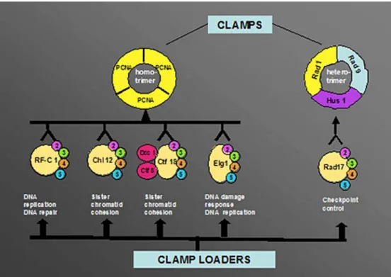
Fig. 3 Alternative clamps and clamp loaders. Under normal conditions, e.g. during replica-tion, the replication factor C (RF-C) heteropentameric clamp-loader (RF-C1–5) loads prolifer-ating cell nuclear antigen (PCNA) in an ATP-dependent manner. In other cellular meta-bolic events such as sister chro-matid cohesion, DNA damage response and checkpoint control alternative clamp loaders (con-sisting of the four small subunits RF-C2–5and an additional pro-tein) can either load PCNA or the alternative clamp, the 9–1–1 complex. For more details see text
Role of polα/primase in the replication checkpoint at the lagging strand
By using a Xenopus egg extract cell-free system, pol α/ primase was found to be indispensable for replication checkpoint induction and this was detected by the intermediary chk1 phosphorylation (Michael et al. 2000). Moreover, evidence was provided that RNA synthesis by the primase subunits alone was sufficient for this effect. The importance of the primase-mediated RNA synthesis in this step might be explained by the transient character of the RNA primer during normal Okazaki fragment matu-ration. Hence, the persistence of such a structure would be a logical signal to activate the checkpoint pathway, thus preventing entrance into mitosis until DNA replication at the lagging strand with its more than 20 million Okazaki fragments has been completed.
Alternative clamp loaders
In addition to the “classical” RF-C protein involved in DNA replication and to the checkpoint clamp-loader Rad17–RF-C2–5, the four small subunits of RF-C are part of other alternative clamp loaders (Fig.3).
First, the sister chromatid cohesion factor, Chl12, exhibits sequence similarity to the large subunit of RF-C. The complex formed by association of Chl12 with the RF-C2–5subunits is structurally similar to RF-C and was shown to be able to load PCNA in an ATP-dependent manner onto circular DNA (Shiomi et al. 2004). The Chl12–RF-C2–5complex could stimulate polδ activity on an M13 circular template in the presence of PCNA and
RP-A. However, this alternative clamp-loader is not capable of replacing RF-C in SV40 DNA replication in vitro. Another set of proteins involved in sister chromatid cohesion, Ctf18, Dcc1 and Ctf8, can also associate with the four small RF-C subunits. Similarly to the previous complex, this complex is also a clamp-loader for PCNA, and can substitute for the replicative RF-C in a pol δ-catalysed DNA replication assay (Shiomi et al. 2004). Taken together, these data suggest that sister chromatid cohesion might be directly linked to DNA replication.
Second, the Elg1 protein shares sequence homology with the large subunit of the clamp-loader RF-C and forms a complex with the RF-C2–5 subunits (Ben-Aroya et al.
2003). Genetic data from yeast studies indicated that Elg1 has a role in DNA damage response and in maintaining the integrity of the genome, as well as in DNA replication (Bellaoui et al. 2003; Kanellis et al. 2003). Elg1 acts in coordination with Rad24, the yeast homologue of human Rad17 in the DNA damage response and contributes to activation of the checkpoint kinase Rad53 (homologue of the human Chk2). Moreover, its physical interaction with the PCNA homologue Pol30 and the FEN-1 homologue Rad27 suggested a role of Elg1 in Okazaki fragment maturation (Kanellis et al. 2003). Finally, Elg1-RF-C2–5 contributes to genome stability via telomere length regulation through a putative replication-mediated path-way. The telomeric function of Elg1 was shown to be independent of recombination and dependent on an active telomerase and polα (Smolikov et al. 2004). These data suggested that Elg1-RF-C2–5 plays a role in checkpoint pathway and DNA replication.
Taken together there is growing evidence that the four small subunits of RF-C are common to all the alternative
Fig. 4 DNA replication pro-teins and their involvement in genomic stability. The replica-tion fork is identical to that in Fig. 1, but in addition the four replication proteins pol α/pri-mase, replication protein A ( RP-A), proliferating cell nuclear antigen (PCNA) and replication factor C (RF-C) are highlighted for their control functions in the cell. For more details see text. (BER base excision repair; MMR mismatch repair;NER nucleo-tide excision repair)
clamp-loaders, and depending whether they are bound to the large subunit of RF-C or to factors such as Rad17, Chl12, Ctf18-Dcc1-Ctf8 or Elg1, play an important role in regulating genomic maintenance processes in coordination with DNA replication (Fig. 3).
Conclusions
The DNA replication machinery is known to be a complex association of proteins tightly regulated in order to ensure faithful and complete DNA replication. However, the complexity of the function of each polypeptide involved in the replisome, and the regulation of DNA replication fork has been underestimated for a long time. Indeed, regula-tion systems exist not only in order to regulate the funcregula-tion of the replicon itself, but also to coordinate it with other events involved in the maintenance of genomic stability (Fig. 4). The challenging task in the future is to depict more accurately the pathways triggering signals leading to assignment of replisomal proteins to different DNA repair tasks under specific genotoxic stress conditions.
Acknowledgements The authors thank Isabelle Frouin for critical reading of the manuscript. The work performed in the authors’ laboratory was supported by the Swiss National Science Foundation and the Kanton of Zürich. M.T. is supported by a FEBS fellowship and in part by the Swiss National Science Foundation.
References
Aboussekhra A, Wood RD (1995) Detection of nucleotide excision repair incisions in human fibroblasts by immunostaining for PCNA. Exp Cell Res 221:326–332
Alleva JL, Doetsch PW (2000) The nature of the 5′-terminus is a major determinant for DNA processing by Schizosaccharomy-ces pombe Rad2p, a FEN-1 family nuclease. Nucleic Acids Res 28:2893–2901
Araki H, Leem SH, Phongdara A, Sugino A (1995) Dpb11, which interacts with DNA polymerase II (epsilon) inSaccharomyces cerevisiae, has a dual role in S-phase progression and at a cell cycle checkpoint. Proc Natl Acad Sci USA 92:11791–11795 Ayyagari R, Gomes XV, Gordenin DA, Burgers PM (2003) Okazaki
fragment maturation in yeast. I. Distribution of functions between FEN1 and DNA2. J Biol Chem 278:1618–1625 Bae SH, Bae KH, Kim JA, Seo YS (2001) RPA governs
endonuclease switching during processing of Okazaki frag-ments in eukaryotes. Nature 412:456–461
Bellaoui M, Chang M, Ou J, Xu H, Boone C, Brown GW (2003) Elg1 forms an alternative RFC complex important for DNA replication and genome integrity. EMBO J 22:4304–4313 Ben-Aroya S, Koren A, Liefshitz B, Steinlauf R, Kupiec M (2003)
ELG1, a yeast gene required for genome stability, forms a complex related to replication factor C. Proc Natl Acad Sci USA 100:9906–9911
Bermudez VP, Lindsey-Boltz LA, Cesare AJ, Maniwa Y, Griffith JD, Hurwitz J, Sancar A (2003) Loading of the human 9–1–1 checkpoint complex onto DNA by the checkpoint clamp loader hRad17-replication factor C complex in vitro. Proc Natl Acad Sci USA 100:1633–1638
Brosh RM Jr, Driscoll HC, Dianov GL, Sommers JA (2002) Biochemical characterization of the WRN-FEN-1 functional interaction. Biochemistry 41:12204–12216
Budd ME, Campbell JL (1997) A yeast replicative helicase, Dna2 helicase, interacts with yeast FEN-1 nuclease in carrying out its essential function. Mol Cell Biol 17:2136–2142
Burtelow MA, Kaufmann SH, Karnitz LM (2000) Retention of the human Rad9 checkpoint complex in extraction-resistant nuclear complexes after DNA damage. J Biol Chem 275:26343–26348 Burtelow MA, Roos-Mattjus PM, Rauen M, Babendure JR, Karnitz LM (2001) Reconstitution and molecular analysis of the hRad9-hHus1-hRad1 (9–1–1) DNA damage responsive check-point complex. J Biol Chem 276:25903–25909
Christmann M, Tomicic MT, Roos WP, Kaina B (2003) Mechanisms of human DNA repair: an update. Toxicology 193:3–34 Clark AB, Valle F, Drotschmann K, Gary RK, Kunkel TA (2000)
Functional interaction of proliferating cell nuclear antigen with MSH2–MSH6 and MSH2–MSH3 complexes. J Biol Chem 275:36498–36501
Dahm K, Hubscher U (2002) Colocalization of human Rad17 and PCNA in late S phase of the cell cycle upon replication block. Oncogene 21:7710–7719
Dianova II, Bohr VA, Dianov GL (2001) Interaction of human AP endonuclease 1 with flap endonuclease 1 and proliferating cell nuclear antigen involved in long-patch base excision repair. Biochemistry 40:12639–12644
Donovan S, Harwood J, Drury LS, Diffley JF (1997) Cdc6p-dependent loading of Mcm proteins onto pre-replicative chromatin in budding yeast. Proc Natl Acad Sci USA 94:5611–5616
Eggler AL, Inman RB, Cox MM (2002) The Rad51-dependent pairing of long DNA substrates is stabilized by replication protein A. J Biol Chem 277:39280–39288
Ellison V, Stillman B (2003) Biochemical characterization of DNA damage checkpoint complexes: clamp loader and clamp complexes with specificity for 5′ recessed DNA. PLoS Biol 1:E33
Feng W, D’Urso G (2001) Schizosaccharomyces pombe cells lacking the amino-terminal catalytic domains of DNA poly-merase epsilon are viable but require the DNA damage checkpoint control. Mol Cell Biol 21:4495–4504
Flores-Rozas H, Clark D, Kolodner RD (2000) Proliferating cell nuclear antigen and Msh2p–Msh6p interact to form an active mispair recognition complex. Nat Genet 26:375–378
Forsburg SL (2004) Eukaryotic MCM proteins: beyond replication initiation. Microbiol Mol Biol Rev 68:109–131 (Table of contents)
Franklin MC, Wang J, Steitz TA (2001) Structure of the replicating complex of a pol alpha family DNA polymerase. Cell 105:657– 667
Furuya K, Poitelea M, Guo L, Caspari T, Carr AM (2004) Chk1 activation requires Rad9 S/TQ-site phosphorylation to promote association with C-terminal BRCT domains of Rad4TOPBP1. Genes Dev 18:1154–1164
Gary R, Ludwig DL, Cornelius HL, MacInnes MA, Park MS (1997) The DNA repair endonuclease XPG binds to proliferating cell nuclear antigen (PCNA) and shares sequence elements with the PCNA-binding regions of FEN-1 and cyclin-dependent kinase inhibitor p21. J Biol Chem 272:24522–24529
Genschel J, Modrich P (2003) Mechanism of 5′-directed excision in human mismatch repair. Mol Cell 12:1077–1086
Goldsby RE, Lawrence NA, Hays LE, Olmsted EA, Chen X, Singh M, Preston BD (2001) Defective DNA polymerase-delta proofreading causes cancer susceptibility in mice. Nat Med 7:638–639
Goldsby RE, Hays LE, Chen X, Olmsted EA, Slayton WB, Spangrude GJ, Preston BD (2002) High incidence of epithelial cancers in mice deficient for DNA polymerase delta proof-reading. Proc Natl Acad Sci USA 99:15560–15565
Greer DA, Besley BD, Kennedy KB, Davey S (2003) hRad9 rapidly binds DNA containing double-strand breaks and is required for damage-dependent topoisomerase II beta binding protein 1 focus formation. Cancer Res 63:4829–4835
Griffith JD, Lindsey-Boltz LA, Sancar A (2002) Structures of the human Rad17-replication factor C and checkpoint Rad 9-1-1 complexes visualized by glycerol spray/low voltage microsco-py. J Biol Chem 277:15233–15236
Gulbis JM, Kelman Z, Hurwitz J, O’Donnell M, Kuriyan J (1996) Structure of the C-terminal region of p21(WAF1/CIP1) complexed with human PCNA. Cell 87:297–306
Haracska L, Johnson RE, Unk I, Phillips B, Hurwitz J, Prakash L, Prakash S (2001a) Physical and functional interactions of human DNA polymerase eta with PCNA. Mol Cell Biol 21:7199–7206
Haracska L, Johnson RE, Unk I, Phillips BB, Hurwitz J, Prakash L, Prakash S (2001b) Targeting of human DNA polymerase iota to the replication machinery via interaction with PCNA. Proc Natl Acad Sci USA 98:14256–14261
Haracska L, Kondratick CM, Unk I, Prakash S, Prakash L (2001c) Interaction with PCNA is essential for yeast DNA polymerase eta function. Mol Cell 8:407–415
Haracska L, Unk I, Johnson RE, Phillips BB, Hurwitz J, Prakash L, Prakash S (2002) Stimulation of DNA synthesis activity of human DNA polymerase kappa by PCNA. Mol Cell Biol 22:784–791
Haracska L, Torres-Ramos CA, Johnson RE, Prakash S, Prakash L (2004) Opposing effects of Ubiquitin conjugation and SUMO modification of PCNA on replicational bypass of DNA lesions inSaccharomyces cerevisiae. Mol Cell Biol 24:4267–4274 Hardy CF, Dryga O, Seematter S, Pahl PM, Sclafani RA (1997)
mcm5/cdc46-bob1 bypasses the requirement for the S phase activator Cdc7p. Proc Natl Acad Sci USA 94:3151–3155 Hasan S, Stucki M, Hassa PO, Imhof R, Gehrig P, Hunziker P,
Hubscher U, Hottiger MO (2001) Regulation of human flap endonuclease-1 activity by acetylation through the transcrip-tional coactivator p300. Mol Cell 7:1221–1231
Hashimoto Y, Takisawa H (2003) Xenopus Cut5 is essential for a CDK-dependent process in the initiation of DNA replication. EMBO J 22:2526–2535
Henneke G, Koundrioukoff S, Hubscher U (2003) Multiple roles for kinases in DNA replication. EMBO Rep 4:252–256
Henricksen LA, Veeraraghavan J, Chafin DR, Bambara RA (2002) DNA ligase I competes with FEN1 to expand repetitive DNA sequences in vitro. J Biol Chem 277:22361–22369
Hoege C, Pfander B, Moldovan GL, Pyrowolakis G, Jentsch S (2002) RAD6-dependent DNA repair is linked to modification of PCNA by ubiquitin and SUMO. Nature 419:135–141 Hoeijmakers JH (2001) Genome maintenance mechanisms for
preventing cancer. Nature 411:366–374
Hozak P, Hassan AB, Jackson DA, Cook PR (1993) Visualization of replication factories attached to nucleoskeleton. Cell 73:361– 373
Hubscher U, Seo YS (2001) Replication of the lagging strand: a concert of at least 23 polypeptides. Mol Cells 12:149–157 Hubscher U, Maga G, Spadari S (2002) Eukaryotic DNA
polymerases. Annu Rev Biochem 71:133–163
Jiang W, McDonald D, Hope TJ, Hunter T (1999) Mammalian Cdc7–Dbf4 protein kinase complex is essential for initiation of DNA replication. EMBO J 18:5703–5713
Jin YH, Ayyagari R, Resnick MA, Gordenin DA, Burgers PM (2003) Okazaki fragment maturation in yeast. II. Cooperation between the polymerase and 3′-5′-exonuclease activities of Pol delta in the creation of a ligatable nick. J Biol Chem 278:1626– 1633
Kai M, Wang TS (2003) Checkpoint activation regulates mutagenic translesion synthesis. Genes Dev 17:64–76
Kamath-Loeb AS, Johansson E, Burgers PM, Loeb LA (2000) Functional interaction between the Werner syndrome protein and DNA polymerase delta. Proc Natl Acad Sci USA 97:4603– 4608
Kamimura Y, Masumoto H, Sugino A, Araki H (1998) Sld2, which interacts with Dpb11 inSaccharomyces cerevisiae, is required for chromosomal DNA replication. Mol Cell Biol 18:6102– 6109
Kanellis P, Agyei R, Durocher D (2003) Elg1 forms an alternative PCNA-interacting RFC complex required to maintain genome stability. Curr Biol 13:1583–1595
Kannouche P, Fernandez de Henestrosa AR, Coull B, Vidal AE, Gray C, Zicha D, Woodgate R, Lehmann AR (2002) Local-ization of DNA polymerases eta and iota to the replication machinery is tightly co-ordinated in human cells. EMBO J 21:6246–6256
Kaur R, Kostrub CF, Enoch T (2001) Structure–function analysis of fission yeast Hus1-Rad1-Rad9 checkpoint complex. Mol Biol Cell 12:3744–3758
Kearsey SE, Cotterill S (2003) Enigmatic variations: divergent modes of regulating eukaryotic DNA replication. Mol Cell 12:1067–1075
Kearsey SE, Montgomery S, Labib K, Lindner K (2000) Chromatin binding of the fission yeast replication factor mcm4 occurs during anaphase and requires ORC and cdc18. EMBO J 19:1681–1690
Kedar PS, Kim SJ, Robertson A, Hou E, Prasad R, Horton JK, Wilson SH (2002) Direct interaction between mammalian DNA polymerase beta and proliferating cell nuclear antigen. J Biol Chem 277:31115–31123
Kleczkowska HE, Marra G, Lettieri T, Jiricny J (2001) hMSH3 and hMSH6 interact with PCNA and colocalize with it to replica-tion foci. Genes Dev 15:724–736
Klungland A, Lindahl T (1997) Second pathway for completion of human DNA base excision-repair: reconstitution with purified proteins and requirement for DNase IV (FEN1). EMBO J 16:3341–3348
Kokoska RJ, Stefanovic L, Tran HT, Resnick MA, Gordenin DA, Petes TD (1998) Destabilization of yeast micro- and minisa-tellite DNA sequences by mutations affecting a nuclease involved in Okazaki fragment processing (rad27) and DNA polymerase delta (pol3-t). Mol Cell Biol 18:2779–2788 Kondo T, Wakayama T, Naiki T, Matsumoto K, Sugimoto K (2001)
Recruitment of Mec1 and Ddc1 checkpoint proteins to double-strand breaks through distinct mechanisms. Science 294:867– 870
Krishna TS, Kong XP, Gary S, Burgers PM, Kuriyan J (1994) Crystal structure of the eukaryotic DNA polymerase processiv-ity factor PCNA. Cell 79:1233–1243
Krokan HE, Otterlei M, Nilsen H, Kavli B, Skorpen F, Andersen S, Skjelbred C, Akbari M, Aas PA, Slupphaug G (2001) Properties and functions of human uracil-DNA glycosylase from the UNG gene. Prog Nucleic Acid Res Mol Biol 68:365– 386
Kubota Y, Takase Y, Komori Y, Hashimoto Y, Arata T, Kamimura Y, Araki H, Takisawa H (2003) A novel ring-like complex of Xenopus proteins essential for the initiation of DNA replica-tion. Genes Dev 17:1141–1152
Lau PJ, Flores-Rozas H, Kolodner RD (2002) Isolation and characterization of new proliferating cell nuclear antigen (POL30) mutator mutants that are defective in DNA mismatch repair. Mol Cell Biol 22:6669–6680
Lebel M, Spillare EA, Harris CC, Leder P (1999) The Werner syndrome gene product co-purifies with the DNA replication complex and interacts with PCNA and topoisomerase I. J Biol Chem 274:37795–37799
Liang C, Weinreich M, Stillman B (1995) ORC and Cdc6p interact and determine the frequency of initiation of DNA replication in the genome. Cell 81:667–676
Lindahl T, Wood RD (1999) Quality control by DNA repair. Science 286:1897–1905
Lindsey-Boltz LA, Bermudez VP, Hurwitz J, Sancar A (2001) Purification and characterization of human DNA damage checkpoint Rad complexes. Proc Natl Acad Sci USA 98:11236–11241
Lucca C, Vanoli F, Cotta-Ramusino C, Pellicioli A, Liberi G, Haber J, Foiani M (2004) Checkpoint-mediated control of replisome-fork association and signalling in response to replication pausing. Oncogene 23:1206–1213
Maga G, Hubscher U (1996) DNA replication machinery: functional characterization of a complex containing DNA polymerase alpha, DNA polymerase delta, and replication factor C suggests an asymmetric DNA polymerase dimer. Biochemistry 35:5764– 5777
Maga G, Hubscher U (2003) Proliferating cell nuclear antigen (PCNA): a dancer with many partners. J Cell Sci 116:3051– 3060
Maga G, Stucki M, Spadari S, Hubscher U (2000) DNA polymerase switching. I. Replication factor C displaces DNA polymerase alpha prior to PCNA loading. J Mol Biol 295:791–801 Maga G, Frouin I, Spadari S, Hubscher U (2001a) Replication
protein A as a“fidelity clamp” for DNA polymerase alpha. J Biol Chem 276:18235–18242
Maga G, Villani G, Tillement V, Stucki M, Locatelli GA, Frouin I, Spadari S, Hubscher U (2001b) Okazaki fragment processing: modulation of the strand displacement activity of DNA polymerase delta by the concerted action of replication protein A, proliferating cell nuclear antigen, and flap endonuclease-1. Proc Natl Acad Sci USA 98:14298–14303
Maga G, Villani G, Ramadan K, Shevelev I, Tanguy Le Gac N, Blanco L, Blanca G, Spadari S, Hubscher U (2002) Human DNA polymerase lambda functionally and physically interacts with proliferating cell nuclear antigen in normal and translesion DNA synthesis. J Biol Chem 277:48434–48440
Maiorano D, Moreau J, Mechali M (2000) XCDT1 is required for the assembly of pre-replicative complexes inXenopus laevis. Nature 404:622–625
Majka J, Burgers PM (2003) Yeast Rad17/Mec3/Ddc1: a sliding clamp for the DNA damage checkpoint. Proc Natl Acad Sci USA 100:2249–2254
Martinho RG, Lindsay HD, Flaggs G, DeMaggio AJ, Hoekstra MF, Carr AM, Bentley NJ (1998) Analysis of Rad3 and Chk1 protein kinases defines different checkpoint responses. EMBO J 17:7239–7249
Meister P, Poidevin M, Francesconi S, Tratner I, Zarzov P, Baldacci G (2003) Nuclear factories for signalling and repairing DNA double strand breaks in living fission yeast. Nucleic Acids Res 31:5064–5073
Melo J, Toczyski D (2002) A unified view of the DNA-damage checkpoint. Curr Opin Cell Biol 14:237–245
Melo JA, Cohen J, Toczyski DP (2001) Two checkpoint complexes are independently recruited to sites of DNA damage in vivo. Genes Dev 15:2809–2821
Michael WM, Ott R, Fanning E, Newport J (2000) Activation of the DNA replication checkpoint through RNA synthesis by primase. Science 289:2133–2137
Mimura S, Takisawa H (1998) Xenopus Cdc45-dependent loading of DNA polymerase alpha onto chromatin under the control of S-phase Cdk. EMBO J 17:5699–5707
Miura M, Sasaki T (1996) Effect of XPA gene mutations on UV-induced immunostaining of PCNA in fibroblasts from xero-derma pigmentosum group A patients. Mutat Res 364:51–56 Mizuno T, Yamagishi K, Miyazawa H, Hanaoka F (1999) Molecular
architecture of the mouse DNA polymerase alpha-primase complex. Mol Cell Biol 19:7886–7896
Mizushima T, Takahashi N, Stillman B (2000) Cdc6p modulates the structure and DNA binding activity of the origin recognition complex in vitro. Genes Dev 14:1631–1641
Morrison A, Johnson AL, Johnston LH, Sugino A (1993) Pathway correcting DNA replication errors inSaccharomyces cerevisiae. EMBO J 12:1467–1473
Mossi R, Keller RC, Ferrari E, Hubscher U (2000) DNA polymerase switching. II. Replication factor C abrogates primer synthesis by DNA polymerase alpha at a critical length. J Mol Biol 295:803–814
Owens JC, Detweiler CS, Li JJ (1997) CDC45 is required in conjunction with CDC7/DBF4 to trigger the initiation of DNA replication. Proc Natl Acad Sci USA 94:12521–12526 Parrilla-Castellar ER, Karnitz LM (2003) Cut5 is required for the
binding of Atr and DNA polymerase alpha to genotoxin-damaged chromatin. J Biol Chem 278:45507–45511
Pascucci B, Stucki M, Jonsson ZO, Dogliotti E, Hubscher U (1999) Long patch base excision repair with purified human proteins. DNA ligase I as patch size mediator for DNA polymerases delta and epsilon. J Biol Chem 274:33696–33702
Podust VN, Hubscher U (1993) Lagging strand DNA synthesis by calf thymus DNA polymerases alpha, beta, delta and epsilon in the presence of auxiliary proteins. Nucleic Acids Res 21:841– 846
Podust VN, Podust LM, Goubin F, Ducommun B, Hubscher U (1995) Mechanism of inhibition of proliferating cell nuclear antigen-dependent DNA synthesis by the cyclin-dependent kinase inhibitor p21. Biochemistry 34:8869–8875
Pollok S, Stoepel J, Bauerschmidt C, Kremmer E, Nasheuer HP (2003) Regulation of eukaryotic DNA replication at the initiation step. Biochem Soc Trans 31:266–269
Post SM, Tomkinson AE, Lee EY (2003) The human checkpoint Rad protein Rad17 is chromatin-associated throughout the cell cycle, localizes to DNA replication sites, and interacts with DNA polymerase epsilon. Nucleic Acids Res 31:5568–5575 Prasad R, Dianov GL, Bohr VA, Wilson SH (2000) FEN1
stimulation of DNA polymerase beta mediates an excision step in mammalian long patch base excision repair. J Biol Chem 275:4460–4466
Reagan MS, Pittenger C, Siede W, Friedberg EC (1995) Characterization of a mutant strain ofSaccharomyces cerevisiae with a deletion of the RAD27 gene, a structural homolog of the RAD2 nucleotide excision repair gene. J Bacteriol 177:364– 371
Reardon JT, Sancar A (2002) Molecular anatomy of the human excision nuclease assembled at sites of DNA damage. Mol Cell Biol 22:5938–5945
Riedl T, Hanaoka F, Egly JM (2003) The comings and goings of nucleotide excision repair factors on damaged DNA. EMBO J 22:5293–5303
Roos-Mattjus P, Vroman BT, Burtelow MA, Rauen M, Eapen AK, Karnitz LM (2002) Genotoxin-induced Rad9-Hus1-Rad1 (9–1– 1) chromatin association is an early checkpoint signalling event. J Biol Chem 277:43809–43812
Schwacha A, Bell SP (2001) Interactions between two catalytically distinct MCM subgroups are essential for coordinated ATP hydrolysis and DNA replication. Mol Cell 8:1093–1104 Shevelev IV, Hubscher U (2002) The 3′→5′ exonucleases. Nat Rev
Mol Cell Biol 3:364–376
Shevelev IV, Ramadan K, Hubscher U (2002) The TREX2 3′→5′ exonuclease physically interacts with DNA polymerase delta and increases its accuracy. ScientificWorldJournal 2:275–281 Shiomi Y, Shinozaki A, Nakada D, Sugimoto K, Usukura J, Obuse
C, Tsurimoto T (2002) Clamp and clamp loader structures of the human checkpoint protein complexes, Rad9-1-1 and Rad17–RFC. Genes Cells 7:861–868
Shiomi Y, Shinozaki A, Sugimoto K, Usukura J, Obuse C, Tsurimoto T (2004) The reconstituted human Chl12–RFC complex functions as a second PCNA loader. Genes Cells 9:279–290
Shivji MK, Ferrari E, Ball K, Hubscher U, Wood RD (1998) Resistance of human nucleotide excision repair synthesis in vitro to p21Cdn1. Oncogene 17:2827–2838
Smolikov S, Mazor Y, Krauskopf A (2004) ELG1, a regulator of genome stability, has a role in telomere length regulation and in silencing. Proc Natl Acad Sci USA 101:1656–1661
Spiro C, Pelletier R, Rolfsmeier ML, Dixon MJ, Lahue RS, Gupta G, Park MS, Chen X, Mariappan SV, McMurray CT (1999) Inhibition of FEN-1 processing by DNA secondary structure at trinucleotide repeats. Mol Cell 4:1079–1085
Stelter P, Ulrich HD (2003) Control of spontaneous and damage-induced mutagenesis by SUMO and ubiquitin conjugation. Nature 425:188–191
Stillman B (1994) Smart machines at the DNA replication fork. Cell 78:725–728
Stucki M, Pascucci B, Parlanti E, Fortini P, Wilson SH, Hubscher U, Dogliotti E (1998) Mammalian base excision repair by DNA polymerases delta and epsilon. Oncogene 17:835–843
Stucki M, Jonsson ZO, Hubscher U (2001a) In eukaryotic flap endonuclease 1, the C terminus is essential for substrate binding. J Biol Chem 276:7843–7849
Stucki M, Stagljar I, Jonsson ZO, Hubscher U (2001b) A coordinated interplay: proteins with multiple functions in DNA replication, DNA repair, cell cycle/checkpoint control, and transcription. Prog Nucleic Acid Res Mol Biol 65:261–298 Takayama Y, Kamimura Y, Okawa M, Muramatsu S, Sugino A, Araki H (2003) GINS, a novel multiprotein complex required for chromosomal DNA replication in budding yeast. Genes Dev 17:1153–1165
Tanaka T, Knapp D, Nasmyth K (1997) Loading of an Mcm protein onto DNA replication origins is regulated by Cdc6p and CDKs. Cell 90:649–660
Thommes P, Kubota Y, Takisawa H, Blow JJ (1997) The RLF-M component of the replication licensing system forms complexes containing all six MCM/P1 polypeptides. EMBO J 16:3312– 3319
Tishkoff DX, Filosi N, Gaida GM, Kolodner RD (1997) A novel mutation avoidance mechanism dependent on S. cerevisiae RAD27 is distinct from DNA mismatch repair. Cell 88:253– 263
Tsurimoto T, Stillman B (1989) Multiple replication factors augment DNA synthesis by the two eukaryotic DNA polymerases, alpha and delta. EMBO J 8:3883–3889
Uchiyama M, Griffiths D, Arai K, Masai H (2001) Essential role of Sna41/Cdc45 in loading of DNA polymerase alpha onto minichromosome maintenance proteins in fission yeast. J Biol Chem 276:26189–26196
Unk I, Haracska L, Gomes XV, Burgers PM, Prakash L, Prakash S (2002) Stimulation of 3′→5′ exonuclease and 3′-phosphodies-terase activities of yeast apn2 by proliferating cell nuclear antigen. Mol Cell Biol 22:6480–6486
Venclovas C, Thelen MP (2000) Structure-based predictions of Rad1, Rad9, Hus1 and Rad17 participation in sliding clamp and clamp-loading complexes. Nucleic Acids Res 28:2481–2493
Waga S, Stillman B (1994) Anatomy of a DNA replication fork revealed by reconstitution of SV40 DNA replication in vitro. Nature 369:207–212
Waga S, Stillman B (1998) The DNA replication fork in eukaryotic cells. Annu Rev Biochem 67:721–751
Wakasugi M, Sancar A (1999) Order of assembly of human DNA repair excision nuclease. J Biol Chem 274:18759–18768 Wang H, Elledge SJ (1999) DRC1, DNA replication and checkpoint
protein 1, functions with DPB11 to control DNA replication and the S-phase checkpoint inSaccharomyces cerevisiae. Proc Natl Acad Sci USA 96:3824–3829
Wang X, Haber JE (2004) Role of Saccharomyces single-stranded DNA-binding protein RPA in the strand invasion step of double-strand break repair. PLoS Biol 2:E21
Yonemasu R, McCready SJ, Murray JM, Osman F, Takao M, Yamamoto K, Lehmann AR, Yasui A (1997) Characterization of the alternative excision repair pathway of UV-damaged DNA in Schizosaccharomyces pombe. Nucleic Acids Res 25:1553– 1558
Yuzhakov A, Kelman Z, Hurwitz J, O’Donnell M (1999) Multiple competition reactions for RPA order the assembly of the DNA polymerase delta holoenzyme. EMBO J 18:6189–6199 Zhang N, Lu X, Legerski RJ (2003) Partial reconstitution of human
interstrand cross-link repair in vitro: characterization of the roles of RPA and PCNA. Biochem Biophys Res Commun 309:71–78
Zhou BB, Elledge SJ (2000) The DNA damage response: putting checkpoints in perspective. Nature 408:433–439
Zou L, Elledge SJ (2003) Sensing DNA damage through ATRIP recognition of RPA-ssDNA complexes. Science 300:1542– 1548
Zou L, Cortez D, Elledge SJ (2002) Regulation of ATR substrate selection by Rad17-dependent loading of Rad9 complexes onto chromatin. Genes Dev 16:198–208
Zou L, Liu D, Elledge SJ (2003) Replication protein A-mediated recruitment and activation of Rad17 complexes. Proc Natl Acad Sci USA 100:13827–13832
![Fig. 2 Switch from the replicative to the translesion DNA polymerase. During DNA replication, the DNA replication machin-ery [schematically drawn as the pol δ / ε holoenzyme ( pol δ/ε , RF-C , and PCNA )], eventually meets lesions on the DNA (represented](https://thumb-eu.123doks.com/thumbv2/123doknet/14864295.636671/6.892.200.694.626.973/replicative-translesion-polymerase-replication-replication-schematically-holoenzyme-represented.webp)