e-Neuroforum 2011 · 2:35–41 DOI 10.1007/s13295-011-0018-1 © Springer-Verlag 2011 H.U. Zeilhofer Institut für Pharmakologie und Toxikologie, Universität Zürich, Zürich
Spinal neuroplasticity
in chronic pain
Introduction
Our body detects potentially noxious stimuli by means of nociceptors, special-ized neurons which translate physical or chemical stimuli into electrical signals and transmit these to the central nervous sys-tem (CNS). The majority of these (prima-ry) nociceptors are non-myelinated slowly conducting C fibers or thinly myelinated fast conducting Aδ fibers, the central end-ings of which end either in the dorsal horn of the spinal cord or in the trigeminal nu-cleus of the brainstem. The initial synaptic integration of nociceptive information oc-curs in these structures. Some of the spi-nal termispi-nals of nociceptors directly stim-ulate projection neurons in the most dor-sal layer of the spinal cord, the lamina I, while other nociceptor endings activate excitatory or inhibitory local interneurons in the same lamina as well as the underly-ing lamina II. Non-nociceptive mechano-sensitive fibers that are activated by mild tactile stimuli end primarily in deep lay-ers of the dorsal horn, where they come in contact with a different class of projection neurons, as well as with excitatory and inhibitory interneurons. In this way, the neuronal network of the dorsal horn in-tegrates peripheral nociceptive and non-nociceptive stimuli with pro- and antino-ciceptive signals from pathways descend-ing from the brainstem and midbrain in-to the spinal cord. The result of this spinal processing is then transmitted via sever-al interconnections by lamina-I projection neurons in lamina I to higher CNS areas, where nociceptive and non-nociceptive stimuli are consciously perceived. Neuro-plastic changes in this network represent an important mechanism of chronic pain.
Synaptic plasticity in lamina-I
projection neurons
Lamina-I projection neurons in the dor-sal horn play a crucial role in the percep-tion of chronic inflammatory and neuro-pathic pain. The majority of these pro-jection neurons are nocispecific neurons which, under physiological conditions, are activated exclusively by noxious stim-uli. They carry neurokinin 1 (NK1) recep-tors for the neuropeptide substance P re-leased from peptidergic C fibers. This lat-ter characlat-teristic has been used to destroy these spinal neurons in a targeted manner. Animals in which a conjugate comprising
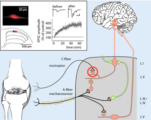
substance P and saporin was injected in the spinal cord were almost completely protected against inflammatory and neu-ropathic hyperalgesia [24]. Pursuing this idea, the plasticity of the synapses between primary nociceptors and these projection neurons have been closely investigated in recent years [13]. It could be demonstrated that intensive stimulation of these synaps-es triggers long-term potentiation (LTP). In particular, synapses with neurons that project to the periaqueductal gray (PAG), undergo LTP already with stimulation fre-quencies mimicking natural C fiber activ-ity (. Fig. 1).
Fig. 1 8 Long-term potentiation at the synapse between primary nociceptors and nocispecific projec-tion neurons in the lamina I. Intense stimulation of nociceptor endings leads to a persistent increase in synaptic transmission onto the projection neuron, as well as to hyperalgesia at the site of nociceptor stimulation. LI–LV indicate the various spinal laminae according to Rexed. (Modified from [13])
Inhibitory interneurons in
spinal control of nociception
Although homosynaptic plasticity at C-fi-ber synapses is able to explain increased
pain perception at the site of nociceptor stimulation (i.e., primary hyperalgesia), it is not able to simply explain why sensitiza-tion to signals from uncondisensitiza-tioned inputs also occurs. Indeed, local C-fiber
stimu-lation—experimentally inducible by, e.g., injection of capsaicin, a selective activa-tor of nocicepactiva-tors—also leads to hyper-sensitivity in unconditioned neighboring tissue and to pain originating from stim-ulation of non-nociceptive (capsaicin-in-sensitive) fibers (secondary hyperalge-sia, . Fig. 2). Interestingly, this second-ary hyperplasia involves exclusively me-chanical stimuli, while thermal sensitivi-ty remains unaltered. This mechanical hy-persensitivity is not based on the sensiti-zation of peripheral nerve fibers, but rath-er is the result of altrath-ered central process-ing of sensory signals. Thus this second-ary hyperplasia resembles heterosynaptic plasticity: intense stimulation of nocicep-tive C fibers leads to the sensitization of the body to signals from other uncondi-tioned fibers. Detailed analysis has shown that, at least in part, these fibers are low-threshold mechanosensitive fibers, i.e., not nociceptors. Sensitization to signals from these fibers leads to the painful per-ception of normally painless, mild tactile stimuli, called allodynia. Neurophysiolog-ically, this phenomenon is explained by the fact that signals from non-nociceptive fibers lead to the excitation of pain-sig-naling neurons. This unphysiological ex-citation of normally nocispecific projec-tion neurons could be the result of gen-erally increased projection neuron excit-ability. Alternatively, secondary hyperal-gesia and allodynia could also be the result of reduced inhibitory control in the dorsal horn. A popular model to explain allodyn-ia assumes the existence of two normally separate pathways for the spinal transmis-sion of nociceptive and non-nociceptive stimuli. While nocispecific neurons are excited exclusively by nociceptive fibers (marked with“N” in . Fig. 2), so-called wide dynamic range (WDR) neurons are excited by both nociceptive and non-no-ciceptive fibers. GABAergic and glyciner-gic neurons keep both pathways function-ally separate in vivo. A critical role of this kind played by inhibitory interneurons in the superficial dorsal horn was already proposed more than 45 years in the“gate control” theory of pain [18] (. Fig. 2a). Indeed, inhibitory γ-aminobutyric acid-releasing (GABAergic) and glycinergic neurons are densely packed in the super-ficial dorsal horn (. Fig. 2b). Both
neu-Fig. 2 8 a Primary and secondary hyperalgesia. In addition to sensitization at the site of nociceptor stimulation (primary hyperalgesia, dark red area), hypersensitivity to signals from non-nociceptive fibers also occurs in neigh-boring unconditioned areas (secondary hyperalgesia, light red area). Under physiological conditions, nociceptor signals are transmitted via nocispecific neurons (N) to supraspinal centers, while non-nociceptive signals are trans-mitted via wide dynamic range neurons in the deep dorsal horn. Left“Gate control” theory of pain (modified according to [18]). Inhibitory interneu- rons in the superficial dorsal horn, the substantia gelatinosa (SG), deter- mine whether signals are transmitted through the spinal transmission sys-tem (T) to the brain. The activity of these inhibitory interneurons is inversely regulated by nociceptors and non-nociceptive fibers. b GABAergic and gly- cinergic interneurons, visualized here by cell type-specific expression of en-hanced green fluorescent protein (EGFP), are abundant in the superficial dorsal horn. c Both transmitters open chloride channels (GABAA and strych- nine-sensitive glycine receptors), which inhibit the activation of postsynap-tic neurons
Review article
rotransmitters inhibit the excitability of spinal neurons by opening chloride chan-nels (. Fig. 2c). In vivo pharmacological blockade of spinal glycine or GABAA
re-ceptors leads to symptoms similar to sec-ondary hyperalgesia [27]. On a cellular level reduced inhibition demasks polysyn-aptic signals from non-nociceptive fibers occurs in previously nocispecific neurons [3, 29]. These findings show that non-no-ciceptive fibers are connected with nor-mally nocispecific neurons by excitatory interneurons, and that these connections are masked by inhibitory neurons un-der physiological conditions. Moreover, GABA and glycine receptor blockade in-creases the response of lamina I neurons to C-fiber stimulation and induces spon-taneous epilepsy-like discharge patterns in these neurons. In the intact organism, such cellular changes lead to correspond-ing sensory changes. Injectcorrespond-ing GABAA or
glycine receptor blockers induces hyper-sensitivity to noxious stimuli (hyperalge-sia), nociceptive reactions following stim-ulation of non-nociceptive fibers (allo-dynia), and behavioral changes suggestive of spontaneous pain. Reducing GABAer-gic or glycinerGABAer-gic inhibition thus recapit-ulates important characteristics of patho-logical pain.
Endogenous mechanisms
of disinhibition
Investigations carried out in recent years by various groups have shown that loss of spinal inhibitory control subsequent to peripheral inflammation, or neuropathies, or following intensive stimulation of noci-ceptors manifests itself endogenously.
Disinhibition in inflammation
In the context of inflammatory reactions, increased production of pronociceptive prostaglandins, in particular of prosta-glandin E2 (PGE2), is seen in
peripher-al inflamed tissue, but peripher-also in the CNS. Induction of cyclooxygenase-2 (COX-2), a key enzyme in prostaglandin syn-thesis, is a key process. On a cellular lev-el, PGE2 production results in facilitated
transmission of signals from sensory af-ferents to second order neurons in the superficial dorsal [20], in direct
depo-larization of neurons in the deep dorsal horn [2], as well as in specific inhibition of glycine receptors in the superficial lay-ers of the dorsal horn, i.e., in the central nociceptive innervation territories [34] (. Fig. 3). PGE2 produced in the spinal
cord activates PGE2 receptors of the EP2
subtype, leading to increased production of cAMP as well as protein kinase A stim-ulation [1]. This activation leads in turn to phosphorylation and inhibition of a par-ticular isoform of glycine receptors con-taining the α3 subunit (GlyRα3 receptors) [9]. The contribution of this process to pain sensitization in the context of various pathologies has been investigated with the help of genetically manipulated mice lack-ing individual elements of this transduc-tion cascade. EP2 receptor- and GlyRα3-deficient mice demonstrated an almost complete absence of nociceptive sensiti-zation following spinal PGE2 injection.
Moreover, they recover far more rapidly from inflammation-induced pain sensiti-zation than wild-type mice [1, 9, 33]. Fur-ther findings support the relevance of this prostaglandin-mediated disinhibition in inflammation-induced pain sensitization. Thus, mice lacking the catalytic subunit of neuronal protein kinase A also showed decreased pain sensitization by spinally injected PGE2 [16]. Conditional
COX-2-deficient mice lacking COX-2 specifically in the nervous system did not develop me-chanical pain sensitization following pe-ripheral inflammation [31]. Interestingly, EP2- and GlyRα3-deficient mice showed no changes in pain reactions in a series of other pain models. In particular, pain sensitization remained unchanged follow-ing peripheral nerve injury and chemical stimulation of nociceptors using capsaicin or formalin [10, 12].
Disinhibition following
nociceptor stimulation
Loss of inhibitory spinal pain control al-so plays an important role in neuropath-ic pain and in pure activity-related forms of pain sensitization. As discussed above, intense stimulation of nociceptors causes not only nociceptive sensitization at the site of stimulation (primary hyperalge-sia), but also in surrounding non-stimu-lated areas (secondary hyperalgesia, [19]).
e-Neuroforum 2011 · 2:35–41 DOI 10.1007/s13295-011-0018-1 © Springer-Verlag 2011 H.U. Zeilhofer
Spinal neuroplasticity
in chronic pain
Abstract Neuroplastic changes play an important role in the generation and maintenance of chron-ic pain syndromes. Such changes occur at all levels of the neuraxis, from the peripheral ter- minals of primary sensory neurons to the ce- rebral cortex. Changes observed in the spi- nal dorsal horn in particular provide a mech-anistic basis for many of the characteristics of chronic pain syndromes. While facilitated synaptic transmission between nociceptive fibers and spinal projection neurons contrib- utes to enhanced perception of noxious stim- uli (hyperalgesia), diminished function of GA- BAergic and glycinergic interneurons not on- ly induces hyperalgesia, but also triggers no-ciceptive reactions on exposure to innocuous stimuli and spontaneous pain behavior in the absence of any sensory stimulation. Spinal disinhibition thus recapitulates typical symp-toms of chronic pathological pain syndromes. Studies performed by various groups over the last 10 years demonstrate that such spi-nal disinhibition occurs naturally in response to peripheral inflammation and nerve dam-age. The present article summarizes current status of this research. Keywords Spinal cord · Pain · GABA · Glycine · Somatosensory processingIf one assumes that this activity- and C fi-ber-dependent form of secondary hyper-algesia also depends on disinhibition, in-tense spinal glutamate release from noci-ceptors should result in reduced synaptic inhibition by glycine or GABA. Hetero-synaptic plasticity of this kind requires the existence of a diffusible messenger that transmits the information from nocicep-tive excitatory synapses to inhibitory syn-apses. Endocannabinoids and CB1 canna-binoid receptors couple intense glutama-tergic excitation to diminished inhibition in numerous CNS regions [5]. The activa-tion of CB1 receptors leads to reduced gly-cine and GABA release also in the dorsal horn of the spinal cord (. Fig. 4). Mice lacking CB1 receptors, either globally or specifically from inhibitory dorsal horn neurons, are largely protected from cap-saicin-induced mechanical pain sensiti-zation [25]. Spinally injected CB1 recep-tor antagonists or group I metabotropic glutamate receptor antagonists can re-verse secondary hyperalgesia in animal models. The pronociceptive effect of en-docannabinoids produced in the spinal cord appears surprising at first glance giv-en the many reports on the analgesic ac-tion of cannabinoids. However, it must
be borne in mind that the reduced pain sensitivity in CB1 receptor-deficient mice and the analgesic action of CB1 receptor antagonists is limited to activity-depen-dent pain sensitization and has not been observed in inflammatory or neuropath-ic pain models. This is supported by find-ings from acute human pain models [15, 22], as well as from clinical studies in post-operative pain patients. In these settings, cannabinoids not only failed to show an analgesic effect, but instead resulted in in-creased pain. The fact that CB1 receptor agonists relieve pain in chronic pain pa-tients could therefore mean that purely ac-tivity-dependent mechanisms of second-ary hyperalgesia play only a minor role in chronic pain.
Disinhibition following
peripheral nerve injury
A third form of pathological pain sensiti-zation occurs following damage to the pe-ripheral or central nervous system. In an experimental setting, neuropathic pain is usually evoked through mechanical le-sion (ligature or partial transsection) of the sciatic nerve. This elicits sensitization of the affected extremity to thermal and
mechanical stimuli, persisting over sever-al weeks. In recent years, it has been ob-served that microglial cells play a cru-cial role in neuropathic pain sensitization (. Fig. 5). Following peripheral nerve le-sion, these cells are recruited in a multi-stage process in the spinal innervation ar-ea of the injured nerve fibers. After neu-ronal damage, primary sensory fibers re-lease the cytokine CCL2 (also known as macrophage chemoattractant protein-1, MCP-1), which binds to CCR2 receptors on microglial cells [28, 36]. Further mi-croglial activation depends on puriner-gic signals, which arise from increased expression of ionotropic P2X4 receptors on microglial cells. In order to cause pain sensitization, microglial activation must lead ultimately to altered neuronal com-munication. This task is fulfilled by brain-derived neurotrophic factor (BDNF), the production and release of which is trig-gered by activation of P2X4 receptors, mi-croglial calcium signals and subsequent activation of the p38 MAP kinase signal-ing pathway [8]. BDNF binds to neuro-nal trk-B receptors and leads to increased expression of the potassium-chloride ex-porter KCC2 [7]. KCC2 keeps the intra-cellular chloride concentration in
neu-Fig. 3 8 Spinal disinhibition as a result of peripheral inflammation. a Strychnine-sensitive (inhibitory) glycine receptors are abundant in the dorsal horn. Those containing the α3 subunit are specifically concentrated in the superficial layers where no-ciceptor endings also lie. Activation of EP2 receptors by PGE2 inhibits this glycine receptor subtype specifically via protein ki- nase A-dependent phosphorylation. b In mice lacking the EP2 receptor or Glyα3 subunit, no reduction in glycinergic inhibi-tion by PGE2 takes place. c These mice recover much faster from inflammatory hyperalgesia than wild-type mice. (a,b Modi-fied according to [9]; c modified according to [33])
Review article
rons low, thus enabling the hyperpolar-izing action of GABA and glycine. Con-versely, reduced KCC2 expression results in an increase in the intracellular chlo-ride concentration and reduced inhibi-tion by GABA and glycine. In extreme cases, GABAergic and glycinergic inhi-bition can even be reversed into depolar-ization and excitation. Neurons in the su-perficial dorsal horn, which have only a relatively low chloride extrusion capaci-ty, appear to be most susceptible to these changes [6]. The question of whether GA-BA indeed acquires an excitatory effect or whether its inhibitory action is merely re-duced is important for possible therapeu-tic implications. The majority of publica-tions report that a local increase in GA-BAergic transmission mitigates neuro-pathic pain [14, 35]. It is difficult to rec-oncile this finding with an excitatory ef-fect of GABA, suggesting rather that the inhibitory action of GABA and glycine is reduced but not changed into one of exci-tation. Alternatively, the analgesic effect of enhanced GABAergic transmission may
result from an increase in shunting con-ductance.
Implications for the
treatment of chronic pain
The findings discussed above demon-strate that pain pathologies of varying or-igin converge on a reduction of inhibito-ry pain control in the spinal cord. Phar-macologically increased GABAergic and glycinergic inhibition in the dorsal horn may therefore represent a new rational ap-proach to the treatment of chronic pain, which should be equally effective irre-spective of the contribution of inflamma-tory or neuropathic components. Indeed, increased synaptic inhibition by GABAA
receptor modulators has a marked anti-hyperalgesic effect in inflammatory and neuropathic pain sensitization [35]. Inves-tigations in GABAA receptor mutant mice
show that these desired spinal effects are based on the activation of specific GABAA
receptor subtypes containing the α2 and/ or α3 subunit [14]. This specificity could prove crucial for the development of new
pharmaceuticals, since the typical unde-sired effects of classic benzodiazepines (sedation, cognitive dysfunction, and ad-diction) require α1-GABAA receptor
ac-tivation. At least in animal models, sub-type-selective activators of GABAA
recep-tors have shown the desired antihyper-analgesic action, while undesired effects have been hitherto less pronounced [35].
In this context, increasing glyciner-gic inhibition pharmacologlyciner-gically could also prove to be very interesting. Cur-rently, there are no compounds available which would do for glycine receptors what benzodiazepines do for GABAA
recep-tors. However, glycine receptors still car-ry binding sites for allosteric modulators such as various cannabinoids [11]. Recent results even suggest that at least part of the analgesic action of cannabis (tetrahydro-cannabinol, THC) is based on direct inter-action with spinal glycine receptors [32].
A model of neuronal connectivity
A mechanistic understanding of the de-velopment of secondary hyperalgesia and
by the cannabinoid recep-tor agonists WIN 55,212-2 (WIN) results in reduced re-lease of GABA and glycine, while glutamate release from excitatory spinal inter-neurons remains unaltered. Inhibition of GABA and gly- cine release by WIN 55,212- 2 is reversed by the CB1 re-ceptor antagonist AM251. Intense glutamate release from nociceptors induces the production and release of endocannabinoids and could thus link intense no- ciceptor stimulation to re-duced synaptic inhibition. Endocannabinoids and CB1 receptors are thus able to function as mediators of secondary C fiber-transmit-ted hyperalgesia. (Modified according to [25])
allodynia crucially depends on identifying the relevant sensory nerve fibers and spi-nal (inter-) neurons, as well as on accurate knowledge of their connectivity. Although we are still far from seeing the full picture, interesting observations have been made in sub-fields in recent years. Thus, a hith-erto unknown fiber class could be identi-fied which, when activated, leads to abnor-mal pain sensation in secondary hyperal-gesic/allodynic areas. This class represents a subpopulation of non-myelinated, low-threshold mechanosensitive fibers charac-terized by the expression of a certain ve-sicular glutamate transporter (VGluT3)
and the absence of typical characteristic peptidergic and non-peptidergic nocicep-tors [4]. The vast majority of these neu-rons bind neither isolectin B4 (IB4) nor antibodies to calcitonin gene-related pep-tide (CGRP), two classical markers of pri-mary nociceptors. VGluT3-deficient mice showed markedly reduced secondary hy-peralgesia following subcutaneous capsa-icin injection, as well as reduced mechani-cal sensitization in the presence of inflam-mation, peripheral nerve lesion, or follow-ing cutaneous injury. These fibers termi-nate in the spinal cord in the lamina I and at the border between lamina II and III, in
the immediate vicinity of protein kinase Cγ (PKCγ)-expressing neurons. These neurons are excitatory interneurons sit-uated at the border between innervation regions of nociceptive and non-nocicep-tive fibers [23, 26]. Remarkably, PKCγ-deficient mice demonstrated strongly re-duced hyperalgesia following peripheral nerve lesion and inflammation [17]. More-over, blockade of PKCγ also prevents gly-cine receptor blockade-induced mechan-ical hyperalgesia. Experiments in which neuronal activation was demonstrated by
c-fos expression also showed that
PKCγ-positive neurons are activated by non-no-ciceptive but not by nonon-no-ciceptive fibers. Although not unequivocally proven to date, it is likely that PKCγ-positive neu-rons project via polysynaptic connections to normally nocispecific neurons. Inter-neurons with dendritic trees extending in a stellate formation (stellate cells) in the superficial posterior horn are possibly in-terconnected between VGlut3-positive fi-bers and PKCγ-positive neurons. The na-ture of the inhibitory neurons controlling this circuit is even less well understood. Functional neurobiological methods de-veloped recently, such as targeted stimu-lation of defined neuronal popustimu-lations by light-activated ion channels (channel rho-dopsins) or their targeted destruction us-ing cell-specific toxins, promise to provide new insights into this exciting field of re-search.
Corresponding address
Prof. Dr. H.U. ZeilhoferInstitut für Pharmakologie und Toxikologie, Universität Zürich Winterthurerstrasse 190, 8057 Zürich Schweiz zeilhofer@pharma.uzh.ch H.U. Zeilhofer Born 1963; studied medicine at the Uni- versity of Erlangen-Nuremberg and Harvard Medi- cal School. Doctorate (1991) and habilitation in phar- macology and toxicology (1997) at the Institute for Ex-perimental and Clinical Pharmacology and Toxicology at the University of Erlangen-Nuremberg. Since 2005, Professor of Pharmacology at the University of Zurich and the ETH Zurich. Acknowledgements. The author’s work on the spinal control of nociception is currently funded by the Swiss National Science Foundation, the German Research Foundation, as well as by a European Research Council (ERC) Advanced Investigator Grant. Fig. 5 8 The role of microglia in the development of hyperalgesia following peripheral nerve lesion. A central neuron is shown (middle) in the superficial dorsal horn, which receives synaptic signals from a damaged sensory fiber (left) and an inhibitory interneuron (right). Microglial cells (bottom) repre- sent the functional connection between damaged nerve fibers and reduced synaptic inhibition. Fol- lowing peripheral nerve lesion, the chemokine CCL2 is released from the endings of sensory nerve fi-bers. CCL2 recruits spinal microglia via activation of CCR2 receptors, additionally leading to increased expression of purinergic P2X2 receptors. Stimulation of the latter by ATP in turn induces the produc-tion and release of BDNF via a p38 MAP kinase-dependent process. BDNF then binds to trkB receptors on neurons in the dorsal horn, leading to reduced expression of the chloride exporter KCC2, which ul-timately results in an increase in the intracellular chloride concentration and attenuated GABAergic and glycinergic inhibition. In this way, BDNF connects microglial activation with reduced synaptic inhi-bition in the dorsal horn
Review article
1. Ahmadi S, Lippross S, Neuhuber WL, Zeilhofer HU (2002) PGE2 selectively blocks inhibitory glyciner-gic neurotransmission onto rat superficial dorsal horn neurons. Nat Neurosci 5:34–40 2. Baba H, Ji RR, Kohno T, Moore KA, Ataka T, Wakai A, Okamoto M, Woolf CJ (2003) Removal of GABAer- gic inhibition facilitates polysynaptic A fiber-medi- ated excitatory transmission to the superficial spi-nal dorsal horn. Mol Cell Neurosci 24:818–830 3. Baba H, Kohno T, Moore KA, Woolf CJ (2001) Di-rect activation of rat spinal dorsal horn neurons by prostaglandin E2. J Neurosci 21:1750–1756 4. Cavanaugh DJ, Lee H, Lo L, Shields SD, Zylka MJ, Basbaum AI, Anderson DJ (2009) Distinct sub- sets of unmyelinated primary sensory fibers me-diate behavioral responses to noxious thermal and mechanical stimuli. Proc Natl Acad Sci U S A 106:9075–9080 5. Chevaleyre V, Takahashi KA, Castillo PE (2006) En-docannabinoid-mediated synaptic plasticity in the CNS. Annu Rev Neurosci 29:37–76 6. Cordero-Erausquin M, Coull JA, Boudreau D, Rol- land M, De Koninck Y (2005) Differential matura-tion of GABA action and anion reversal potential in spinal lamina I neurons: impact of chloride extru-sion capacity. J Neurosci 25:9613–9623 7. Coull JA, Beggs S, Boudreau D, Boivin D, Tsuda M, Inoue K, Gravel C, Salter MW, De Koninck Y (2005) BDNF from microglia causes the shift in neuronal anion gradient underlying neuropathic pain. Na-ture 438:1017–-1021 8. Coull JA, Boudreau D, Bachand K, Prescott SA, Nault F, Sik A, De Koninck P, De Koninck Y (2003) Trans-synaptic shift in anion gradient in spinal lamina I neurons as a mechanism of neuropathic pain. Nature 424:938–942 9. Harvey RJ, Depner UB, Wässle H, Ahmadi S, Heindl C, Reinold H, Smart TG, Harvey K, Schütz B, Abo- Salem OM, Zimmer A, Poisbeau P, Welzl H, Wolf-er DP, Betz H, Zeilhofer HU, Müller U (2004) GlyRα3: an essential target for spinal PGE2-mediated in-flammatory pain sensitization. Science 304:884– 887 10. Harvey VL, Caley A, Müller UC, Harvey RJ, Dicken- son AH (2009) A selective role for α3 subunit gly-cine receptors in inflammatory pain. Front Mol Neurosci 2:14 11. Hejazi N, Zhou C, Oz M, Sun H, Ye JH, Zhang L (2006) Delta9-tetrahydrocannabinol and endoge-nous cannabinoid anandamide directly potentiate the function of glycine receptors. Mol Pharmacol 69:991–997 12. Hösl K, Reinold H, Harvey RJ, Müller U, Narumiya S, Zeilhofer HU (2006) Spinal prostaglandin E re- ceptors of the EP2 subtype and the glycine recep- tor α3 subunit, which mediate central inflamma-tory hyperalgesia, do not contribute to pain after peripheral nerve injury or formalin injection. Pain 126:46–53 13. Ikeda H, Stark J, Fischer H, Wagner M, Drdla R, Jag- er T, Sandkuhler J (2006) Synaptic amplifier of in-flammatory pain in the spinal dorsal horn. Science 312:1659–1662 14. Knabl J, Witschi R, Hösl K, Reinold H, Zeilhofer UB, Ahmadi S, Brockhaus J, Sergejeva M, Hess A, Brune K, Fritschy J-M, Rudolph U, Möhler H, Zeilhofer HU (2008) Reversal of pathological pain through specific spinal GABAA receptor subtypes. Nature 451:330–334 flammatory pain and hyperalgesia in volunteers. Anesthesiology 109:101–110 16. Malmberg AB, Brandon EP, Idzerda RL, Liu H, McK- night GS, Basbaum AI (1997a) Diminished inflam-mation and nociceptive pain with preservation of neuropathic pain in mice with a targeted mutation of the type I regulatory subunit of cAMP-depen-dent protein kinase. J Neurosci 17:7462–7470 17. Malmberg AB, Chen C, Tonegawa S, Basbaum AI (1997b) Preserved acute pain and reduced neuro-pathic pain in mice lacking PKCγ. Science 278:279– 283 18. Melzack R, Wall PD (1965) Pain mechanisms: a new theory. Science 150:971–979 19. Meyer R, Ringkamp M, Campbell J, Raja S (2006) Peripheral mechanisms of cutaneous nociception. In: McMahon SB, Koltzenburg M (eds) Textbook of pain. Churchill Livingstone/Elsevier, pp 3-34 20. Minami T, Okuda-Ashitaka E, Hori Y, Sakuma S, Sugimoto T, Sakimura K, Mishina M, Ito S (1999) In- volvement of primary afferent C-fibres in touch-evoked pain (allodynia) induced by prostaglandin E2. Eur J Neurosci 11:1849–1856 21. Miraucourt LS, Dallel R, Voisin DL (2007) Glycine in-hibitory dysfunction turns touch into pain through PKCγ interneurons. PLoS ONE 2:e1116 22. Naef M, Curatolo M, Petersen-Felix S, Arendt- Nielsen L, Zbinden A, Brenneisen R (2003) The an-algesic effect of oral delta-9-tetrahydrocannabinol (THC), morphine, and a THC-morphine combina-tion in healthy subjects under experimental pain conditions. Pain 105:79–88 23. Neumann S, Braz JM, Skinner K, Llewellyn-Smith IJ, Basbaum AI (2008) Innocuous, not noxious, in- put activates PKCγ interneurons of the spinal dor-sal horn via myelinated afferent fibers. J Neurosci 28:7936–7944 24. Nichols ML, Allen BJ, Rogers SD, Ghilardi JR, Honore P, Luger NM, Finke MP, Li J, Lappi DA, Sim- one DA, Mantyh PW (1999) Transmission of chron-ic nociception by spinal neurons expressing the substance P receptor. Science 286:1558–1561 25. Pernia-Andrade AJ, Kato A, Witschi R, Nyilas R, Ka-tona I, Freund TF, Watanabe M, Filitz J, Koppert W, Schüttler J, Ji G, Neugebauer V, Marsicano G, Lutz B, Vanegas H, Zeilhofer HU (2009) Spinal endocan- nabinoids and CB1 receptors mediate C-fiber-in-duced heterosynaptic pain sensitization. Science 325:760–764 26. Polgar E, Fowler JH, McGill MM, Todd AJ (1999) The types of neuron which contain protein kinase Cγ in rat spinal cord. Brain Res 833:71–80 27. Sivilotti L, Woolf CJ (1994) The contribution of GABAA and glycine receptors to central sensitiza-tion: disinhibition and touch-evoked allodynia in the spinal cord. J Neurophysiol 72:169–179 28. Thacker MA, Clark AK, Bishop T, Grist J, Yip PK, Moon LD, Thompson SW, Marchand F, McMa- hon SB (2009) CCL2 is a key mediator of microg-lia activation in neuropathic pain states. Eur J Pain 13:263–272 29. Torsney C, MacDermott AB (2006) Disinhibition opens the gate to pathological pain signaling in superficial neurokinin 1 receptor-expressing neu-rons in rat spinal cord. J Neurosci 26:1833–1843 30. Tsuda M, Shigemoto-Mogami Y, Koizumi S, Mizo-koshi A, Kohsaka S, Salter MW, Inoue K (2003) P2X4 receptors induced in spinal microglia gate tactile allodynia after nerve injury. Nature 424:778–783 matory pain hypersensitivity in mice. J Clin Invest 119:287–294 32. Xiong W, Cheng K, Cui T, Godlewski G, Rice KC, Xu Y, Zhang L (2011) Cannabinoid potentiation of gly-cine receptors contributes to cannabis-induced analgesia. Nat Chem Biol 7:296–303 33. Zeilhofer HU (2007) Prostanoids in nociception and pain. Biochem Pharmacol 73:165–174 34. Zeilhofer HU (2005) The glycinergic control of spi-nal pain processing. Cell Mol Life Sci 62:2027–2035 35. Zeilhofer HU, Möhler H, Di Lio A (2009) GABAer-gic analgesia: new insights from mutant mice and subtype-selective agonists. Trends Pharmacol Sci 30:397–402 36. Zhang J, De Koninck Y (2006) Spatial and tempo- ral relationship between monocyte chemoattrac- tant protein-1 expression and spinal glial activa- tion following peripheral nerve injury. J Neuro-chem 97:772–783