Molecular Biotechnology© 2004 Humana Press Inc. All rights of any nature whatsoever reserved. 1073–6085/2004/28:2/147–166/$25.00
Abstract
*Author to whom all correspondence and reprint requests should be addressed:1London Research Institute Lab 214, Cancer Research UK, 61 Lincoln’s Inn Fields, London WC2A 3PX. :2Institute of Cell Biology, Department of Biology, ETH Zurich, 8093 Zurich, Switzerland.E-mail: [email protected]
Wound-Healing Studies in Transgenic and Knockout Mice
Richard Grose*
,1,2and Sabine Werner
2Injury to the skin initiates a cascade of events including inflammation, new tissue formation, and tissue remodeling, that finally lead to at least partial reconstruction of the original tissue. Historically, animal models of repair have taught us much about how this repair process is orchestrated and, over recent years, the use of genetically modified mice has helped define the roles of many key molecules. Aside from conven-tional knockout technology, many ingenious approaches have been adopted, allowing researchers to circum-vent such problems as embryonic lethality, or to affect gene function in a tissue- or temporal-specific manner. Together, these studies provide us with a growing source of information describing, to date, the in vivo function of nearly 100 proteins in the context of wound repair.
This article focuses on the studies in which genetically modified mouse models have helped elucidate the roles that many soluble mediators play during wound repair, encompassing the fibroblast growth factor (FGF) and transforming growth factor-β (TGF-β) families and also data on cytokines and chemokines. Fi-nally, we include a table summarizing all of the currently published data in this rapidly growing field. For a regularly updated web archive of studies, we have constructed a Compendium of Published Wound Healing Studies on Genetically Modified Mice which is avaialble at http://icbxs.ethz.ch/members/grose/ woundtransgenic/home.html.
Index Entries: Wound healing; mouse; gene targeting; growth factor; cytokine. 1. Introduction
Wound healing is a highly ordered and well-coordinated process that involves inflammation, cell proliferation, matrix deposition, and tissue re-modeling (1). During the past few years, a series of candidate key players in the wound-healing scenario have been identified. These include not only a variety of different growth factors and cytokines, but also molecules that are involved in cell–cell and cell–matrix interactions, and pro-teins responsible for cell stability and cell migra-tion. In most cases, the suggested function of these molecules is based on descriptive expres-sion studies and/or functional in vitro studies. By contrast, their in vivo function in wound repair has been poorly defined.
The development of transgenic mouse tech-nologies has already provided new insights into the function of many different genes during
em-bryonic development. These technologies allow gain-of-function experiments (overexpression of ligands and receptors) as well as loss-of-function experiments (gene knockouts by homologous re-combination in embryonic stem cells or overex– pression of dominant-negative-acting molecules). Although many unconditional knockout and transgenic animals die during embryonic develop-ment, spatial and temporal control of gene abla-tion and overexpression, using both inducible and cre-lox technologies, makes it possible to investi-gate the functions of proteins formerly precluded owing to developmental lethality. A large number of viable genetically modified mice are now avail-able that should not only be useful in determining the role of the targeted or overexpressed genes in normal physiology, but also for different types of repair processes. Indeed, the past 5 yr have seen an exponential growth in the number of transgenic
mice used for wound-healing experiments, and these studies have provided interesting and, in many cases, unexpected results concerning the in vivo function of growth factors, extracellular ma-trix molecules, proteinases, and structural proteins in wound repair. The list of transgenic wound-healing studies continues to expand, and in an at-tempt to retain an overview of the field, we have therefore established a regularly updated
Compen-dium of Genetically Modified Mouse Wound Heal-ing Studies available at http://icbxs.ethz.ch/
members/grose/woundtransgenic/home.html. In this chapter, we focus on the studies in which genetically modified mouse models have helped elucidate the roles that many soluble mediators play during wound repair. We begin by covering the fibroblast growth factor (FGF) family, move
on to the transforming growth factor-β (TGF-β)
superfamily, and then address the data from cytokine and chemokine studies. Finally, Table 1 summarizes all of the currently published data in this rapidly growing field.
2. Fibroblast Growth Factors
FGFs comprise a growing family of structur-ally related polypeptide growth factors, currently consisting of 22 members (2). During embryo-genesis, FGFs play key roles in regulating cell proliferation, migration, and differentiation. In adult tissues, FGFs have a diverse range of ef-fects, including mediating angiogenesis and neuroprotection, in addition to their stimulatory effects during wound repair (3). FGFs transduce their signals through four high-affinity transmem-brane protein tyrosine kinases, FGF receptors 1–4 (FGFR1–4) (4). Many of the FGFs and their re-ceptors show specific expression patterns in both developing and adult skin (3), where they are par-ticularly involved in the regulation of hair growth
(5). Being potent mitogenic and chemotactic
fac-tors, FGFs are clear candidates for contributing to the wound-healing response. This hypothesis has been corroborated by a number of studies in which local application of FGFs stimulated tissue repair
(6).
2.1. Keratinocyte Growth Factor
2.1.1. Epidermal Expression of a Dominant-Negative Keratinocyte Growth Factor Impairs Wound Reepithelialization
Keratinocyte growth factor (KGF, FGF-7) binds exclusively to a splice variant of FGF re-ceptor 2 (FGFR2IIIb), a transmembrane protein tyrosine kinase receptor that is present only on epithelial cells (7). In previous studies, a strong upregulation of KGF expression has been ob-served after skin injury in mice and humans (8,9), with mouse studies revealing KGF mRNA levels more than 150-fold higher compared with the basal levels at d 1 after injury (8). KGF expression at the wound site is restricted to dermal fibroblasts and γδ T cells, and, thus, it was hypothesized to act in a paracrine manner, stimulating keratinocytes to proliferate and migrate and thereby effect wound reepithelialization. By targeting expression of dominant-negative FGFR2IIIb to the epidermis, we could block KGF receptor signaling at the wound site and clearly demonstrate a reepithelialization defect in transgenic mice. The mutant receptor lacks a functional tyrosine kinase domain (10,11) and, on ligand binding, forms nonfunctional heterodimers with full-length wild-type recep-tors, thereby blocking signal transduction (10–12). The truncated form of the KGF receptor is known to bind KGF; FGF-10; FGF-1; FGF-3; and, al-though with lower affinity, FGF-2 (7,13,14). Therefore, it should inhibit the action of all these ligands.
Although the transgenic mice generated by Werner et al. (15) appeared macroscopically nor-mal, a histological analysis of their skin revealed epidermal atrophy, disorganization of the epider-mis, abnormal hair follicle morphology, and a 60– 80% reduction in the number of hair follicles. Finally, these mice revealed dermal hyper-thick-ening with a gradual replacement of adipose tis-sue by connective tistis-sue. Histological analysis of full-thickness excisional wounds, where back skin was excised to the level including the panniculus carnosus muscle, revealed a severe delay in wound
Table 1
Wound-Healing Studies on Transgenic and Knockout Micea
Gene Strategy Ref.
Activin A Overexpressor - Keratin 14 promoter (59)
BMP-6 Overexpressor - Keratin 10 promoter (62)
BPAG1 Knockout (85)
Cathepsin G Knockout (86)
CD44 Antisense knockdown - Keratin 5 promoter (87)
CDK4 Knockout (115) Connexin 30/31/43 Knockouts (116) CXCR2 Knockout (75) Cyr61 Knockout (117) E2F-1 Knockout (118) EGFR Knockout (88) FGF-1/2 Double knockout (119) FGF-2 Knockout (27) Fibrinogen Knockout (89) Fibrinogen Knockout (120)
Plasma Fibronectin Knockout (121)
Fibronectin EDA Knockout (122)
Follistatin Overexpressor - Keratin 14 promoter (123)
Glucocorticoid receptor Knock-in mutation (124)
GM-CSF Overexpressor - Keratin 5 promoter (125)
HGF Overexpressor - Metallothionein promoter (126)
HGFL Knockout (90)
Hoxb13 Knockout (127)
β2-Integrin Knockout (128)
β1-Integrin Conditional knockout - Keratin 5 Cre (129)
β5-Integrin Knockout (91)
Interleukin-6 Knockout (66)
Interleukin-6 Knockout (130)
Interleukin-10 Knockout (77)
IP-10 Overexpressor - Keratin 5 promoter (72)
c-Jun Conditional knockout - Keratin 14 Cre (131)
c-Jun Conditional knockout - Keratin 5 Cre (132)
Keratin 6a Knockout (92)
Keratin 8 Knockout (80)
KGF Knockout (16)
KGFR Dominant negative - Keratin 14 promoter (15)
Krox 24/20 Knockouts (133)
MCP-1 / MIP-1α Knockout (134)
MMP-1 Overexpressor - Haptoglobin promoter (93)
MMP-3 Knockout (94)
MMP-9 Knockout (135)
c-Myc-ER Overexpressor - Keratin 14 promoter (136)
Nf-1 Knockout (95)
eNOS Knockout (96)
iNOS Knockout (97)
iNOS Knockout (137)
NPY(2) receptor Knockout (138)
Nrf-2 Knockout (139)
Osteopontin Knockout (98)
PAI-1 Knockout (140)
PAI-1 Knockout (141)
PAI-2 and PAI-1/2 Knockout / double knockout (99)
PAR-1 Knockout (100)
PDGF-B Knockout (142)
PKC-ε Knockout (143)
Placenta growth factor Knockout (144)
Plasminogen Knockout (101)
Plasminogen / Fibrinogen Double knockout (89)
PPAR α and β Knockouts (145)
PPAR β/δ Knockout (146)
PU.1 Knockout (147)
P and E Selectin Double knockout (102)
Skn-1a/i Knockout (103)
SLPI Knockout (104)
Smad-3 Knockout (54)
Smad-3 Knockout (148)
Stat-3 Conditional knockout - Keratin 5 Cre (68)
Syndecan-1 Knockout (149)
Syndecan-4 Knockout (105)
Tachykinin / Neurokinin-1 receptor Knockout (150)
γ/δT cell receptor Knockout (151)
Telomerase Overexpressor - Keratin 5 promoter (152)
Tenascin C Knockout (106) TGFα Knockout (153) TGFα Knockout (154) TGFα Knockout (155) TGFα/KGF Double knockout (16) TGFβ Knockout (40) TGFβ Knockout (46) TGFβ Knockout (156)
TGFβ Overexpressor - Albumin promoter (47)
TGFβ Overexpressor - Keratin 14 promoter (157)
TGFβ Overexpressor - Keratin 14 promoter (158)
Type II TGFβ receptor Dominant negative - Keratin 5 promoter (159)
Thrombomodulin Knockout (107)
Thrombomodulin Overexpressor- Keratin 14 promoter (108)
Thrombospondin-1 Overexpressor- Keratin 14 promoter (109)
Thrombospondin-2 Knockout (110)
TNF receptor p55 Knockout (160)
tPA / uPA Double knockout (111)
tPA / uPAR Double knockout (112)
Transglutaminase-1 Knockout (113)
Vimentin Knockout (78)
Vitronectin Knockout (114)
aRegularly updated version available at http://icbxs.ethz.ch/members/grose/woundtransgenic/home.html
Table 1 (continued)
Wound-Healing Studies on Transgenic and Knockout Micea
reepithelialization in the transgenic mice compared with control littermates (15). On d 5 after injury, the number of proliferating keratinocytes in the hyperproliferative epithelium was 80–90% reduced compared with control mice. These results dem-onstrated an important role for KGF receptor sig-naling in wound repair, although the type of KGF receptor ligand responsible for this defect was not defined.
2.1.2. KGF-Deficient Mice Show No Defect in Wound Healing
To determine further the role of KGF in devel-opment and in repair processes, Guo et al. (16) used embryonic stem cell technology to generate mice lacking KGF. The obtained knockout mice revealed no obvious defects, with the exception of the fur, which appeared matted and greasy, es-pecially in male animals. Although KGF is widely expressed during development and in the adult animal, no histological defects could be detected in the KGF knockout mice. Most surprising, even the healing process of full-thickness incisional wounds was normal. Thus, no histological differ-ences could be determined between normal mice and those lacking KGF, and the proliferation rate of the keratinocytes at the wound edge was not altered. These data demonstrate that incisional wounds can heal in the absence of KGF. It would, however, be interesting to study the healing pro-cess of excisional wounds in these animals, since the extent of reepithelialization is much higher in excisional than in incisional wounds.
The lack of obvious phenotypic abnormalities in the KGF knockout mice is in contrast to the re-sults obtained with the dominant-negative KGF receptor (see this section, above). Although it might be possible that KGF is indeed not involved in these processes, this seems highly unlikely, since the pattern of KGF expression correlates very well with its postulated functions in normal and wounded skin. The most likely explanation for the discrepancies between the knockout and the dominant-negative receptor results is a redun-dancy in ligand signaling. Although KGF might
normally be the most important KGF receptor ligand in normal and wounded skin, the lack of this gene in KGF knockout mice might be com-pensated for by other known KGF receptor ligands. More recent data from our laboratory and from oth-ers suggest that FGF-10 is the principal candidate for effecting this compensation since it is also ex-pressed in the mesenchyme of normal and wounded skin (14,17). Studies using neutralizing KGF and/ or FGF-10 antibodies during wound repair should help to clarify this issue further. Furthermore, the tissue-specific knockout of the KGF receptor splice variant of FGFR2 as well as double knock-outs of different ligands of this receptor will shed light on the role of the KGF receptor and the vari-ous types of FGF in normal and wounded skin.
2.2. Wound Healing in Mice Deficient in TGF-α
As already described, the inhibition of KGF re-ceptor signaling in basal keratinocytes of transgenic mice caused a severe delay in reepithelialization
(15). However, the wounds in these animals finally
healed (unpublished finding), demonstrating the presence of other epithelial mitogens in the wound that can at least partially compensate for the defect in KGF receptor signaling. One of these factors might be TGF-α, a strong mitogen and chemo–at-tractant for many different cell types, including epidermal keratinocytes (18). Furthermore,
ex-pression of TGF-α is strongly upregulated in the
wound tissue on d 1 after injury (16), and addi-tion of exogenous TGF-α to a wound has been shown to enhance epithelial wound healing (19).
To determine the role of endogenous TGF-α in
wound repair, two groups generated mice lacking this growth factor (20,21). Surprisingly, these mice appeared normal with the exception of eye abnormalities and waviness of whiskers and fur. By contrast, the epidermis of these mice was in-distinguishable from that of control mice. Most interestingly, no significant wound-healing abnor-malities were observed in these mice, whereby two different wound-healing models (full-thick-ness back skin excisions and wounds generated by
tail amputation) were used. However, one group observed more variability in the rate of wound
clo-sure in TGF-α null mice (21), suggesting that the
lack of this mitogen can be compensated for to a variable extent by other growth factors. Such a compensation could be achieved by growth
fac-tors that, like TGF-α, bind to the epidermal
growth factor (EGF) receptor. The most likely candidates are EGF and heparin-binding EGF, which are present at high levels in wound fluid
(22). This hypothesis is supported by the severe
phenotypic abnormalities of mice lacking the EGF receptor (23,24) and by transgenic mice express-ing a dominant-negative EGF receptor in the epi-dermis (25), although the wound-healing process in these animals has not been analyzed yet. By
contrast, the lack of TGF-α is unlikely to be
com-pensated for exclusively by KGF, since incisional wound healing also appeared normal in mice lack-ing both TGF-α and KGF (16).
2.3. Mice Lacking FGF-2 Show Delayed Reepithelialization
In contrast to the restricted pattern characteris-tic of many of the FGFs, FGF-2 is expressed in many different tissues and cell types. It exerts a plethora of effects, both in vivo and in vitro, in-cluding mitogenic, chemotactic, angiogenic, and developmental activities. Thus, it is reported to act as a survival factor in many models of tissue re-pair, ranging from neural injury models to corneal and skin wounds (26). Surprisingly, considering the myriad of potential functions, mice lacking FGF-2 appeared superficially indistinguishable from wild-type littermates. However, when these mice were challenged by full-thickness excisional wounding, they showed delayed healing (27). In addition to a retardation in the rate of reepithelialization, mice null for FGF-2 show re-duced collagen deposition at the wound site and also have thicker scabs. Expression of FGF-2 is known to be enhanced following injury (8,28), and topical application of FGF-2 has been reported to accelerate both dermal and epidermal repair (29–
31). In addition, neutralizing antibodies to FGF-2
were shown to inhibit granulation tissue forma-tion in sponges implanted into rats (32). Taken
together, these findings suggest a specific role for FGF-2 during wound healing that, despite the ap-parent redundancy of FGF signaling, cannot be covered for by other FGF family members.
3. TGF-β Superfamily Members and Downstream Signaling Molecules
The TGF-β superfamily encompasses a diverse range of proteins, many of which play important roles during development and differentiation.
Mammalian members include TGF-β1-3, bone
morphogenetic proteins (BMPs), Mullerian inhib-iting substance, inhibins, and activins (33). Their biological effects are mediated by heteromeric re-ceptor complexes, which signal via activation of intracellular Smad signaling pathways (34). TGF-β is one of the most studied molecules in the wound-healing scenario. This growth and differ-entiation factor is found in large amounts in plate-lets and is also produced by several cell types that are present in a wound, including activated mac-rophages, fibroblasts, and keratinocytes (35). Three TGF-β isoforms (TGF-β1, TGF-β2, and TGF-β3) are present in mammals and have both distinct and overlapping functions. In vitro, these molecules have been shown to be mitogenic for fibroblasts, but they inhibit proliferation of most other cells. Furthermore, TGF-βs modulate differ-entiation processes and are very potent stimula-tors of the expression of extracellular matrix (ECM) proteins and integrins (33). Therefore, they have the properties expected of wound cytokines. Indeed, a series of studies has demon-strated a beneficial effect of exogenous TGF-β for wound repair (35). Furthermore, endogenous
TGF-β is likely to play an important role in wound
healing, since all three types of mammalian TGF-β are expressed during repair, with each isoform having a characteristic distribution in the wound
tissue (36,37). TGF-β induction is modulated in a
complicated manner by systemic glucocorticoid treatment of wild-type mice, suggesting that aber-rant expression of TGF-β1, TGF-β2, and TGF-β3 is associated with the wound-healing defect seen
in these mice (37). Additionally, TGF-β is
particu-larly important for the scarring response; it has
cu-taneous scarring, whereas TGF-β3 seems to in-hibit this effect (38,39).
3.1. TGF-β1-Deficient Mice Show Severely Impaired Late-Stage Wound Repair
To clarify further the role of the TGF-β1
isoform in wound repair, Brown et al. (40) wounded transgenic mice deficient in TGF-β1 owing to a targeted disruption of the TGF-β1 gene
(41,42). These mice exhibit no obvious
develop-mental abnormalities and appear phenotypically normal. However, at approx 3 wk of age, they de-velop a severe wasting syndrome, which is accom-panied by a pronounced multifocal inflammatory response and tissue necrosis, resulting in multi-system organ failure and death (41,42). To over-come this problem, the animals were wounded at d 10 after birth.
Full-thickness excisional wounds were created
on the backs of TGF-β1 null mice and control
mice and covered with a nonabsorbent dressing. The percentage of wound closure was determined at different time points after injury. Mice were sacrificed at d 10 after wounding and the wounds were analyzed histologically. Surprisingly, early wound healing proceeded almost normally in the
TGF-β1-deficient mice, suggesting that other
TGF-β isoforms or even different growth factors can compensate for the lack of TGF-β1 (40). Al-ternatively, maternal rescue in utero by
transpla-cental transfer of TGF-β1 and postnatally by
transmission in the milk (43) might explain the lack of abnormalities in early wounds. However, the lack of TGF-β1 ultimately caused a severe in-flammatory response in the wound, as well as in many other tissues and organs, which is likely to be responsible for the wound-healing abnormali-ties seen at later stages. Thus, histological analy-sis of the wounds at d 10 after injury revealed a thinner, less vascular granulation tissue in the knockout mice, which was dominated by a marked inflammatory cell infiltrate. Furthermore, de-creased reepithelialization and dede-creased collagen deposition were observed in mutant animals when compared with control mice (40). These defects in wound repair are likely to be a secondary effect of the severe wasting syndrome observed in these
mice. Malnutrition and weight loss have been as-sociated with impaired wound healing (44,45), and the weight loss that accompanies the inflam-matory response is likely to exert an adverse ef-fect on repair.
In summary, Brown et al. (40) demonstrated
that the lack of TGF-β1 can be compensated in
the early stage of wound repair. However, the se-vere inflammation seen in the mice ultimately caused a severe wound-healing defect.
3.2. Immunodeficient Mice Lacking TGF-β1 Show Retarded Healing
To try to dissect the TGF-β1-dependent wound-healing defects from the effects of severe
inflam-mation, Crowe et al. (46) crossed TGF-β1 null
mice onto the immunodeficient Scid–/–
back-ground (46). Scid–/– mice lack T- and B-cells and
therefore do not have the machinery to mount the large inflammatory response seen in
non-immunocompromised mice lacking TGF-β1 (40).
In contrast to what was predicted, the absence of
inflammation in TGF-β1–/– Scid–/– mice resulted
in a major delay in all the primary phases of repair
by around a week compared to TGF-β1+/+ Scid–/–
controls. This delay was not singly owing to
ei-ther the lack of TGF-β1 or the lack of
lympho-cytes, but to the combination of the two. This suggests that TGF-β1 and lymphocytes may af-fect compensatory pathways during repair. Alter-natively, the delay may be a side effect of the
absence of TGF-β1 in wounds leading to delayed
expression of the other two β isoforms, TGF-β2 and TGF-β3. Although unable to distinguish between which of these hypotheses may be true, Crowe et al.’s (46) study presents an elegant method for bypassing a knockout phenotype that would otherwise mask a defect in wound repair.
3.3. Mice Overexpressing TGF-β1 Show Severely Impaired Late-Stage Wound Repair
In contrast to the knockout approaches de-scribed above, Shah et al. (47) investigated the effect of excess levels of TGF-β1 on wound re-pair. Their hypothesis was that elevated levels of
also enhance scarring. Mice with elevated plasma
levels of active TGF-β1 were generated by cloning
a modified porcine TGF-β1 construct, generating
constitutively active TGF-β1, downstream of the
mouse albumin promoter region. Using a dorsal incisional wounding model, complemented by ven-tral subcutaneous implantation of polyvinyl alco-hol (PVA) sponges, Shah et al. (47) were able to study both normal cutaneous wound repair and cel-lular infiltration as a model of granulation tissue formation.
Surprisingly, they found that while the PVA sponges yielded the expected results, with in-creased cellularity, granulation tissue formation, and collagen deposition in transgenic animals, lo-cal TGF-β1 levels were lower in the incisional wounds of transgenic mice than in their control littermates. As such, the data show that increased
circulating levels of TGF-β1 do not necessarily
lead to increased levels of TGF-β1 at the wound site. Concomitant with the decreased TGF-β1 level in transgenic wounds, an increase in levels
of TGF-β3 and type II TGF-β-receptor at the
wound site were observed, and this resulted in an improved neodermal architecture in the healed transgenic wounds.
3.4. Smad3 Null Mice Show Accelerated Cutaneous Wound Healing With Increased Rate of Reepithelialization and Reduced Inflammation
TGF-βs and activin, both of which regulate key cellular functions during cutaneous wound repair, are known to require the nuclear transcriptional ac-tivators Smad2 and Smad3 for their intracellular signaling functions (48–50). Smad2 and Smad3 proteins are recruited to ligand-bound TGF-β and activin receptor complexes, where they are phos-phorylated by the type I receptor. The phosphory-lated Smads 2 and 3 undergo a conformational change, which allows them to bind to cytoplasmic Smad4, after which they are able to translocate to the nucleus and activate their downstream targets
(51).
In contrast to Smad2 null mice, which die dur-ing embryogenesis (52), mice lackdur-ing functional Smad3 survive into adulthood (53). Following
full-thickness incisional wounding, Smad3 null mice show a marked augmentation in repair. This accelerated healing was shown to be characterized by an increased rate of reepithelialization and a reduced local inflammatory infiltrate (54). In ad-dition to neutrophils and monocytes being almost absent in the Smad3 knockout wounds, there was a dramatic decrease in granulation tissue forma-tion, resulting in an overall decrease in wound area. Wounds of Smad3 knockout mice were found to have significantly lower levels of TGF-β expression, likely owing to the decreased mono-cyte concentration, since these cells form a major
supply line delivering TGF-β to the early wound.
To determine whether the lack of TGF-β was a cause of rather than an effect of the lack of
inflam-matory response, exogenous TGF-β1 was applied
to the wounds of control and Smad3 null mice. While this treatment resulted in an augmented neu-trophil infiltration into the wounds of control mice, it failed to rescue the inflammatory response in Smad3 null animals, indicating that Smad3
signal-ing may underpin TGF-β1-mediated inflammatory
cell chemotaxis. Contrastingly, exogenous TGF-β1
did rescue the granulation tissue phenotype, result-ing in a stimulation of matrix production in the wounds of Smad3 null mice, though the fibroblast
numbers were not increased. Thus, TGF-
β1-depen-dent matrix deposition seems to function in a Smad3-independent fashion in these mice, in agree-ment with previous studies suggesting a c-Jun-de-pendent pathway (55).
Overall, the data suggest that Smad3 signaling plays an inhibitory role during wound repair, since its abrogation leads to enhanced reepithelialization and contraction of wound areas, at least in an incisional wound-healing scenario. As with the KGF knockout mice, it would be interesting to see how efficiently Smad3 null mice manage to repair full-thickness excisional wounds, where the granu-lation tissue formation is thought to play a more important role.
3.5. Overexpression of Activin A in Basal Keratinocytes Stimulates Wound Repair
Activin A, a TGF-β superfamily member, is a
monomers connected by disulfide linkage. That it might play a role in the skin was first suggested by knockout mouse studies, of activin βA (56) and of the activin antagonist follistatin (57), which both showed clear phenotypes in hair follicle de-velopment. Further studies from our laboratory
demonstrated activin βA to be strongly induced
following wounding (58). Therefore, mice overexpressing the human activin βA chain in the epidermis, under the control of the keratin 14 pro-moter, were generated to further investigate its role in tissue repair (59).
A study by Munz et al. (59) found that un-wounded mice overexpressing activin displayed epidermal hyperthickening and dermal fibrosis. Following full-thickness excisional wounding, the mice also showed enhanced granulation tissue for-mation and more rapid wound reepithelialization
(Fig. 1). This augmentation of the granulation
tis-sue was accompanied by an earlier increase in ex-pression of the ECM molecules fibronectin and tenascin-C, although collagen expression remained unaffected. Thus, in addition to revealing novel ac-tivities of activin in keratinocyte differentiation and dermal fibrosis, this study implicates activin as a stimulatory factor during wound repair.
3.6. Epidermal Overexpression of BMP-6 Inhibits Wound Reepithelialization
BMP-6 is strongly expressed in the developing murine epidermis, with mRNA levels falling after 6 d postpartum to a low level in adult skin (60). As such, it is closely associated with the most ac-tive phases of skin proliferation. To address fur-ther the role of BMP-6 in the skin, Blessing et al.
(61) engineered transgenic mouse lines overex–
Fig. 1. Wound-healing phenotype of activin-overexpressing mice. Full-thickness excisional wounds were made on the back of 3-mo-old female transgenic mice (wt/tg) and female control littermates (wt/wt). Mice were killed on d 5 after injury. Sections (6 mm) from the middle of the wound were stained with hematoxylin and eosin. G, granulation tissue; HE, hyperproliferative epithelium. Note the larger area of granulation tissue in the transgenic mice. Magnification: ×25. (Reprinted from ref. 59. Copyright 1999 Oxford University Press.)
pressing BMP-6 in the suprabasal layers of the epidermis, using the keratin 10 promoter. Different lines with varied patterns of transgene expression showed completely opposite skin phenotypes. Strong and uniform expression of the BMP-6 transgene inhibited cell proliferation but had little effect on differentiation, whereas weak and patchy expression evoked strong hyperproliferation and parakeratosis in adult epidermis and severe per-turbations of the usual pattern of differentiation, resulting in a psoriasis-like phenotype.
Since BMP-6 was found to be upregulated in human skin ulcers, where it may be involved in the inhibition of reepithelialization, the same laboratory decided to investigate wound repair in these mice strongly overexpressing BMP-6
(62). Although all the major phases of tissue
re-pair could be observed in transgenic mice, they showed significant delays in eschar detachment, reepithelialization, and granulation tissue matu-ration. Thus, BMP-6 may well be a causal factor in the failure of repair seen in chronic wounds.
4. Cytokines and Chemokines
Cytokines are small, secreted proteins of up to 20 kDa that affect the behavior of immune cells as well as other cells. They include the interleukins, lymphokines, and several related signaling mol-ecules, such as tumor necrosis factor-α (TNF-α),
TNF-β, and interferons. Chemokines are a subset
of cytokines that stimulate chemotaxis and ex-travasation of immune cells via binding to G-pro-tein-coupled receptors on the surface of target cells. It has long been thought that the proinflammatory
cytokines, including interleukin-1α (IL-1α) and
IL-1β, IL-6, and TNFα, play an important role in
wound repair. They likely influence a series of crucial biological effects at the wound site, includ-ing stimulation of keratinocyte and fibroblast pro-liferation, synthesis and breakdown of ECM proteins, fibroblast chemotaxis, and regulation of the immune response.
4.1. IL-6 Knockout Mice Show Severe Deficits in Cutaneous Repair
IL-6 expression is rapidly upregulated follow-ing woundfollow-ing, befollow-ing produced mainly by keratinocytes, but also by macrophages,
Langer-hans cells, and fibroblasts (63). A mitogen for keratinocytes (64), its overexpression is associ-ated with several skin pathologies, including psoriasis (65). Using a full-thickness punch bi-opsy wounding model on IL-6 knockout mice, Gallucci et al. (66) showed that IL-6 is essential for reepithelialization, inflammation, and granu-lation tissue formation.
Excisional wounds to IL-6 null mice took up to three times longer to heal than those of wild-type controls and were characterized by a dramatic de-lay in reepithelialization and granulation tissue formation. This impaired phenotype was com-pletely rescued by administration of recombinant murine IL-6 protein 1 h prior to wounding. Thus, it appears that IL-6 is crucial for kick-starting the wound response, both via its mitogenic effects on wound edge keratinocytes and via its chemoat– tractive effect on neutrophils, the first inflamma-tory cells to reach the clot.
4.2. Stat-3-Mediated Transduction of IL-6 Signaling Is Essential for Tissue Repair
STATs (signal transducers and activators of transcription) are cytoplasmic molecules that transduce signals from a variety of growth factors, cytokines, and hormones. Once activated by ty-rosine phosphorylation, they dimerize and trans-locate to the nucleus, where they bind to specific DNA elements and thus activate target gene ex-pression (67). Stat-3 is activated by IL-6 signal-ing and is thus a likely candidate for a role in wound repair. Since Stat-3 null mice die during embryogenesis (68), Sano et al. (69) used a Cre-lox approach to knock out Stat-3 in keratinocytes. Consistent with a low level of IL-6 expression in normal skin, they saw no effect on skin morpho-genesis. However, following full-thickness excisional wounding, the healing process was se-verely compromised, with dramatically reduced reepithelialization (Fig. 2), showing clear simi-larities to the reepithelialization phenotype of the IL-6 knockout mice. The overall effect on repair was less dramatic than in the IL-6 null mice, since the cell types involved in both the inflammatory response and granulation tissue formation were unaffected by the tissue-specific approach.
4.3. Epidermal Overexpression of IP-10 Delays Wound Repair
Interferon-γ-inducible protein-10 (IP-10) is a chemokine that is detected at high levels in sev-eral chronic inflammatory conditions, including psoriasis. It is a member of the CXC family of chemokines and acts primarily in the recruitment of neutrophils and lymphocytes (70). It is also one of a group of several chemokines that are upregulated following wounding, with an ex-pression pattern that correlates well with recruit-ment of inflammatory cells to the wound site
(71). To determine whether IP-10 could
modu-late an in vivo inflammatory response, Luster et al. (72) engineered mice that constitutively
ex-press IP-10 in keratinocytes. These mice showed no obvious abnormalities, until they were sub-jected to full-thickness excisional wounding. Following injury, IP-10-overexpressing mice showed a more intense inflammatory phase, de-layed reepithelialization, and a prolonged, disor-ganized granulation phase with impaired angiogenesis compared with control littermates. These data suggest that IP-10 is able to inhibit wound repair by disrupting the normal develop-ment of the granulation tissue.
4.4. CXCR2 Null Mice Show Multiple Defects in Wound Healing
IP-10 exerts its biological effects via binding to the CXCR3 chemokine receptor. This receptor is reported to antagonize the signal transduction pathway downstream of another chemokine re-ceptor, CXCR2 (72). CXCR2 receptors are ex-pressed on keratinocytes, neovascularizing endothelial cells, and neutrophils and bind the chemokines MIP-2 and KC, both of which are upregulated in mouse wounds (73). To determine their role in wound repair, Devalaraja et al. (74) made full-thickness excisional punch biopsy wounds in mice lacking CXCR2. Following wounding, these mice exhibited defective neutro-phil recruitment, delayed monocyte recruitment, and decreased secretion of the proinflammatory
cytokine IL-1β. Histologically, they also showed
delayed reepithelialization and decreased neovascularization (75).
4.5. IL-10 Causes Scarring in a Model of Fetal Wound Repair
Fetal wound healing is characterized by rapid reepithelialization, minimal inflammation, and scar-free repair (76). Fetal wounds also show di-minished expression of the proinflammatory cytokines IL-6 and IL-8, a phenomenon that was hypothesized to be owing to their negative regula-tion by the anti-inflammatory cytokine IL-10. To test this hypothesis, Liechty et al. (77) wounded embryonic skin from IL-10 null mice that had been grafted onto strain-matched adult mice. Wounds of control embryonic skin grafts showed little inflammation and normal restoration of
der-Fig. 2. Retardation of skin wound healing in Stat3-disrupted mice. A comparison of skin wound healing in a Stat3-disrupted mouse (–/–, right) and a control littermate (+/+, left) is shown. The photograph was taken on d 8 after wounding. (Figure courtesy of Prof. Junji Takeda. Reprinted from ref. 69 by permission. Copyright 1999 Oxford University Press.)
mal architecture. However, wounded IL-10 null grafts showed significantly higher inflammatory cell infiltration and collagen deposition more akin to the scarring associated with adult repair. This study suggests that IL-10 plays an important role in regulating the expression of proinflammatory cytokines at the fetal wound site, and thus modu-lates downstream matrix deposition that leads to scar-free repair.
4.6. Embryo Studies Address Roles
for Intermediate Filaments in Wound Repair
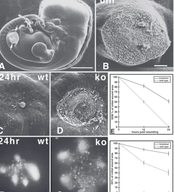
In a further study into the mechanisms behind the perfect healing that occurs in the embryo, Eckes et al. (78) wounded midgestational mouse embryos lacking the intermediate filament protein vimentin. Embryonic day 11.5 mouse embryos are capable of healing an excisional hind leg amputa-tion wound within 24 h (79). Another study, using the same model, had revealed that one of the ma-jor intermediate filaments in early embryonic skin, keratin 8, was not essential for normal em-bryonic repair (80). Using DiI-labeling of wound margin mesenchymal cells, Eckes et al. (78) showed that, while reepithelialization proceeded normally, vimentin null embryos failed to contract the mesenchyme of their wound bed (Fig. 3). Thus, vimentin is essential for the generation of the tractional forces that drive mesenchymal con-traction in the embryonic wound. This defect in repair was also seen in adult wounds to vimentin null mice, again in a defect limited to the connec-tive tissue, which displayed delayed granulation tissue formation and contraction.
5. Conclusion
Studies of wound healing using genetically modified mice have already revealed crucial roles for several genes in the repair process. However, some of the normal functions of the genes targeted might not be revealed owing to redundancy or compensation. This hypothesis is supported by the lack of obvious wound-healing abnormalities in various knockout mice, such as mice deficient in
KGF or TGF-α (16). Although it cannot be
ex-cluded that these proteins are indeed not impor-tant for wound repair, their strong induction in healing skin wounds supports their functional
sig-nificance. In the case of TGF-α and KGF, other growth factors, which bind to the same receptor, might compensate for the lack of these mitogens. Wound-healing studies using animals deficient in two or more homologous molecules, as well as studies with dominant-negative-acting molecules that can inhibit the function of several members of a protein family, will be very useful in answer-ing these areas of question.
At the other extreme, secondary effects, which are owing to systemic defects caused by the transgene, or to transgene-mediated defects in nonwounded skin, might obscure the normal func-tion of a gene in wound repair. Thus, it has long been known that the wound-healing process is sig-nificantly impaired by systemic abnormalities such as malnutrition, weight loss, impaired oxy-genation, and ageing (44,45). This was also the
case in the TGF-β1 knockout mice, which
devel-oped a severe wasting syndrome at approx 3 wk of age, accompanied by a severe inflammatory response in various tissues and organs, including the wound. These abnormalities are likely to be responsible for the impaired wound healing seen in these mice, making it impossible to study the local effects of the lack of TGF-β1 on wound re-pair in this model. One approach to circumvent this problem, as mentioned under Subheading
3.2., was to cross the mice onto an
immunodefi-cient background. However, these problems might also be solved by the generation of mice that have a tissue-specific knockout or tissue-specific overexpression of a transgene (81). Ideally, induc-ible systems that allow the induction of a transgene or the deletion of an endogenous gene in a time- and tissue-specific manner should be used. Such systems thus allow the study of the role of a particular gene under specific conditions such as during wound repair.
The first successful results with inducible sys-tems in the skin have recently been published. Two have adopted an estrogen receptor-based ap-proach, where Cre recombinase was fused in frame with the tamoxifen-responsive hormone-binding domain of the estrogen receptor. This fu-sion protein was expressed under the control of the Keratin 5 promoter (82) or Keratin 14
pro-Fig. 3. Wound closure in wild-type and vimentin-deficient embryos. (A) Scanning electron micrograph of embryonic d 11.5 mouse embryo with left hind limb bud amputated to leave oval-shaped excisional wound (ar-rows). (B) Higher magnification detail of this 0-h wound. (C) After 24 h the wild-type wound is closed. (D) By contrast, at 24 h postwounding the vimentin-deficient wound is still open. (E) Graphic representation of wound closure (reepithelialization + connective-tissue contraction) as measured from scanning electron micrographs. Shaded and solid symbols indicate area measurements in wild-type and vimentin-deficient embryos, respectively, during the 24-h culture period. Error bars are SEMs. (F) Twenty-four hours postwounding, the wild-type controls have significantly contracted their DiI-marked wound mesenchyme.(G) By contrast, at the same time point, the vimentin-deficient wound has barely contracted. (H) Graphic representation of connective-tissue contraction com-ponent of wound closure as measured from DiI-marked specimens. Shaded and solid symbols indicate area mea-surements in wild-type and vimentin-deficient embryos, respectively, during the 24-h culture period. Error bars are SEMs. Scale bars: (A) 1 mm; (B, C, D, F, G) 100 mm. (Figure courtesy of Dr. Paul Martin. Reprinted from ref. 78 by permission. Copyright 2000 Company of Biologists Ltd.)
moter (83). Cre-mediated recombination of loxP sites flanking the target gene thus results in tem-porally and spatially restricted knockout of the
tar-get gene. In a different approach, Wang et al. (84) have used topical application of antiprogestin to induce expression of target genes. This method
works via skin-specific expression of a fusion pro-tein, under the control of the loricrin promoter, containing a truncated progesterone receptor fused to the yeast GAL4 transcription factor. Thus, by engineering a GAL4-binding domain, normally absent in mammalian cells, upstream of the target gene, transcription can be activated in a tissue-specific and temporally controlled manner. Use of these types of systems promises to yield extremely valuable data on the actions of many genes crucial to wound repair, but formerly im-possible to study.
Acknowledgments
We are particularly grateful to Prof. Anita Rob-erts, NIH Bethesda; Prof. Junji Takeda, Osaka University Medical School; and Dr. Paul Martin, University College London, for kindly providing
Figs. 1–3. We also thank Prof. Manfred Blessing
and Dr. Gillian Ashcroft for critical reading of the manuscript, and our coworkers for providing un-published data. Work in Sabine Werner’s labora-tory is supported by the ETH Zurich, the Swiss National Science Foundation (grant no. 31-61358.00), the German Ministry for Education and Research (BMBF), and the Stiftung Verum.
References
1. Clark, R. A. F. (1996) Wound repair: Overview and general considerations, in The Molecular and Cellu-lar Biology of Wound Repair, 2nd ed. (CCellu-lark, R. A. F., ed.) Plenum, New York, pp. 3-50.
2. Ornitz, D. M. and Itoph, N. (2001) Fibroblast growth factors. Genome Biol. 2, 3005.1-3005.12.
3. Werner, S. (1998) The role of fibroblast growth fac-tors in skin morphogenesis and wound repair, in Epi-thelial Morphogenesis in Development and Disease (Birchmeier, C. and Birchmeier, W., eds.), Harwood Academic, GMBH, Chur, Switzerland, p. 233. 4. Johnson, D. E. and Williams, L. T. (1993) Structural
and functional diversity in the FGF receptor multigene family. Adv. Cancer Res. 60, 1-41.
5. Rosenquist, T. A. and Martin, G. R. (1996) Fibroblast growth factor signalling in the hair growth cycle: ex-pression of the fibroblast growth factor receptor and ligand genes in the murine hair follicle. Dev. Dyn.
205, 379-386.
6. Abraham, L. A. and Klagsbrun, M. (1996) Modula-tion of wound repair by members of the fibroblast growth factor family, in The Molecular and Cellular Biology of Wound Repair, 2nd ed. (Clark, R. A. F., ed.), Plenum, New York, pp. 195–248.
7. Miki, T., Bottaro, D. P., Fleming, T. P., Smith, C. L., Burgess, W. H., Chan, A. M., and Aaronson, S. A. (1992) Determination of ligand-binding specificity by alternative splicing: two distinct growth factor recep-tors encoded by a single gene. Proc. Natl. Acad. Sci. USA 89, 246–250.
8. Werner, S., Peters, K. G., Longaker, M. T., Fuller-Pace, F., Banda, M., and Williams, L. T. (1992) Large induction of keratinocyte growth factor expression in the dermis during wound healing. Proc. Natl. Acad. Sci. USA 89, 6896–6900.
9. Marchese, C., Chedid, M., Dirsch, O. R., Csaky, K. G., Santanelli, F., Latini, C., LaRochelle, W. J., Torrisi, M. R., and Aaronson, S. A. (1995) Modula-tion of keratinocyte growth factor and its receptor in re-epithelialising human skin. J. Exp. Med. 182, 1369–1376.
10. Ueno, H., Colbert, H., Escobedo, J. A., and Williams, L. T. (1991) Inhibition of PDGF b-receptor by co-expres-sion of a truncated receptor. Science 252, 844–848. 11. Honegger, A. M., Schmidt, A., Ullrich, A., and
Schlessinger, J. (1990) Evidence for epidermal growth factor (EGF)-induced intermolecular autophosphorylation of the EGF receptors in living cells. Mol. Cell Biol. 10, 4035–4044.
12. Kashles, O., Yarden, Y., Fischer, R., Ullrich, A., and Schlessinger, J. (1991) A dominant negative mutation suppresses the function of normal epidermal growth factor receptors by heterodimerisation. Mol. Cell Biol.
11, 1454–1463.
13. Mathieu, M., Chatelain, E., Ornitz, D., Bresnick, J., Mason, I., Kiefer, P., and Dickson, C. (1995) Recep-tor binding and mitogenic properties of mouse fibro-blast growth factor 3: modulation of response by heparin. J. Biol. Chem. 41, 24, 197–24,203.
14. Yamasaki, M., Miyake, A., Tagashira, S., and Itoh, N. (1996) Structure and expression of the rat mRNA encoding a novel member of the fibroblast growth fac-tor family. J. Biol. Chem. 271, 15,918–15,921. 15. Werner, S., Smola, H., Liao, X., Longaker, M. T.,
Krieg, T., Hofschneider, P. H., and Williams, L. T. (1994) The function of KGF in epithelial morphogen-esis and wound re-epithelialisation. Science 266, 819–822.
16. Guo, L., Degenstein, L., and Fuchs, E. (1996) Keratinocyte growth factor is required for hair devel-opment but not for wound healing. Genes Dev. 10, 165–175.
17. Beer, H. D., Florence, C., Dammeier, J., McGuire, L., Werner, S., and Duan, D. R. (1997) Mouse fibroblast growth factor 10: cDNA cloning, protein character-ization, and regulation of mRNA expression. Oncogene 15, 2211–2218.
18. Barrandon, Y. and Green, H. (1987) Cell migration is essential for sustained growth of keratinocyte colo-nies: the roles of transforming growth factor-a and epidermal growth factor. Cell 50, 1131–1137.
2 5 7 8 9 10 11 12 14 15 16 17 18
19. Schultz, G. S., White, M., Mitchell, R., Brown, G., Lynch, J., Twardzik, D. R., and Todaro, G. J. (1987) Epithelial wound healing enhanced by transforming growth factor-a and vaccinia growth factor. Science
235, 350–352.
20. Mann, G. B., Fowler, K. J., Gabriel, A., Nice, E. C., Williams, R. L., and Dunn, A. R. (1993) Mice with a null mutation of the TGFa gene have abnormal skin architecture, wavy hair, and curly whiskers and often develop corneal inflammation. Cell 73, 249–261. 21. Luetteke, N. C., Qiu, T. H., Peiffer, R. L., Oliver, P.,
Smithies, O., and Lee, D. C. (1993) TGFa deficiency results in hair follicle and eye abnormalities in tar-geted and waved-1 mice. Cell 73, 263–278.
22. Marikovsky, M., Breuing, K., Liu, P. Y., Eriksson, E., Higashiyama, S., Farber, P., Abraham, J., and Klagsbrun, M. (1993) Appearance of heparin-binding EGF-like growth factor in wound fluid as a response to injury. Proc. Natl. Acad. Sci. USA 90, 3889–3893. 23. Sibilia, M. and Wagner, E. F. (1995)
Strain-depen-dent epithelial defects in mice lacking the EGF recep-tor. Science 269, 234–238.
24. Miettinen, P. J., Berger, J. E., Meneses, J., Phung, Y., Pedersen, R. A., Werb, Z., and Derynck, R. (1995) Epi-thelial immaturity and multiorgan failure in mice lacking epidermal growth factor receptor. Nature 376, 337–341. 25. Murillas, R., Larcher, F., Conti, C. J., Santos, M., Ullrich, A., and Jorcano, J. L.(1995) Expression of a dominant negative mutant of epidermal growth factor receptor in the epidermis of transgenic mice elicits striking alterations in hair follicle development and skin structure. EMBO J. 14, 5216–5223.
26. Bikfalvi, A., Klein, S., Pintucci, G., and Rifkin, D. B. (1997) Biological roles of fibroblast growth factor-2. Endocr. Rev. 18, 26–45.
27. Ortega, S., Ittmann, M., Tsang, S. H., Ehrlich, M., and Basilico, C. (1998) Neuronal defects and delayed wound healing in mice lacking fibroblast growth fac-tor 2. Proc. Natl. Acad. Sci. USA 95, 5672–5677. 28. Gibran, N. S., Isik, F. F., Heimbach, D. M., and
Gor-don, D. (1994) Basic fibroblast growth factor in the early human burn wound. J. Surg. Res. 56, 226–234. 29. McGee, G. S., Davidson, J. M., Buckley, A., Sommer, A., Woodward, S. C., Aquino, A. M., Barbour, R., and Demetriou, A. A. (1988) Recombinant basic fibro-blast growth factor accelerates wound healing. J. Surg. Res. 45, 145–153.
30. Hebda, P. A., Klingbeil, C. K., Abraham, J. A., and Fiddes, J. C. (1990) Basic fibroblast growth factor stimulation of epidermal wound healing in pigs. J. Invest. Dermatol. 95, 626–631.
31. Tsuboi, R. and Rifkin, D. B. (1990) Recombinant ba-sic fibroblast growth factor stimulates wound healing in healing-impaired db/db mice. J. Exp. Med. 172, 245–251.
32. Broadley, K. N., Aquino, A. M., Woodward, S. C., Buckley-Sturrock, A., Sato, Y., Rifkin, D. B., and
Davidson, J. M. (1989) Monospecific antibodies im-plicate basic fibroblast growth factor in normal wound repair. Lab. Invest. 61, 571–575.
33. Massagué, J. (1990) The transforming growth factor-b family. Annu. Rev. Cell Biol. 6, 597–641. 34. Massague, J. and Wotton, D. (2000) Transcriptional
control by the TGF-beta/Smad signaling system. EMBO J. 19, 1745–1754.
35. Roberts, A. B. and Sporn, M. B. (2001) Transforming growth factor-β, in The Molecular and Cellular Biol-ogy of Wound Repair, 2nd ed. (Clark, R. A. F., ed.), Plenum, New York, pp. 275–308.
36. Levine, J. H., Moses, H. L., Gold, L. I., and Nanney, L. B. (1993) Spatial and temporal patterns of immu-noreactive TGF-β1, β2, β3 during excisional wound repair. Am. J. Pathol. 143, 368–380.
37. Frank, S., Madlener, M., and Werner, S. (1996) Trans-forming growth factors β1, β2, and β3 and their re-ceptors are differentially regulated during normal and impaired wound healing. J. Biol. Chem. 271, 10, 188– 10,193.
38. Shah, M., Foreman, D. M., and Ferguson, M. W. J. (1994) Neutralising antibody to TGF-β1,2 reduces cutaneous scarring in adult rodents. J. Cell Sci. 107, 1137–1157.
39. Shah, M., Foreman, D. M., and Ferguson, M. W. J. (1995) Neutralisation of TGF-b1 and TGF-b2 or exog-enous addition of TGF-β3 to cutaneous rat wounds re-duces scarring. J. Cell Sci. 108, 985–1002.
40. Brown, R. L., Ormsby, I., Doetschman, T. C., and Greenhalgh, D. G. (1995) Wound healing in the trans-forming growth factor-b1-deficient mouse. Wound Rep. Reg. 3, 25–36.
41. Shull, M. M., Ormsby, I., Kier, A. B., Pawlowski, S., Diebold, R. J., Yin, M., Allen, R., Sidman, C., Proetzel, G., Calvin, D., Annunziata, N., and Doetschman, T. (1992) Targeted disruption of the mouse transforming growth factor-β1 gene results in multifocal inflamma-tory disease. Nature 359, 693–699.
42. Kulkarni, A. B., Huh, C.-G., Becker, D., Geiser, A., Lyght, M., Flanders, K. C., Roberts, A. B., Sporn, M. B., Ward, J. M., and Karlsson, S. (1993) Transform-ing growth factor β1 null mutation in mice causes ex-cessive inflammatory response and early death. Proc. Natl. Acad. Sci. USA 90, 770–774.
43. Letterio, J. J., Geiser, A. G., Kulkarni, A. B, Roche, N. S., Sporn, M. B., and Roberts, A. B. (1994) Mater-nal rescue of transforming growth factor-β1 null mice. Science 264, 1936–1938.
44. Greenhalgh, D. G. and Gamelli, R. L. (1987) Is im-paired wound healing caused by infection or nutri-tional depletion? Surgery 102, 306–312.
45. Reed, B. R. and Clark, R. A. (1985) Cutaneous tissue repair: practical implications of current knowledge. II. J. Am. Acad. Dermatol. 13, 919–941.
46. Crowe, M. J., Doetschman, T., and Greenhalgh, D. G. (2000) Delayed wound healing in immunodeficient
19 20 21 22 23 24 25 26 27 28 29 30 31 32 34 36 38 39 40 41 42 43 44 45 46
TGF-β 1 knockout mice. J. Invest. Dermatol. 115, 3–11.
47. Shah, M., Revis, D., Herrick, S., Baillie, R., Thorgeirson, S., Ferguson, M., and Roberts, A. (1999) Role of elevated plasma transforming growth factor-β1 levels in wound healing. Am. J. Pathol. 154, 1115–1124.
48. Massague, J. (1998) TGF-β signal transduction. Annu. Rev. Biochem. 67, 753–791.
49. Derynck, R., Zhang, Y., and Feng, X. H. (1998) Smads: transcriptional activators of TGF-β responses. Cell 95, 737–740.
50. Ashcroft, G. S. and Roberts, A. B. (2000) Loss of Smad3 modulates wound healing. Cytokine Growth Factor Rev. 11, 125–131.
51. Christian, J. L. and Nakayama, T. (1999) Can’t get no SMADisfaction: Smad proteins as positive and nega-tive regulators of TGF-beta family signals. Bioessays
21, 382–390.
52. Weinstein, M., Yang, X., Li, C., Xu, X., Gotay, J., and Deng, C. X. (1998) Failure of egg cylinder elon-gation and mesoderm induction in mouse embryos lacking the tumor suppressor smad2. Proc. Natl. Acad. Sci. USA 95, 9378–9383.
53. Yang, X., Castilla, L. H., Xu, X., Li, C., Gotay, J., Weinstein, M., Liu, P. P., and Deng, C. X. (1999) Angiogenesis defects and mesenchymal apoptosis in mice lacking SMAD5. Development 126, 1571–1580. 54. Ashcroft, G. S., Yang, X., Glick, A. B., Weinstein, M., Letterio, J. L., Mizel, D. E.,Anzano, M., Greenwell-Wild, T., Wahl, S. M., Deng, C., and Rob-erts, A. B. (1999) Mice lacking Smad3 show acceler-ated wound healing and an impaired local inflammatory response. Nat. Cell Biol. 1, 260–266. 55. Hocevar, B. A., Brown T. L., and Howe, P. H. (1999)
TGF-beta induces fibronectin synthesis through a c-Jun N-terminal kinase-dependent, Smad4-indepen-dent pathway. EMBO J. 18, 1345–1356.
56. Matzuk, M. M., Kumar, T. R., Vassalli, A., Bickenbach, J. R., Roop, D. R., Jaenisch, R., and Bra-dley, A. (1995) Functional analysis of activins during mammalian development. Nature 374, 354–356. 57. Matzuk, M. M., Lu, N., Vogel, H., Sellheyer, K.,
Roop, D. R., and Bradley, A. (1995) Multiple defects and perinatal death in mice deficient in follistatin. Nature 374, 360–363.
58. Hübner, G., Hu, Q., Smola, H., and Werner, S. (1996) Strong induction of activin expression after injury suggests an important role of activin in wound repair. Dev. Biol. 173, 490–498.
59. Munz, B., Smola, H., Engelhardt, F., Bleuel, K., Brauchle, M., Lein, I., Evans, L. W., Huylebroeck, D., Balling, R., and Werner, S. (1999) Overexpression of activin A in the skin of transgenic mice reveals new activities of activin in epidermal morphogenesis, der-mal fibrosis and wound repair. EMBO J. 18, 5205– 5215.
60. Lyons, K. M., Pelton, R. W., and Hogan, B. L. (1989) Patterns of expression of murine Vgr-1 and BMP-2a RNA suggest that transforming growth factor-beta-like genes coordinately regulate aspects of embryonic development. Genes Dev. 3, 1657–1668.
61. Blessing, M., Schirmacher, P., and Kaiser, S. (1996) Overexpression of bone morphogenetic protein-6 (BMP-6) in the epidermis of transgenic mice: inhibi-tion or stimulainhibi-tion of proliferainhibi-tion depending on the pattern of transgene expression and formation of pso-riatic lesions. J. Cell Biol. 135, 227–239.
62. Kaiser, S., Schirmacher, P., Philipp, A., Protschka, M., Moll, I., Nicol, K., and Blessing, M. (1998) In-duction of bone morphogenetic protein-6 in skin wounds: delayed reepitheliazation and scar formation in BMP-6 overexpressing transgenic mice. J. Invest. Dermatol. 111, 1145–1152.
63. Paquet, P. and Pierard, G. E. (1996) Interleukin-6 and the skin. Int. Arch. Allergy Immunol. 109, 308–317. 64. Sato, M., Sawamura, D., Ina, S., Yaguchi, T., Hanada, K., and Hashimoto, I. (1999) In vivo introduction of the interleukin 6 gene into human keratinocytes: in-duction of epidermal proliferation by the fully spliced form of interleukin 6, but not by the alternatively spliced form. Arch. Dermatol. Res. 291, 400–404. 65. Grossman, R. M., Krueger, J., Yourish, D.,
Granelli-Piperno, A., Murphy D. P., May, L. T., Kupper, T. S., Sehgal, P. B., and Gottlieb, A. B. (1989) Interleukin 6 is expressed in high levels in psoriatic skin and stimu-lates proliferation of cultured human keratinocytes. Proc. Natl. Acad. Sci. USA 86, 6367–6371.
66. Gallucci, R. M., Simeonova, P. P., Matheson, J. M., Kommineni, C., Guriel, J. L., Sugawara, T., and Lus-ter, M. I. (2000) Impaired cutaneous wound healing in interleukin-6-deficient and immunosuppressed mice. FASEB J. 14, 2525–2531.
67. Bromberg, J. (2001) Activation of STAT proteins and growth control. Bioessays 23, 161–169.
68. Takeda, K., Noguchi, K., Shi, W., Tanaka, T., Matsumoto, M., Yoshida, N., Kishimoto, T., and Akira, S. (1997) Targeted disruption of the mouse Stat3 gene leads to early embryonic lethality. Proc. Natl. Acad. Sci. USA 94, 3801–3804.
69. Sano, S., Itami, S., Takeda, K., Tarutani, M., Yamaguchi, Y., Miura, H., Yoshikawa, K., Akira, S., and Takeda, J. (1999) Keratinocyte-specific ablation of Stat3 exhibits impaired skin remodeling, but does not affect skin morphogenesis. EMBO J. 18, 4657–4668. 70. Devalaraja, M. N. and Richmond, A. (1999) Multiple chemotactic factors: fine control or redundancy? Trends Pharmacol. Sci. 20, 151–156.
71. Engelhardt, E., Toksoy, A., Goebeler, M., Debus, S., Brocker, E. B., and Gillitzer, R. (1998) Chemokines IL-8, GROalpha, MCP-1, IP-10, and Mig are sequen-tially and differensequen-tially expressed during phase-spe-cific infiltration of leukocyte subsets in human wound healing. Am. J. Pathol. 153, 1849–1860.
47 48 49 50 51 52 53 54 55 56 57 59 60 61 62 63 64 65 66 67 68 69 70 71
72. Luster, A. D., Cardiff, R. D., MacLean, J. A., Crowe, K., and Granstein, R. D. (1998) Delayed wound heal-ing and disorganized neovascularization in transgenic mice expressing the IP-10 chemokine. Proc. Assoc. Am. Physicians 110, 183–196.
73. Fivenson, D. P., Faria, D. T., Nickoloff, B. J., Poverini, P. J., Kunkel, S., Burdick, M., and Streiter, R. M. (1997) Chemokine and inflammatory cytokine changes during chronic wound healing. Wound Rep. Reg. 5, 310–322.
74. Cacalano, G., Lee, J., Kikly, K., Ryan, A. M., Pitts-Meek, S., Hultgren, B., Wood, W. I., and Moore, M. W. (1994) Neutrophil and B cell expansion in mice that lack the murine IL-8 receptor homolog. Science
265, 682–684.
75. Devalaraja, R. M., Nanney, L. B., Qian, Q., Du, J., Yu, Y., Devalaraja, M. N., and Richmond, A. (2000) Delayed wound healing in CXCR2 knockout mice. J. Invest. Dermatol. 115, 234–244.
76. Martin, P. (1997) Wound healing-aiming for perfect skin regeneration. Science 276, 75–81.
77. Liechty, K. W., Kim, H. B., Adzick, N. S., and Crombleholme, T. M. (2000) Fetal wound repair re-sults in scar formation in interleukin-10-deficient mice in a syngeneic murine model of scarless fetal wound repair. J. Pediatr. Surg. 35, 866–872; discus-sion: 872,873.
78. Eckes, B., Colucci-Guyon, E., Smola, H., Nodder, S., Babinet, C., Krieg, T., and Martin, P. (2000) Impaired wound healing in embryonic and adult mice lacking vimentin. J. Cell Sci. 113, 2455–2462.
79. McCluskey, J. and Martin, P. (1995) Analysis of the tissue movements of embryonic wound healing-DiI studies in the limb bud stage mouse embryo. Dev. Biol. 170, 102–114.
80. Brock, J., McCluskey, J., Baribault, H., and Martin, P. (1996) Perfect wound healing in the keratin 8 deficient mouse embryo. Cell. Motil. Cytoskel. 35, 358–366. 81. Rajewsky, K., Gu, H., Kuhn, R., Betz, U. A., Muller,
W., Roes, J., and Schwenk, F. (1996) Conditional gene targeting. J. Clin. Invest. 98, 600–603. 82. Indra, A. K., Warot, X., Brocard, J., Bornert, J. M.,
Xiao, J. H., Chambon, P., and Metzger, D. (1999) Temporally-controlled site-specific mutagenesis in the basal layer of the epidermis: comparison of the recombinase activity of the tamoxifen- inducible Cre-ER(T) and Cre-ER(T2) recombinases. Nucleic Acids Res. 27, 4324–4327.
83. Vasioukhin, V., Degenstein, L., Wise, B., and Fuchs, E. (1999) The magical touch: genome targeting in epider-mal stem cells induced by tamoxifen application to mouse skin. Proc. Natl. Acad. Sci. USA 96, 8551–8556. 84. Wang, X. J., Liefer, K. M., Tsai, S., O’Malley, B. W., and Roop, D. R. (1999) Development of gene-switch transgenic mice that inducibly express transforming growth factor beta1 in the epidermis. Proc. Natl. Acad. Sci. USA 96, 8483–8488.
85. Guo, L., Degenstein, L., Dowling, J., Yu, Q.-C., Wollman, R., Perman, B., and Fuchs, E. (1995) Gene targeting of BPAG1: abnormalities in mechanical strength and cell migration in stratified epithelia and neurologic degeneration. Cell 81, 233–243.
86. Abbott, R. E., Corral, C. J., MacIvor, D. M., Lin, X., Ley, T. J., and Mustoe, T. A. (1998) Augmented inflam-matory responses and altered wound healing in cathep-sin G-deficient mice. Arch. Surg. 133, 1002–1006. 87. Kaya, G., Rodriguez, I., Jorcano, J. L., Vassalli, P., and
Stamenkovic, I. (1997) Selective suppression of CD44 in keratinocytes of mice bearing an antisense CD44 transgene driven by a tissue-specific promoter disrupts hyaluronate metabolism in the skin and impairs keratinocyte proliferation. Genes Dev. 11, 996–1007. 88. Hansen, L. A., Alexander, N., Hogan, M. E., Sundberg, J. P., Dlugosz, A., Threadgill, D. W., Magnuson, T., and Yuspa, S. H. (1997) Genetically null mice reveal a central role for epidermal growth factor receptor in the differentiation of the hair fol-licle and normal hair development. Am. J. Pathol.
150, 1959–1975.
89. Bugge, T. H., Kombrinck, K. W., Flick, M. J., Daugherty, C. C., Danton, M. J., and Degen, J. L. (1996) Loss of fibrinogen rescues mice from the pleiotropic ef-fects of plasminogen deficiency. Cell 87, 709–719. 90. Bezerra, J. A., Carrick, T. L., Degen, J. L., Witte, D.,
and Degen, S. J. F. (1998) Biological effects of tar-geted inactivation of hepatocyte growth factor-like protein in mice. J. Clin. Invest. 101, 1175–1183. 91. Huang, X., Griffiths, M., Wu, J., Farese, R.V. Jr., and
Sheppard, D. (2000) Normal development, wound healing, and adenovirus susceptibility in beta5-defi-cient mice. Mol. Cell. Biol. 20, 755–759.
92. Wojcik, S. M., Bundman, D. S., and Roop, D. R. (2000) Delayed wound healing in keratin 6a knock-out mice. Mol. Cell. Biol. 20, 5248–5255.
93. Di Colandrea, T., Wang, L., Wille, J., D’Armiento, J., and Chada, K. K. (1998) Epidermal expression of col-lagenase delays wound-healing in transgenic mice. J. Invest. Dermatol. 111, 1029–1033.
94. Bullard, K. M., Lund, L., Mudgett, J. S., Mellin, T. N., Hunt, T. K., Murphy, B., Ronan, J., Werb, Z., and Banda, M. J. (1999) Impaired wound contraction in stromelysin-1-deficient mice. Ann. Surg. 230, 260–265.
95. Atit, R. P., Crowe, M. J., Greenhalgh, D. G., Wenstrup, R. J., and Ratner, N. (1999) The Nf1 tumor suppressor regulates mouse skin wound healing, broblast proliferation, and collagen deposited by fi-broblasts. J. Invest. Dermatol. 112, 835–842. 96. Lee, P. C., Salyapongse, A. N., Bragdon, G. A.,
Shears, L. L. 2nd, Watkins, S. C., Edington, H. D., and Billiar, T. R. (1999) Impaired wound healing and angiogenesis in eNOS-deficient mice. Am. J. Physiol.
277, H1600–H1608.
97. Yamasaki, K., Edington, H. D., McClosky, C., Tzeng, E., Lizonova, A., Kovesdi, I., Steed, D. L., and Billiar,
72 73 74 75 76 77 78 79 80 81 82 83 84 85 86 87 88 89 90 91 92 93 94 95 96 97