Exp Brain Res (2004) 158: 308–316 DOI 10.1007/s00221-004-1904-3
R E S E A R C H A RT I C L E S
V. Dietz . G. Colombo . R. Müller
Single joint perturbation during gait: neuronal control
of movement trajectory
Received: 28 October 2003 / Accepted: 2 February 2004 / Published online: 27 April 2004 # Springer-Verlag 2004
Abstract The aim of this study was to investigate the effect of single joint displacement on the pattern of leg muscle electromyographic (EMG) activity during loco-motion. For the first time, unilateral rotational hip or knee joint displacements were applied by a driven orthotic device at three phases of swing during locomotion on a treadmill. The response pattern of bilateral leg muscle activation with respect to the timing and selection of muscles was almost identical for displacements of upper (hip joint) or lower (knee joint) leg. The leg muscle EMG responses were much stronger when the displacement was directed against the physiological movement trajectory, compared with when the displacement was reinforcing, especially during mid swing. It is suggested that these response patterns are designed to restore physiological movement trajectory rather than to correct a single joint position. Displacements released at initial or terminal swing, assisting or resisting the physiological movement trajectory, were followed by similar and rather unspecific response patterns. This was interpreted as being directed to stabilise body equilibrium.
Keywords Compensatory EMG responses . Interlimb co-ordination . Locomotion . Movement synergies .
Movement trajectory . Neuronal control . Single joint displacement
Introduction
Many similarities exist in the way humans and other mammals co-ordinate their limb movements in response to disturbance during walking (for review see Dietz 2002). Unilateral leg displacements of support during stance and
gait evoke a bilateral response pattern with a spinal onset latency on both sides (Dietz et al.1986a,1989). Therefore, it was assumed that a purposeful activation pattern of synergistic leg muscles becomes released by the so-called spinal central pattern generator (for review see Dietz 1992). According to this concept, focal muscle responses to a local displacement play a minor role for the compensatory reaction. From a functional point of view, this interlimb co-ordination is necessary to maintain body equilibrium (Dietz et al.1989; for review see Dietz1992). In most studies on the bilateral co-ordination of leg movements in cat and man one or both legs become perturbed. By such an approach several joints of a limb become displaced. Therefore, the source of the relevant afferent input for the bilateral co-ordination of leg muscle activation is not known. It is an unanswered question whether this input is provided by the displacement of a single joint or by the combination of the afferent input from many sensors within joints and muscles activated by the leg displacement. Nevertheless, there are observations which point out the importance of hip position for initiating the stance to swing transition with an appropriate leg muscle activation for human infant stepping (Pang and Yang 2000). The significance of hip joint afferents for locomotion was also emphasised for the chronic spinal cat (Grillner and Rossignol 1978). Furthermore, entrainment of a locomotor rhythm was obtained by using rhythmic hip movements in immobilised spinal (Andersson and Grillner 1983) and decerebrate (Kriellaars et al. 1994) cats.
The aim of this study was to evaluate the relevance of input related to different leg joints for the generation of the locomotor pattern. Therefore, the effect of single joint displacements on the leg muscle activity during locomo-tion was studied using a novel approach. For the first time, a driven gait orthosis (DGO) was applied, which allowed induction of a displacement at the hip or knee joint during the step cycle. It was hypothesised that hip and knee joint-related afferent input differentially contributes to the bilateral co-ordination of leg muscle activation.
V. Dietz (*) . G. Colombo . R. Müller
Spinal Cord Injury Center, Balgrist University Hospital, Forchstrasse 340,
CH-8008 Zurich, Switzerland e-mail: [email protected] Tel.: +41-1-3863901
Materials and methods
With the permission from the local Ethical Committee and the informed consent of the volunteers, the leg muscle electromyographic (EMG) responses to unilateral hip or knee perturbations during locomotion were analysed in nine healthy subjects (age 29±5 years). Surface EMG recordings were made from the activity of representative right and left as well as proximal and distal leg muscles (rectus femoris (RF), biceps femoris (BF), tibialis anterior (TA) and medial gastrocnemius (GM)). Subjects walked with the DGO Lokomat (Hocoma AG, Zurich, Switzer-land) on a treadmill.
Driven gait orthosis
A detailed description of the device can be found elsewhere (Colombo et al. 2000). Briefly, the DGO provides drives for the hip and knee joint movements of both legs, whereas the feet can move freely (Fig.1). Four separate position controllers implemented in a computer based real-time system control the angles of the hip and knee joints. The leg joint trajectories are taken from a database of healthy subjects walking within the DGO (Fig.2A) and are identical for all subjects. Feedback of the actual angle is provided by potentiometers attached to the lateral aspect of the hip and knee joints of the orthosis on both sides.
The DGO is fixed to the treadmill by a flexible parallelogram. A compensation mechanism for the weight of the orthosis is provided. The subjects are fixed to the DGO by straps around the waist, the thighs and the shanks. The orthosis can be adjusted in size at the different
segments and, therefore, can be adapted to the different subjects.
Walking within the DGO
During treadmill walking, speed was kept constant at 0.53 m/s (1.9 km/h) for all trials. Cadence had to be slightly adjusted according to the leg length of the different subjects. Mean cycle time for the subjects was 2.2 s. After 5 min of habituation within the orthosis, subjects reported few restrictions compared with their normal walking movements. Although there were differ-ences between the locomotor pattern obtained during walking within the DGO and normal walking on the treadmill (Fig. 2B), the timing of EMG activity of the respective leg muscles was largely unaffected. The low walking speed was chosen to enable comparisons with similar measurements in paraplegic patients where this is the normal therapeutic treadmill speed. In addition there are technical restrictions within the DGO for speeds higher than 0.7 m/s.
Unilateral joint perturbations
Custom made software was used for the generation of the unilateral joint perturbations released at different phases of the gait cycle. While normal walking movements were always provided for the right leg, perturbations were released in a random order to the left leg. Extension or flexion displacements, assisting or resisting the physio-logical movement, applied at the left hip or knee joint, were released at three different phases during swing phase of the gait cycle. The 12 modes of perturbations were released only during the swing phase. This is because during the stance phase, interaction with the moving treadmill belt occurs, making it difficult to control. Each perturbation was followed by three to five normal gait cycles, and every condition was applied five times. The duration of one experiment amounted to around 10 min; subjects did not experience fatigue within this time.
Joint displacements were induced by switching from a closed loop position control, using the desired angle values, to an open loop control for 100 ms. After 100 ms the DGO was set back to the closed loop position control. The normal movement trajectory was reached about 200 ms after the onset of displacement; i.e. the displace-ment did not change the overall duration of the swing phase. The software generated an analog trigger with different amplitudes encoding all conditions and indicating the beginning of the stance phase of the left leg.
The open loop control was the same for all perturba-tions. The application of the maximal current of 15 A for the motor at the corresponding joint resulted, on average, in a change in angular momentum of about 6 Nms at the hip and 3 Nms at the knee joint, respectively (measured by built-in force sensors). Some existing variability in angular momentum was due to the different movement directions Fig. 1 Experimental set up. Walking within a DGO
and velocities, as well as to different inertia of the perturbed part of the limb. Therefore, the influence of the impulse strength on the EMG responses was tested only in five subjects by the application of two weaker impulses (75 and 50% of the original one). These additional measurements with less impulse strength were made for
the knee flexion perturbation during mid swing to get not only data with comparable perturbation input, but also data with comparable perturbation output, i.e. displacement amplitude.
Figure 2A shows the timing and direction of all perturbations superimposed on the normal movement Fig. 2A, B Stepping within the
DGO. Joint angle and EMG recordings during one normal gait cycle during stepping with-in the DGO. A Normal hip and knee joint movements are pro-vided by the DGO; the direction and time frame of the perturba-tions are superposed on the normal joint angle diagram (b, arrows) and the leg positions at the beginning of the perturba-tions are indicated by a sche-matic stick diagram (a, left pertubated leg in black). B EMG activity of upper and lower leg muscles stepping within and without the DGO. The exten-sion/flexion displacements ap-plied to the left hip or knee at various phases of the step cycle are indicated by thin vertical lines
trajectories of hip and knee joints during the gait cycle. The resulting displacements depended on the direction of the impulse with respect to the physiological leg move-ment trajectory and the phase of the step cycle in which the perturbation was released.
Data analysis
For data recording and signal analysis the Soleasy Software (ALEA Solutions GmbH, Zurich, Switzerland) was used. EMG recordings were amplified, band-pass filtered (30–1,000 Hz), rectified and low-pass filtered (10 Hz). All data (EMG, actual hip and knee joint angle trajectories of both legs of the orthosis, and the trigger signal) were sampled at 2,000 Hz.
The data for each perturbation was normalised in time to one gait cycle, and an average over the five repetitions was calculated. In order to get the net effect of the different perturbations on the leg muscle EMG activity, the averaged data of normal walking within the DGO was subtracted from the averaged data of each perturbation condition for every subject. These differences were then normalised in amplitude to the mean EMG activity over one gait cycle of normal walking. The population mean values were calculated over the averages obtained from five subjects for each of the perturbation conditions; with an additional four subjects being included for the mid swing conditions, due to this being the focus of interest. The maximal EMG amplitude within a time frame of 200 ms after perturbation onset was calculated for every muscle in every condition. Differences between
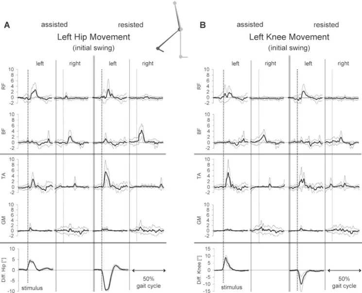
perturba-Fig. 3A, B Population means (with SD) of the rectified and averaged (n=9) net EMG responses in RF, BF, TA, GM of both legs to assisting and resisting displacements, of A the left hip and B the left knee movement, released during mid swing (cycle duration 2.2 s). The individual EMG amplitudes were normalised to the mean EMG activity of the respective muscle during unperturbed
locomo-tion. Below the EMG traces, the net hip or knee deflection movement induced by the displacement is displayed. The release of the displacement at the left leg is indicated by the dotted vertical lines on the left and right leg, respectively. In the schematic diagram the leg position (left perturbated leg in black) is shown for the condition mid swing
tion conditions were tested using the Wilcoxon signed-ranks test with level of significance set to p<0.05.
The latency between onset of joint displacement and EMG response was determined by the time interval between the deflection of the joint trajectory and the occurrence of an increase in EMG amplitude of twice background level. Visual inspection was used to judge when a signal-to-background EMG ratio of more than two was reached.
Results
The displacements at the hip and knee joints were released at three phases of the swing phase of the left leg (Fig.2A). During swing, the hip and knee joints both exert first a flexion and then an extension movement whereby this transition occurs at different time points. During mid swing, the hip is still flexing, whereas the knee is already extending. Displacements directed with and against phys-iological movement direction were called “assisted” and
“resisted”, respectively. The displacements at the left hip or knee were perceived by the subjects as weak or moderate perturbations of gait. Most displacements were followed by bilateral EMG responses in upper and lower muscles of both legs. The response pattern also involved muscles which were not affected by the displacements. For a better comparison, the corresponding conditions for hip or knee displacements (i.e. assisting and resisting hip and knee displacement at one phase of swing) were taken together. In Figs.3,4and5, the impulse strength applied to the left hip or knee was the same in all conditions. Therefore, the resulting joint displacement could differ, i.e. it was smaller in some conditions during assisting displacements.
Figure 3 shows the population mean (with SD) of the bilateral net EMG and joint responses to assisting and resisting hip (Fig. 3A) and knee (Fig.3B) displacements released during mid swing. Figure 3shows that when the movement was“resisted” during mid swing, i.e. when the displacement applied was directed against the physiolog-ical movement, the amplitude of the responses was
stronger compared with the condition when the displace-ment was assisted, i.e. had the same direction as the physiological movement. A bilateral response pattern was obtained following displacements that resisted the move-ments with an early activation ipsilaterally of m. rectus femoris (RF) and m. tibialis anterior (TA) and contralat-erally of m. biceps femoris (BF) and m. gastrocnemius medialis (GM). The variability of the response pattern among the subjects was rather small. Such a pattern makes sense as the intended movement of the ipsilateral leg becomes restored by such a muscle activation. At the contralateral standing leg, a BF and GM activation took place. This provides enhanced support for balance correction and represents the appropriate compensation of a forward disturbance of body equilibrium evoked by the hip or knee displacement of the swing leg.
When the movement was assisted by the displacement, released during mid swing, the response amplitudes in the muscles of both legs were small or almost absent. Although both impulse energy and displacement ampli-tude were about the same during the hip displacements, the distinct response pattern during resisting displacements
switched to an almost negligible one with small EMG responses (e.g. ipsilateral BF following hip and ipsilateral TA following knee perturbations) during assisting dis-placements. This difference was statistically significant (p<0.01) for the EMG amplitudes of ipsilateral RF and TA as well as the contralateral BF for both hip and knee displacements during mid swing.
Figures4and5show the population mean (with SD) of the EMG responses to assisting and resisting hip and knee displacements induced during initial (Fig.4) and terminal (Fig. 5) swing. The responses were smaller and rather independent of the direction of the displacement (i.e. were rather unspecific). They mainly consisted of an ipsilateral RF or RF/TA (initial swing) and BF/GM-activation (terminal swing), respectively. Small but significant (p<0.05) differences in EMG amplitude were found only following hip perturbations for ipsilateral TA (initial and terminal swing), contralateral BF (initial swing) and contralateral RF (terminal swing), respectively. They all showed larger amplitudes in the resisting condition. No difference in EMG amplitudes was found following assisted and resisted knee displacements.
There was a striking similarity between the response pattern following corresponding hip and knee displace-ments during all three phases of swing. This similarity is primarily based on visual inspection and concerned the muscles involved as well as the whole pattern of muscle activation in both legs, while the amplitudes of the EMG responses showed some difference. The latencies of the responses were in the range of 50 to 90 ms (e.g. left RF and TA mid swing) or in the range of 150 to 200 ms (e.g. left RF initial swing, GM terminal swing).
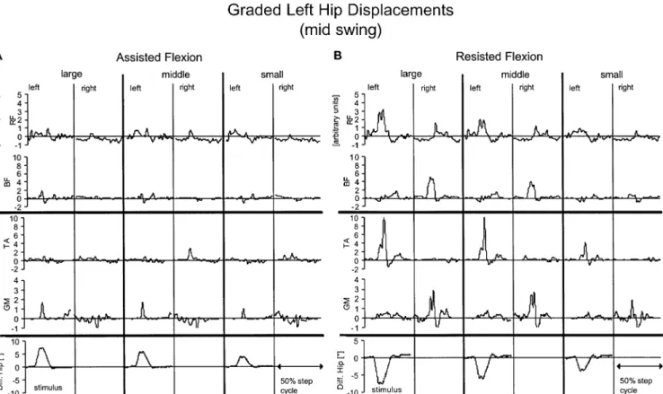
The constant impulse strength (change in angular momentum) resulted in different displacement amplitudes that depended on several factors such as the limb inertia and the direction of the displacement (see Methods). Figure 6 shows the influence of three impulse strengths (100, 75 and 50%) leading to different displacement amplitudes during assisted (Fig.6A) and resisted (Fig.6B) hip flexion movements on the response pattern (population mean of five subjects). In general there was a moderate influence of displacement amplitude on the strength of leg muscle response. However, the bilateral organisation of the response pattern remained unchanged. If the displace-ment amplitudes were less than about 5 deg, even in the resisting displacements no distinct EMG responses occurred.
Discussion
The aim of this study was to evaluate the effect of single joint displacement on the activation of leg muscles during locomotion. To our knowledge, the present work repre-sents the first study on the response pattern to isolated hip or knee displacements during human locomotion. How-ever, one has to take into account some technical restrictions of the present approach such as the low locomotion speed and the limitations in displacement amplitude for a few perturbation conditions. Furthermore, the application of a disturbance to a single joint, as in the present approach, means it is also likely that sensors around the displaced joint (i.e. skin receptors, force receptors in muscles that cross the joints) become activated. Therefore, the interpretation of some measures has to be made with caution.
The main results obtained were the following: 1) Unilateral hip and knee displacements were followed by a similarly organised pattern of leg muscle activation; 2) During mid-swing the pattern depended on the direction of the displacement with respect to the physiological move-ment trajectory; 3) Displacemove-ments released at initial or terminal swing were followed by a rather “unspecific” response pattern; i.e. the pattern depended little on the displacement direction. The results will be discussed with respect to the relevant afferent input and their possible functional significance.
Fig. 6A, B Effect of displacement amplitude on the response pattern. Bilateral net EMG and joint responses to three different amplitudes of A assisting and B resisting displacements of left hip
movements (100%, i.e. same impulse as applied in Fig. 3; 75% and 50%) released during mid swing. Mean values of five subjects
Specific reactions
The response patterns obtained for displacements during mid swing were basically different dependent on whether the movements were assisted or resisted. They were called ‘specific reactions’. In contrast, the hip and knee displacements released at initial and end of swing evoked similar responses. These were called ‘unspecific reac-tions’. There was a striking similarity in the response pattern following hip or knee joint displacements for corresponding physiological movement conditions. Mid swing represents the phase just prior to the transition of hip joint movement from flexing to extending and just after this transition of the knee joint (see Fig. 2A). Therefore, different functions of the muscles around the two joints were affected. When displacements resisted these movements, i.e. when a hip extension or a knee flexion displacement was applied, the same pattern of bilateral leg muscle activation occurred. This pattern can be regarded as being compensatory for the respective displacement, i.e. by the RF and TA activation of the displaced leg, the intended movement trajectory becomes restored. On the contralateral, non-displaced leg the BF and GM activation can lead to an extension movement and might provide compensation for perturbation and en-hanced body support. Allthough any conclusion has to be drawn with caution because of the biarticular function of RF, BF and GM muscles, the response pattern seems to reflect the need to maintain body equilibrium simulta-neously with the requirement to maintain the locomotor rhythm. This observation stands in contrast to isolated ankle joint displacements, which were followed only by local responses (Sinkjaer et al. 1996, and personal communication).
The difference from the work of Sinkjaer et al. is understandable given that perturbations to more proximal joints have greater intersegmental effects. The question of why some EMG responses in proximal or distal muscles of both sides appeared with a short (spinal) or longer latency, respectively, cannot be answered by this study. The pathways possibly mediating these responses were not an aim of this study. However, according to the latencies, the early responses might be mediated on a polysynaptic spinal level. Such a pathway has also been suggested for corresponding compensatory leg muscle responses follow-ing whole leg perturbation durfollow-ing gait (Dietz et al.1986b; 1987; Gollhofer et al.1986).
In contrast, almost no distinct EMG responses appeared when assisted displacements were released during mid swing. For the knee joint this observation might partly be due to the smaller displacement amplitude in this condi-tion. Nevertheless, in general, displacement amplitude had only a moderate influence on the response amplitude (cf. Fig.6).
On the basis of the observations made here one might suggest that if a displacement goes in the same direction as the motor program, it does not need to be modified. On the contrary, if the displacement disrupts the global motor output, a complex feedback reaction on both legs is
induced. In addition, in such a condition, the input from activated receptors can be assumed to be fundamentally different, e.g. some muscle afferents are probably only activated during stretch of the contracting muscle. In addition, spindle afferents may act in a task-specific manner during such a functional motor condition. The pattern of leg muscle activation observed here (e.g. following hip/knee movement resisting displacement) is quite similar to that observed during forward displacement of the support (see Fig. 5, Gollhofer et al. 1986), or following obstruction of the swing phase of the whole leg (see Fig. 1, Dietz et al. 1986b) during gait. Also, these response patterns were interpreted as being compensatory for the displacement. Nevertheless, the two conditions of perturbation, i.e. single joint during swing versus—the more natural—whole leg perturbation during stance or swing, can only be compared with caution.
Alternative explanations would be, firstly, that the response pattern represents an attempt to stiffen the leg in order to resist the displacement—this would require both hip and knee movements because of the interaction torques—or secondly, that the reaction reflects a more generalised startle response to the displacement. Due to the fact that the response pattern was purposeful in restoring the normal movement trajectory and concerns the activation of selected proximal and distal leg muscles of both sides, these alternative explanations seem to be rather unlikely. Furthermore, a startle response would be expected to appear not only in two of the 12 randomly released displacement conditions.
One has to be aware that several muscles such as rectus femoris cross hip and knee joints. This fact alone can, however, not explain the specific response pattern, as EMG responses appeared also in muscles neither directly affected by hip or by knee joint displacements (e.g. ipsilateral TA or contralateral EMG responses). Never-theless, the double joint issue makes it difficult to determine exactly the mechanical effect of the EMG responses in these muscles. This restricts the interpretation of the data to some extent.
On the basis of the observations made here it is supposed that the movement trajectory of the whole leg is controlled by the nervous system rather than the position of a single joint. This is surprising in view of earlier studies indicating the significance of hip joint afferent for initiating the stance to swing transition in infancy stepping (Pang and Yang 2000) and cat locomotion (Grillner and Rossignol 1978; Andersson and Grillner 1983; Kriellaars et al.1994). The discrepancy to the present results might be due to the differences in geometry and function of the legs during adult bipedal and quadrupedal locomotion, respectively (for review see Dietz2002).
Non-specific reactions
A rather non-specific response pattern was obtained when displacements were applied to the hip or the knee at initial or end of the swing phase. This means that the pattern of
muscle activation in both legs was similar in the conditions of knee or hip movement assisting or resisting displacements in contrast to the displacements released during mid swing. Initial and mid swing are critical phases of the step cycle in so far that body equilibrium becomes transferred from one leg to the other. Displacements released during this phase were associated with a RF and TA activation (initial swing) and a more or less pronounced co-activation pattern of the muscles (terminal swing) of the perturbed leg as well as a small BF and GM activation or negligible EMG responses in the contralateral leg. This pattern might represent a more unspecific reaction with a stiffening of both legs. A similar co-activation of antagonistic muscles, reported for infants (Okamoto and Goto1985), was suggested to represent an effective way to minimise the threat to equilibrium (Misiaszek et al.2000).
Bilateral co-ordination
The observation of a bilateral pattern of compensatory EMG responses made here for unilateral hip or knee displacements is in line with earlier observations on the co-ordination of stepping in cat (Grillner and Rossignol 1978; Gorassini et al. 1994; Hiebert et al. 1994, 1996; Schomburg et al. 1998) and infants (Yang et al. 1998; Pang and Yang2000,2001). This pattern is based on the organisation of the central pattern generator. The responses to the single joint displacements applied here were similar in their organisation to those seen during unilateral whole limb perturbations during gait in adults (Dietz et al.1987; Ghori and Luckwill1989; Prokop et al.1995; for review see Dietz1992) and infants (Pang and Yang2000,2001). However, in the present study, the strength of bilateral leg muscle activation depended on the direction of the displacement. In contrast, the EMG responses to whole leg perturbations were of similar size independent of the direction of perturbation (Dietz et al.1987). This discrep-ancy might be due to the fact that 1) a displacement of the whole leg is more threatening to body equilibrium compared with the displacement of a single joint and 2) displacements here were applied during the swing phase, while those in the earlier studies were applied at mid-stance phase.
According to the observations made here, one might assume that the bilateral response pattern described for whole limb displacements during locomotion can at least partially be attributed to a task-dependent response pattern based on hip and knee joint-related afferent input. Acknowledgements This work was supported by the Swiss National Science Foundation (No 31-53526.98 and No. 3152-062025) and the NCCR on Neural Plasticity and Repair.
References
Andersson O, Grillner S (1983) Peripheral control of the cat’s step cycle. II. Entrainment of the central pattern generators for locomotion by sinusoidal hip movements during “fictive locomotion”. Acta Physiol Scand 118:229–239
Colombo G, Joerg M, Schreier R, Dietz V (2000) Treadmill training of paraplegic patients using a robotic orthosis. J Rehabil Res Dev 37:693–700
Dietz V (1992) Human neuronal control of automatic functional movements. Interaction between central programs and afferent input. Physiol Rev 72:33–69
Dietz V (2002) Do human bipeds use quadrupedal coordination? TINS 25:462–467
Dietz V, Quintern J, Berger W (1986a) Stumbling reactions in man: Release of a ballistic movement pattern. Brain Res 362:355– 357
Dietz V, Quintern J, Boos G, Berger W (1986b) Obstruction of the swing phase during gait: phase-dependent bilateral leg muscle coordination. Brain Res 384:166–169
Dietz V, Quintern J, Sillem M (1987) Stumbling reactions in man— significance of proprioceptive and pre-programmed mechan-isms. J Physiol 386:149–163
Dietz V, Horstmann G, Berger W (1989) Interlimb co-ordination of leg muscle activation during perturbation of stance in humans. J Neurophysiol 62:680–693
Ghori GMU, Luckwill RG (1989) Pattern of reflex responses in lower-limb muscles to a resistance in walking man. Eur J Appl Physiol 58:852–857
Gollhofer A, Schmidtbleicher D, Quintern J, Dietz V (1986) Compensatory movements following gait perturbations: chang-es in kinematic and muscular activation patterns. Int J Sports Med 7:325–329
Gorassini MA, Prochazka A, Hiebert GW, Gauthier MJA (1994) Corrective responses to loss of ground support during walking. I. Intact cats. J Neurophysiol 71:603–610
Grillner S, Rossignol S (1978) On the initiation of the swing phase of locomotion in chronic spinal cats. Brain Res 146:269–277 Hiebert GW, Gorassini MA, Jiang A, Prochazka A, Pearson KG
(1994) Corrective responses to loss of ground support during walking. II. Comparison of intact and chronic spinal cats. J Neurophysiol 71:611–622
Hiebert GW, Whelan PJ, Prochazka A, Pearson KG (1996) Contribution of hind limb flexor muscle afferents to the timing of phase transitions in the cat step cycle. J Neurophysiol 75:1126–1137
Kriellaars DJ, Brownstone RM, Noga BR, Jordan LM (1994) Mechanical entrainment of fictive locomotion in the decere-brate cat. J Neurophysiol 71:2074–2086
Misiaszek JE, Stephens MJ, Yang JF, Pearson KG (2000) Early corrective reactions of the leg to perturbations at the torso during walking in humans. Exp Brain Res 131:511–523 Okamoto T, Goto Y (1985) Human infant pre-independent and
independent walking. In: Kondo S (ed) Primate morphophy-siology, locomotor analysis and human bipedalism. University of Tokyo Press, Tokyo
Pang MYC, Yang JF (2000) The initiation of the swing phase in human infant stepping: Importance of hip position and leg loading. J Physiol 528:389–404
Pang MYC, Yang JF (2001) Interlimb co-ordination in human infant stepping. J Physiol 533:617–625
Prokop T, Berger W, Zijlstra W, Dietz V (1995) Adaptational and learning processes during split-belt locomotion: interaction between central mechanisms and afferent input. Exp Brain Res 106:449–456
Schomburg ED, Peteresen N, Barajon I, Hultborn H (1998) Flexor reflex afferents reset the step cycle during fictive locomotion in the cat. Exp Brain Res 122:339–350
Sinkjaer T, Andersen JB, Larsen B (1996) Soleus stretch reflex modulation during gait in humans. J Neurophysiol 76:1112– 1120
Yang JF, Stephens MJ, Vishram R (1998) Transient disturbances to one limb produce coordinated, bilateral responses during infant stepping. J Neurophysiol 79:2329–2337