ORIGINAL PAPER
Isolation and characterization of major histocompatibility
complex (MHC) class II B genes in the Barn owl
(Aves:
Tyto alba)
Reto Burri&Hélène Niculita-Hirzel&Alexandre Roulin&
Luca Fumagalli
Received: 22 February 2008 / Accepted: 19 May 2008 / Published online: 12 June 2008 # Springer-Verlag 2008
Abstract We isolated major histocompatibility complex class II B (MHCIIB) genes in the Barn owl (Tyto alba). A PCR-based approach combined with primer walking on genomic and complementary DNA as well as Southern blot analyses revealed the presence of two MHCIIB genes, both being expressed in spleen, liver, and blood. Characteristic structural features of MHCIIB genes as well as their expression and high non-synonymous substitution rates in the region involved in antigen binding suggest that both genes are functional. MHC organization in the Barn owl is simple compared to passerine species that show multiple duplications, and resembles the minimal essential MHC of chicken.
Keywords MHC class II . Avian MHC . Positive selection . Concerted evolution . Tyto alba
Introduction
Genes of the major histocompatibility complex (MHC) are placed among the best candidates for the study of adaptive genetic diversity. The cell-surface proteins encoded by MHC class I bind antigens derived from intra-cellular pathogens, such as viruses, whereas class II molecules bind antigens derived from extra-cellular pathogens. Their important role in the activation of the adaptive immune response links MHC genes directly to individual fitness. Moreover, MHC genes exhibit the highest levels of polymorphism known to date for vertebrates, with human populations exhibiting over 500 alleles at a single locus (Robinson et al.2003). This high level of polymorphism is maintained by balancing selection resulting from heterozy-gote or rare-allele advantage (Takahata and Nei 1990), as the ability of MHC genes to screen broad arrays of pathogens is related to a high allelic sequence variation in the region coding for the antigen binding sites (ABS) (Doherty and Zinkernagel 1975).
Despite a growing body of empirical data, the role of pathogens in maintaining a high level of MHC diversity in natural populations is still debated and needs further investigation (Penn et al. 2002; Zelano and Edwards 2002; Milinski 2006; Piertney and Oliver 2006). Elucida-tion of these issues calls for the isolaElucida-tion of MHC markers in non-model species, which however until recently was hampered by the tremendous inter-specific variation in MHC architecture. Since species vary considerably in the number of functional and non-functional MHC genes, an important prerequisite to study MHC diversity is to know how many duplications of MHC genes are present in the species of interest, and whether or not these loci are expressed.
Here, we isolated and characterized MHC class II B genes in the Barn owl (Tyto alba). Given its worldwide distribution Data deposition: Nucleotide sequence data reported in this paper have
been deposited in the GenBank database and have accession numbers EU442602–EU442607.
Electronic supplementary material The online version of this article (doi:10.1007/s00251-008-0308-0) contains supplementary material, which is available to authorized users.
R. Burri (*)
:
L. FumagalliLaboratory for Conservation Biology, Department of Ecology and Evolution, Biophore, University of Lausanne,
1015 Lausanne, Switzerland e-mail: [email protected] H. Niculita-Hirzel
:
A. RoulinDepartment of Ecology and Evolution, Biophore, University of Lausanne,
(Del Hoyo et al.2000), the species presumably is exposed to a variety of pathogens. Furthermore, this bird can display assortative mating with respect to an ornamental trait that is associated to parasite resistance and offspring fitness (Roulin 1999; Roulin et al. 2001, 2003), all pointing to still unanswered questions upon the role of pathogen-driven and sexual selection in the maintenance of MHC diversity.
Materials and methods
DNA extraction, RNA extraction, and reverse transcription Total genomic DNA was extracted from blood from five Swiss Barn owls (Tyto alba, Tytonidae, Strigiformes) using the DNeasy Tissue Kit (Qiagen, Switzerland). For expres-sion analysis, total RNA was isolated from the spleen and liver obtained from a Swiss Barn owl roadkill frozen at −20°C immediately after collection and from three individ-uals’ blood stored on liquid nitrogen directly after sam-pling, using TRIzol (Invitrogen, Switzerland). After DNase treatment using the RNeasy Mini Kit (Qiagen), first-strand cDNA was synthesized from 1μg of total RNA using the SuperScript™ III Reverse Transcriptase (Invitrogen). Isolation of MHC class II B genes
Primers used to amplify MHCIIB exon 2 in other avian species were aligned with MHCIIB sequences of diverse
bird species, Spectacled caiman (Caiman crocodilus, accession no. AF256652) and human (accession no. NM_002124). Primers pen-1 and pen-4 (Table 1, Tsuda et al. 2001), which matched the conserved regions in exon 2 best, were applied in initial PCR reactions on genomic DNA. PCR reactions were carried out on a Biometra T1 gradient thermocycler in a final volume of 25μl containing 1× buffer Gold 10×, 2.0 mM MgCl2, 0.2 mM dNTP,
0.5 μM each primers, and 2 U AmpliTaq Gold (Applied Biosystems, Switzerland). PCR conditions included an initial denaturation step at 95°C for 5 min, 35 cycles of denaturation at 95°C for 1 min, primer annealing at 55°C for 1 min, and primer extension at 72°C for 2 min. A final step at 72°C for 7 min was used to complete primer extension. To confirm transcription of the gene, a PCR applying the same conditions as above with an annealing temperature of 54°C was carried out on cDNA. Amplicons of both genomic DNA and cDNA were run on a 2% agarose gel. Bands of expected size were excised and purified using the QIAquick Gel Extraction Kit (Qiagen), and sequenced directly.
In order to isolate the entire gene sequence, we applied a primer walking strategy, starting from exon 2 (for primers see Supplementary Material Table S1). First, the Genome Walker™ Kit (BD Biosciences Clontech, Germany) was applied in both 5′- and 3′-direction on genomic DNA. Candidate bands were sequenced directly and cloned using the pGEM®-T Easy Vector System (Promega, Switzerland). This method was successful in 5′-direction and yielded a Table 1 Primers used for amplification of MHCIIB genes in Barn owls
Primer Sequence Locus/allele PCR Reference
pen-1 AAC GGC ACC GAG CGG GYG AGG T Tyal-DAB1 Exon 2: initial PCR on gDNA and cDNA/Exon 2–3 on cDNA
Tsuda et al.2001
(slightly modified) pen-4 CCC GTA GTT GTG TTG GCA G Tyal-DAB1 Exon 2: initial PCR
on gDNA and cDNA
Tsuda et al.2001
EX3R-aves AGC ACC TGG TAG GTC CAG TC Tyal-DAB1/2 Exon 2–3 on cDNA Ekblom et al.2003
(slightly modified) Tyal-ex1F TGT GGG GGG AGT TGG GGC TGT
GCT G
Tyal-DAB1/2 Exon 1–3 on cDNA/intron 1 Present study Tyal-ex3-3′R AGG CTG ACG TGC TCC ACC TG Tyal-DAB1/2 Exon 1–3 on cDNA Present study Tyal-int1F CCA TGA CCG ACC TCC CTA TG Tyal-DAB1 Exon 2 DAB1 on gDNA/
introns 2 and 3
Present study Tyal-ex2R AAA CCT CAT AGT TGT GTC GGC AG Tyal-DAB1/2 Exon 2 on gDNA and cDNA Present study DAB1*01R TCC ACT GCG GCT CTT TTA CG Tyal-DAB1*01 Allele-specific PCR Present study DAB1*04R TCC ACC GCG GCT CTT GCA TC Tyal-DAB1*04 Allele-specific PCR Present study Tyal-DAB2-F ATA TCC AGT TCC AGT TTA AGG
GCG
Tyal-DAB2*01 Locus-specific PCR/introns 2 and 3
Present study Tyal-DAB2-R TGT AGT TGT TTC GGC AGA CC Tyal-DAB2*01 Locus-specific PCR Present study Tyt-DAB1-int1R CTG TTT GCA TAG GGA GGT CGG TC Tyal-DAB1 Intron 1 Present study Tyt-DAB2-ex2R GTC GCC CTT AAA CTG GAA CTG G Tyal-DAB2 Intron 1 Present study Tyal-ex4R GAG TCC CAG CAC GAA GCC CCC
CAC
sequence extended into intron 1. To extend the sequence, we used the PCR-based DNA Walking SpeedUp™ Premix Kit (Seegene, Korea). Primers designed from intron 1 and exon 2 were used to walk in the 5′- and 3′-direction, respectively. Direct sequencing of the resulting bands yielded a sequence encompassing intron 1 and exon 1 up to the putative start codon.
As primer walking on genomic DNA was not successful in 3′-direction, the next steps were performed on cDNA. Primers pen-1 and EX3R-aves (Table1), situated in exons 2 and 3 respectively, were applied in a PCR with the conditions indicated above. Based on the obtained se-quence, primers situated in exon 3 were designed and applied in 3′-direction with the DNA Walking SpeedUp™ Premix Kit. Direct sequencing of the candidate bands yielded the lacking part of the coding sequence down to the putative stop codon.
RT-PCR, cloning, and intron isolation
For the isolation of introns, information on polymorphic sites in exon 2 and exon 3 was needed to construct primers situated in conserved exon regions. To obtain allelic sequences of exon 2 and exon 3, we therefore designed primers Tyal-ex1F in exon 1 and Tyal-ex3–3′ R in exon 3 (Table1). RT-PCR was performed for four individuals with the conditions indicated above and an annealing tempera-ture of 68°C. Amplicons were migrated on a 1% agarose gel. Bands of expected length were excised, purified with the QIAquick Gel Extraction Kit (Qiagen), and cloned using the pGEM®-T Easy Vector System (Promega). Ten positive clones per individual were sequenced.
To isolate intron 1, we used primer pairs Tyal-ex1F/Tyt-DAB1-int1R and Tyal-ex1F/Tyt-DAB2-ex2R (Table 1). Tyal-int1F/Tyal-ex4R and Tyal-DAB2-F/Tyal-ex4R (Table1) were used to isolate introns 2 and 3. PCR reactions were done on a Biometra T1 gradient thermocycler in a final volume of 25 μl containing 1× buffer Gold 10×, 2.0 mM MgCl2, 1× Q Solution (Qiagen), 0.2 mM dNTP, 0.5 μM
each primers, and 1 U AmpliTaq Gold (Applied Biosys-tems). PCR conditions included an initial denaturation step at 95°C for 7 min, 35 cycles of denaturation at 95°C for 40 s, primer annealing at primer specific annealing temperatures for 40 s, and primer extension at 72°C for 2 min. Annealing temperatures were 58°C and 68°C for intron 1 and intron 2/3 PCRs, respectively. A final step at 72°C for 7 min was used to complete primer extension.
Southern blot analyses
The MHC is characterized by high rates of gene duplication and pseudo-gene formation. To obtain an overview of the number of MHCIIB genes present in the Barn owl, we thus performed a Southern blot analysis. High molecular weight DNA was extracted from the liver of the individual used in primer walking, using the DNeasy Tissue Kit (Qiagen). DNA was digested overnight with one of the five enzymes AleI, AflIII, Bsu36I, EcoRI, and SacI (see Fig. 1). Ten micrograms of digested DNA was migrated on a 1% agarose gel in TBE buffer for up to 20 h at 1.14 V/cm and then transferred to a nylon membrane by Southern blotting. Two probes were constructed from a cDNA clone, using primer combinations pen-1/Tyal-ex2R (exon 2 probe) and Tyal-ex3–3′ F/Tyal-ex3–5′ R (exon 3 probe). Probes were marked with DIG-dUTP using the PCR DIG Probe Synthesis Kit (Roche) and purified from gel using the QIAquick Gel Extraction Kit (Qiagen). Hybridization was carried out overnight in DIG easy Hyb (Roche) at optimal temperature (45°C). Membranes were washed twice 5 min in 2× standard saline citrate (SSC)/0.1% sodium dodecyl sulfate (SDS) at room temperature and twice in 0.5× SSC/ 0.2% SDS at 65°C for 15 min and then exposed 5 h to overnight to Kodak Biomax MR film.
Sequence analyses
Sequences were assembled in Aligner 2.0.1 (CodonCode) and aligned using the ClustalW algorithm. Cloned cDNA
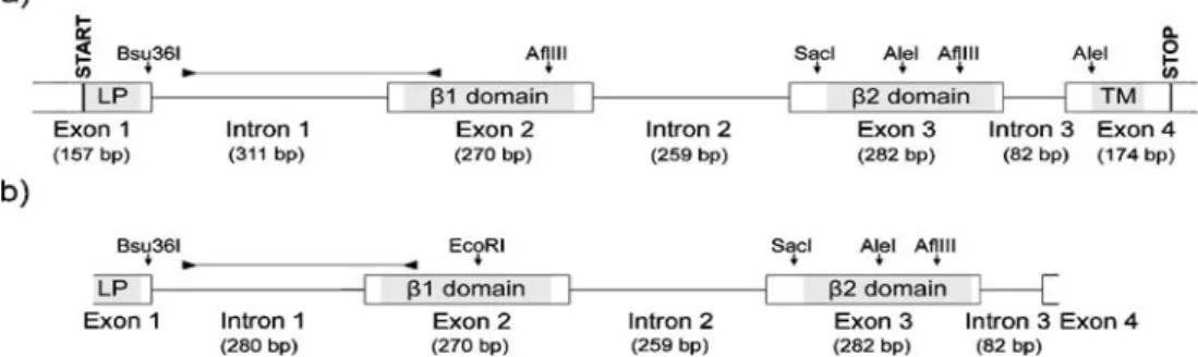
Fig. 1 Schematic illustration and restriction maps of the Barn owl MHCIIB genes Tyal-DAB1 (a) and Tyal-DAB2 (b). Boxes represent exons. Functional domains are indicated in light grey. The highly divergent regions encompassing intron 1 and the region of exon 2
containing the 18-bp locus signature are indicated by horizontal lines delimited by arrow heads. LP, leader peptide; TM, trans-membrane domain
sequences were cross-checked with the ones from uncloned PCR products, and only those represented by more than one clone were considered in further analyses.
To confirm the identity of the coding sequence isolated from Barn owl, we performed BLAST searches with nucleotide (megablast) and protein sequences (blastp). CD-Search (Marchler-Bauer and Bryant2004) and SMART (Schultz et al. 1998) were used to identify signaling domains. Additionally, sequences were manually checked for features typical to functional MHCIIB genes, like the intra-domain cysteine salt bridges, the conserved glycosyl-ation site (NGT) in theβ1 domain, the conserved residues in the β1 domain characteristic of classical MHCIIB molecules (Kaufman et al. 1994), and the conserved residues implicated in CD4 binding in the human DRB molecule (Wang et al. 2001). For each intron, BLAST searches (blastn) were done separately.
Mean pairwise differences between sequences (π) were calculated in Arlequin 3.0 (Excoffier et al.2005). Analyses of selection were performed in CodeML (Yang and Nielsen 2000). To test for differences of selective pressures among sites in exon 2 and identify those sites that were potentially under positive selection, we performed maximum likeli-hood (ML) ratio tests comparing model M1a with M2a and model M7 with M8. Models M1a and M7 are neutral, while models M2a and M8 allow for a proportion of sites under positive selection. For a detailed description of these models see Nielsen and Yang (1998) and Yang and Nielsen (2000).
Results
Isolation and characterization of MHC class II B genes Making use of a PCR-based approach combined with primer walking on genomic and complementary DNA, we isolated two functional MHCIIB genes (accession numbers of isolated sequences at GenBank are EU442602– EU442607). According to Klein et al. (1990), we designat-ed those two loci, MhcTyal-DAB1 and MhcTyal-DAB2, with alleles denoted by the gene name followed by an asterisk and sequential allele numbers. The two genes are highly similar over large stretches, including introns 2 and 3. However, they are distinguished by the highly divergent intron 1 and a locus-specific 18 bp signature within the first 70 nucleotide sites of exon 2 (see below, Figs. 1 and 2). Mean pairwise differences between cloned sequences from the different loci were 49.00±2.73, and are significantly larger between than within loci when the locus signature is considered (Kruskal–Wallis test, p<0.01) and marginally significant when the 18 nucleotide sites of the signature are removed (Kruskal–Wallis test, p=0.06).
The coding sequence of Tyal-DAB1 encompasses 783 bp (261 amino acid residues) from start to stop codon and is partitioned among four exons (Fig. 1). For Tyal-DAB2, 652 bp of coding sequence (217 amino acid residues) were obtained by PCR.
Expression analyses for the two loci were performed through RT-PCR. Sequencing of the cloned RT-PCR products from four individuals resulted in a total of six alleles, i.e., four for Tyal-DAB1 and two for Tyal-DAB2. Their successful amplification from spleen, liver, and blood shows that both genes are expressed in all these tissues.
Protein–protein BLAST searches produced significant hits with known avian and mammalian MHCIIB protein sequences. Tyal-DAB1 and Tyal-DAB2 proteins contain the features expected for classical MHCIIB molecules (Fig.2). CD-Search (Marchler-Bauer and Bryant 2004) identified a β1 domain in exon 2 and a IGc1 domain in exon 3 (Figs.1 and 2). SMART confirmed these domains, and identified a signal peptide and a trans-membrane domain (Figs. 1 and 2). The trans-membrane domain was confirmed by the TMpred (Hofmann and Stoffel 1993) and Phobius soft-wares (Kall et al.2004). The manual survey confirmed the conserved intra-domain cysteine salt bridges withinβ1 and β2 domains. In the conserved glycosylation site (NGT), the threonine is replaced conservatively (Hanada et al.2007) by a serine. All but two of the conserved residues in the β1 domain characteristic of classical MHCIIB molecules (Kaufman et al. 1994) are conserved, as are the conserved residues implicated in CD4 binding in the human DRB molecule (Wang et al.2001).
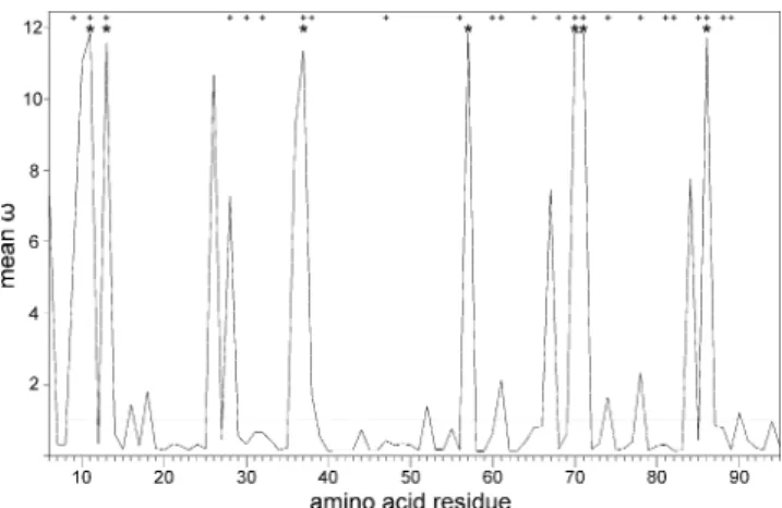
Positive selection on the sites involved in antigen-binding is expected to result in considerable variation in exon 2 of functional MHCIIB genes. We therefore estimated variability in the cloned sequences and tested if positive selection was acting on the Barn owl MHCIIB. Cloning yielded four DAB1 alleles and two Tyal-DAB2 alleles. Exon 2 harbors most of the polymorphic sites within and among genes (92%, 65 out of 71 sites). About half (31 sites) are located in sites corresponding to human ABS (Brown et al. 1993). Mean pairwise differ-ences between alleles were 35.37±17.78 overall, 21.83± 12.28 within Tyal-DAB1 and six between Tyal-DAB2 alleles. ML analyses show that positive selection is acting on the Barn owl MHC. They reveal considerable variation in selective pressures among sites in exon 2. Seven sites were detected (M2a,ω=11.73±0.20) that are under positive selection, of which six correspond to human ABS (Brown et al. 1993; however, for an alternative model of antigen binding see Tong et al.2006) (Fig.3).
Using locus-specific primers situated in exons, we successfully amplified all introns of both Tyal-DAB genes. Introns vary from 82 bp (intron 3) to 311 bp (DAB1 intron 1) in length (Fig. 1), and all contain the classical splicing
signal 5′-GT-intron-AG-3′. Introns 2 and 3 are identical between Tyal-DAB genes. Intron 1 is identical at only the first 40 nucleotide sites and completely divergent between genes for the rest of its sequence, and 32 bp longer in Tyal-DAB1 than in Tyal-DAB2 (Figs.1and S1).
Number of MHCIIB genes in the Barn owl
To confirm the presence of the two genes isolated by PCR and primer walking and obtain an estimate for the number
of MHCIIB genes present in Barn owl, we performed simple PCR-based tests and Southern blot analyses. The presence of the two genes Tyal-DAB1 and Tyal-DAB2 was confirmed by successful locus- and allele-specific PCR reactions in the individual on which the isolation was performed (for primers see Table1). Southern blot analyses confirmed this result and gave no indication for the presence of further MHCIIB genes (Fig. 4). The number of detected fragments corresponded to fragments predicted from the restriction map (Fig.1), except for the hybridiza-Fig. 2 Amino acid alignment
of Barn owl MHCIIB sequences with chicken B-Lb I (Gallus gallus, accession no. AL023516) and Tuatara (Sphenodon punctatus, accession no. DQ124232). Dots indicate identity with the Tyal-DAB1*01 sequence; dashes indicate gaps. Exon boundaries are indicated by black arrow heads and exon number. Conserved residues characteristic of classical MHCIIB chain molecules (Kaufman et al.1994) and highly conserved residues in exon 3 implicated in CD4 binding in humans (Wang et al.
2001) are shaded. Cysteine bridges in theβ1 and β2 domains are indicated by brackets
tion of the EcoRI restriction with the exon 3 probe that produced a single band, while two were expected. The presence of only one band in this hybridization could be due to an approximately equal length of the fragments expected for the two genes or to a tandem architecture with Tyal-DAB2 in 5′-position of Tyal-DAB1. The weakness of three bands in AflIII-restricted DNA and of the short fragment in the AleI restriction can be explained by small overlaps of these fragments with the probes. Results for AleI, AflIII, and EcoRI are shown in Fig. 4; those for Bsu36I and SacI confirmed the first three and are not shown.
Discussion
Genomic organization of the Barn owl MHCIIB
Using PCR-based and Southern blot analyses, we isolated and characterized two MHCIIB genes in the Barn owl. As recently demonstrated, these genes are orthologs of Strigi-dae owl and Great snipe (Gallinago media) MHCIIB genes (Burri et al.2008). Several observations are consistent with the hypothesis that both loci are functional: (1) they are expressed in several tissues; (2) show high non-synony-mous substitution rates indicative of positive selection; (3) have no frameshift mutations and stop codons. This provides strong evidence for a simple genomic organization of the Barn owl MHCIIB, similar to the simple chicken MHCIIB (Zoorob et al. 1990; Kaufman et al. 1999). Our results thus constitute the first conclusive support for the suggestion made by Alcaide et al. (2007) that in birds of prey MHC organization might be simple. These authors applied a PCR-based survey, admitting that such an
approach is prone to the underestimation of the number of genes (Wagner et al.1994). Accordingly, for the Barn owl, their approach confirmed our genomic sequence of Tyal-DAB1 but did not detect the presence of Tyal-DAB2. Concerted evolution of the Barn Owl MHCIIB
Concerted evolution has acted to a limited extent between Tyal-DAB genes, contrary to most avian species (Hess and Edwards 2002). Tyal-DAB genes are highly similar over most of their sequence, presumably owing to homogeniza-tion by gene conversion. However, 340 nucleotide sites, comprising intron 1 and 70 nucleotide sites of exon 2 (Figs.1and S1), seem to have escaped concerted evolution so far. These positions differ substantially between Tyal-DAB1 and Tyal-DAB2. Despite the high overall divergence of intron 1 between the two genes, this intron’s first 40 nucleotide sites are identical. Moreover, this region is the most conserved in intron 1 among all birds of prey (Fig. S1), among songbirds, and among chicken and pheasant (Wittzel et al. 1994; Edwards et al. 1998). The inter-specific conservation was previously interpreted in terms of selective constraints, concerted evolution, or recent duplication (Wittzel et al. 1994; Edwards et al. 1998). It seems unlikely that selective constraints result from selection on the intron itself. Given the frequency of
Fig. 4 Banding pattern for the Southern blot analyses with the exon 2 (a) and the exon 3 (b) probes. Flashes highlight weak bands Fig. 3 Meanω (dN/dS) along the Barn owl MHCIIB exon 2 sequence
as calculated in CodeML (model M2a). The horizontal line denotes a ω of 1, indicating neutral evolution. Residue numbers are given according to Brown et al. (1993). * Sites identified to be under positive selection in Barn owl. Significance was tested by the BEB procedure. + Sites corresponding to human antigen-binding sites (Brown et al.1993)
deletions in the intron 1 of birds of prey (Fig. S1), the conservation of the exon-flanking sequences might rather represent a byproduct of selection eliminating deletions reaching into exon 2. Most probably, however, it might also be a result of convergent evolution.
MHC evolutionary ecology
The study of the MHC evolutionary ecology in birds is usually hampered by the presence of multiple gene duplicates, and typically alleles cannot be attributed to loci (Hess and Edwards 2002). Therefore, studies considering only a subset of the loci present in a given species may miss important information or suffer from low statistical power, and data from non-functional loci may even yield misleading results. The present study provides the essential basics for immunogenetic studies in the Barn owl. The low number of loci found in this species allows for studies considering the complete individual MHCIIB genotypes. Together with the possibility to attribute the alleles to specific loci, this will strongly facilitate the study of MHC evolutionary ecology in the Barn owl compared to most avian species.
Acknowledgements We thank Catherine Berney for technical assistance in the lab, Anne-Lyse Ducrest for helpful discussions, Philippe Christe for providing samples, and two anonymous reviewers for helpful comments on earlier versions of the manuscript. The study was supported by the Swiss National Science Foundation grants 3100A0-109852/1 to LF and PP00A-102913 to AR.
References
Alcaide M, Edwards S, Negro J (2007) Characterization, polymor-phism, and evolution of MHC Class II B genes in birds of prey. J Mol Evol 65:541–554
Brown JH, Jardetzky TS, Gorga JC, Stern LJ, Urban RG, Strominger JL et al (1993) Three-dimensional structure of the human class II histocompatibility antigen HLA-DR1. Nature 364:33–39 Burri R, Niculita-Hirzel H, Salamin N, Roulin A, Fumagalli L (2008)
Evolutionary patterns of MHC class II B in owls and their implications for the understanding of avian MHC evolution. Mol Biol Evol 25:1180–1191
Del Hoyo J, Elliott A, Sargatal J (2000) Barn owls to hummingbirds. Handbook of the birds of the world. Lynx Editions, Barcelona, p 759
Doherty PC, Zinkernagel RM (1975) Enhanced immunological surveillance in mice heterozygous at the H-2 gene complex. Nature 256:50–52
Edwards S, Gasper J, March M (1998) Genomics and polymorphism of Agph-DAB1, an Mhc class II B gene in red-winged blackbirds (Agelaius phoeniceus). Mol Biol Evol 15:236–250
Ekblom R, Grahn M, Höglund J (2003) Patterns of polymorphism in the MHC class II of a non-passerine bird, the great snipe (Gallinago media). Immunogenetics 54:734–741
Excoffier L, Laval G, Schneider S (2005) Arlequin (version 3.0): an integrated software for population genetics data analysis. Evol Bioinformatics 1:47–50
Hanada K, Shiu S-H, Li W-H (2007) The nonsynonymous/synonymous substitution rate ratio versus the radical/conservative replacement rate ratio in the evolution of mammalian genes. Mol Biol Evol 24:2235–2241
Hess CM, Edwards SV (2002) The evolution of the major histocom-patibility complex in birds. Bioscience 52:423–431
Hofmann K, Stoffel W (1993) TMbase—a database of membrane spanning proteins segments. Biol Chem Hoppe Seyler 374:166 Kall L, Krogh A, Sonnhammer ELL (2004) A combined
transmem-brane topology and signal peptide prediction method. J Mol Biol 338:1027–1036
Kaufman J, Milne S, Gobel TWF, Walker BA, Jacob JP, Auffray C et al (1999) The chicken B locus is a minimal essential major histocompatibility complex. Nature 401:923–925
Kaufman J, Salomonsen J, Flajnik M (1994) Evolutionary conserva-tion of MHC class I and class II molecules—different yet the same. Semin Immunol 6:411–424
Klein J, Bontrop R, Dawkins RL, Erlich HA, Gyllensten UB, Heise ER et al (1990) Nomenclature for major histocompatibility complexes of different species: a proposal. Immunogenetics 31:217–219
Marchler-Bauer A, Bryant SH (2004) CD-Search: protein domain annotations on the fly. Nucleic Acids Res 32:W327–W331 Milinski M (2006) The major histocompatibility complex, sexual
selection, and mate choice. Annu Rev Ecol Evol Syst 37:159–186 Nielsen R, Yang Z (1998) Likelihood models for detecting positively
selected amino acid sites and applications to the HIV-1 envelope gene. Genetics 148:929–936
Penn DJ, Damjanovich K, Potts WK (2002) MHC heterozygosity confers a selective advantage against multiple-strain infections. Proc Natl Acad Sci USA 99:11260–11264
Piertney SB, Oliver MK (2006) The evolutionary ecology of the major histocompatibility complex. Heredity 96:7–21
Robinson J, Waller MJ, Parham P, de Groot N, Bontrop R, Kennedy LJ et al (2003) IMGT/HLA and IMGT/MHC: sequence databases for the study of the major histocompatibility complex. Nucleic Acids Res 31:311–314
Roulin A (1999) Nonrandom pairing by male Barn owls (Tyto alba) with respect to a female plumage trait. Behav Ecol 10:688–695 Roulin A, Ducrest A-L, Balloux F, Dijkstra C, Riols C (2003) A
female melanin ornament signals offspring fluctuating asymme-try in the Barn owl. P Roy Soc B-Biol Sci 270:167–171 Roulin A, Riols C, Dijkstra C, Ducrest A-L (2001) Female plumage
spottiness signals parasite resistance in the Barn owl (Tyto alba). Behav Ecol 12:103–110
Schultz J, Milpetz F, Bork P, Ponting CP (1998) SMART, a simple modular architecture research tool: identification of signaling domains. Proc Natl Acad Sci USA 95:5857–5864
Takahata N, Nei M (1990) Allelic genealogy under overdominant and frequency-dependent selection and polymorphism of major histocompatibility complex loci. Genetics 124:967–978 Tong JC, Bramson J, Kanduc D, Chow S, Sinha AA, Ranganathan S
(2006) Modeling the bound conformation of Pemphigus vulgaris-associated peptides to MHC class II DR and DQ alleles. Immunome Res 2:1
Tsuda T, Tsuda M, Naruse T, Kawata H, Ando A, Shiina T et al (2001) Phylogenetic analysis of penguin (Spheniscidae) species based on sequence variation in MHC class II genes. Immunogenetics 53:712–716
Wagner A, Blackstone N, Cartwright P, Dick M, Misof B, Snow P et al (1994) Surveys of gene families using polymerase chain reaction: PCR selection and PCR drift. Syst Biol 43:250–261 Wang J-H, Meijers R, Xiong Y, Liu J-h, Sakihama T, Zhang R et al
(2001) Crystal structure of the human CD4 N-terminal two-domain fragment complexed to a class II MHC molecule. Proc Natl Acad Sci USA 98:10799–10804
Wittzel H, von Schantz T, Zoorob R, Auffray C (1994) Molecular characterization of three Mhc class II B haplotypes in the ring-necked pheasant. Immunogenetics 39:395–403
Yang Z, Nielsen R (2000) Estimating synonymous and nonsynon-ymous substitution rates under realistic evolutionary models. Mol Biol Evol 17:32–43
Zelano B, Edwards SV (2002) An Mhc component to kin recognition and mate choice in birds: predictions, progress, and prospects. Am Nat 160:S225–S237
Zoorob R, Béhar G, Kroemer G, Auffray C (1990) Organisation of a functional chicken class II B gene. Immunogenetics 31:179– 187