J Orofac Orthop 2011; 72:371-380 DOI 10.1007/s00056-011-0041-4
Shear bond strengths of seven self-etching primers after
thermo-cycling
Scherkräfte von sieben selbstätzenden Primern nach
Thermozyklierung
Abstract
Objective. Self-etching primers are widely used in restorative dentistry. However, only one primer has been developed for or-thodontic applications. The objective of this study was to assess the suitabiliy of restorative self-etching primers on unabraded enamel.
Material and Methods. Seven self-etching primers (Trans-bond
™
SEP+ , Xeno®
III, Clearfil™
SE, Futurabond®
NR, One Coat, M-Bond and OptiBond®
) were compared with two conventional-ly-etched control groups (Transbond™
MIP). All groups with the exception of one of the two control groups underwent thermo-cycling (1000x, 5–55°C). Bovine teeth stored in physiological sa-line solution were used for the purpose of this study.Results. Both before and after thermo-cycling, the control groups presented very similar shear strengths which did not differ significantly from each other or from the Transbond
™
SEP+ and Clearfil™
SE specimens. During thermo-cycling, all groups except the control group and the Transbond™
SEP+ specimens displayed losses from the shear bodies. These losses occurred on all of the OptiBond®
samples. The adhesive remnant index (ARI) and enam-el tear-outs correnam-elated closenam-ely with the adhesive values.Conclusions. Our adhesive value assessments support the con-clusion that only Transbond
™
SEP+ and Clearfil™
SE can be re-commended for clinical use.Keywords
Shear strengths · Self-etching primers · Etching pattern
Zusammenfassung
Ziel. Selbstätzende Primer werden in der restaurativen Zahn-heilkunde breit eingesetzt. Speziell für die kieferorthopädische Verwendung wurde allerdings nur ein einzelner Primer entwick-elt. Ziel der vorliegenden Studie war es, die Eignung restaurativer selbstätzender Primer auf unbeschliffenem Schmelz zu unter-suchen.
Material und Methodik. Sieben selbstätzende Primer (Trans-bond
™
SEP+, Xeno™
III, Clearfil™
SE, Futurabond NR, One Coat, M-Bond und OptiBond®
) wurden mit 2 konventionell geätzten Kontrollgruppen (Transbond™
MIP) verglichen. Alle Gruppen außer einer Kontrollgruppe wurden einer Thermozyklierung (1000x, 5–55°C) unterzogen. Für die Untersuchung wurden Rind-erzähne verwendet, die in physiologischer Kochsalzlösung ge-lagert wurden.Ergebnisse. Die Kontrollgruppen zeigten vor und nach Ther-mozyklierung sehr ähnliche Scherkräfte, die sich untereinander und gegenüber Transbond
™
SEP+ und Clearfil™
SE nicht signi-fikant unterschieden. Während der Thermozyklierung wurden für alle Gruppen außer der Kontrollgruppe und Transbond™
SEP+ Verluste der Scherkörper registriert. Bei OptiBond®
fanden sich diese bei sämtlichen Proben. Der Adhäsiv-Rest-Index (ARI) sowie Schmelzausbrüche korrelierten gut mit den Haftwerten.Schlussfolgerungen. Nur Transbond
™
SEP+ und Clearfil™
SE können aufgrund der Haftwertuntersuchungen für den klinischen Einsatz empfohlen werden.Schlüsselwörter
Scherkräfte · Selbstätzende Primer · Ätzmuster
Lorenz Brauchli
1, Marlis Zeller
1, Andrea Wichelhaus
21 Clinic of Orthodontics and Pedodontics, University of Basel,
Switzer-land
2 Department of Orthodontics, Ludwig-Maximilian University Munich,
Germany
Received: January 17, 2011; accepted: July 15, 2011; published online: October 6, 2011
Brauchli L et al. Scherkräfte – selbstätzende Primer
Introduction
Adhesive technologies in dentisty and orthodontics have at-tracted serious interest since the introduction of acid etching by Buonocore [7] and bonding techniques introduced by Newman [33]. Since then, seven generations of adhesives have been described [11, 29]. The first generation was the classic BIS-GMA, which remains a chief component of today's bond-ing systems; however, its main problem lay in its low adhesion to dentin. More hydrophile methacrylates were later developed which improved adhesion to dentin. It was not until the fourth generation that today's classic system encompassed hy-drophilic primer, ambiphilic adhesive and hydrophobic bond-ing. After a renewed combination of the three substances in a single bottle system (fifth generation), the sixth generation de-livered the self-etching primer. Most of these systems use a methacrylate-based phosphoric acid ester. With the seventh generation, an attempt was made to include anti-bacterial, anti-inflammatory and desensitizing substances; however, the effect of these substances is controversial [11, 25, 36].
The main advantages of self-etching primers are that they re-duce the likelihood of error and save time [15, 21, 49]. Their etching pattern is frequently less distinctive than with conven-tional phosphoric-acid etching [20, 37, 42]. However, there is no proven correlation with adhesive forces [27]. This could be due to the identical depth of penetration of the methacrylate and etching acid when self-etching primers are used, something which cannot be guaranteed by conventional etching [13]. With self-etching primers, the etching process is stopped by dissolved calcium ions, increasing viscosity and ultimately by light-curing due to the inclusion of acid groups in the polymer matrix of the methacrylate [13]. Contact with oxygen causes incomplete hardening with active, residual acids on the surface of self-etch-ing primers. These prevent combination with acid-sensitive, chemically-hardening resins. There is apparently no reduction in adhesion in conjunction with the combination with light-curing products [12, 41]. The bond between the self-etching primer and adhesive may weaken due to the accumulation of osmotically-active, dissolved hydroxyapatite in the unpolymer-ized surface layer [46, 47].
Transbond
™
SEP+ (3M Unitek, Monrovia, CA, USA) was one of the first self-etching primers in orthodontics proven to be a suitable substitute for conventional etching methods in several clinical studies [9, 15, 36, 38]. Yet some authors reported re-duced adhesive values [5, 14, 26, 30]. There is much less data on other self-etching primers, and their suitability for use on un-treated enamel surfaces remains controversial [3, 6, 35, 42, 43].In this study we examined the shear adhesion on unabraded enamel surfaces after thermo-cycling of the self-etching primers employed in conservative dentistry. Our objective was not pri-marily to re-examine the proven primer Transbond
™
SEP+ , but to establish the suitability of alternative primers for orthodontic applications.Einleitung
Seit der Einführung der Säureätzung durch Buonocore [7] und der Klebetechniken in der Kieferorthopädie durch Newman [33] sind Adhäsivtechnologien für Zahnärzte und Kieferor-thopäden von großem Interesse. Seither wurden 7 Generatio-nen von Adhäsivstoffen beschrieben [11, 29]. Die erste Gene-ration war das klassische BIS-GMA, das immer noch eine Hauptkomponente der heutigen Bondingsysteme darstellt. Das Hauptproblem lag in einer niedrigen Haftung auf Dentin. Nachfolgend wurden hydrophilere Methacrylate entwickelt, welche die Haftung auf Dentin verbessern konnten. Erst in der vierten Generation wurde das heute klassische System mit hy-drophilem Primer, ambiphilem Adhäsiv und hydrophobem Bonding beschrieben. Nach erneuter Kombination der 3 Subs-tanzen in einem Einflaschensystem (fünfte Generation), brachte die sechste Generation die selbstätzenden Primer. Die meisten Systeme verwenden dabei einen Phosphorsäureester auf Methacrylatbasis. In der siebten Generation wurde ver-sucht, antibakterielle, antiinflammatorische und desensibilisie-rende Substanzen beizufügen. Diese sind in ihrer Wirkung je-doch umstritten [11, 25, 36].
Der Hauptvorteil der selbstätzenden Primer besteht in der Reduktion der Fehlermöglichkeiten und einer Zeitersparnis [15, 21, 49]. Das Ätzmuster der selbstätzenden Primer ist oft schwächer ausgeprägt als bei konventioneller Phosphorsäure-ätzung [20, 37, 42]. Allerdings wurde keine klare Korrelation zu den Haftkräften gefunden [27]. Dies könnte darauf beruhen, dass bei selbstätzenden Primern die Penetrationstiefe der Me-thacrylate mit jener der Ätzmittel identisch ist, was bei konven-tioneller Ätzung nicht gewährleistet werden kann [13]. Der Ätz-prozess bei selbstätzenden Primern wird über herausgelöste Kalziumionen, eine zunehmende Viskosität und schließlich bei Lichthärtung durch den Einschluss der Säuregruppen in der Polymermatrix des Methacrylats gestoppt [13]. An der Ober-fläche der selbstätzenden Primer entsteht durch den Kontakt mit Sauerstoff eine unvollständige Härtung mit weiterhin akti-ven Säuregruppen. Diese verhindern die Kombination mit den säuresensitiven, chemisch härtenden Kunststoffen. Für die Kombination mit lichthärtenden Produkten fand sich keine Haftminderung [12, 41]. Eine Schwächung der Verbindung zwischen selbstätzendem Primer und verwendetem Adhäsiv könnte sich auch über eine Akkumulation von osmotisch akti-vem, gelöstem Hydroxylapatit in der unpolymerisierten Ober-flächenschicht ergeben [46, 47].
In der Kieferorthopädie erwies sich Transbond
™
SEP+ (3M Unitek, Monrovia, CA, USA), einer der ersten selbstätzenden Primer, in mehreren klinischen Studien als geeigneter Ersatz für konventionelle Ätzmethoden [9, 15, 36, 38]. Einige Autoren fan-den aber auch verminderte Haftwerte [5, 14, 26, 30]. Für andere selbstätzende Primer bestehen deutlich weniger und kontrover-se Daten zu deren Eignung auf unbehandelten Schmelzoberflä-chen [3, 6, 35, 42, 43].Die vorliegende Untersuchung prüfte selbstätzende Primer aus der konservierenden Zahnheilkunde bezüglich der
Scher-Material and methods
The shear strengths of seven self-etching primers were studied and compared with conventionally etched groups: Trans-bond
™
SEP+ (3M Unitek, Monrovia, CA, USA), Xeno®
III (Dentsply, Konstanz, Germany), Clearfil™
SE (Kuraray, Frank-furt, Germany), Futurabond®
NR (VOCO, Cuxhaven, Ger-many), One Coat (Coltène/Whaledent, Cuyahoga Falls, OH, USA), M-Bond (Tokuyama, Burlingame, CA, USA) and Opti-Bond®
(Kerr GmbH, Bioggio, Switzerland). Each group con-sisted of 20 specimens, and the self-etching primers were ad-ministered observing manufacturer instructions. The self-etching primers were compared with two conventionally-etched groups using 35% orthophosphoric acid (Unitek™
Etching Gel; 3M Unitek, Monrovia, CA, USA) and Trans-bond
™
MIP.Bovine incisors were used for the enamel substrate in compli-ance with ISO 11405/TS. The only teeth used were those whose macroscopic enamel surfaces were in perfect condition. The dental pulp was removed and the teeth stored temporarily prior to the shear tests in physiological saline solution.
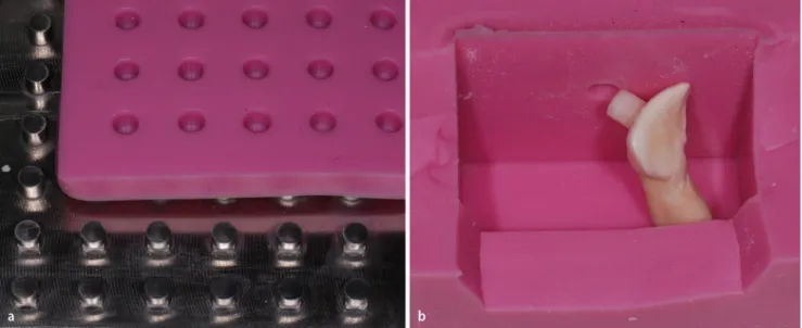
We used shear bodies made of Grandio Flow (Grandio Flow; VOCO, Cuxhaven, Germany) in a cylindrical form with a base surface of 12.6 mm2 in all the specimens. For this purpose, a CNC milling machine (Picomax 60-M/HSC; Fehlmann AG, Seon, Switzerland) was used to mill a stainless-steel positive form base plate with 49 cylinders protruding from it. Using fast-flowing silicone (Finosil; Fino GMBH, Schweinfurt, Germany) the cylinders were molded, creating a negative form. The hollow cylindrical forms in the silicone mold were filled with Grandio Flow to cast the shear bodies (Figure 1). Finally, the composite
haftwerte auf ungeschliffenen Schmelzoberflächen nach Ther-mozyklierung. Ziel der Studie war weniger die erneute Über-prüfung des bewährten Primers Transbond
™
SEP+, sondern vielmehr die Eignung alternativer Primer zur kieferorthopädi-schen Anwendung.Material und Methodik
Die Scherhaftkräfte von 7 selbstätzenden Primern wurden untersucht und mit konventionell geätzten Gruppen vergli-chen: Transbond
™
SEP+ (3M Unitek, Monrovia, CA, USA), Xeno®
III (Dentsply, Konstanz, Deutschland), Clearfil™
SE (Kuraray, Frankfurt, Deutschland), Futurabond®
NR (VOCO, Cuxhaven, Deutschland), One Coat (Coltène/Whaledent, Cuyahoga Falls, Ohio, USA), M-Bond (Tokuyama, Burlin-game, CA, USA) und OptiBond®
(Kerr GmbH, Bioggio, Schweiz). Jede Gruppe bestand aus 20 Proben, die selbstätzen-den Primer wurselbstätzen-den gemäß Herstellerangaben appliziert. Die selbstätzenden Primer wurden mit 2 konventionell geätzten Gruppen unter Verwendung einer 35%igen Orthophosphor-säure (Unitek™
Etching Gel; 3M Unitek, Monrovia, CA, USA) und Transbond™
MIP verglichen.Als Schmelzsubstrat wurden gemäß ISO 11405/TS Rinderin-zisivi verwendet. Es wurden ausschließlich Zähne mit makros-kopisch einwandfreien Schmelzflächen verwendet. Die Pulpa der Zähne wurde entfernt und die Zähne bis zur Schertestung in physiologischer Kochsalzlösung zwischengelagert.
Für alle Proben wurden Abscherkörper aus Grandio Flow (Grandio Flow; VOCO, Cuxhaven, Deutschland) mit zylindri-scher Form und einer Grundfläche von 12,6 mm2 verwendet. Da-zu wurde über eine CNC-Fräse (Picomax 60-M/HSC; Fehlmann
Figure 1. Specimen fabrication: the shear bodies were made by pouring a silicone mold of a CNC milled steel plate (a). A special single-bed form (b) allowed the shearing cylinder to be oriented parallel to the base surface of the test specimen
Abbildung 1. Probenherstellung: Die Abscherkörper wurden durch Ausgießen einer Silikonabformung einer CNC-gefrästen Stahlplat-te ersStahlplat-tellt (a). Eine spezielle Einbettform (b) ermöglichStahlplat-te die parallele Ausrichtung des Scherzylinders zur Grundfläche des Prüfkörpers
Brauchli L et al. Scherkräfte – selbstätzende Primer
cylinders were light-cured for 40 s at 800 mW/cm2 (bluephase
®
C8; Ivoclar Vivadent, Schaan, Liechtenstein).After bonding the composite cylinders onto the prepared enamel specimens using Transbond
™
XT (3M Unitek, Monro-via, CA, USA) composite, and another curing period of 40 s (bluephase®
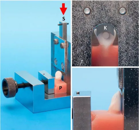
C8; Ivoclar Vivadent, Liechtenstein), we placed all the self-etching primer samples and one of the two control groups in a contrast bath set at 1000 thermo-cycles between 5 and 55°C for a period of 50 h [Circulator C-85 (Techne, Brou-wer, Luzern, Switzerland) and Julabo UC and 5B (Julabo Labortechnik, Seelbach, Germany)]. The second control group was not thermo-cycled.We ensured that the enamel adhesive bond was parallel to the shear direction by using a custom-made single-bed form (Fig-ure 1). Polymethacrylate was po(Fig-ured around the roots of the teeth (Technovit; Heraeus Kulzer, Wehrheim, Germany) and the resulting specimens were sheared off using a universal test-ing machine (Instron 4444; Instron Corp., Wilmtest-ington, DE, USA). The distance of the shear blade from the enamel surface was set at 0.5 mm (Figure 2). These data were recorded auto-matically at a shear speed of 0.1 mm/s on a PC.
The adhesive remnant index (ARI) developed by Artun [2] was documented at a 3.5-fold magnification in all specimens.
Statistical evaluation of the adhesive strengths (Figure 2) was carried out using GraphPad Instat 3 (GraphPad Software Inc., San Diego, CA, USA). Normal distribution was calculated ac-cording to Kolmogorov-Smirnov. The descriptive statistics were
AG, Seon, Schweiz) aus Edelstahl eine Positivform-Grundplatte mit 49 daraus hervorragenden Zylindern gefräst. Mittels eines dünnfließenden Silikons (Finosil; Fino GMBH, Schweinfurt, Deutschland) wurden die Zylinder abgeformt und eine Negativ-form erstellt. Die ZylinderhohlNegativ-formen in der SilikonabNegativ-formung wurden zur Herstellung der Abscherkörper mit Grandio Flow ausgegossen (Abbildung 1). Abschließend erfolgte eine Lichthär-tung der Kompositzylinder über 40 s bei 800 mW/cm2 (bluepha-se
®
C8; Ivoclar Vivadent, Schaan, Liechtenstein).Nach Bonding der Kompositzylinder auf die vorbereiteten Schmelzproben mittels des Komposits Transbond
™
XT (3M Unitek, Monrovia, CA, USA) und erneuter Härtung für 40 s (bluephase®
C8; Ivoclar Vivadent, Liechtenstein) wurden sämt-liche Proben der selbstätzenden Primer und eine der beiden Kontrollgruppen in einem Wechselbad einer 1000-fachen Ther-mozyklierung von 5–55°C über eine Laufzeit von 50 h ausge-setzt (Circulator C-85, Techne, Brouwer, Luzern, Schweiz; Jula-bo UC und 5B, JulaJula-bo LaJula-bortechnik, Seelbach, Deutschland). Die zweite Kontrollgruppe wurde nicht thermozykliert.Die Parallelität der Schmelzadhäsivverbindung mit der Scherrichtung wurde durch die Verwendung einer eigens gefer-tigten Einbettform gewährleistet (Abbildung 1). Die Wurzeln der Zähne wurden mit einem Polymethacrylat umgossen (Technovit; Heraeus Kulzer, Wehrheim, Deutschland) und die so hergestellten Proben mit einer Universalprüfmaschine (In-stron 4444; In(In-stron Corp., Wilmington, DE, USA) abgeschert. Der Abstand der Scherklinge zur Schmelzoberfläche wurde auf
Figure 2. Shear assembly: the overview shows the shear plunger (S), specimen (P) and direction of pres-sure of the universal testing machine (arrow). The de-tailed images illustrate the form of the shear blade (K), the minimum clearance and its parallelism to the enamel surface
Abbildung 2. Schervorrichtung: Die Übersicht zeigt den Scherstempel (S), die Probe (P) und die Druckrich-tung der Universalprüfmaschine (Pfeil). Auf den De-tailaufnahmen sind die Gestaltung der Scherklinge (K) sowie deren minimaler Abstand und Parallelität zur Schmelzoberfläche ersichtlich
performed calculating the mean, median, standard deviation and the standard error of the mean. The Kruskal-Wallis and Dunn's post-hoc test with a significance level set at p ≤ 0.05 were used for the comparative statistics.
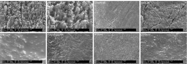
In addition, the pH value of all the self-etching primers was measured using test strips featuring 0.5-stage pH segments (Acilit pH 0–6; Merck KGaG, Darmstadt, Germany). Further-more, we examined the etching patterns of three additional specimens of each etchant under a scanning electron micro-scope in a vacuum at 20,000-fold magnification (ESEM, Philips 30; Royal Philips Electronics, The Netherlands), capturing typi-cal sections (Figure 3).
Results
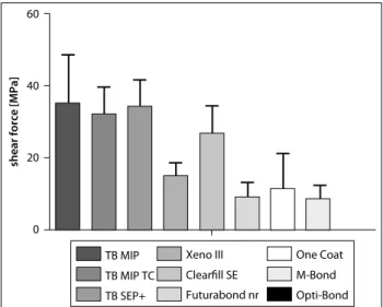
Our results are shown in Table 1, Table 2, Figure 3 and Figure 4. The difference in the adhesion values of the control group with thermo-cycling (x 32.3 MPa) and without thermo-cycling (‒x = 35.5 MPa) was very small and statistically not significant (Table 1, Figure 2). Transbond
™
SEP+ revealed nearly identical values (‒‒x = 34.2 MPa). Even the difference compared to Clear-fil™
SE (‒x = 26.8 MPa) was statistically insignificant. The re-maining self-etching primers demonstrated significantly lower adhesive values (Xeno®
III: ‒x = 15.2 MPa, One Coat: ‒x = 11.5 MPa, Futurabond®
NR: ‒x = 9.0 MPa, M-Bond: ‒x = 8.6 MPa, OptiBond®
: ‒x = 0.0 MPa). All groups aside from the control group and Transbond™
SEP+ , presented spontaneous losses from the shear bodies during thermo-cycling. The frequency of adhesion losses was inversely proportional to the adhesion val-ues. In the OptiBond®
group, all the specimens suffered losses from shear bodies during thermo-cycling, thus that group could no longer undergo evaluation. No etching relief was visible when the specimens with shear body losses were inspected.0,5 mm festgelegt (Abbildung 2). Die Datenaufzeichnung er-folgte automatisiert bei einer Schergeschwindigkeit von 0,1 mm/s über einen PC.
Für sämtliche Proben wurde unter 3,5-facher Vergrößerung der Adhäsiv-Rest-Index (ARI) nach Artun [2] festgehalten.
Die statistische Auswertung der Haftkräfte (Tabelle 1) und der ARI-Werte (Tabelle 2) erfolgte mithilfe der Software Gra-phPad Instat 3 (GraGra-phPad Software Inc., San Diego, CA, USA). Die Normalverteilung wurde nach Kolmogorov-Smirnov be-rechnet. Die deskriptive Statistik erfolgte unter Berechnung von Mittelwert, Median, Standardabweichung und Standardmittel-wertfehler. Die vergleichende Statistik wurde mit dem Kruskal-Wallis-Test und dem Post-hoc-Test nach Dunn durchgeführt, wobei das Signifikanzniveau auf p ≤0,05 festgelegt wurde.
Zusätzlich wurden von allen selbstätzenden Primern pH-Messungen mittels Teststreifen mit 0,5-stufiger pH-Einteilung (Acilit pH 0–6; Merck KGaG, Darmstadt, Deutschland) durch-geführt. Außerdem wurden die Ätzmuster von jeweils 3 zusätz-lichen Proben im Rasterelektronenmikroskop unter Vakuum und bei 20.000-facher Vergrößerung dargestellt (ESEM, Philips 30; Royal Philips Electronics, Niederlande) und typische Berei-che festgehalten (Abbildung 3).
Ergebnisse
Die Resultate sind übersichtlich in Tabelle 1 und Tabelle 2 sowie in Abbildung 3 und Abbildung 4 dargestellt.
Der Unterschied der Haftwerte der Kontrollgruppen ohne Thermozyklierung (‒x = 35,5 MPa) und mit Thermozyklierung (‒x = 32,3 MPa) war sehr klein und statistisch nicht signifikant (Tabelle 1, Abbildung 2). Transbond
™
SEP+ wies fast identische Werte auf (‒x = 34,2 MPa). Auch der Unterschied zu Clearfil™
SE (‒x = 26,8 MPa) war statistisch nicht signifikant. Die restlichen selbstätzenden Primer zeigten signifikant verringerte HaftwerteFigure 3. Enamel surfaces after etching with various primers as well as H3PO4. Obvious differences in quality are visible in the depth of
the etching relief. The sequence corresponds to the shear strengths achieved
Abbildung 3. Schmelzoberflächen nach Ätzung mit unterschiedlichen Primern, sowie H3PO4. Es sind deutliche qualitative Differenzen
Brauchli L et al. Scherkräfte – selbstätzende Primer
All groups except the One Coat group demonstrated normal distribution in the statistical evaluation. OptiBond
®
was not analyzed.The ARI correlated closely with the adhesion values meas-ured, while the specimens with high adhesion values revealed a high ARI (Table 2). Statistical analysis of the ARI yielded a sig-nificant difference among the three Transbond
™
products and remaining self-etching primers, which did not significantly dif-fer from one another. Optibond®
was not included in the analy-sis. Enamel tear-outs were only observed in the Transbond™
groups; they also showed agreement with the adhesion values obtained.
The pH measurements differed considerably: Transbond
™
SEP+ and Xeno
®
III revealed the lowest pH values (pH = 1), fol-lowed by Futurabond®
NR and M-Bond (pH = 1.5) and Clear-fil™
SE, One Coat and OptiBond®
(pH = 2).Conventional etching and Transbond
™
SEP+ demonstrated the most obvious etching patterns. Xeno®
III also showed clear etching patterns. Surface roughness from the remaining prim-ers was only moderate; OptiBond®
and One Coat revealed hardly any etching pattern (Figure 4).Discussion
The outer layer of enamel displayed some differences com-pared to the deeper enamel layers. Morphologically this con-sists of a non-prismatic layer [40, 50] which appears poorly suited for adhesive procedures [27, 32] and especially for the use of self-etching primers [10, 27, 37]. In addition, the outer enamel layer contains a modified ratio of fluorapatite and hy-droxyapatite, whereby the more acid-resistant fluorapatite oc-cupies a higher percentage [45]. The etching of the surface
(Xeno
®
III: ‒x = 15,2 MPa, One Coat: ‒x = 11,5 MPa, Futurabond®
NR: ‒x = 9,0 MPa, M-Bond: ‒x = 8,6 MPa, OptiBond
®
: ‒x = 0,0 MPa). Alle Gruppen außer der Kontrollgruppe und Trans-bond™
SEP + wiesen spontane Verluste von Scherkörpern wäh-rend der Thermozyklierung auf. Die Häufigkeit der Haftverlus-te stand in umgekehrt proportionalem Verhältnis zu den Haft-werten. In der OptiBond®
-Gruppe erlitten sämtliche Proben Verluste von Scherkörpern unter Thermozyklierung, weshalb die Gruppe nicht weiter ausgewertet werden konnte. In der vi-suellen Überprüfung der Proben mit Scherkörperverlusten war kein Ätzrelief ersichtlich.Die statistische Auswertung der Daten ergab für alle Gruppen außer One Coat eine Normalverteilung. OptiBond
®
wurde nicht ausgewertet.Der ARI wies eine deutliche Abhängigkeit von den gemesse-nen Haftwerten auf, wobei sich für Proben mit hohen Haftwer-ten ein hoher ARI ergab (Tabelle 2). Die statistische Auswertung des ARI ergab einen signifikanten Unterschied zwischen den 3 Transbond
™
-Produkten und den restlichen selbstätzenden Pri-mern, die sich ihrerseits nicht signifikant voneinander unter-schieden. OptiBond®
wurde nicht in die Auswertung einbezo-gen. Schmelzausrisse wurden nur in den Transbond™
-Gruppen beobachtet und wiesen ebenfalls eine Übereinstimmung mit den erzielten Haftwerten auf.Die pH-Messungen ergaben deutliche Unterschiede: Trans-bond
™
SEP+ und Xeno®
III wiesen die niedrigsten pH-Werte auf (pH=1). Es folgten Futurabond®
NR und M-Bond (pH=1,5) sowie Clearfil™
SE, One Coat und OptiBond®
(pH=2).Die konventionelle Ätzung sowie Transbond
™
SEP+ wiesen die ausgeprägtesten Ätzstrukturen auf. Auch Xeno®
III zeigte deutliche Ätzmuster. Die Oberflächenaufrauung der restlichenTable 1. Shear strengths of the seven self-etching (SE) primers in comparison with the control group Transbond™ XT. Mean values, standard deviation (SD), standard error of the mean (SEM), median, normal distribution according to Kolmogorov-Smirnov, ranking, significance and spontaneous detachments after thermo-cycling Tabelle 1. Scherkräfte der 7 SE-Primer im Vergleich zur Kontrollgruppe Transbond™ XT. Mittelwerte, Standardabweichung (SD), Mittelwertfehler (SEM), Median, Normalverteilung nach Kolmogorov-Smirnov, Rangfolge, Signifikanz und spontane Haftverluste nach Thermozyklierung
Primer Group N# Mean (MPa) SD SEM Median (MPa) Kolmogorov-Smirnov, p value Ranking (Kruskal-Wallis) Significance (Dunn, p ≤ 0.05) Spontaneous detachments Transbond™ MIP No thermo cycles 1 35.3 13.4 3.0 36.2 > 0.1 1 4, 6, 7, 8, 9 0 Transbond™ MIP With thermo cycles 2 32.3 7.2 1.6 32.6 > 0.1 1 4, 6, 7, 8, 9 0 Transbond™ SEP plus 3 34.2 7.3 1.6 34.7 > 0.1 1 4, 6, 7, 8, 9 0 Xeno® III 4 15.2 3.2 0.8 15.2 > 0.1 3 1, 2, 3 5 Clearfil™ SE 5 26.8 7.7 1.8 27 0.1 2 6, 7, 8, 9 1 Futurabond® NR 6 9.0 4.2 1.1 7.9 > 0.1 4 1, 2, 3, 5 7 One Coat 7 11.5 9.8 2.5 11.1 0.0001 4 1, 2, 3, 5 5 M-Bond 8 8.6 3.7 1.0 6.6 > 0.1 4 1, 2, 3, 5 7 OptiBond® 9 0.0 – – – – 5 1–8 20
enamel with weak acids may thus be reduced, as confirmed in several studies [23, 37] and rejected in another [37]. This study has not enabled us to provide a definitive answer to this ques-tion either. On the one hand, Transbond
™
SEP+ (pH = 1) re-vealed better adhesion values than primers with higher pH values such as Futurabond®
(pH = 1.5), One Coat (pH = 2), M-Bond (pH = 1.5) and OptiM-Bond®
(pH = 2), but Clearfil™
SE (pH = 2) obtained very high adhesion values—clearly ahead of Xeno®
III (pH = 1). The good adhesion strength of Clearfil™
with a relatively high pH value was confirmed in an earlier study [34] and is probably related to the adhesive components used, 10-MDP [48]. From studies on the correlation between adhesion values and the depth of etching relief, we learned that the infiltration depth of the primer is more important than just the depth of the etching pattern [8, 22, 44].
We observed no clear correlation between adhesion values and the acids used. Most primers contain phosphoric acid ester (Transbond
™
SEP+ , XENO®
III, Clearfil™
SE, Futurabond®
NR, M-Bond). Only OptiBond
®
(methylic acid) and One Coat (acrylamide sulfonic acid) used other groups of acid. While we noted deep but not significantly different adhesion values in as-sociation with M-Bond and One Coat, OptiBond®
could not be analyzed due to complete adhesion failure during thermo-cy-cling. The barely perceptible etching patterns caused by One Coat and OptiBond®
(Figure 4) were striking. These substances are apparently either not, or only partially, suitable for use on surface enamel.The specimens in the present study were exposed to 1000-fold thermo-cycling between 5 and 55°C, which according to earlier studies can lead to reduced adhesion values [16, 18]. One can assume that this enables a better approximation of the intraoral situation [4]. The control group without thermo-cycling acted as a control for the load procedure. The considerable drop in adhesion strength after thermo-cycling could be attributed to overload caused by excessive thermal fluctuations. However, in
Primer war nur mäßig ausgeprägt, bei OptiBond
®
und One Coat zeigte sich kaum eine Ätzstruktur (Abbildung 4).Diskussion
Die äußere Schmelzschicht weist einige Unterschiede zu tiefer liegenden Schmelzschichten auf. Morphologisch besteht sie aus einer aprismatischen Schicht [40, 50], die für Haftverfahren [27, 32] und insbesondere die Verwendung von selbstätzenden Pri-mern ungünstig erscheint [10, 27, 37]. Zudem enthält die äu-ßere Schmelzschicht ein verändertes Verhältnis von Fluorapatit und Hydroxylapatit, wobei säureresistenter Fluorapatit zu einem erhöhten Prozentsatz vorkommt [45]. Die Ätzung von ober-flächlichem Schmelz mit schwachen Säuren könnte deshalb ver-mindert ausfallen, was in einigen Studien bestätigt werden konnte [23, 37] und in einer anderen verworfen wurde [37]. Auch die vorliegende Arbeit kann keine abschließende Antwort liefern. Transbond
™
SEP+ (pH = 1) wies zwar bessere Haftwerte als Primer mit höheren pH-Werten wie Futurabond®
(pH = 1,5), One Coat (pH = 2), M-Bond (pH = 1,5) und OptiBond®
(pH = 2) auf, allerdings erreichte Clearfil™
SE (pH = 2) sehr hohe Haft-werte, die deutlich über denen von Xeno III (pH = 1) lagen. Die gute Haftkraft von Clearfil™
bei relativ hohem pH-Wert fand sich in einer früheren Studie bestätigt [34] und steht wahr-scheinlich im Zusammenhang mit der verwendeten Adhäsiv-komponente 10-MDP [48]. In Studien zur Korrelation der Haft-werte mit der Tiefe des Ätzreliefs konnte gezeigt werden, dass die Infiltrationstiefe des Primers wichtiger ist als die alleinige Tiefe des Ätzmusters [8, 22, 44].Des Weiteren konnte keine klare Korrelation zwischen Haft-werten und verwendeter Säure gefunden werden. Die meisten Primer enthielten Phosphorsäureester (Transbond
™
SEP +, Xe-no®
III, Clearfil™
SE, Futurabond®
NR, M-Bond). Nur Opti-Bond®
(Methylsäure) und One Coat (Acrylamidsulfonsäure) verwendeten andere Säuregruppen. Während für M-Bond undTable 2. Adhesive remnant index (ARI): mean values, standard deviation, ranking and significance. The ARI values correlate closely with the shear strengths meas-ured
Tabelle 2. Adhäsiv-Rest-Index (ARI): Mittelwerte, Standardabweichung, Rangfolge und Signifikanz. Die ARI-Werte standen in engem Zusammenhang mit den gemessenen Scherkräften
Group
N# Mean Enamel fracture Rank (Kruskal-Wallis) Significance (Dunn, p ≤ 0.05)
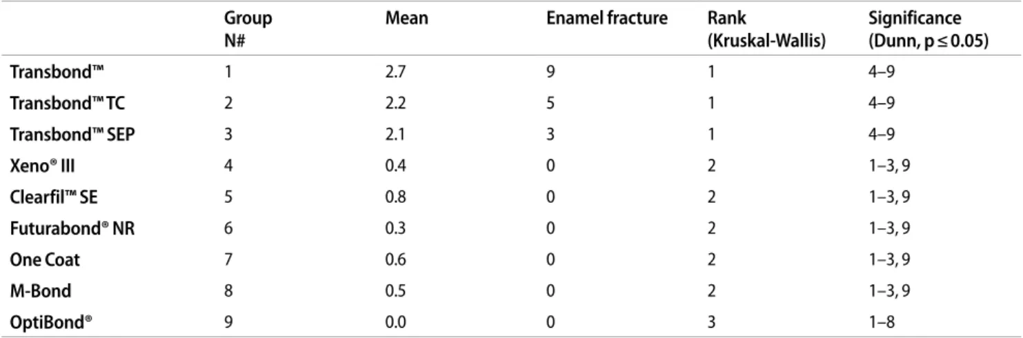
Transbond™ 1 2.7 9 1 4–9 Transbond™ TC 2 2.2 5 1 4–9 Transbond™ SEP 3 2.1 3 1 4–9 Xeno® III 4 0.4 0 2 1–3, 9 Clearfil™ SE 5 0.8 0 2 1–3, 9 Futurabond® NR 6 0.3 0 2 1–3, 9 One Coat 7 0.6 0 2 1–3, 9 M-Bond 8 0.5 0 2 1–3, 9 OptiBond® 9 0.0 0 3 1–8
Brauchli L et al. Scherkräfte – selbstätzende Primer
comparison to the control groups with and without thermo-cy-cling, we observed no significant adhesion loss. We thus assume that the thermo-cycling range we employed was reasonable. Un-like earlier studies without thermo-cycling, several self-etching primers showed unreliable adhesion values [1, 8, 10]—the number of spontaneous adhesion losses caused by thermo-cy-cling was particularly astonishing. Besides the Transbond
™
products, only Clearfil
™
SE revealed acceptable stress behavior with a single spontaneous fracture, while all the OptiBond®
specimens displayed complete adhesion loss during thermo-cycling. The clinical applicability of the products with high spontaneous adhesion losses is questionable. Adhesion loss in the oral cavity is caused by thermal fluctuations and repetitive mechanical loads, fluid absorption, and biodegradation [10, 24, 28, 31]. Hence, adhesion losses of as high as 30% have been measured over a period of 6 months [19].
The ARI values observed in this study agree well with the ad-hesion values we obtained. While the Transbond
™
groups' val-ues fell between 2.1 and 2.7, indicating cohesive failure of the adhesive bond, the self-etching primers with deep adhesion strengths showed ARI values ≤ 0.8, demonstrating a weak point in the enamel-adhesive bond (Table 2). Our Transbond™
groups' ARI values and adhesion values with over 30 MPa are thus clearly higher than findings in the literature, which reveal adhesive failure in the bracket-composite interface [10, 43]. This may be related to the shear bodies used. When brackets are used, the adhesive break usually occurs between the bracket base and composite [3, 17]. This is unfavorable when testing enamel ad-hesion using self-etching primers, as what is being tested instead is the adhesion of the resin to the bracket base. We circumvented
One Coat tiefe, aber nicht signifikant unterschiedliche Haftwer-te vorlagen, konnHaftwer-te OptiBond
®
aufgrund der kompletten Haft-verluste während der Thermozyklierung nicht weiter ausgewer-tet werden. Auffällig waren die kaum ausgeprägten Ätzmuster bei One Coat und OptiBond®
(Abbildung 4). Die Substanzen scheinen für die Anwendung auf oberflächlichem Schmelz nicht oder nur bedingt geeignet.Die Proben wurden in der vorliegenden Untersuchung einer 1000-fachen Thermozyklierung von 5–55°C unterzogen, was früheren Studien gemäß zu einer Minderung der Haftwerte führen kann [16, 18]. Es ist davon auszugehen, dass die intra-orale Situation dadurch besser repräsentiert wird [4]. Die Kont-rollgruppe ohne Thermozyklierung diente der Überprüfung des Belastungsverfahrens. Ein starker Abfall der Haftkraft nach Thermozyklierung könnte auf eine Überlastung durch exzessi-ve thermische Wechselspannungen zurückzuführen sein. Im Vergleich der Kontrollgruppen mit und ohne Thermozyklie-rung konnte allerdings kein signifikanter Haftverlust festgestellt werden. Es ist deshalb davon auszugehen, dass die angewandte Thermozyklierung in einem sinnvollen Bereich liegt. Demge-genüber wiesen einige selbstätzende Primer im Vergleich zu früheren Studien ohne Thermozyklierung ungünstige Haftwer-te auf [1, 8, 10]. Erstaunlich war insbesondere die Anzahl spon-taner Haftverluste durch die Thermozyklierung. Neben den Transbond
™
-Produkten zeigte nur Clearfil™
SE mit einer ein-zelnen spontanen Fraktur ein akzeptables Stressverhalten, wäh-rend bei OptiBond®
sämtliche Proben während den Thermo-zyklen einen kompletten Haftverlust aufwiesen. Die klinische Anwendbarkeit der Produkte mit hohen spontanen Haftverlus-ten ist fraglich. In der Mundhöhle führen neben thermischen Schwankungen auch repetitive mechanische Belastungen, Flüs-sigkeitsaufnahme und Biodegradation zu einem Haftverlust [10, 24, 28, 31]. So wurden Haftverluste bis 30% über 6 Monate gemessen [19].Die in der Studie gefundenen ARI-Werte standen in guter Übereinstimmung mit den gefundenen Haftwerten. Während die Transbond
™
-Gruppen Werte zwischen 2,1–2,7 aufwiesen, die auf ein kohäsives Versagen des Haftverbunds hindeuten, zeigten selbstätzende Primer mit tiefen Haftkräften ARI-Werte ≤0,8, was auf eine Schwachstelle im Schmelz-Adhäsiv-Verbund hindeutet (Tabelle 2). Die ARI-Werte der Transbond™
-Gruppe und die Haftwerte von über 30 MPa lagen damit deutlich über den in der Literatur gefundenen Angaben, die auf ein adhäsives Versagen des Bracket-Komposit-Übergangs hindeuten [10, 43]. Dies könnte mit den verwendeten Scherkörpern zusammen-hängen. Bei Verwendung von Brackets tritt meist ein adhäsiver Bruch zwischen Bracketbasis und Komposit auf [3, 17]. Für die Prüfung der Schmelzadhäsion mittels selbstätzender Primer ist dies ungünstig, da vielmehr die Adhäsion des verwendeten Kunststoffs zur Bracketbasis getestet wird. Die Problematik konnte mit den Kompositscherkörpern umgangen werden. Die z. T. sehr hohen Haftwerte mit großem Anteil an Schmelzausris-sen dürften neben dem geringen Abstand der Scherklinge zur Schmelzoberfläche mit daraus folgender hoher Scher- und tie-fer Schälkomponente auf der Unterbindung einesFrakturver-Figure 4. Shear strengths shown as mean values and standard deviations. As all specimens of the primer OptiBond
®
displayed spontaneous adhesion losses after thermo-cycling, shear strengths could not be measuredAbbildung 3. Scherkräfte, dargestellt als Mittelwerte und Standardabwei-chungen. Bei allen Proben des Primers OptiBond
®
fanden sich spontane Haft-verluste nach der Thermozyklierung, weshalb keine Scherkräfte gemessen werden konnten60
40
20
0
shear force [MPa
] TB MIP TB MIP TC TB SEP+ Xeno III Clearfill SE Futurabond nr One Coat M-Bond Opti-Bond
laufs zwischen Bracketbasis und Komposit beruhen. Schmel-zausrisse wurden nur bei den klinisch weitverbreiteten Trans-bond
™
-Produkten und erst bei einer mehr als doppelt so hohen Kohäsionskraft des Schmelzes von 14 MPa [39] beobachtet. Die Problematik der Schmelzausrisse ist in der klinischen Anwen-dung nicht in diesem Maße bekannt. Im Vergleich zur vorlie-genden Untersuchung wird in der klinischen Verwendung der Haftverbund nicht ausschließlich durch eine reine Scherwir-kung erreicht. Zudem wird der Haftverbund vor Debonding über bereits beschriebene Stresseinwirkungen reduziert. Den-noch sollte bei Debonding gerade bei vielen selbstligierenden Brackets mit einer sehr starren Basis bzw. bei Keramikbrackets, die sich nicht durch Kompression der Flügel verformen und ab-schälen lassen, auf ein vorsichtiges Vorgehen geachtet werden.Schlussfolgerung
Nur Transbond
™
SEP+ und Clearfil™
SE erreichten Haftwerte, die denen der konventionellen Klebetechniken entsprachen. Die restlichen selbstätzenden Primer konnten auch aufgrund der hohen vollständigen Haftverlustraten während der Ther-mozyklierung nicht überzeugen.Danksagung
Wir möchten uns bei den Firmen bedanken, die uns die Untersuchungsmaterialien kostenfrei zur Verfügung gestellt haben.
Interessenkonflikt
Der korrespondierende Autor gibt an, dass kein Interessen-konflikt besteht.
this problem by using composite shear bodies. Our occasionally very high adhesion values with a large percentage of enamel tear-outs are doubtless due to having prevented a fracture line between the bracket base and composite, in addition to the small distance between the shear blade and enamel surface and thus high shearing and deeper peeling components. Enamel tear-outs were only observed in the widely-used Transbond
™
products, and only in conjunction with forces having more than twice the cohesion strength of enamel (14 MPa) [39]. The enam-el tear-out problem is not known in the clinical context to this extent. In comparison to the present study, the adhesive bond in clinical use is not attained solely by shearing effect. Moreover, the adhesive bond is weakened before debonding by the afore-mentioned stress effects. Nevertheless, special care must be taken when debonding, especially when dealing with many self-ligating brackets with a highly rigid base, or ceramic brackets that cannot be deformed and peeled off in response to wing compression.
Conclusions
Only Transbond
™
SEP+ and Clearfil™
SE achieved adhesive values corresponding to those of conventional adhesive tech-niques. The remaining self-etching primers failed to convince due to the high rate of complete adhesive failures during thermo-cycling.Acknowledgments
We would like to thank the companies who provided us with the materials needed for the study free of charge.
Conflict of interest
The corresponding author states that there are no conflicts of interest.
Brauchli L et al. Scherkräfte – selbstätzende Primer
References
1. Arnold RW, Combe EC, Warford JH (2002) Bonding of stainless steel brackets to enamel with a new self-etching primer. Am J Orthod Dentofacial Orthop 122:274–276
2. Artun J, Bergland S (1984) Clinical trials with crystal growth conditioning as an alternative to acid-etch enamel pretreatment. Am J Orthod 85:333–340 3. Attar N, Taner TU, Tülümen E, Korkmaz Y (2007) Shear bond strength of
ortho-dontic brackets bonded using conventional vs one and two step self-etching/ adhesive systems. Angle Orthod 77:518–523
4. Bishara SE, Ajlouni R, Laffoon JF (2003) Effect of thermocycling on the shear bond strength of a cyanoacrylate orthodontic adhesive. Am J Orthod Dentofa-cial Orthop 123:21–24
5. Bishara SE, Ajlouni R, Laffoon JF, Warren JJ (2006) Comparison of shear bond strength of two self-etch primer/adhesive systems. Angle Orthod 76:123–126 6. Bishara SE, Otsby AW, Ajlouni R et al (2008) A new premixed self-etch adhesive
for bonding orthodontic brackets. Angle Orthod 78:1101–1104
7. Buonocore MG (1955) A simple method of increasing the adhesion of acrylic filling materials to enamel surfaces. J Dent Res 34:849–853
8. Buyukyilmaz T, Usumez S, Karaman AI (2003) Effects of self-etching primers on bond strength—are they reliable? Angle Orthod 73:64–70
9. Cal-Neto JP, Quintao CA, Oliveira Almelda MA, Miguel JAM (2009) Bond failure rates with a self-etching primer: a randomized controlled trial. Am J Orthod Dentofacial Orthop 135:782–786
10. Cehreli ZC, Kecik D, Kocadereli I (2005) Effect of self-etching primer and adhesi-ve formulations on the shear bond strength of orthodontic brackets. Am J Ort-hod Dentofacial Orthop 127:573–579
11. Cehreli ZC, Stephan A, Sener B (2003) Antimicrobial properties of self-etching primer-bonding systems. Oper Dent 28:143–148
12. Cheong C, King NM, Pashley DH et al (2003) Incompatibility of self-etching ad-hesives with chemical/dual cured composites: two-step vs. one-step systems. Oper Dent 28:747–755
13. Cinader D (2001) Chemical processes and performance comparisons of Trans-bond Plus self-etching primer. Orthod perspect 8:5–6
14. Elekdag-Turk S, Cakmak F, Isci D, Turk T (2008) 12-Month self-ligating bracket failure rate with a self-etching primer. Angle Orthod 78:1095–1100
15. Elekdag-Turk S, Isci D, Turk T, Cakmak F (2008) Six-month bracket failure rate evaluation of a self-etching primer. Eur J Orthod 30:211–216
16. Elekdag-Turk S, Turk T, Isci D, Ozkalyci N (2008) Thermocycling effects on shear bond strength of a self-etching primer. Angle Orthod 78:351–356
17. Eminkahyagil N, Korkmaz Y, Gokalp S, Baseren M (2005) Shear bond strength of orthodontic brackets with newly developed antibacterial self-etch adhesive. Angle Orthod 75:843–848
18. Faltermeier A, Behr M, Müßig D (2007) A comparative evaluation of bracket bonding with 1-, 2- and 3-component adhesive systems. Am J Orthod Dentofa-cial Orthop 132:144.e1–5
19. Ferracane JL, Berge HX, Condon JR (1998) In vitro aging of dental composites in water—effect of degree of conversion, filler volume and filler/matrix coupling. J Biomed Mater Res 42:465–472
20. Fjeld M, Ogaard B (2006) Scanning electron microscopic evaluation of enamel surfaces exposed to 3 orthodontic bonding systems. Am J Orthod Dentofacial Orthop 130:575–581
21. Fritz UB, Diedrich P, Finger WJ (2001) Self-etching primers—an alternative to the conventional acid etch technique? J Orofac Orthop 3:238–245
22. Galan D, Williams PT, Kasloff Z (1991) Effects of warm air-drying and spreading on resin bonding. Am J Dent 4:277–280
23. Glasspoole EA, Erickson RL, Davidson CL (2001) Effect of enamel pretreatments on bond strength of compomer. Dent Mater 17:402–408
24. Gwinnett AJ, Yu S (1995) Effect of long-term water storage on dentin bonding. Am J Dent 8:109–111
25. Imazato S, Kuramoto A, Kaneko T et al (2002) Comparison of antibacterial activi-ty of simplified adhesive systems. Am J Dent 15:356–360
26. Ireland AJ, Knight H, Sherriff M (2003) An in vivo investigation into bond failure rates with a new self-etching primer system. Am J Orthod Dentofacial Orthop 124:323–326
27. Kanemura N, Sano H, Tagami J (1999) Tensile bond strength and SEM evaluation of ground and intact enamel surfaces. J Dent 27:523–530
28. Kitsako Y, Burrow MF, Nikaido T, Tagami J (2000) The influence of storage soluti-on soluti-on dentin bsoluti-ond durability of resin cement. J Dent 16:1–6
29. Kugel G, Ferrari M (2000) The science of bonding: from first to sixth generation. J Am Dent Assoc 13[Suppl]:20S–25S
30. Murfitt PG, Quick AN, Swain MV, Herbison GP (2006) A randomised clinical trial to investigate bond failure rates using a self-etching primer. Eur J Orthod 28:444–449
31. Murray SD, Hobson RS (2003) Comparison of in vivo and in vitro shear bond strength. Am J Orthod Dentofacial Orthop 123:2–9
32. Nathanson D, Bodkin JL, Evans JR (1982) SEM of etching patterns in surface and subsurface enamel. J Pedod 7:11–17
33. Newman GV (1965) Epoxy adhesives for orthodontic attachments: progress re-port. Am J Orthod 51:901–912
34. Ostby AW, Bishara SE, Denehy GE et al (2008) Effect of self-etchant pH on the shear bond strength of orthodontic brackets. Am J Orthod Dentofacial Orthop 134:203–208
35. Pandis N, Eliades T (2005) A comparative in vivo assessment of the long-term failure rate of 2 self-etching primers. Am J Orthod Dentofacial Orthop 128:96– 98
36. Paschos E, Kurochkina N, Huth KC et al (2009) Failure rate of brackets bonded with antimicrobial and fluoride-releasing, self-etching primer and the effect on prevention of enamel demineralization. Am J Orthod Dentofacial Orthop 135:613–620
37. Pashley DH, Tay FR (2001) Aggressiveness of contemporary self-etching adhesi-ves. Part II: Etching effects on unground enamel. Dent Mater 17:430–444 38. Pasquale A, Weinstein M, Borislow AJ, Braitman LE (2007) In-vivo prospective
comparison of bond failure rates of 2 self-etching primer/adhesive systems. Am J Orthod Dentofacial Orthop 671–674
39. Retief DH (1974) Failure at the dentin adhesive-etched enamel interface. J Oral Rehabil 1:265–284
40. Ripa LW, Gwinnet AJ, Buonocore MG (1966) The 'prismless' outer layer of deci-duous and permanent enamel. Arch Oral Biol 11:41–48
41. Sanares AM, Itthagarun A, King NM et al (2001) Adverse surface interactions between one-bottle light-cured adhesives and chemical-cured composites. Dent Mater 17:542–556
42. Scougall-Vilchis RJ, Ohashi SH, Yamamoto K (2009) Effects of 6 self-etching pri-mers on shear bond strength of orthodontic brackets. Am J Orthod Dentofacial Orthop 135:424e1–7
43. Scougall-Vilchis RJ, Yamamoto S, Kitai N, Yamamoto K (2009) Shear bond strength of orthodontic brackets bonded with different self-etching adhesives. Am J Orthod Dentofacial Orthop 136:425–430
44. Shinchi MJ, Soma K, Nakabayashi N (2000) The effect of phosphoric acid con-centration on resin tag length and bond strength of a photo-cured resin to acid-etched enamel. Dent Mater 16:324–329
45. Sturdevant CM, Barton RE, Sockwell CL, Strickland WD (1985) The art and scien-ce of operative dentistry, 2nd edn. W.B. Saunders, Philadelphia, pp 54–55 46. Tay FR, Pashley DH, Carvalho RM, Itthagarun A (2002) Single-step adhesives are
permeable membranes. J Dent 30:371–382
47. Tay FR, Pashley DH, Suh B et al (2004) Single-step, self-etch adhesives behave as permeable membranes after polymerization. Part I. Bond strength and mor-phologic evidence. Am J Dent 17:271–278
48. Van Landuyt KL, Yoshida Y, Hirata I et al (2008) Influence of the chemical structu-re of functional monomers on their adhesive performance. J Dent Res 87:757– 761
49. Velo S, Carano A (2002) Self-etching versus traditional bonding systems in or-thodontics: an in vitro study. Orthod Craniofac Res 5:166–169
50 Whittaker DK (1982) Structural variations in the surface zone of human tooth enamel observed by scanning electron microscopy. Arch Oral Biol 27:383–392
Correspondence address
Dr. Dr. Lorenz BrauchliKlinik für Kieferorthopädie und Kinderzahnmedizin Hebelstrasse 3
4056 Basel Switzerland