HAL Id: inserm-02348330
https://www.hal.inserm.fr/inserm-02348330
Submitted on 5 Nov 2019
HAL is a multi-disciplinary open access
archive for the deposit and dissemination of
sci-entific research documents, whether they are
pub-lished or not. The documents may come from
teaching and research institutions in France or
abroad, or from public or private research centers.
L’archive ouverte pluridisciplinaire HAL, est
destinée au dépôt et à la diffusion de documents
scientifiques de niveau recherche, publiés ou non,
émanant des établissements d’enseignement et de
recherche français ou étrangers, des laboratoires
publics ou privés.
therapeutic strategies of anti-tau immunotherapy
Morvane Colin, Simon Dujardin, Susanna Schraen-Maschke, Guy
Meno-Tetang, Charles Duyckaerts, Jean-Philippe Courade, Luc Buee
To cite this version:
Morvane Colin, Simon Dujardin, Susanna Schraen-Maschke, Guy Meno-Tetang, Charles Duyckaerts,
et al.. From the prion-like propagation hypothesis to therapeutic strategies of anti-tau immunotherapy.
Acta Neuropathologica, Springer Verlag, 2019, Epub ahead of print. �10.1007/s00401-019-02087-9�.
�inserm-02348330�
1 23
Acta Neuropathologica
Pathology and Mechanisms of
Neurological Disease
ISSN 0001-6322
Acta Neuropathol
DOI 10.1007/s00401-019-02087-9
From the prion-like propagation hypothesis
to therapeutic strategies of anti-tau
immunotherapy
Morvane Colin, Simon Dujardin,
Susanna Schraen-Maschke, Guy
Meno-Tetang, Charles Duyckaerts,
1 23
Commons Attribution license which allows
users to read, copy, distribute and make
derivative works, as long as the author of
the original work is cited. You may
self-archive this article on your own website, an
institutional repository or funder’s repository
and make it publicly available immediately.
https://doi.org/10.1007/s00401-019-02087-9
REVIEW
From the prion‑like propagation hypothesis to therapeutic strategies
of anti‑tau immunotherapy
Morvane Colin
1,6· Simon Dujardin
1,2· Susanna Schraen‑Maschke
1· Guy Meno‑Tetang
3· Charles Duyckaerts
4·
Jean‑Philippe Courade
5· Luc Buée
1,6Received: 28 May 2019 / Revised: 18 October 2019 / Accepted: 19 October 2019 © The Author(s) 2019
Abstract
The term “propagon” is used to define proteins that may transmit misfolding in vitro, in tissues or in organisms. Among
propagons, misfolded tau is thought to be involved in the pathogenic mechanisms of various “tauopathies” that include
Alz-heimer’s disease, progressive supranuclear palsy, and argyrophilic grain disease. Here, we review the available data in the
literature and point out how the prion-like tau propagation has been extended from Alzheimer’s disease to tauopathies. First,
in Alzheimer’s disease, the progression of tau aggregation follows stereotypical anatomical stages which may be considered
as spreading. The mechanisms of the propagation are now subject to intensive and controversial research. It has been shown
that tau may be secreted in the interstitial fluid in an active manner as reflected by high and constant concentration of
extra-cellular tau during Alzheimer’s pathology. Animal and cell models have been devised to mimic tau seeding and propagation,
and despite their limitations, they have further supported to the prion-like propagation hypothesis. Finally, such new ways of
thinking have led to different therapeutic strategies in anti-tau immunotherapy among tauopathies and have stimulated new
clinical trials. However, it appears that the prion-like propagation hypothesis mainly relies on data obtained in Alzheimer’s
disease. From this review, it appears that further studies are needed (1) to characterize extracellular tau species, (2) to find
the right pathological tau species to target, (3) to follow in vivo tau pathology by brain imaging and biomarkers and (4) to
interpret current clinical trial results aimed at reducing the progression of these pathologies. Such inputs will be essential to
have a comprehensive view of these promising therapeutic strategies in tauopathies.
Keywords
Immunotherapy · Alzheimer’s disease · Seeding · Progressive supranuclear palsy · Secretion · CSF · Plasma ·
Tau
Introduction
Alzheimer’s disease (AD) is a genuine challenge for the
pharmaceutical industry. The current drugs on the market
show only low efficacy, and the clinical trials of the last
20 years have all been negative in phase 3. With the
hegem-ony of the amyloid cascade hypothesis and the focus on
Morvane Colin, Simon Dujardin and Susanna Schraen-Maschke co-first authors.
* Morvane Colin
morvane.colin@inserm.fr * Luc Buée
luc.buee@inserm.fr
1 Univ. Lille, Inserm, CHU-Lille, Lille Neuroscience
and Cognition, Place de Verdun, 59045 Lille, France
2 Mass General Institute for Neurodegenerative Disease,
Massachusetts General Hospital, Harvard Medical School, Charlestown, MA, USA
3 UCB Celltech, 208 Bath Rd, Slough SL1 3WE, UK 4 Department of Neuropathology, Sorbonne Université,
Assistance Publique Hôpitaux de Paris, Inserm, CNRS, Institut du Cerveau Et de La Moelle, Hôpital Pitié-Salpêtrière, 75013 Paris, France
5 UCB Biopharma, Chemin du Foriest, 1420 Braine l’Alleud,
Belgium
6 Inserm UMR-S 1172, ‘Alzheimer and Tauopathies’, Bâtiment
amyloid precursor protein (APP) and its metabolites (Aβ and
amyloid precursor protein intracellular domain: AICD),
ther-apeutic strategies targeting tau protein, a major component
of neurofibrillary tangles and neuropil threads, have emerged
only in recent years. However, tau aggregates not only in
AD, but also in many other highly heterogeneous
patholo-gies called tauopathies. Understanding tau and tauopathies is
essential before designing a therapeutic approach. Tau may
be an excellent therapeutic target, but should the strategy be
similar among tauopathies? The hypothesis of the amyloid
cascade has oriented most of the research towards APP and
Aβ; in the tau field, the hypothesis of a prion-like
propaga-tion has similarly captured the research effort and oriented
the new therapeutic approaches such as immunotherapy into
new directions.
In this review, we combined the data obtained in humans
and those generated from experimental models in the
con-text of the prion-like propagation hypothesis for
tauopa-thies. We evaluated the current knowledge of tau biology in
intra- and extracellular compartments (brain and peripheral
fluids), described recent breakthroughs, and highlighted
some unanswered questions. Finally, with this knowledge,
we wondered whether we could predict the mode of action
and the target engagement of immunotherapy approaches
among tauopathies.
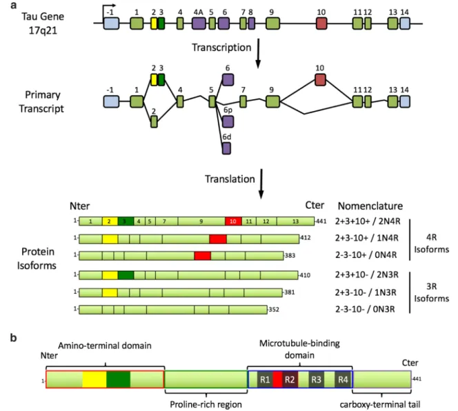
Tau isoforms and inclusions
The advance of immunohistochemistry has revealed
that approximately 20 neurodegenerative diseases,
named tauopathies, are characterized by the
accumula-tion of hyperphosphorylated tau (pTau) [
165
]. Tau is
encoded by a single gene (MAPT) located on
chromo-some 17. The MAPT gene is mostly expressed in
neu-rons [
27
], and due to alternative splicing of exons 2, 3
and 10, six main isoforms are found in the adult brain:
2 – 3 − 10− (0N3R), 2 + 3 − 10− (1N3R), 2 + 3 + 10− (2N3R),
2 − 3 − 10 + (0N4R), 2 + 3 − 10 + (1N4R), and
2 + 3 + 10 + (2N4R) [
5
,
81
] (Fig.
1
a).
Tau protein has four domains with unique biochemical
characteristics and specific functions: (i) an acidic
amino-terminal domain, (ii) a proline-rich region followed by (iii)
microtubule-binding regions (MTBR) and (iv) a
carboxy-terminal tail. The microtubule-binding regions contain
three or four repeat domains (depending on the inclusion
of exon 10) [
38
,
82
,
90
,
114
] (Fig.
1
b). Several
post-trans-lational modifications (PTMs) have been described on tau
proteins [
87
]; phosphorylation dynamically regulates the
physiological functions of tau [
133
,
160
]. In tauopathies,
tau is excessively and abnormally phosphorylated [
10
].
Other PTMs (acetylation, glycation, glycosylation,
meth-ylation, SUMOmeth-ylation, truncation, ubiquitinmeth-ylation, etc.)
have also been described; some of them, such as
acetyla-tion, glycosylation and truncaacetyla-tion, may also be related to
the pathology and are considered as therapeutic targets
[
100
].
pTau may accumulate in the cell bodies of neurons
without forming fibrillary aggregates—a change called a
“pre-tangle”. Moreover, pTau may aggregate in the cell
bodies of neurons (neurofibrillary tangles = NFTs) or in
the cell processes (neuropil threads = NT), and NT may be
axonal (as in the corona of the senile plaque) or dendritic.
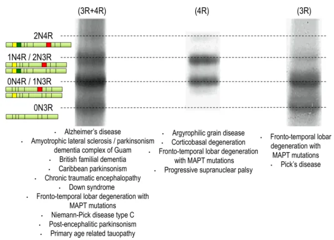
Under electron microscopy, tau aggregates are principally
made of paired helical filaments (PHF) in AD (3R and 4R),
which is also the case in ‘primary age related tauopathy’
(PART) in chronic traumatic encephalopathy (CTE) and
in some less-common disorders (see Fig.
2
). In
progres-sive supranuclear palsy (PSP) and cortico-basal
degen-eration (CBD), tau aggregates are found both in neurons
and glia and are made of 4R straight tau filaments. Other
specific neuronal tau inclusions are Pick bodies (seen in
Pick disease), in which 3R tau aggregates in the neuronal
cell body adopt a spherical shape, and argyrophilic grains
made of 4R tau (seen in argyrophilic grain disease (AGD))
are located in presynaptic terminals. The glial inclusions
may involve astrocytes: in astrocytic tufts, suggestive of
PSP, all processes of the astrocyte are filled with pTau;
in astrocytic plaques, seen in CBD, pTau
immunoreactiv-ity is found at the end of the astrocytic processes [
108
,
110
]; and they may involve oligodendrocytes where they
form coiled bodies (abundant in PSP, CBD and AGD). The
ratio between 3 and 4R tau explains why specific sets of
migration bands have been recognized by Western
blot-ting (Fig.
2
). Recent structural studies by cryo-electron
microscopy have confirmed the presence of different tau
structures among AD, Pick’s disease and CTE [
67
,
68
,
71
].
Genetic frontotemporal lobar degeneration
(FTLD-MAPT formerly called FTDP-17, [
74
]) is of particular
inter-est. It is caused by autosomal dominant MAPT mutations
mainly located in the sequence regulating exon 10
alterna-tive splicing or encoding microtubule-binding regions. Most
coding region mutations often act on both nucleation and
fibrillogenesis [
12
,
36
]. They are widely used for studying
the pathological mechanisms of tauopathies, especially in
animal models [
61
].
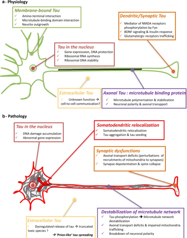
Altogether, tauopathies are the consequence of several
molecular dysfunctions likely due to PTM and
protein-fold-ing deregulations. These deregulations are associated with
the development of conformational changes, oligomerization
and finally, intracytoplasmic aggregation of tau. All these
processes have a significant impact on cell physiology and
particularly on tau functions [
164
] (Fig.
3
).
Neuropathol-ogy, however, shows the large variety of cerebral areas and
cell populations (glial or neuronal) affected; it also
indi-cates that the type of tau aggregation is diverse, as shown
by the multiplicity of tau inclusions and their molecular
composition.
Prion‑like propagation hypothesis
Tauopathies and other neurodegenerative disorders are
remi-niscent of prion diseases. The latter are indeed related to
a pathological change (misfolding) in the molecular
con-formation of the normally present prion protein and to the
capacity of misfolded prions to induce misfolding by contact
with a normally folded prion. Interestingly, different strains
of prion exist with dissimilar fibre conformations that are
transmissible [
175
]. There are at least two steps in the
devel-opment of prion diseases: (1) misfolding induction and (2)
transport of the misfolded prion that permits contact with
still normal prions. Step 1 is called seeding, and step 2 is
propagation. Propagation can take place by diffusion in the
extracellular space or along the axonal paths.
Seeding and propagation have been reported not only with
prions, but also with Aβ [
102
] and alpha-synuclein [
97
].
The term “prion-like” has been attributed to this molecular
Fig. 1 a Schematic presentation of the MAPT gene, its primary
tran-script and the six protein isoforms expressed in the human brain. The MAPT gene is composed of 16 exons. In the brain, exons 4A and 8 are excluded from the primary transcript. Exons 1, 4, 5, 7, 9, 11, 12 and 13 are constitutive, whereas exons 2, 3, 6 and 10 are alternative. Exon 3 never appears independently of exon 2. Exons 1 and 14 are present in the mRNA, but are never translated. Six main transcripts are present in the adult brain: 2 – 3 – 10 − or 0N3R; 2 + 3 − 10 − or
1N3R; 2 + 3 + 10 − or 2N3R; 2 – 3 − 10 + or 0N4R; 2 + 3 − 10 + or 1N4R; 2 + 3 + 10 + or 2N4R. b Tau structure. Four domains with dif-ferent biochemical properties can be retrieved in tau protein: an acidic amino terminal region (corresponding to the expression of exons 1–5), a proline-rich domain (corresponding to the expression of exons 7 and 9), the MTBR with four repeated sequences (R1–R4), and a carboxy-terminal tail (exon 13). Modified from [28]
behaviour when applied to proteins other than prions; such
proteins have been called “propagons” and may induce
pro-tein misfolding in test-tubes, tissues, organisms or between
subjects (infectious). Although there is no clear evidence of
such interindividual transmission for tau, in several
tauopa-thies, tau pathology progresses by well-defined “stages”,
which may support prion-like propagation. Here, the term
“prion-like propagation hypothesis” includes the two steps:
seeding and propagation.
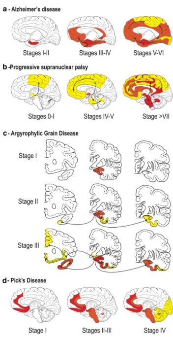
Pathology stages and the “propagation hypothesis”
in humans
In AD, tauopathy is found sequentially in the
transentorhi-nal–entorhinal areas, hippocampus and limbic areas and
finally in the associative and then primary neocortical areas
[
20
] (Fig.
4
a). In PSP, the tauopathy is initially confined
to the pallido-luyso-nigral system and then involves the
basal ganglia, the pontine nuclei and the dentate nucleus,
and finally the frontal and parietal lobes—with not only
the extent but also the severity of the involvement
increas-ing with time [
181
,
189
] (Fig.
4
b). In AGD, the tauopathy
is initially limited to the ambient gyrus and its vicinity; it
involves secondarily the temporal pole, the subiculum and
the entorhinal cortex and finally, the septum, insular cortex,
and anterior cingulate gyrus [
152
] (Fig.
4
c). In Pick disease,
Pick bodies are initially seen in limbic regions;
subcorti-cal regions (the thalamus, striatum, locus coeruleus, raphe
nuclei, dorsal motor nucleus of the vagus nerve, and reticular
formation) as well as the primary sensory cortex are affected
secondarily, followed by the primary motor cortex, the
infe-rior olivary nucleus, and finally, the primary visual cortex
[
96
] (Fig.
4
d).
The active cell-to-cell transfer of tau has not yet been
visualized in the human brain, even if some positron
emis-sion tomography (PET) and magnetic resonance imaging
(MRI) studies suggest that it may occur. Similar to the Braak
stages, there are strong arguments to support a hierarchical
pathway of neurodegeneration along neural networks in AD
[
42
,
48
,
91
].
The progression by stages in those tauopathies has
sug-gested that two mechanisms, not necessarily antagonists,
could be involved: progression of a pathogenic molecule
along neuronal connections or selective vulnerability
(patho-clisis). Given the complexity of the neuroanatomy of the
central nervous system, the existence of connections between
Fig. 2 Tauopathy ‘barcode’. Western blots showing the electropho-retic profile observed with tau protein aggregates from patients with different tauopathies. In AD-like, the six tau isoforms are present in
the aggregates. In PSP-like, only the 4R-tau proteins are aggregated. In Pick’s disease, only the 3R isoforms are aggregating. Modified from [160]
Fig. 3 Functions and dysfunctions of tau proteins a Physiologically, tau protein is mainly located in the cytoplasm of axons to stabilize the microtubules. Other minor locations of tau can be observed, such as in the nucleus [117], bound to the membranes [23] and in den-drites [99]. These locations are associated with atypical functions of tau [164], such as structuring chromatin and protecting nucleic acids from oxidative stress [15, 39, 120, 144, 169, 182, 183], insulin sig-nalling by binding to PTEN in the somato-dendritic compartment [121], mediating neuronal activity via the Fyn kinase and NMDA
receptors in dendrites [29, 99, 129]. Tau proteins are also retrieved in extracellular fluids. b During tauopathies, tau proteins are excessively and abnormally phosphorylated and then aggregate, leading to a sub-stantial loss of function. In particular, the microtubule network is destabilized, tau proteins are relocalized and synaptic deficits appear. Extracellular tau proteins are modified, and their functions are not completely understood even if they could participate in tau pathology propagation in the brain
two regions of the brain that are successively involved is a
necessary condition, but not proof, of the progression along
neuronal connections. On the other hand, the presence of
tau pathology in astroglial cells cannot be directly explained
by the connections. In fact, in PSP, at transcriptomic level,
tufted astrocytes appear to be associated with a microglial
gene-enriched immune network whereas neurofibrillary
tangles would be linked to a brain co-expression network
enriched for synaptic and PSP candidate risk genes [
3
].
Numerous uncertainties remain concerning the progression
of the tauopathies and neurodegenerative diseases in general,
which explains the current controversies [
65
,
113
,
177
].
Notably, the diversity of the tauopathies likely implies a
diversity of pathogenic mechanisms. It appears implausible
that one single treatment could apply to all of them.
Con-versely, a progression through connections like in AD would
be compatible with a prion-like mechanism and thus it is fair
to explore this hypothesis among tauopathies.
The most striking evidence of transmission of the
pathol-ogy through connection is found in AD. Connectivity is
explicitly implicated by Braak and Braak in the description
of their stages [
20
]. In addition, an interesting autopsied AD
case has also been reported showing that a 27-year
meningi-oma removal by neurosurgery had massively disconnected
a piece of cortex. A local dissociation between neuritic and
Aβ pathologies was seen. Neuropathological analyses fully
confirmed AD diagnosis with numerous amyloid deposts
and NFTs. However, in the disconnected cortical brain
region where amyloid deposits were numerous, no
neurofi-brillary tangle was observed suggesting that their presence
is determined by the neural connections [
64
].
However, other hypotheses have been proposed.
Accord-ing to one of them, AD would be a phylogenic
neurode-generative disease of higher primates following an
evolu-tionary change in the primate genome [
145
]. According to
another hypothesis, the pattern of neurodegeneration may
be explained by the possible existence of chemically defined
neuronal subpopulations that are highly vulnerable in AD
[
92
]. In fact, this neurodegeneration pathway bears a striking
Fig. 4 a Staging of tau pathology in AD. Topographic distribution
of tau lesions at the different stages of tau pathology in schemes of brains in medial views. Stages I and II, tau lesions invade entorhinal and transentorhinal regions. Stages III and IV: lesions involve the associative areas of the neocortex, and finally, during stages V and VI, tau lesions invade all the primary and secondary neocortical areas. From [22]. b Staging of tau pathology in PSP. Topographical distribution of tau lesions at the different neuropathological stages of PSP in schematic brain representations in medial views. Stages 0/I—Only the pallido-luyso-nigral complex shows tau pathology with weak involvement of the premotor cortex. Stage II/III—Tau pathol-ogy reaches the basal ganglia, pedunculopontine nucleus and den-tate nucleus. Stages IV/V—Frontal and temporal lobes are involved. Stages VI/VII—Subthalamic nucleus, substantia nigra, internal glo-bus pallidus, neocortical areas, pedunculopontine nucleus and cer-ebellum are more severely affected. Modified from [189]. c Staging of tau pathology in AGD. Topographical distribution of argyrophilic grains at the different stages of tau pathology evolution in three cor-onal sections. Stage I—argyrophilic grains are located in the ambi-ens gyrus, anterior CA1, anterior entorhinal area and amygdala. The stage II—medial temporal lobe is more affected by the involvement of the posterior subiculum, entorhinal and transentorhinal cortices. Stage III grains invade the anterior cingulate gyrus, septum, accum-bens nucleus, rectus gyrus, insular cortex and hypothalamus. Modi-fied from [152]. d Staging of tau pathology in Pick’s disease (PiD). Topographical distribution of Pick bodies at the different stages of tau pathology evolution in schematic brain representations in medial views. Stage I Tau pathology is deposited in the limbic and neocor-tical frontotemporal regions as well as the angular gyrus. Stages II/ III—White matter tracts, subcortical structures, serotonergic/noradr-energic brainstem nuclei are affected, followed by the primary motor cortex and pre-cerebellar nuclei. Finally, in stage IV, tau is deposited in the visual cortex as well as in the cerebellar granular layer and brainstem white matter. Modified from [96]
resemblance to the inverse sequence of cortical myelination
[
19
].
Recently, the hypothesis of prion-like propagation has
offered an explanation for this hierarchical pathway of
neu-rodegeneration [
21
]. The relevance of the widening of this
concept is still being discussed. There is still controversy
about tau propagation in CBD, Pick’s disease and PSP [
24
,
48
,
59
,
73
,
96
,
137
,
181
,
189
]. The recent use of tau PET
imaging will be helpful in bringing new insights into such
debate [
112
].
Human tau in peripheral fluids: a new argument
for the prion‑like propagation hypothesis?
The propagation, included in the prion-like hypothesis,
implies tau passage in the extracellular space. The passage
of misfolded tau in the extracellular space could be passive
and related to neuronal death. Currently, there is evidence
that misfolded tau is actually secreted [
194
,
195
]. As a
con-sequence of the secretion step, tau is found in
extracellu-lar fluids (in a vesicuextracellu-lar or non-vesicuextracellu-lar form) [
130
,
140
].
The interstitial fluid (ISF) that is found in the brain between
the cells may be directly sampled and analysed by
microdi-alysis. In humans, microdialysis has been used in cases of
acute brain trauma to monitor brain metabolism [
89
], but the
technique cannot be applied to neurodegenerative diseases
such as tauopathies. Approximately, 20% of the
cerebro-spinal fluid (CSF) originates from ISF [
157
]; changes in
ISF protein concentrations are thus probably reflected by
changes in the CSF. Tau proteins originating mainly from
neurons have decreasing concentrations from ventricles
(320 pg/mL) to lumbar subarachnoid space (210 pg/mL)
[
147
]. This observation suggests that CSF tau originates
mainly from ISF and CSF is the extracellular compartment
closest to the brain, which is most accessible for sampling.
CSF tau has been extensively studied to investigate the
pathophysiological processes occurring in
neurodegen-erative diseases in living patients. The presence of tau in
CSF was identified by electrophoresis in the mid-80s [
37
,
77
] and quantified from 1993 by sandwich enzyme-linked
immunosorbent assay [
180
]. Human CSF contains tau
lev-els typically ranging from approximately 100 to 1200 pg/
mL [
123
]. Recently, CSF tau kinetics were quantified by
stable isotope labelling kinetics (SILK) labelling in healthy
individuals (half-life = 23 ± 6.4 days, tau production rate of
26.3 pg/mL/day ± 9.2 and CSF level of 812.7 ± 186 pg/mL)
[
158
]. Regarding CSF tau quantification in tauopathies, only
individuals with AD clearly display increased tau
concentra-tions in CSF [
14
].
Until very recently, tau could not be detected in the
plasma, because of its very low concentration compared
with that in CSF. Although protein transfer from ISF and
CSF to blood is very low, and its mechanisms are not well
known (efflux across the blood–brain barrier [
11
], along the
glymphatic paravascular pathway [
95
], clearance through
the dural lymphatic system in blood [
139
]). Tau levels in
plasma might thus also be altered by changes in the brain
[
11
] (Table
1
).
Altogether, the prion-like propagation hypothesis is
fur-ther supported by the increased and stable concentrations of
extracellular tau in AD. In addition, recent work has shown
that human CSF from AD patients is able to induce tau
seed-ing in experimental models [
163
]. However, without a full
characterization of tau species present in human ISF, it is
hazardous to infer its concentration from that in CSF or in
plasma. Animal models offer the possibility to recover ISF
[
192
,
193
].
Modelling of the prion‑like propagation
Data from humans allowed the development of
experi-mental models to assess whether the ‘prion-like’
propaga-tion hypothesis is implicated in tauopathies and especially
whether extracellular tau is relevant to this hypothesis.
These models were thus designed to investigate the prion
paradigm in regard to human neuropathology. For instance,
with the upregulation of Rab7A in AD patients [
78
–
80
],
tau secretion seems to be regulated by this small GTPase
involved in the trafficking of endosomes, autophagosomes,
and lysosomes [
149
]. Similarly, tau secretion is correlated
with Golgi dynamics [
128
], consistent with its fragmentation
noted in AD [
167
]. This hypothesis involves a multi-step
mechanism (tau secretion, uptake and subsequent seeding
processes) that has been widely explored.
Tau secretion
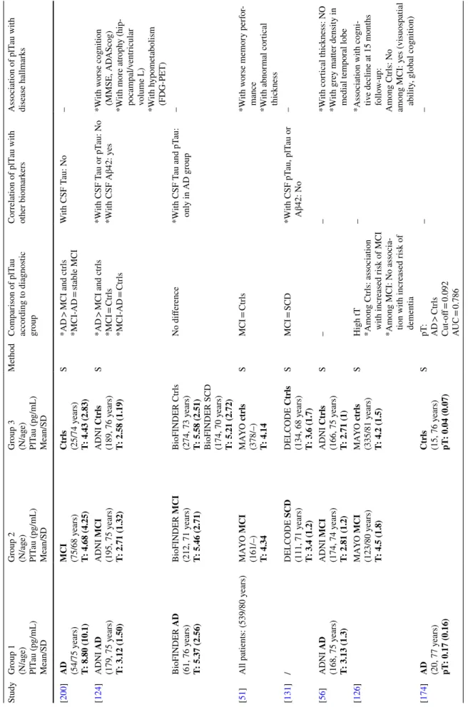
First, is it possible to identify a mechanism by which tau
is found in peripheral fluids? The nature of secreted tau is
debated in the literature [
130
,
141
] (Fig.
5
). Tau is secreted
in a free form [
60
,
103
,
125
,
185
], but it is also found inside
nanotubes [
1
,
173
] or associated with extracellular vesicles
(EVs) [
140
]. Whereas nanotubes may be difficult to visualize
in the human brain, secreted tau in EVs is found not only
in experimental models but also in peripheral fluids (CSF
[
153
,
185
] and blood [
70
,
85
,
190
]) in AD patients.
How-ever, immunoassays in cell models revealed that whereas
tau is mainly secreted in a free form in conditioned medium,
only 10–20% of EVs contain tau [
60
,
185
].
In this regard, tau is a cytosolic protein and does not
follow the classical secretory pathway. It is translated into
the cytosol, and the mechanisms modulating tau secretion
are numerous and include tau oligomerization, tau
trunca-tion and mutatrunca-tions, tau interactrunca-tion with plasma and
orga-nelle membranes and tau hyperphosphorylation [
142
]. For
Table 1 Plasma t au le vels b y diagnos tic g
roup in AD/MCI (mild cognitiv
e im pair ment) and FTD (fr ont otem por al deg ener ation) s tudies Study Gr oup 1 (N/ag e) PlT au (pg/mL) Mean/SD Gr oup 2 (N/ag e) PlT au (pg/mL) Mean/SD Gr oup 3 (N/ag e) PlT au (pg/mL) Mean/SD Me thod Com par ison of plT au accor ding t o diagnos tic gr oup Cor relation of plT au wit h ot her biomar kers Association of plT au wit h disease hallmar ks [ 200 ] AD (54/75 y ears) T: 8.80 (10.1) MCI (75/68 y ears) T: 4.68 (4.25) Ctr ls (25/74 y ears) T: 4.43 (2.83) S *AD > MCI and ctr ls *MCI-AD = st able MCI W ith CSF T au: N o – [ 124 ] ADNI AD (179, 75 y ears) T: 3.12 (1.50) ADNI MCI (195, 75 y ears) T: 2.71 (1.32) ADNI Ctr ls (189, 76 y ears) T: 2.58 (1.19) S *AD > MCI and ctr ls *MCI = Ctr ls *MCI-AD = Ctr ls *W ith CSF T au or pT au: N o *W ith CSF Aβ42: y es *W ith w orse cognition (MMSE, AD AScog) *W ith mor e atr oph y (hip -pocam pal/v entr icular volume L) *With h ypome tabolism (FDG-PET) BioFINDER AD (61, 76 y ears) T: 5.37 (2.56) BioFINDER MCI (212, 71 y ears) T: 5.46 (2.71) BioFINDER Ctr ls (274, 73 y ears) T: 5.58 (2.51) BioFINDER SCD (174, 70 y ears) T: 5.21 (2.72) No differ ence *W ith CSF T au and pT au: onl y in AD g roup – [ 51 ]
All patients: (539/80 y
ears) MA YO MCI (161/–) T: 4.34 MA YO ctr ls (378/–) T: 4.14 S MCI = Ctr ls *W ith w orse memor y per for
-mance *With abnor
mal cor tical thic kness [ 131 ] / DEL CODE SCD (111, 71 y ears) T: 3.4 (1.2) DEL CODE Ctr ls (134, 68 y ears) T: 3.6 (1.7) S MCI = SCD *W ith CSF pT au, plT au or Aβ42: N o – [ 56 ] ADNI AD (168, 75 y ears) T: 3.13 (1.3) ADNI MCI (174, 74 y ears) T: 2.81 (1.2) ADNI Ctr ls (166, 75 y ears) T: 2.71 (1) S – – *W ith cor tical t hic kness: N O *W ith g re y matter density in medial tem por al lobe [ 126 ] MA YO MCI (123/80 y ears) T: 4.5 (1.8) MA YO ctr ls (335/81 y ears) T: 4.2 (1.5) S High tT *Among Ctr ls: association wit h incr eased r isk of MCI *Among MCI: N o associa -tion wit h incr eased r isk of dementia – *Association wit h cogni -tiv e decline at 15 mont hs follo w-up: Among Ctr ls: N o among MCI: y es (visuospatial ability , g lobal cognition) [ 174 ] AD (20, 77 y ears) pT : 0.17 (0.16) Ctr ls (15, 76 y ears) pT : 0.04 (0.07) S pT: AD > Ctr ls Cut-off = 0.092 AUC = 0.786 – –
Table 1 (continued) Study Gr oup 1 (N/ag e) PlT au (pg/mL) Mean/SD Gr oup 2 (N/ag e) PlT au (pg/mL) Mean/SD Gr oup 3 (N/ag e) PlT au (pg/mL) Mean/SD Me thod Com par ison of plT au accor ding t o diagnos tic gr oup Cor relation of plT au wit h ot her biomar kers Association of plT au wit h disease hallmar ks [ 72 ] bvFTLD (71, 64 y ears) T: 1.96 (1.07) PPA (83, 67 y ears) T: 2.65 (2.15) Ctr ls (22, 69 y ears) T: 1.67 (0.50) S *b vFL TD and PP A > ctr ls *Ser um NfL: N o *Br ain v olume: no *Disease dur ation: no Gene tic subgr oups: MAPT (12): T: 2.62 (1.39) GRN (9): T: 2.22 (1.60) C9ORF (15): T: 1.93 (0.70) *MAPT > GRN and C9ORF [ 127 ] MA YO AD (40, 68 y ears) T: 7.2 (2.8) pT: 11.6 (4.1) MA YO MCI (57, 71 y ears) T: 5.9 (2.8) pT: 9.0 (13.9) MA YO Ctr ls (172, 72 y ears) T: 5.9 (61.9) pT: 6.4 (6.4) S M T: AD > MCI = Ctr ls pT : AD > Ctr ls –
Higher plT associated wit
h
Aβ PET and T
au PET [ 40 ] AD Disco ver y cohor t (25, 61 y ears) Validation cohor t (23, 72 y ears) MCI-AD Disco ver y cohor t (21, 65 y ears) NtT : 4.85 Validation cohor t (22, 73 y ears) NtT : 3.42 Ctr ls Disco ver y cohor t (9/10, 60/70 y ears) NtT : 5.12 Validation cohor t (41, 72 y ears) NtT : 3.40 S NtT separ ate ctr ls fr om disco ver y cohor t/v alidation cohor t: MCI-AD (A UC = 0.88/0.79) AD (A UC = 0.96/0.75) – – [ 138 ] FHS Incident dementia (134, –) Incident AD subg roup (105, –) Aut opsy sam ple subgr oup (42, 82 y ears) Confir med AD (11, –) FHS Ctr ls (1319, 75 y ears) S T > median associated wit h: *Gr eater r isk of dementia (HR = 1.62) *Gr eater r isk of AD (HR = 1.76) *P oor er cognitiv e per for
-mance *Smaller hippocam
pal v
olume
*Higher bur
den of NFT in
the medial tem
por al lobe (aut opsy subg roup) [ 41 ] Middle-ag ed Ctr ls (56, 58 y ears) T: 14.3 (4.9) Old Ctr ls (70, 74 y ears) T: 18.1 (7.3) I *Old ctr l > middle-ag ed ctr l *A ge w as positiv ely associ -ated wit h tT – *V olume of subcor tical br ain str uctur es: N o *Thic kness of cor tical regions: N o [ 118 ] BSHRI AD (16, 82 y ears) T: 34.5 (3.8) NTUH AD (31, 72 y ears) T: 52.5 (2.7) – BSHRI Ctr ls (16, 82.5 y ears) T: 20.5 (1.2) NTUH Ctr ls (61, 64 y ears) T: 14.0 (1.9) I AD > Ctr ls (p < 0.02) Cut-off: 25 pg/mL AUC = 0.97, Se = 89%, spe = 94% *Combined wit h plAβ42: AUC = 0.98, Se = 94%, spe = 92% – [ 196 ] Mild AD T: 37.5 (12.3) pT: 6.1 (1.6) MCI due t o AD T: 33.0 (10.2) pT: 4.4 (1.8) Ctr ls T: 18.8 (10.2) pT: 2.5 (1.1) I T: MCI-AD > Ctr ls pT : AD > MCI-D > Ctr ls – –
Table 1 (continued) Study Gr oup 1 (N/ag e) PlT au (pg/mL) Mean/SD Gr oup 2 (N/ag e) PlT au (pg/mL) Mean/SD Gr oup 3 (N/ag e) PlT au (pg/mL) Mean/SD Me thod Com par ison of plT au accor ding t o diagnos tic gr oup Cor relation of plT au wit h ot her biomar kers Association of plT au wit h disease hallmar ks [ 197 ] AD (29, 72 y ears) T: 55.4 (22.4) FTLD (26, 62) T: 41.3 (20) MCI-AD (24, 71 y ears) T: 33.3 (7.8) Ctr ls (66, 65 y ears) T: 13.4 (7.8) I AD, MCI-AD and FTD > Ctr ls cut-off: 17.4 pg/mL AUC = 1.0 (f or AD and MCI-AD), AUC = 0.96 (for FTLD) – – [ 115 ] PSP (6, 67 y ears) T: 18.9 (2.1) pT: 3.8 (0.7) CBD (3, 62 y ears) T: 14.7 (1.0) pT: 3.2 (0.2) FTLD wit hout P ar kinsonism (25, 59 y ears) T: 41.5 (1.1) pT: 6.8 (0.3) FTLD-P ar k (6, 58 y ears) T: 24.1 (2.0) pT: 6.8 (0.2) Ctr ls (35, 63 y ears) T: 12.1 (1.0) pT: 2.5 (1.1) I
*T and pT in all disease groups
> ctr ls *T FTLD > FTLD-P ar k – – Column 1: s tudy r ef er
ence; columns 2, 3 and 4: patient g
roups and plT au le vels in bold (T : t ot al t au, pT : phospho t au Thr181, NtT : N-ter minal fr agment of t au); column 5: me thod used f or plasma t au anal ysis (S: SIMO A (sing le molecule ar ra y tec hnology); I: IMR (immunomagne tic reduction); M: MSD (Mesoscale Disco ver y); columns 6, 7, 8: results accor ding t o patient g roups, ot her biomar
kers and disease hallmar
ks, r espectiv ely PlT au plasma t au, pT au phospho t au, BSHRI
Banner Sun Healt
h Ins titute (U nited S tates), NTUH N ational T aiw an U niv ersity Hospit al (T aiw an), AUC Ar ea U nder t he Cur ve (R OC anal y-sis), ADNI Alzheimer ’s Disease N eur oimaging Initiativ e (Amer ican R esear ch Pr og ram on Alzheimer ’s Disease), BioFINDER Biomar kers F or Identifying N eur odeg ener ativ e Disor ders Ear ly and R eliabl y (Sw edish Study), M AYO Ma yo Clinic (R oc hes ter , Minneso ta, US A), DEL CODE DZNE-Longitudinal Cognitiv e Im pair ment and Dementia Study (DEL CODE) conducted by the DZNE (Ger man Center f or N eur odeg ener ativ e Diseases), FHS F ramigham Hear t S
tudy (US community
-based cohor t), SCD Subjectiv e Cognitiv e Decline, PPA Pr imar y Pr og ressiv e Aphasia
instance, non-conventional secretion of cytosolic proteins
such as fibroblast-growth factor 2 (FGF-2) and interleukin
1β (IL1β) include oligomerization and truncation,
respec-tively [
119
,
166
]. Similar to FGF-2, soluble
hyperphos-phorylated tau oligomers may directly translocate across
the plasma membrane upon binding to PI(4.5)P
2, followed
by retention on the cell surface through heparan sulfate
proteoglycans (HSPG) [
103
,
125
]. IL1ß secretion is also
an interesting model for tau secretion. First, extrinsic
fac-tors, such as inflammation, are likely to modulate this
non-conventional secretion. Additionally, a sequence containing
human tau residues (amino acid 18 to amino acid 28) was
recently shown to act as a binding motif for end binding
proteins [
159
]. These proteins belong to the group of
micro-tubule plus-end tracking proteins that have been implicated
in the secretion of IL1β [
184
] and might also regulate tau
Fig. 5 How is tau secreted and transferred into recipient cells? Tau secretion-Yellow-Tau protein could be carried by EVs, and the most investigated proteins are the exosomes, which are small vesicles (50– 150 nm) coming from a subpopulation of intraluminal multivesicular bodies vesicles. Orange-Tau protein could also be carried by larger EVs named ectosomes (150–1000 nm) coming from the direct bud-ding of the plasma membrane. Ectosome budbud-ding is regulated at least by calcium and oxidative stress, which are deregulated in many neu-rodegenerative disorders. Violet-Finally, tau protein is mainly found in a free form in extracellular fluids. How tau is secreted is not well documented, but a few papers are now investigating this mechanism and its regulation. Regardless of the shuttles and depending on the models used, tau has been identified in many forms in the extracel-lular compartment, and to date, no one has been able to decipher the toxic/propagative forms. Are those secreted species cleared from the interstitial fluid? Are they transferred to other brain cells to propagate the pathology? Is this information implied in normal brain cell-to-cell communications? Tau transfer-How is tau taken up and handled by the receiving cells? Whether tau transfer requires the synapse remains
a matter of debate, and there is now some evidence that it might sup-port the process. Nevertheless, the co-existence of the lateral trans-mission process should not be excluded. Black-Tau protein may move from cell to cell via nanotubes, membranous actin-rich structures that form between two cells inducing cytoplasmic and membrane exchanges. Yellow-to deliver tau, exosomes may be taken up by endocytosis in receiving cells. However, in this manner, the rest of the process is unclear. Is tau targeted to intracellular degradative com-partments, such as lysosomes, to generate tau seeds that will in turn convert the non-pathological receiving cell into a pathological state? Is tau transferred to a third population due to the endosomal pathway? The exosomal transfer from the first to the third neurons via exosomes seems to be linked to the hijacking of secretory endosomes. The way ectosomes are taken up by receiving cells has not yet been investi-gated. Red-Tau proteins could also be internalized in the secondary neuron via an endocytosis mechanism. Such a process might be regu-lated by Bin1 and PICALM proteins, as both have been identified by GWAS as binding partners of tau
secretion. The inflammasome may trigger a pathological
cas-cade leading to enzyme activation, tau truncation and
secre-tion. In human CSF, 99.9% of tau is truncated at the
C-ter-minal domain, and major cleavage seems to occur around
amino acids 210–230 [
158
]. This finding was confirmed by
Cicognola and collaborators who described an important
cleavage site at amino acid 224 [
43
]. In the presence of Aβ,
such a mechanism may be exacerbated [
158
], supporting the
prion-like propagation hypothesis in AD.
Tau uptake/transfer
In the propagation hypothesis, secreted tau must be taken
up by cells (Fig.
5
). In vivo models recapitulating
patho-logical propagation have been developed. Focal
intraronal expression of tau was obtained using either a
neu-ropsin promoter [
55
,
116
,
187
] or viral vectors [
6
,
30
,
59
,
60
,
186
]. Although both models show cell-to-cell transfer
of tau, thorough controls are required to exclude transgene
leakage [
198
], viral diffusion throughout the brain [
63
] or
side-effects related to tau overexpression. Regarding this
latter, the recent humanization of the murine MAPT gene
clearly indicates that prion-like propagation occurs without
overexpression [
151
]. Many other in vivo models
investi-gating tau propagation are based on intracranial delivery of
pathological material (recombinant oligomeric or fibrillar
tau, human or mouse brain-derived material, etc.) [
130
] or
peripheral administration [
46
] in transgenic and
non-trans-genic mice. These models bypass how tau is secreted from
donor cells and focus on how tau is captured and lead to
seeding in receiving cells. Some of these models suggest
that over time, tau pathology appears in distant, synaptically
connected areas, which suggests cell-to-cell propagation.
However, the exact role of synapses in the tau propagation
process remains under investigation, although seed
compe-tent tau was recently shown to be enriched in the synaptic
fraction of AD brain-derived materials [
57
]. Nonetheless,
we cannot exclude the possibility that the spread of tau
pathology may occur via a number of pathways, including
synaptic transfer, interstitial diffusion and even glial
(astro-cytes [
109
,
122
], oligodendrocytes [
132
] and microglia [
6
])
intervention.
Many in vitro models have been developed to
under-stand such uptake. Free aggregates present in the culture
medium seem to be internalized via endocytosis [
66
,
75
,
86
,
93
,
106
,
143
,
156
,
191
,
194
], and this uptake might be
mediated by HSPGs [
93
,
146
,
168
,
188
]. The extracellular
region of APP also seems to be involved in the uptake of
tau fibrils in neuroblastoma cell lines [
170
], but other more
specific receptors are also likely implicated in these
mecha-nisms. Two entry mechanisms were recently described: the
monomer of tau uses a slow actin-dependent
micropinocy-tosis pathway, whereas aggregated tau is taken up in a rapid
way independent of actin polymerization but dependent
on dynamin, consistent with an endocytosis process [
66
].
Bin1, a neuronal amphyphisin2 isoform that is
downregu-lated in AD brains, inhibits endocytic activity and might be
involved in the propagation of tau pathology [
32
]. The use
of double or triple microfluidic chambers is informative for
studying cell-to-cell transfer as it directly links the donor to
the receiving neuronal populations, emphasizing the role of
synapses in cell transfer [
31
,
63
,
143
,
171
,
185
].
Altogether, the hierarchical pathway of
neurodegen-eration in the prion-like propagation hypothesis strongly
suggests a highly regulated mechanism allowing specific
targeting of neuronal populations. In light of the available
data, this selectivity is likely to rely on receptor-mediated
endocytosis.
Tau seeding
The prion-like seeding implies that abnormal proteins can
convert normal proteins into a pathological form. In this
regard, Clavaguera and co-workers showed that injecting
tau aggregates extracted from mice overexpressing mutated
tau (P301S) into mice overexpressing human wild-type tau
was sufficient to induce tau pathology [
45
]. When a
tau-immunodepleted extract is injected, no pathology can be
detected, showing that tau is the responsible factor as
con-firmed later by other groups [
134
,
179
,
194
]. However, this
seeding activity may also require the presence of different
co-factors such as polyanions [
58
,
201
]. The same group
obtained similar results by injecting human brain lysates of
different tauopathies and reproducing the specific
morphol-ogy of the lesions seen in the human diseases [
44
],
support-ing the concept of propagons and tau strains.
In vitro, even if a few studies showed that extracellular
tau may bind to neuronal receptors (muscarinic [
83
], Na
+/
K
+ATPase (NKA) [
161
]…) and have toxic effects, most
have shown that incubated aggregates/seeds are internalized
by endocytosis and promote aggregation of overexpressed
tau in cell lines [
66
,
75
,
86
,
93
,
94
,
134
,
135
,
148
,
156
,
170
–
172
]. Nevertheless, most seeding assays use
transfec-tion reagents and thus bypass receptor-mediated
endocyto-sis, which may be a critical step in prion-like propagation.
In this regard, are the propagons in a free form or membrane
associated? It is known that exosomes containing tau with
seeding activity have been isolated from the brains of tau
transgenic mice [
9
]. In addition, seed-competent tau
spe-cies, in both free and vesicular forms, have been detected in
CSF and ISF from experimental models and CSF from AD
patients [
85
,
171
]. These studies imply that tau in EVs may
be endocytosed and act as a seed and therefore contribute
to prion-like propagation of tau pathology. Nonetheless, as
indicated above, tau in EVs is minor and immunodepleted
brain extracts does not exhibit any seeding competencies.
Following the work of Clavaguera and collaborators,
many subsequent studies have shown that injections of
cer-ebral lysates or synthetic tau fibres in transgenic mice
poten-tiate transmissibility [
130
]. In such experiments,
human-mutated tau seeds are already overexpressed in a murine
tau model, and additional seeds are injected. These works
further not only support tau seeding but also indicate that it
happens outside of the strict prion definition. In any case,
regardless of the seeds used (artificial or brain-derived),
seeding is efficient and supports this step of the prion-like
propagation hypothesis [
130
].
Collectively, with their strengths and weaknesses, all the
data generated in experimental models have greatly
strength-ened the prion-like propagation hypothesis. In addition, in
line with the prion-like propagation hypothesis and the
iden-tification of tau in the extracellular space, free extracellular
tau species are now considered key drivers in the
pathol-ogy propagation by the scientific community, making them
attractive targets for therapeutic approaches.
From experimental models to clinical trials
Historically, tau therapeutic interventions were designed to
target intraneuronal mechanisms such as modulating PTMs,
breaking tau aggregates or decreasing tau concentrations
[
100
]. Many of them have already failed and a global tau
silencing may have side-effects due to multiple tau
func-tions [
100
,
164
]. More recently, tau immunotherapy showed
promising preclinical results. While different mechanisms,
such as microglial activation and the generation of different
anti-tau antibodies, are involved in vaccination, we focus
here on mechanisms involved in passive immunotherapy
that have been widely explored in different experimental
models [
2
,
7
,
17
,
33
–
35
,
47
,
50
,
52
–
54
,
155
,
176
,
178
,
179
,
194
]. Early studies have shown that tau vaccination and tau
immunotherapy could reduce intraneuronal tau pathology.
Most of them decreased the amount of insoluble tau
mate-rials [
7
,
176
,
194
]. Cognitive decline was also reduced in
some [
176
,
194
], including those using MC1, an antibody
recognizing a conformational epitope involving the amino
terminus of tau [
101
] and HJ8.5, an antibody recognizing
the amino acids 25–30. Both have been further developed
for human immunotherapy and are now in clinical trials
(LY3303560, ABBV 8E12-Table
2
). Peripheral
administra-tion of this MC1 antibody in murine models of tauopathy
has not only reduced tau pathology quantified by
biochemi-cal and immunohistochemibiochemi-cal analyses but also delayed the
onset of decreased motor impairment and weight loss [
35
].
Such experiments were also performed with a
phospho-dependent antibody (anti-pS422, also further developed for
human immunotherapy, RG7345-Table
2
) showing similar
effects with the involvement of the endosome-lysosome
pathway [
47
]. The finding of tau secretion and its
com-patibility with the prion-like propagation hypothesis have
modified the strategy of immune therapy: research was then
oriented towards antibodies that did not penetrate the cells
and principally acted in the extracellular space.
The Fc part of immunoglobulins G (IgGs) binds to
specific Fc gamma receptors (FcγR), which are variously
expressed in neurons, microglia or astrocytes. Different
classes of FcγR have been described with high affinity
(bind-ing to monomeric IgG complexes) or low affinity (bind(bind-ing
only to multimeric IgG complexes) to different Fc.
Micro-glial cells highly express all classes of FcγR [
4
], whereas
only FcγRI has been reported in astrocytes. In neurons, the
expression of FcγR is still debated [
136
], but neurons [
69
]
and microglia [
4
] seem to upregulate FcγR in response to
extracellular IgG with a functional Fc domain.
The different isotypes of human IgG (IgG1, IgG2, IgG3
and IgG4) have, on the other hand, different affinities for the
receptors. For example, human IgG1 has a higher affinity for
activating FcγRs present on microglia compared to human
IgG4 and may induce a more pro-inflammatory response
[
26
]. IgG4 may thus theoretically bind to its antigen,
low-ering its toxicity, while at the same time, limiting its cell
internalization through Fc receptors and the potential
inflam-mation, it may induce [
111
]. Most tau antibodies currently
being tested in clinical trials use this isotype to target
extra-cellular tau (Table
2
). Nevertheless, most of the preliminary
studies are performed in mice, and it is often difficult to
translate these data to humans. In mice, four classes are also
found, but they do not correspond to human IgG. In fact,
murine IgG1 is closer to human IgG4 than murine IgG2.
Thus, when murine antibodies and their effector functions
are tested in animal models, it is always difficult to draw
conclusions. For instance, Funk and collaborators showed
that the murine version of ABBV 8E12 (HJ8.5 IgG2a) drives
uptake of tau species into BV2 microglial cells [
76
].
How-ever, single-chain HJ8.5 antibodies without Fc fragment
(scFvs) significantly reduced levels of hyperphosphorylated,
aggregated tau in brain tissue of tau transgenic mice [
98
].
Such data suggest that the Fc fragment may not be required
for the efficacy of this family of anti-tau antibodies in tau
immunotherapy. The use of different approaches and
differ-ent models may thus sometimes lead to differdiffer-ent conclusions
on the impact of Fc fragment and receptor.
The most widely accepted scenario is that therapeutic
antibodies bind extracellular pathological tau species in the
ISF responsible for the spread of pathology but are not
nec-essarily required to directly bind intraneuronal tau. The
anti-bodies would block the seeding activity of extracellular tau,
most likely by blocking the initial uptake of these seeds and
slowing down the spread of pathology [
134
]. Nevertheless,
the future of such anti-tau antibody-tau complexes and their
clearance are still puzzling, and many hypotheses have been
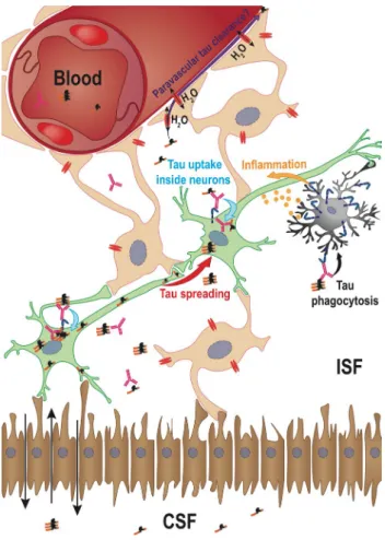
proposed, as summarized in Fig.
6
.
Therapeutic antibodies currently in clinical
develop-ment have shown their capability to target extracellular tau
(by modulating cell-to-cell tau transfer) and/or to reduce
tau seeding in experimental models. The murine version
of BIIB076 has been shown to block neuronal uptake of
human tau species in microfluidic chambers [
134
]. BIIB076
was described to be more efficient in blocking tau uptake
than an antibody directed against the C-terminus of tau
protein [
134
]. The murine version of UCB0107 (binding a
region before the first MTBR) displayed superior efficacy
in an in vitro model of seeding and aggregation induced by
human AD tau than antibodies targeting N-terminal, pS202/
pT205, or pS422 conformational epitopes [
49
]. It also
effec-tively prevented the induction of tau pathology in the brain
of transgenic mice injected with human AD brain extracts,
in contrast to an amino-terminal tau antibody recognizing
the same epitope as BIIB092 [
2
]. Other antibodies have
also been tested by Vandermeeren and collaborators from
Janssen [
179
]. Immunodepletion assays strongly suggest
that difference exist between epitopes. In fact, antibodies
that recognized the mild-region of tau are most effective
while N-terminal antibodies could not fully block seeding
in vitro (FRET assays) as well as in vivo (AD brain material
enriched for PHFs injected into the hippocampus of P301L
transgenic tau mouse to spur fibrillization immunized by
various antibodies). Together, these results suggest that
the murine version of JNJ-63733657 which is targeting the
pS217, a central part of tau may also act in this way [
107
,
150
]. Extracellular tau may also lead to neuronal
hyperactiv-ity and facilitate Aß peptide secretion; in this paradigm, it
was reported that the murine version of BIIB092, IPN002,
is protective by blocking extracellular tau [
25
].
Finally, all these antibodies have shown some effects in
mouse models, although few reports are available in humans.
The next challenge is to validate the target engagement and
efficacy of drug candidates.
Some studies on transgenic models suggest that plasma
tau is a potential biomarker for therapeutic monitoring.
Indeed, the active immunization of tau transgenic mice with
peptides containing a pathological phosphorylated epitope
(pS422) reduced pathological tau species in the brain and
Table 2 Clinical trials for tau immunotherapy: to facilitate the reading, the name of antibodies currently tested in clinical trials are given, but it should be kept in mind that their murine versions have been used in experimental models to assess their mode of action
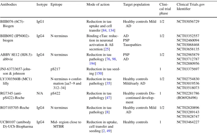
The most advanced clinical trials (phases 1 and 2) include vaccination (AADvac1 and ACI-35, not shown) and passive immunotherapy (BIIB076, BIIB092 (Gosuranemab), ABBV-8E12 (Tilavonemab), JNJ-63733657, LY3303560 (Zagotenemab), RO7105705, UCB0107) and Lu-AF87908.
Antibodies Isotype Epitope Mode of action Target population Clini-cal trial phase
Clinical Trials.gov Identifier
BIIB076
(6C5)-Biogen IgG1 Reduction in tau uptake and cell transfer [84, 134]
Healthy controls Mild
AD 1/2 NCT03056729 BIIB092
(IPN002)-biogen IgG4 N-terminus Binding eTau: reduc-tion in neuronal activation & Aß secretion [25] AD PSP Tauopathies 1/2 NCT03352557 NCT02460094 NCT03068468 NCT03658135 ABBV 8E12
(HJ8.5)-abbvie IgG4 N-terminus Reduction in tau pathology [76, 98,
194]
PSP
AD 1/2 NCT02985879NCT03712787 NCT02880956
JNJ-63733657-john-son & johnJNJ-63733657-john-son pS217 Reduction in tau seed-ing [150] 1 NCT03375697 LY3303560B (MC1)
lilly N-terminus + confor-mation [aa7–9 and 312–34]
Reduction in tau
pathology [35] Healthy controlsMild/early AD 1/2 NCT02754830NCT03019536 NCT03518073 RG7345
(anti-pS422)-Roche N/A pS422 Reduction in tau pathology [47] Healthy controls Dis-continued develop-ment
1 NCT02281786 (RO6926496) RO7105705-Roche IgG4 N-terminus Reduction in tau
pathology [8] Healthy controls Mild AD 1/2 NCT02820896NCT03289143 NCT03828747 UCB0107 (antibody
D)-UCb Biopharma IgG4 Mid- region close to MTBR Reduction in uptake, cell transfer and seeding [2, 49]
delayed cognitive deficits but was importantly associated
with a significant increase in plasma tau concentrations
[
176
]. Similarly, it was reported at the 2017 AD/PD
confer-ence that injection of RO7105705 in tau transgenic mice
(13 weeks treatment/3–30 mg/kg) increased plasma tau [
8
].
This concept of tau/Ab complex stabilization in the plasma
has also been validated by another study demonstrating that
peripheral administration of the anti-tau antibody HJ8.5
(ABBV 8E12) increased plasma tau not only in transgenic
mice, but also in PSP patients [
195
]. Such stabilization in
plasma may be explained by high plasma concentrations of
therapeutic antibodies. For instance, at the 2017 AD/PD and
AAIC conferences, it was shown that BIIB076
concentra-tions were 1000-fold higher in plasma than in CSF when
a single injection was administered in blood in
cynomol-gus monkeys [
84
]. Tau analysis is also performed in CSF
in clinical trials and may help to show target engagement.
For instance, the BIIB092 antibody in a phase 1b clinical
trial in PSP is well tolerated, and its complex with tau is
found in CSF [
18
]. In fact, most of the antibodies described
in Table
2
are in clinical trials, and their outcome should be
published in the months to come. Nevertheless, we know
that the ABBV 8E12 antibody has already been discontinued
in a phase 2 clinical trial in PSP. However, it is still ongoing
in AD clinical trials. BIIB092, RO7105705 and ABBV 8E12
target the amino terminal domain of tau proteins, whereas
UCB0107 recognizes an epitope in the mid-region before the
repeats. Only JNJ-63733657 [
107
] and LY3303560B [
35
]
target so-called pathological intracellular epitopes. Knowing
the molecular diversity of tau species among tauopathies,
do we have the right tool for the right disease? In fact, the
main question is to define which species have seeding- and
propagative-competent properties.
Tau diversity in brain and biological fluids
Since it is now acknowledged that extracellular tau (eTau)
is a therapeutic target, the next generation of antibodies has
to take into account the heterogeneity of tau species. This
heterogeneity exists at the level of protein composition (tau
sequence and PTMs), compartment (free or vesicular) and
seeding competency. In fact, high heterogeneity of
aggre-gated tau species is encountered in the human brain among
tauopathies, and some of them may be released in ISF/CSF.
As revealed by 2D electrophoresis studies, the tau proteome
in CSF is at least as complex as that in the AD autopsy brain
[
88
]. The pattern is composed of tau proteolytic fragments
[
16
,
162
,
199
]. Barthelemy and collaborators [
13
,
14
]
iden-tified peptides in the CSF of AD where they are abundant
but also in the CSF of PSP, Lewy body dementia (LBD)
and control groups where tau concentrations are very low.
Among the identified peptides, those with the central part
of tau (amino acids 126–234) were the most abundant ones,
4R-specific MTBR peptides were below the lower limit of
detection, and the 2 N peptides were poorly detected, which
might suggest that the 1N3R isoform is the most abundant
isoform in CSF. When comparing pathologies, the central
peptides (amino acids 126–234) were more abundant in
the AD group than in the control, PSP and LBD groups.
Fig. 6 Tau clearance mechanisms-The potential for targeting extracel-lular tau from ISF to prevent tau pathology spreading led research-ers to investigate how antibodies might be able to clear tau from the brain. Very few data are available, but among them, three major hypotheses have emerged. (1) Internalization inside neurons (blue arrow)-The first mechanism that has been described is the endocyto-sis of tau-Ab complexes inside neurons after binding to FcγRII/III, which might target tau to lysosomes for intracellular degradation. This degradation might amplify the propagation process by generat-ing news seeds (red arrow). (2) Phagocytosis inside microglia (black arrow)—This second hypothesis is related to the microglial activity in the brain that might phagocytose tau and degrade it. This mode of action might be very deleterious for neighbouring neurons as the microglial phagocytosis process will generate a strong inflammatory response (yellow arrow). (3) Internalization inside astrocytes: the glymphatic tau clearance hypothesis (purple arrow). The ability of the brain to clear tau from ISF without the help of neurons and microglia is the more recent hypothesis. In this system, tau is cleared using the CSF flow through the water channel aquaporin 4 that is expressed on astrocytes and cells connected to the blood and CSF circulation via their basal ends