HAL Id: inserm-00869961
https://www.hal.inserm.fr/inserm-00869961
Submitted on 4 Oct 2013
HAL is a multi-disciplinary open access
archive for the deposit and dissemination of
sci-entific research documents, whether they are
pub-lished or not. The documents may come from
teaching and research institutions in France or
abroad, or from public or private research centers.
L’archive ouverte pluridisciplinaire HAL, est
destinée au dépôt et à la diffusion de documents
scientifiques de niveau recherche, publiés ou non,
émanant des établissements d’enseignement et de
recherche français ou étrangers, des laboratoires
publics ou privés.
Inactivation of the Celf1 gene that encodes an
RNA-binding protein delays the first wave of
spermatogenesis in mice.
Marie Cibois, Gaëlla Boulanger, Yann Audic, Luc Paillard, Carole
Gautier-Courteille
To cite this version:
Marie Cibois, Gaëlla Boulanger, Yann Audic, Luc Paillard, Carole Gautier-Courteille. Inactivation
of the Celf1 gene that encodes an RNA-binding protein delays the first wave of spermatogenesis in
mice.. PLoS ONE, Public Library of Science, 2012, 7 (10), pp.e46337. �10.1371/journal.pone.0046337�.
�inserm-00869961�
Binding Protein Delays the First Wave of
Spermatogenesis in Mice
Marie Cibois1,2.¤, Gaella Boulanger1,2., Yann Audic1,2, Luc Paillard1,2, Carole Gautier-Courteille1,2*
1 Universite´ de Rennes 1, Universite´ Europe´enne de Bretagne, Biosit, Rennes, France, 2 Institut de Ge´ne´tique et De´veloppement de Rennes, CNRS UMR6290, Rennes, France
Abstract
Background:The first wave of spermatogenesis in mammals is characterized by a sequential and synchronous appearance of germ cells in the prepubertal testis. Post-transcriptional controls of gene expression play important roles in this process but the molecular actors that underlie them are poorly known.
Methodology/principal findings:We evaluated the requirement for the RNA-binding protein CELF1 during the first wave of spermatogenesis in mice. Mice inactivated for Celf1 gene were not viable on pure genetic backgrounds. On a mixed background, we observed by histology and gene profiling by RT-qPCR that the testes of inactivated prepubertal mice were characterized by several features. (i) Spermiogenesis (differentiation of post-meiotic cells) was blocked in a subset of prepubertal inactivated mice. (ii) The appearance of the different stages of germ cell development was delayed by several days. (iii) The expression of markers of Leydig cells functions was similarly delayed.
Conclusions/significance:Celf1 disruption is responsible for a blockage of spermiogenesis both in adults and in prepubertal males. Hence, the spermiogenesis defects found in Celf1-inactivated adults appear from the first wave of spermiogenesis. The disruption of Celf1 gene is also responsible for a fully penetrant delayed first wave of spermatogenesis, and a delay of steroidogenesis may be the cause for the delay of germ cells differentiation.
Citation: Cibois M, Boulanger G, Audic Y, Paillard L, Gautier-Courteille C (2012) Inactivation of the Celf1 Gene that Encodes an RNA-Binding Protein Delays the First Wave of Spermatogenesis in Mice. PLoS ONE 7(10): e46337. doi:10.1371/journal.pone.0046337
Editor: Christoph Englert, Leibniz Institute for Age Research - Fritz Lipmann Institute (FLI), Germany Received April 5, 2012; Accepted August 31, 2012; Published October 2, 2012
Copyright: ß 2012 Cibois et al. This is an open-access article distributed under the terms of the Creative Commons Attribution License, which permits unrestricted use, distribution, and reproduction in any medium, provided the original author and source are credited.
Funding: This work was supported by grants from the Association pour la Recherche Contre le Cancer [ARC4003 to LP, http://www.arc-cancer.net/] and Agence Nationale de la Recherche [ANR-07-JCJC-0097-01 to LP, http://www.agence-nationale-recherche.fr/]. The funders had no role in study design, data collection and analysis, decision to publish, or preparation of the manuscript.
Competing Interests: The authors have declared that no competing interests exist. * E-mail: carole.gautier@univ-rennes1.fr
.These authors contributed equally to this work.
¤ Current address: Institut de biologie du de´veloppement de Marseille, CNRS UMR6216, Universite´ de la Me´diterrane´e, Marseille Luminy, France
Introduction
Mammalian spermatogenesis is a complex process that can be divided into three stages. During the pre-meiotic stage, spermato-gonia divide actively by mitosis. The cells entering the meiotic phase are named spermatocytes. It is during the meiotic phase that recombinations occur, more specifically in pachytene spermato-cytes [1]. Finally, the post-meiotic phase, or spermiogenesis, is characterized by deep morphological and structural modifications of germ cells that transform round spermatids into elongated spermatids and finally spermatozoa [2].
This complex differentiation process requires stage-specific expression of several gene products, and relies on tightly controlled gene expression. A prerequisite to understand these regulations is to characterize the stage-specific transcriptomes of germ cells. This was achieved by microarray analysis starting from enriched germ cell populations [3], but also starting from different ages of prepubertal testes. In mice, the first wave of spermatogenesis in prepubertal males extends over a period of 35 days after birth [4]
and corresponds to the appearance of each germ cell stage within seminiferous tubules in a sequential manner. Therefore, whereas spermatozoa are produced asynchronously during postpubertal life, germ cells differentiate synchronously during the first wave of spermatogenesis. Hence, prepubertal testes of a given age have homogeneous contents, and comparing the transcriptome contents of gonads at different ages allowed the identification of stage-specific transcripts [5,6], and putative transcriptional networks were proposed from these data [7].
During spermatogenesis, there exist two stages when transcription is blocked, in pachytene spermatocytes and during the late steps of spermiogenesis (reviewed in [8]). Hence, post-transcriptional regulations (controls exerted on pre-mRNAs or mRNAs) are probably particularly important. Indeed, high-throughput analyses of gene expression have highlighted adult testis as one of the organs with the highest level of alternative splicing events [9,10]. In the developing testis, more than 700 mRNAs are translationally regulated [11], but very little is known about the molecular actors of the
post-transcriptional controls active during the first wave of spermatogenesis.
The goal of the present work was to evaluate if the RNA-binding protein CELF1 was required for the first wave of spermatogenesis in mice. CELF1 (CUGBP1 and ETR3 like factor 1, also named CUGBP1 or EDEN-BP) is a member of the vertebrate CELF family of RNA-Binding proteins that play several roles in post-transcriptional controls. In the nucleus, it regulates alternative splicing by stimulating either the inclusion or the skipping of non constitutive exons. In the cytoplasm, it regulates the translation and stability of bound mRNAs [12,13,14]. Xenopus CELF1 binds to the 39 untranslated region (39UTR) of certain mRNAs via specific sequence elements leading to the rapid deadenylation, destabilisation and translational repression of these mRNAs [15,16,17]. In a previous study [18], we showed that Celf1 inactivated mice had growth, viability and fertility defects. In males, hypofertility or sterility is associated with defects of spermiogenesis that can reach a complete blockage at stage 7 of round spermatids [18]. In the present article, we analysed the first wave of spermatogenesis in Celf1 inactivated mice to test if the spermiogenesis defects in adults arise from a defective mainte-nance of spermiogenesis or are set up during prepubertal life. We found that the inactivation of Celf1 hampers spermiogenesis in prepubertal animals like is adults, but also delays the first wave of spermatogenesis at both the germ cells and Leydig cells levels.
Results
The Disrupted Allele of Celf1 is not Viable on 129SvPas Nor C57BL/6N Backgrounds
We have previously shown that the sterility phenotype of
Celf1-inactivated mice (Celf1tm1Cba/tm1Cba, hereafter Celf12/2or 2/2)
was not fully penetrant. In males, 5/15 and 4/152/2 mice were respectively completely sterile and fully fertile, while the remaining 6/15 males had intermediate fertilities. This was attributed to the mixed genetic background of the mice [18]. In an attempt to fix that variability, we transferred the disrupted allele of Celf1 on 129SvPas and C57BL/6N genetic backgrounds. We next crossed
heterozygous mice (Celf1+/2, +/2). We frequently found dead
newborns that all had a 2/2 genotype; by contrast, none of the pups that were alive at 8–10 days post-partum (dpp) had that genotype (Table 1). Taking into account all the born pups (live and dead), the proportion of 2/2 animals did not differ from the expected Mendalian ones, which does not support a hypothetical
prenatal morbidity. Hence, homozygous Celf12/2mice on pure
129SvPas or C57BL/6N backgrounds die within the first day after birth. Consequently, we made the following experiments with the same mixed 129SvPas*C57BL/6N background as previously described [18].
Spermiogenesis Blockage in Celf12/2Mice During the First Wave of Spermatogenesis
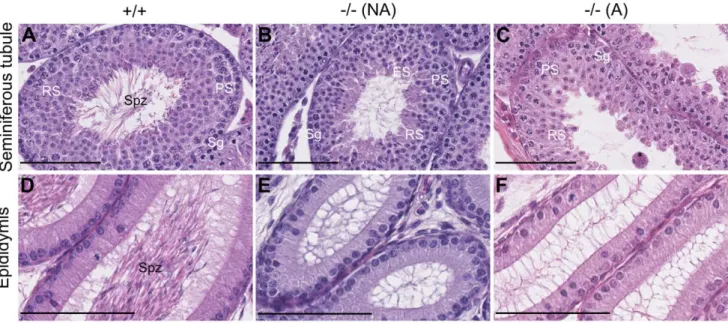
To investigate if the spermiogenesis blockage found in adult mice [18] already occurs during the first wave of spermatogenesis, we compared sections of seminiferous tubules from prepubertal +/ + and 2/2 mice. As expected for 42 dpp males [19], tubules and epididimes from +/+ mice contained spermatozoa (Figures 1A, 1D). The tubules from 2/2 animals either contained elongated spermatids (Figure 1B), revealing spermiogenesis set-up, or only contained round spermatids (Figure 1C), suggesting a spermio-genesis blockage. The tubules devoid of elongated spermatids contained multinuclear giant cells (Figure 1C), possibly as a consequence of the spermiogenesis blockage as observed in adults
[18]. The ratio between mice with tubules containing or not Table
1. Homozygous Celf1 2 /2 mice are not viable on 129SvPas nor C57BL/6N pure backgrounds. Background Number of females Number of litters Number of live pups Number of dead pups Likelihood of Mendelian segregation (P-value), live pups only Likelihood of Mendelian segregation (P-value), all pups 2 /2 other 2 /2 other 129SvPas 6 1 2 0 51 12 0 3.7e-05 0.27 C57BL/6N 3 6 0 2 6 6 0 3.2e-03 0.41 We separately crossed heterozygous Celf1 + /2 mice from two different inbred strains (129SvPas and C 57BL/6N) to obtain F1 litters. We genotyped pups found dead a fter birth, and pups alive a t 8 –10 d p p. We show here the number of 2 /2 and o ther (+ /+ and + /2 ) pups. P -values for agreement with the Mendelian ratio (1/4 of 2 /2 pups) w ere calculated by chi 2test for goodness o f fit taking into account either only the live p ups o r all the pups. doi:10.1371/journal.pone. 0046337.t001
elongated spermatids was roughly 1:1. Hence, the first wave of spermatogenesis is blocked at the round spermatid stage in about half of the 2/2 mice.
The First Wave of Spermatogenesis is Delayed in Celf12/2 Mice
Although the 42 dpp 2/2 mice were splited between those that encountered spermiogenesis during the first wave of spermato-genesis and those that did not, we observed that none of the 42 dpp 2/2 seminiferous tubules contained spermatozoa, the last stage of spermiogenesis (Figure 1B–C). Consistently, 2/2 epididimes were empty, contrasting with +/+ epididimes that started accumulating spermatozoa at this age (Figures 1 D–F). This suggests that the first wave of spermatogenesis is delayed in 2/2 animals. To characterize that delay, we compared histological sections of testes from younger mice. Representative photographs are shown in Figure 2. At 7 dpp, +/+ and 2/2 seminiferous tubules were mainly composed of Sertoli cells (St) and spermato-gonia (Sg) at the periphery (Figure 2A). However, gonocytes (G, foetal germ cells) were found in 7 dpp 2/2 testes (right panel), but not in 7 dpp +/+ testes (left panel). At 15 dpp, meiosis reached the pachytene spermatocyte (PS) stage in +/+ animals whereas the most differentiated cells in 2/2 mice were zygotene spermato-cytes (ZS, Figure 2B). At 24 dpp, round spermatids were present in the seminiferous tubules of +/+ mice (Figure 2C, left panel), demonstrating the completion of meiosis. We only observed pachytene spermatocytes in 2/2 mice at the same age (Figure 2C right panel). The ages at which the first round spermatids were observed were 21 dpp in +/+ animals and 31 dpp in 2/2 mice (see Figure 3A). In 35 dpp +/+ mice, we observed elongated spermatids in all the seminiferous tubules (Figure 2D left panel). By contrast, the 2/2 tubules at the same age only harboured round spermatids as the most advanced stages (Figure 2D right panel).
The above data suggest that, compared with Celf1+/+mice, the
onset of the first wave of spermatogenesis is markedly delayed in
Celf12/2 mice. To obtain a quantitative view of this delay, we
analysed histological sections of testes of several +/+ and 2/2 mice and, for each testis section, we determined the most advanced stage of spermatogenesis. Next, we measured within each testis the percentage of seminiferous tubules that contained germ cells at that most advanced stage, and we plotted it against the age (Figure 3A). The first wave of spermatogenesis was as previously described in +/+ animals [19], and was delayed in all the 2/2 mice. This delay was on average 7 days in young mice. For example, 20% of the seminiferous tubules of +/+ mice taken around 17 dpp contained pachytene spermatocytes, whereas this value was only observed in 2/2 mice around 24 dpp. The delay increased with the age of the mice, as 60% of seminiferous tubules contained round spermatids around 24 dpp in +/+ animals but only at 31 to 42 dpp in 2/2 animals. As previously noticed (Figures 1B–C), the most advanced stage of germ cell differenti-ation in 42 dpp 2/2 testes was either round or elongated spermatids. Noteworthingly, all the 2/2 testes older than 31 dpp contained round spermatids. This confirms that the 42 dpp 2/2 testes devoid of elongated spermatids suffer from a blocked spermiogenesis, and not from a longer delay of germ cell differentiation. In contrast to 42 dpp 2/2 testes, the testicular
contents of different Celf12/2mice up to 35 dpp were
homoge-neous, and it was not possible to predict, among the 35 dpp mice, which ones would have supported spermiogenesis if they had been allowed to grow older. Together, these data show that the first wave of spermatogenesis is delayed by several days in 2/2 mice in a fully penetrant manner, while spermiogenesis is blocked during the first wave in 2/2 mice in an incompletely penetrant manner.
We next asked if the first wave of spermatogenesis differed in 2/2 and +/+ mice by additional criteria. To do this, we compared the expression of several germ cell markers previously shown to be down-regulated in adult 2/2 testes [18]. Preliminary experiments revealed significantly reduced expression levels of these markers in prepubertal 2/2 testes as compared with +/+
Figure 1. Incompletely penetrant blockage of spermiogenesis in Celf12/2 mice during the first wave of spermatogenesis. Representative histological sections of testes of+/+ (left panels) and 2/2 (two right panels) mice at 42 days post-partum (dpp). A–C, seminiferous tubules.D–F, epididimes of the same respective mice. Depending on the presence of elongated spermatids in seminiferous tubules, KO mice were classified as ‘‘non affected’’ (B, E) or ‘‘affected’’ (C, F). Sg, Spermatogonia; PS, Pachytene Spermatocyte; RS, Round Spermatid; ES, Elongated Spermatid; Spz, Spermatozoa; *, multinuclear giant cell. Scale bars, 100 mm (A–C), 80 mm (D–F).
testes taken at the same age (data not shown), but these results might simply be due to the delayed testicular development. Hence, we measured the expression of these markers in +/+ and 2/2 testes that had reached similar stages of spermatogenesis. On the
one hand we compared 17 dpp Celf1+/+ with 24 dpp Celf12/2
animals, where 20% of seminiferous tubules contained pachytene spermatocytes as the most advanced stage (see Figure 3A). On the
other hand, we compared 24 dpp Celf1+/+ males with 42 dpp
Celf12/2 males (devoid of elongated spermatids) where 60% of
seminiferous tubules contained round spermatids (see Figure 3A).
Except for Acr in testes where 60% of seminiferous tubules contained round spermatids, none of the tested germ cell markers was expressed at different levels in 2/2 and +/+ males (Figure 3B). This contrasts with the situation in adults where Hspa2, Ldhc and Piwil1 are down-regulated in 2/2 testes [18]. This suggests that, except for the delay and the spermatogenesis blockage in part of the animals, Celf1 inactivation does not dramatically affect gametogenesis during the first wave of spermatogenesis.
Figure 2. The first wave of spermatogenesis is delayed in Celf12/2mice. Representative histological sections of testes of homozygous Celf1+/+mice (left panels) and Celf12/2mice (right panels) at different prepubertal ages.A, 7 dpp; B, 15 dpp; C, 24 dpp; D, 35 dpp. G, Gonocyte; Sg, Spermatogonia; St, Sertoli cell; ZS, Zygotene Spermatocyte; PS, Pachytene Spermatocyte; RS, Round Spermatid; ES, Elongated Spermatid. Scale bars, 50 mm.
doi:10.1371/journal.pone.0046337.g002
Figure 3. Quantification of the delay of the first wave of spermatogenesis. A, For each mouse, we analysed 300 seminiferous tubules in 15 testis sections. We classified each tubule according to the most differentiated germ cells that it contained (ZS, Zygotene Spermatocyte; PS, Pachytene Spermatocyte; RS, Round Spermatid; ES, Elongated Spermatid; Spz, Spermatozoa). Next, we calculated for each mouse the percentage of seminiferous tubules of each class, and we plotted the percentage of seminiferous tubules of the most advanced class against the age. Blue diamonds and orange squares correspond to individual Celf1+/+and Celf12/2mice respectively.B, We quantified by real-time RT-PCR the relative amounts of the indicated mRNAs (Mouse Vasa Homolog, Ddx4; Proacrosine, Acr; Heat Shock Protein 70.2, Hspa2; Lactate Dehydrogenase 3, Ldhc; Miwi, Piwil1) for+/+ and 2/2 testes of similar stages of spermatogenesis but different ages: lower panels and inserts, 17 dpp +/+ (blue bars) and 24 dpp 2/2 (orange bars) testes with 20% of seminiferous tubules of the PS class; upper panels, 24 dpp+/+ (blue bars) and 42 dpp 2/2 (orange bars) testes with 60% of seminiferous tubules of the RS class. Results are expressed as the means of 3–5 animals for each genotype. Error bars are standard deviations. We used a Student’s t-test to statistically compare the+/+ and the 2/2 genotypes, and the p-values below 0.1 are given on the right of the corresponding bars.
The Setting-up of Steroidogenesis is Delayed in Celf12/2 Mice
The delay of gametogenesis in 2/2 mice may be specific for germ-cells differentiation, but it may also reflect a more general delay of testicular development. To discriminate between these possibilities, we compared in +/+ and 2/2 prepubertal mice the expression of the Lhr gene that encodes the luteinizing hormone receptor (Figure 4A), and of steroidogenic enzymes that metab-olize cholesterol into testosterone in several steps (Figure 4B). The expressions of these genes are markers of Leydig cells maturation and functions. They increased with the age of the animals both in +/+ and 2/2 males but with different kinetics. Between 17 and 24 dpp, the Cyp11a1, Hsd3b6 and Cyp17a1 mRNAs began accumulating in +/+ animals, whereas they remained at low levels in 2/2 animals. All the tested genes were stimulated between 24 and 35 dpp irrespective of the genotype. We classified 42 dpp 2/2 mice as supporting or not spermiogenesis (see above), and we analysed separately these two classes. In 2/2 animals, the expression of 5 genes about 7 (Lhr, Star, Cyp11a1, Hsd3b1 and Hsd17b3) was stimulated between 35 dpp and at least one class of 42 dpp mice, whereas it was the case for only 1 gene (Star) in +/+ animals. Together, these data show that genes expressing steroidogenic enzymes start being expressed and reach a plateau earlier in +/+ than in 2/2 mice, demonstrating that the setting-up of steroidogenesis is delayed in 2/2 males. However, at 42 dpp, the difference of expression between +/+ and affected 2/2 mice is weakly significant for only two genes, Star and Cyp11a1, showing that the tested genes are less differentially expressed at 42 dpp than at earlier ages. This suggests that 2/2 mice catch up their delay by the end of prepubertal life.
We compared more thoroughly the two classes of 42 dpp 2/2 males (Figure 4). Four genes (Lhr, Cyp11a1, Hsd3b6 and Cyp17a1) were expressed at very similar levels. Among the three other genes, two (Star and Hsd17b3) were possibly more strongly expressed in testes with elongated spermatids than in testes without elongated spermatids while the third one (Hsd3b1) was potentially more strongly expressed in testes without elongated spermatids. There is therefore no correlation between the capacity of 2/2 testes to fulfil spermiogenesis and the expression of Leydig cells markers taken as a whole.
Discussion
Previously we have shown that Celf12/2mice are barely viable, with
about one-third of mice on a mixed 129SvPas*C57BL/6N back-ground being alive 8–10 days after birth [18]. Here, we have been
unable to identify any live Celf12/2mice on 129SvPas or C57BL/6N
backgrounds. A similar genetic background-dependent viability has been described for other gene disruptions in mice. For example, homozygous inactivation of Hsf1 is virtually lethal in a 129Sv background, but not in other, mixed backgrounds [20]. Consequently,
Celf12/2mice can be analysed only on a mixed background, and this is
probably at the origin of a high phenotypic variability. Indeed, the different traits of 2/2 mice have different penetrances. During the first wave of spermatogenesis, all the 2/2 mice show a delayed gametogenesis and a delayed expression of Leydig cells markers and
Figure 4. The expression of Leydig cells markers is delayed in prepubertalCelf12/2mice. We quantified by real-time RT-PCR the relative amounts of the indicated mRNAs in+/+ (blue diamonds) and 2/2 (orange squares) testes at the indicated prepubertal ages. NA (not affected) and A (affected) refer to 42 dpp 2/2 mice with and without
elongated spermatids respectively based on histological analyses.A, Lhr. B, The main pathway of steroidogenesis in rodents and the corresponding enzymes [37]. Hsd3b1 and Hsd3b6 have different expression patterns but encode enzymes with similar activities [22]. Results are expressed as the means of 3–5 animals for each age and genotype. Error bars are standard deviations. We used a Student’s t-test to statistically compare the different genotypes of identical ages, and we show the p-values below 0.1 above the corresponding symbols. doi:10.1371/journal.pone.0046337.g004
steroidogenic enzymes, but spermiogenesis is only blocked in a fraction of them. We have previously shown that spermiogenesis is also blocked in a fraction of adult males [18]. Hence, fully penetrant traits are the delayed spermatogenesis and steroidogenesis in prepubertal animals, while an incompletely penetrant trait is the spermiogenesis arrest during the first wave of spermatogenesis and in adults. Spermatogenesis is highly dependant on testosterone [21]. Hence, delayed steroidogen-esis might be a major cause for delayed gametogensteroidogen-esis, and the observation that these traits both are completely penetrant is consistent with this hypothesis. However, there are probably additional causes for the delay of germ cell differentiation as it is already observed at 7 dpp, before the onset of steroidogenesis [22].
Another fully penetrant trait of 2/2 mice is their reduced size [18], and this raises the question of a potential link between CELF1, delayed steroidogenesis and spermatogenesis, and reduced size. In humans, there is apparently no large-scale correlation between the size at birth, the prepubertal and postpubertal growth and the age of puberty in boys [23]. However, in particular genetic backgrounds, relationships exist between growth and onset of puberty. The Laron syndrome is a dwarfism due to a mutation is the Growth Hormone (GH) receptor. Sexual maturation is delayed in men with Laron syndrome [24], whereas for boys with a homozygous deletion of exon 3 (a gain-of-function mutation) pubertal onset is at a younger age [25]. In model animals, puberty is also delayed in male mice inactivated for GH receptor gene [26], and this may be due to a delayed maturation of Leydig cells, as GH stimulates the maturation of Leydig cells, both directly [27] and through IGF-1 [28]. Futhermore, according to a systematic analysis of gene expression in human [29], CELF1 is virtually ubiquitously expressed, including in the pituitary gland and the hypothalamus. Hence, it is tempting to speculate that the homozygous inactivation of Celf1 results in fully penetrant defects in the GH/IGF-1 pathway that would reduce body size and delay puberty by delaying the maturation of Leydig cells. and testing this hypothesis will require additional experiments. A spermiogenesis blockage has been reported in mice that suffer from impaired Leydig cells functions due to an inactivation of the LH receptor gene [30,31] or androgen receptor gene in Leydig cells [32]. It is therefore tempting to hypothesize a supplemental link between Leydig cells dysfunctions and spermatogenesis defects in 2/2 males: not only delayed Leydig cells maturation would cause delayed gametogenesis during the first wave, but defective Leydig cells functions would block spermiogenesis in part of the 2/2 mice. We found no global correlation between the expression of steroidogenic enzymes and the capacity of 2/2 testes to fulfil spermiogenesis during the first wave, ruling out that a major dysfunction of Leydig cells would be the cause of the spermiogenesis blockage. However, Star and Hsd17b3 tended to be more strongly expressed in 42 dpp 2/2 testes with elongated spermatids (Figure 4), and other genes involved in Leydig cells functions are also probably differentially expressed in 2/2 testes supporting or not spermiogenesis. The spermatogenesis defects in part of the
Celf12/2mice may therefore be due to subtle defects of Leydig cells
that could be investigated by systematic approaches.
Germ cell markers are down-regulated in adults with a blocked spermiogenesis [18], but not in prepubertal 2/2 mice compared with wild-type mice of similar development (Figure 3B). In adults, intercellular cross-talks maintain a critical cell ratio between the different germ cell stages, but these cross-talks are not set up during the first wave of spermatogenesis [33]. We can therefore propose a hypothetical link between the occurence of cross-talks and the down-regulation of germ cell markers. Specifically, the down-down-regulation probably reflects a partial depletion rather than a dysfunction of germ cells, because these genes are differently regulated and the correspond-ing gene products are involved in different molecular pathways.
Consequently, the fall in number of elongated spermatids due to spermiogenesis blockage in 2/2 mice may reduce the number of more immature germ cells owing to intercellular cross-talks in adults, but not during the first wave. Hence, the inactivation of Celf1 would reduce the amount of germ cells in adults as a consequence of the spermiogenesis blockage rather than through a direct effect on cell proliferation.
Materials and Methods Ethics Statements
Animals were bred in the Biosit animal facilities as approved by the French animal care agency (Direction des Services ve´te´r-inaires, approval number A3523840). Experiments were made according to standard procedures after acceptance by the local ethics commitee (Comite´ rennais d’e´thique en matie`re d’expe´ri-mentation animale, approval number R-2011-CGC-01).
Animals
The tm1Cba allele of the Celf1 gene was shown to be null [18] and is noted - (minus) throughout this manuscript. This allele is present on three genetic backgrounds. The mixed background (C57BL/6N*129SvPas) has been described [18]. We obtained backgrounds congenic to 129SvPas and C57BL/6N strains by more than 10 backcrosses. We intercrossed heterozygous mice to
obtain Celf12/2animals and we genotyped them at 8–10 dpp by
PCR on tail tips as described [18].
For each animal one testis associated with the epididymis was removed and fixed during four to six hours at RT in Bouin’s fluid,
dehydrated and embedded in paraffin wax. Sections of 7mm were
made and stained with hematoxylin and eosine (Shandon). The other testis was snap frozen into liquid nitrogen and crushed in Trireagent (Euromedex. 1 ml Trireagent/testis). RNA was recov-ered according to the manufacturer instructions and quantified by spectrophometry (Nanodrop).
Real-Time RT-PCR
Reverse transcription was made following standard procedures using random primers and Superscript II reverse transcriptase (Invitrogen), except for ‘‘RT-’’ controls where the enzyme was omitted. We made real-time PCR with an ABI Prism 7900 device (Applied) using SybrGreen mastermix and the primers given below. For each mRNA sample from individual testes, quantifications were made in triplicate. We checked that the RT- controls gave no amplification, or at a Ct far above that obtained with the corresponding RT+ conditions. Relative mRNA quantities were given by the difference of the Ct with the Ct of Hprt gene according to the formula [relative quantity = 2exp(CtHprt-Ctgene)], or by a double normalisation with Hprt and Beta-2 microglobulin (B2m) genes as described [34].
Primer sequences for Acr (D00754), Ldhc (X04752), Hspa2 (BC052350), Piwil1 (NM_021311), Gapdh (XM_354654), Lhr (M81310), Cyp11a1 (AF195119) and Hsd17b3 (U66827) have been published [18,22,35,36]. The sequences of the other primers are: Ddx4 TACCTATGTGCCTCCCAGCTTC and TGTATT-CAACGTGTGCTTTGCC;
Hprt CTGGTGAAAAGGACCTCTCG and TCAAGGGCA TATCCAACAACAAAC;
B2m TGGTGCTTGTCTCACTGACC and CCGTTCTTCAG CATTTGGAT;
Hsd3b6 TCCCCATTCAGAGCATGTATAGC and TTTTT TTGAGGTATTGACAAGTATTTATTG;
Hsd3b1 CTCAGTTCTTAGGCTTCAGCAATTAC and CC AAAGGCAGGATATGATTTAGGA.
Acknowledgments
We thank the Biosit structure for technological support: mice husbandry, histopathology (H2P2) and microscopy (MRic). We sincerely thank Dr HB Osborne for helpful discussions on the manuscript.
Author Contributions
Conceived and designed the experiments: LP CGC. Performed the experiments: MC GB YA CGC. Analyzed the data: MC GB LP CGC. Contributed reagents/materials/analysis tools: YA LP. Wrote the paper: LP CGC.
References
1. Russell L, Ettlin R, SinhaHikim A, Clegg E (1990) Histological and histopathological evaluation of the testis. St Louis: Cache River Press. 2. Yan W (2009) Male infertility caused by spermiogenic defects: lessons from gene
knockouts. Mol Cell Endocrinol 306: 24–32.
3. Chalmel F, Rolland AD, Niederhauser-Wiederkehr C, Chung SS, Demougin P, et al. (2007) The conserved transcriptome in human and rodent male gametogenesis. Proc Natl Acad Sci U S A 104: 8346–8351.
4. Itman C, Mendis S, Barakat B, Loveland KL (2006) All in the family: TGF-beta family action in testis development. Reproduction 132: 233–246.
5. Shima JE, McLean DJ, McCarrey JR, Griswold MD (2004) The murine testicular transcriptome: characterizing gene expression in the testis during the progression of spermatogenesis. Biol Reprod 71: 319–330.
6. Schultz N, Hamra FK, Garbers DL (2003) A multitude of genes expressed solely in meiotic or postmeiotic spermatogenic cells offers a myriad of contraceptive targets. Proc Natl Acad Sci U S A 100: 12201–12206.
7. Lalancette C, Platts AE, Lu Y, Lu S, Krawetz SA (2008) Computational identification of transcription frameworks of early committed spermatogenic cells. Mol Genet Genomics 280: 263–274.
8. Paronetto MP, Sette C (2009) Role of RNA-binding proteins in mammalian spermatogenesis. Int J Androl 33: 2–12.
9. Grosso AR, Gomes AQ, Barbosa-Morais NL, Caldeira S, Thorne NP, et al. (2008) Tissue-specific splicing factor gene expression signatures. Nucleic Acids Res 36: 4823–4832.
10. Yeo G, Holste D, Kreiman G, Burge CB (2004) Variation in alternative splicing across human tissues. Genome Biol 5: R74.
11. Iguchi N, Tobias JW, Hecht NB (2006) Expression profiling reveals meiotic male germ cell mRNAs that are translationally up- and down-regulated. Proc Natl Acad Sci U S A 103: 7712–7717.
12. Barreau C, Paillard L, Mereau A, Osborne HB (2006) Mammalian CELF/ Bruno-like RNA-binding proteins: molecular characteristics and biological functions. Biochimie 88: 515–525.
13. Vlasova IA, Tahoe NM, Fan D, Larsson O, Rattenbacher B, et al. (2008) Conserved GU-Rich Elements Mediate mRNA Decay by Binding to CUG-Binding Protein 1. Mol Cell 29: 263–270.
14. Dasgupta T, Ladd AN (2012) The importance of CELF control: molecular and biological roles of the CUG-BP, Elav-like family of RNA-binding proteins. Wiley Interdiscip Rev RNA 3: 104–121.
15. Gautier-Courteille C, Le Clainche C, Barreau C, Audic Y, Graindorge A, et al. (2004) EDEN-BP-dependent post-transcriptional regulation of gene expression in Xenopus somitic segmentation. Development 131: 6107–6117.
16. Ezzeddine N, Paillard L, Capri M, Maniey D, Bassez T, et al. (2002) EDEN dependent translational repression of maternal mRNAs is conserved between Xenopus and Drosophila. Proc Natl Acad Sci U S A 99: 257–262.
17. Cibois M, Gautier-Courteille C, Vallee A, Paillard L (2010) A strategy to analyze the phenotypic consequences of inhibiting the association of an RNA-binding protein with a specific RNA. Rna 16: 10–15.
18. Kress C, Gautier-Courteille C, Osborne HB, Babinet C, Paillard L (2007) Inactivation of CUG-BP1/CELF1 causes growth, viability, and spermatogenesis defects in mice. Mol Cell Biol 27: 1146–1157.
19. de Rooij DG, Russell LD (2000) All you wanted to know about spermatogonia but were afraid to ask. J Androl 21: 776–798.
20. Xiao X, Zuo X, Davis AA, McMillan DR, Curry BB, et al. (1999) HSF1 is required for extra-embryonic development, postnatal growth and protection during inflammatory responses in mice. Embo J 18: 5943–5952.
21. Sofikitis N, Giotitsas N, Tsounapi P, Baltogiannis D, Giannakis D, et al. (2008) Hormonal regulation of spermatogenesis and spermiogenesis. J Steroid Biochem Mol Biol 109: 323–330.
22. O’Shaughnessy PJ, Willerton L, Baker PJ (2002) Changes in Leydig cell gene expression during development in the mouse. Biol Reprod 66: 966–975. 23. Ibanez L, de Zegher F (2006) Puberty and prenatal growth. Mol Cell Endocrinol
254–255: 22–25.
24. Chandrashekar V, Zaczek D, Bartke A (2004) The consequences of altered somatotropic system on reproduction. Biol Reprod 71: 17–27.
25. Sorensen K, Aksglaede L, Petersen JH, Leffers H, Juul A (2010) The exon 3 deleted growth hormone receptor gene is associated with small birth size and early pubertal onset in healthy boys. J Clin Endocrinol Metab 95: 2819–2826. 26. Keene DE, Suescun MO, Bostwick MG, Chandrashekar V, Bartke A, et al.
(2002) Puberty is delayed in male growth hormone receptor gene-disrupted mice. J Androl 23: 661–668.
27. Kanzaki M, Morris PL (1999) Growth hormone regulates steroidogenic acute regulatory protein expression and steroidogenesis in Leydig cell progenitors. Endocrinology 140: 1681–1686.
28. Wang GM, O’Shaughnessy PJ, Chubb C, Robaire B, Hardy MP (2003) Effects of insulin-like growth factor I on steroidogenic enzyme expression levels in mouse leydig cells. Endocrinology 144: 5058–5064.
29. Lukk M, Kapushesky M, Nikkila¨ J, Parkinson H, Goncalves A, et al. (2010) A global map of human gene expression. Nature Biotech 28: 322–4.
30. Zhang FP, Poutanen M, Wilbertz J, Huhtaniemi I (2001) Normal prenatal but arrested postnatal sexual development of luteinizing hormone receptor knockout (LuRKO) mice. Mol Endocrinol 15: 172–183.
31. Zhang FP, Pakarainen T, Zhu F, Poutanen M, Huhtaniemi I (2004) Molecular characterization of postnatal development of testicular steroidogenesis in luteinizing hormone receptor knockout mice. Endocrinology 145: 1453–1463. 32. Xu Q, Lin HY, Yeh SD, Yu IC, Wang RS, et al. (2007) Infertility with defective
spermatogenesis and steroidogenesis in male mice lacking androgen receptor in Leydig cells. Endocrine 32: 96–106.
33. Rodriguez I, Ody C, Araki K, Garcia I, Vassalli P (1997) An early and massive wave of germinal cell apoptosis is required for the development of functional spermatogenesis. Embo J 16: 2262–2270.
34. Vandesompele J, De Preter K, Pattyn F, Poppe B, Van Roy N, et al. (2002) Accurate normalization of real-time quantitative RT-PCR data by geometric averaging of multiple internal control genes. Genome Biol 3: RESEARCH0034. 35. Lu JY, Sadri N, Schneider RJ (2006) Endotoxic shock in AUF1 knockout mice mediated by failure to degrade proinflammatory cytokine mRNAs. Genes Dev 20: 3174–3184.
36. Hecht NB (1998) Molecular mechanisms of male germ cell differentiation. Bioessays 20: 555–561.
37. Eacker SM, Agrawal N, Qian K, Dichek HL, Gong EY, et al. (2008) Hormonal regulation of testicular steroid and cholesterol homeostasis. Mol Endocrinol 22: 623–635.