The Disque Platform for the Investigation of Islet Differentiation
to Study, Treat, and Cure Type 1 Diabetes
by
Peter Anthony Jones B.S. Plastics Engineering
University of Massachusetts Lowell, 2014 B.A. International Business Administration
American University of Paris, 2006
SUBMITTED TO THE PROGRAM IN HEALTH SCIENCES AND TECHNOLOGY I FULFILLMENT OF THE REQUIREMENTS FOR THE DEGREE OF MASTER OF SCIENCE IN HEALTH SCIENCES AND TECHNOLOGY,
CONCENTRATION AREA: MECHANICAL ENGINEERING AT THE
MASSACHUSETTS INSTITUTE OF TECHNOLOGY JUNE 2017
Peter Anthony Jones. All rights reserved.
Signature of Autho
Certified by:
The author hereby grants to MIT permission to reproduce and to distribute publicly paper and electronic copies of this thesis document in whole or in part
in any medium now known or hereafter created.
r:Signature
redacted
Sig
dI
N PARTIAL MASSACHUSETTS INSTITUTE OF TECHNOLOGYJUN 12 2017
LIBRARIES
ARCHIVES
rbgram in Health Sciences and Technology May 22, 2017
nature redacted
7 Jeffrey M. Karp
Associate Professor of Medicine, Brigham and Women's Hospital, Harvard Medical School
- Thesis Supervisor
Accepted
Signature redacted
Emery N. Brown, MD, PhD Director, Harvard-MIT Program in Health Sciences and Technology Professor of Computational Neuroscience and Health Sciences and Technology
The Disque Platform for the Investigation of Islet Differentiation
to Study, Treat, and Cure Type 1 Diabetes
by
Peter Anthony Jones
Submitted to the Program in Health Sciences and Technology on May 22, 2017 in Partial Fulfillment of the
Requirements for the Degree of Master of Science in Health Sciences and Technology
ABSTRACT
There is a critical health care need to generate large numbers of beta cells for transplantation. In Type 1 Diabetes (TID), insulin-producing beta cells in the islets of Langerhans within the pancreas, which support glucose homeostasis, are destroyed in an autoimmune attack. The ensuing loss of glycemic control leads to serious complications, requiring life-long insulin injections and close monitoring, while shortening lifespan by 11-13 years. In the face of a three percent annual increase in T1D incidence, there is a grave lack of transplantable material, and very few patients are able to receive an islet transplant each year. Recent advances in stem cell differentiation have enabled the production of large quantities of insulin-producing beta-like cells in vitro, bringing hope to the field. However, the efficiency and yield of such production methods remains unacceptably low, with high batch-to-batch variability, and the function of these cells is unstable. Moreover, the ability to probe the conditions that affect differentiation outcomes is limited by the scale, cost, and complexity of existing culture systems.
The present work focuses on the Disque Platform, a biomimetic screening platform for the investigation of islet cell differentiation. The Disque Platform allows for the formation of differentiating 3D micro-tissues within an automation-friendly design, and is capable of systematically manipulating the developing stem cell niche in order to identify chemical and physical cues that enhance beta cell production. Significantly, the Disque Platform consistently differentiates beta-like cells from pancreatic progenitor cells, with similar efficacy to existing high-volume production methods. Furthermore, it achieves superior differentiation outcomes compared to the 2D culture systems tested, and is able to respond to interventions when conventional systems cannot produce a clear signal or readout. Together, these data support the ability of the Disque Platform to investigate specific interventions to enhance beta-cell differentiation. It is hoped that the Disque Platform can serve as a springboard for beta cell and islet study within the diabetes community, and that these advances can contribute towards a cure for Type 1 Diabetes.
Thesis Supervisor: Jeffrey M. Karp
Title: Associate Professor of Medicine, Brigham and Women's Hospital, Harvard Medical School
Table of Contents
BACKGROUND & SIGNIFICANCE ... 4
Beta Cell Loss in Type 1 and Type 2 Diabetes...4
Beta Cell Replacement Therapy: Promise and Limitations...4
A Device to Interrogate Methods to Enhance Beta Cell Differentiation from Stem Cells... 5
SPECIFIC AIM 1: To CREATE A 3D CULTURE SYSTEM FOR BETA CELL DIFFERENTIATION, WITH CONTROLLED PRESENTATION OF MICRO-ENV IRO N M ENTA L CU ES. ... 5
SPECIFIC AIM 2: To UTILIZE THE 3D CULTURE SYSTEM AS A SCREENING TOOL TO INTERROGATE VARIOUS MICROENVIRONMENTS (CHEMICAL AND MECHANICAL CUES) AND CULTURE CONDITIONS TO ENHANCE THE BETA-CELL DIFFERENTIATION EFFICIENCY AND YIELD..6
RESULTS ... 6
SPECIFIC AIM 1: BUILDING A 3D CULTURE SYSTEM FOR BETA CELL DIFFERENTIATION ... 6
The Disque Platform for Beta Cell Differentiation...6
Diffusion Gradients and Suspension Culture Differentiation of Beta-Cell Clusters ... 7
Optimization of 3D Culture Conditions for Beta Cell Differentiation ... 8
Cell Discs Form Cell-Cell Junctions Criticalfor Islet Function...10
The Disque Platform Supports High Viability During Multi-Week Culture ... 11
The Disque Platform Replicates Key Outcomes of Spinner Flask Differentiation ... 12
The Disque Platform Shows More Promise for Screening Versus Simpler Culture Methods... 13
The Disque Platform: A Springboard for the Study of Islet Differentiation ... 15
SPECIFIC AiM 2: IDENTIFYING MICROENVIRONMENTS TO ENHANCE BETA CELL DIFFERENTIATION ... 15
Mechanical and ECM Microenvironments and Beta Cell Differentiation...15
RADA16-l Oligopeptide Gels Mimic Pancreatic Stiffness ... 16
DISCUSSION ... 17
FUTUR E WORK ... 18
Simultaneous Differentiation of Beta, Alpha, and Delta Cells...18
Oxidative Sterss and Beta Cell Differentiation ... 19
Glucose and Beta Cell Differentiation ... 19
Investigating Developmental Pathways and Cell-Cell Signaling ... 19
Generation of Transplantable Material to Cure T1D In Vivo...20
Disease Modeling of Beta Cells in T1D: Redox Imbalance and Beta Cell Death... 20
METHODS...21
Manufacturing Custom Cell Culture Inserts with Reducing Washer ... 21
Gel Preparation and ECM Coating ... 21
Cell Seeding: Forming a Thin Cell Disc within the Disque Platform... 21
Cell Disc Removalfor Downstream Analysis ... 21
Cryosectioning and IH C ... 22
96-Well Multi-Color Flow Cytometry ... 22
Measurement of Gel Modulus ... 22
ACKNOW LEDGEMENTS...23
STUDENT'S INVOLVEMENT IN THE RESEARCH ... 23
SUPPLEMENTA L DATA ... 24
Temperature- and Time-Dependent Shear Properties of PuraMatrixm and Matrigel* ... 24
Expression of Other Differentiation Markers on the Disque Platform ... 26
Pilot Testing of Notch Activator and Inhibitor Cocktails ... 27
Effect of Handling and Transportation on Beta Cell Differentiation ... 27
BACKGROUND & SIGNIFICANCE
Beta Cell Loss in Type 1 and Type 2 Diabetes
Pancreatic beta cell loss is a common feature of both T1D and advanced T2D1, leading to the gradual loss of insulin production and of glycemic control2. Patients who have lost their insulin-producing 0 cells live with a constant risk of ketoacidosis and hypoglycemic coma, and often suffer from long-term complications such as heart disease, stroke, kidney failure, foot ulcers, diabetic retinopathy, and shortened lifespan by 11-13 years. Diabetes burdened the U.S. economy at a level equal to 2% of annual GDP in 2012, or $322 billion, of which $6.2 billion alone was due to injectable insulin, and this cost increases steadily each year.3
Beta Cell Replacement Therapy: Promise and Limitations
There is currently no durable and effective treatment for
B-cell
loss4, and constant monitoring and insulin injections are mandatory for life. Whole pancreas or islet transplantation can represent a multi-year cure for insulin-dependent patients, however there is an extremely limited supply of transplantable material. This lack of material had held back the promise of@-cell replacement therapy, until the recent development of protocols to differentiate stem @-cells into @ cells, by Melton Lab at Harvard University and other researchers in the field.5'6
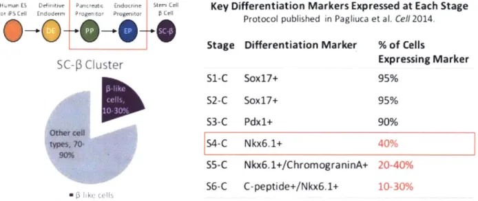
Although these @ cell protocols are capable of producing large quantities of
transplantable material in spinner flasks or bioreactors, they have been faced with several limitations. Notably, there is wide batch-to-batch variability, with final yields between 10% and
30% 1-like cells per batch as compared to ~70%
1
cells on average in native human islets (Fig.1). These stem-cell-derived 1-like cells (SC-@ cells) lack complete and stable function.
Furthermore, the remaining 70% to 90% of non--like cells present at the end of the process include unidentified phenotypes that are not controlled by current protocols. Finally, current protocols focus on differentiating one islet cell type at a time and are missing glucagon-producing alpha cells and somatostatin-glucagon-producing delta cells, which work together with beta cells to control blood sugar in a paracrine feedback system. Such drawbacks severely limit the utility of SC-@ cells for transplantation and in vitro disease modeling.
There is an urgent need to develop more efficient and effective stem cell differentiation protocols in order to improve the supply of functional
1
cells for transplantation. Unfortunately, screening for methods to enhance 1-cell differentiation directly in spinner flask culture is costly and time-consuming due to its scale. In addition, current 2D culture systems are only capable of measuring immediate effects (due to a dramatic increase in variability after only 3-4 days), while existing 3D culture systems are complex and not amenable to automation and medium-to high-throughput screening. A three-dimensional culture system is a requirement fordifferentiation, since tissue morphogenesis during development depends on both cell-cell and cell-environment interactions. In the case of islet differentiation, it has been reported that hESCs cultured within a soft Matrigel-Collagen type I scaffold expressed higher levels of pancreatic and endocrine markers versus 2D culture.
-J
1uman ES Definitive Pancreatx Endacrint t Key Differentiation Markers Expressed at Each Stage
Protocol published in Pagliuca et al. Cell 2014.
0 -0'- Stage Differentiation Marker % of Cells
SC-3 Cluster Expressing Marker
S1-C Sox17+ 95% S2-C Soxl7+ 95% S3-C Pdx1+ 90% Oth4~t cei~ typcs. YO 90% 54-C Nkx6.1+ 40% 55-C Nkx6.1+/ChromograninA+ 20-40% 56-C C-peptide+/Nkx6.1+ 10-30%
Figure 1. The Melton Lab -cell differentiation protocol summary and key outcomes. The protocol is organized into stages, and key differentiation markers are assessed at the end of each stage. Stage 4 day 1 (S4dl) corresponds approximately in vitro to the beginning of the pancreatic progenitor stage, when nearly all cells are pancreatic in origin (express Pdx1). By Stage 4 Complete (S4-C), on average ~40% of cells express Nkx6.1, an insulin gene promoting transcription factor. Nkx6.1 expression in a cell is a pre-requisite for subsequent insulin expression.
A Device to Interrogate Methods to Enhance Beta Cell Differentiation from Stem Cells In light of the limitations to studying 1-cell differentiation in currently-available cell culture devices, we set out to create a platform to interrogate soluble and mechanical cues within the differentiating stem cell niche that play a central role in determining
1
cell differentiation outcomes. Since1
cells require cell-cell contact to survive and function, and since 0-cell differentiation generally occurs more efficiently in 3D versus 2D culture9, we identified the need for a 3D biomimetic culture system that would allow us to study the entire course of the differentiation and to develop fully functional1
cell micro-tissues.This automation-friendly, 3D culture platform can be utilized to enhance the function and viability of differentiated SC- cells in vitro. These cells have the potential to transform
1
cell replacement into a viable therapy for insulin-dependent diabetics. In addition, such a system may be uniquely suited for: the direct generation of transplantable material; diseasemodeling of immune-@ cell interactions in TID and 1-cell death in T1D/T2D; and drug discovery and toxicology.
Specific Aim 1: To create a 3D culture system for Beta cell differentiation, with controlled presentation of micro-environmental cues.
0 Culture System Requirements:
o The system should form 1-cell micro-tissues with cell-cell junctions, with the ability to incorporate extracellular matrix components.
o The system must maintain high cell viability for multi-week differentiations.
o The system must have reliable and consistent read-outs by imaging and high-throughput phenotypic analysis.
o The system should match (gold standard) spinner flask differentiation outcomes in terms of the expression of key pancreatic endocrine markers.
o The system should produce measurable changes in differentiation outcomes in response to changes in the microenvironment.
" Hypothesis 1: Differentiating pancreatic progenitor cells in direct contact will lead to the formation of a 3D micro-tissue with cell-cell junctions.
" Hypothesis 2: A 3D culture system with controlled diffusion distance will enhance cell viability and lead to more better differentiation outcomes.
* Hypothesis 3: A 3D culture system based on these design principles will better predict differentiation outcomes compared to 2D culture systems.
Specific Aim 2: To utilize the 3D culture system as a screening tool to interrogate various microenvironments (chemical and mechanical cues) and culture conditions to enhance the beta-cell differentiation efficiency and yield.
* A wide variety of chemical and mechanical factors within the developing stem cell microenvironment can impact cell fate decisions, including: exogenous cytokines and growth factors, ECM composition, stiffness, juxtacrine and paracrine interactions with neighboring cells such as endothelial cells, temperature, pH, media composition, perfusion, oxygen tension, cell-cell junctions, redox balance, etc.
RESULTS
Specific Aim 1: Building a 3D Culture System for Beta Cell Differentiation
"To create an 3D culture system for beta-cell differentiation, with controlled presentation of microenvironmental cues."
The Disque Platform for Beta Cell Differentiation
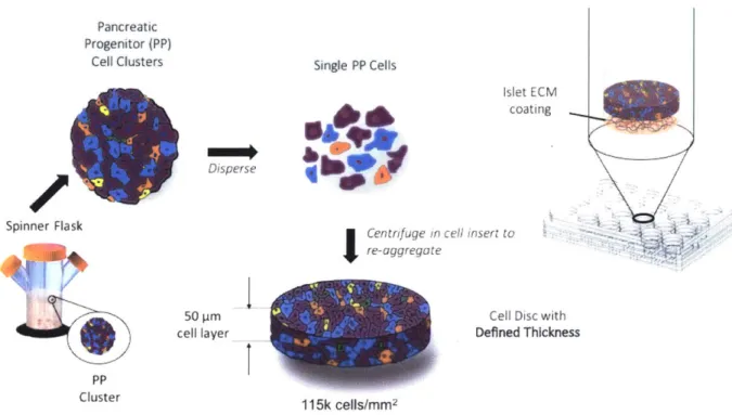
The Disque Platform is an in vitro culture system designed to create 3D biomimetic pseudo-islets for improved pancreatic -cell differentiation. Dispersed pancreatic progenitor cells are re-aggregated into cell discs, which are situated in a custom cell culture insert in a standard 24-well plate, amenable to medium-throughput manipulations. (Fig. 2) The thick cell layer allows cell-cell contact, which is essential for juxtacrine (contact-dependent) signaling such as Notch signaling in development, and for gap junction communication for coordinating insulin secretion in mature @ cells.
Pancreatic Progenitor (PP) Cell Clusters Spinner Flask c PP Cluster Single PP Cells Islet E coati 0-0 A11111i Ah.-'mow III*#* 40 ?F Disperse
Centrifuge in cell insert to
re-aggregate
50 pm ell layer
I
I
Cell Disc with Defined Thickness
115k cells/mm2
Figure 2. The Disque Platform
The Disque Platform enables the formation of thin cell discs made up of [-like cells for the study of [-cell differentiation. (Fig. 3) A custom reducing washer can be set into the bottom of the cell culture insert, which reduces the cell culture area and allows for simple ejection of the disc and the underlying membrane by applying a downward force on the edges of the washer. If desired, extracellular matrix components and gels with defined mechanical properties can be integrated into the cell disc at the time of seeding. Soluble cues can be integrated into the culture medium both above and below the cell disc. The cell discs can be sectioned or left intact for imaging.
Representative sample from n = 1 biological batch.
Figure 3. Cross-section of a thin 1 cell disc on the Disque Platform, illustrating the expression of key 1-cell markers. Pdx-1 expression indicates pancreatic origin; Nkx-6.1 is an insulin gene-promoting transcription factor; and C-peptide is co-expressed with insulin in 1 cells. The image was generated by confocal microscopy of a 15-pm cross section of a cell disc, stained by immunohistochemistry.
Diffusion Gradients and Suspension Culture Differentiation of Beta-Cell Clusters
Gradients in morphogen concentrations and central hypoxia are inherent challenges to cultured embryoid bodies and cell spheroids, since gradients over length scales of just 100 to 200 p.m are known to drive cell specification in development.1012 Indeed, attempts by others to
cM
reduce the effects of morphogen gradients have led to more uniform differentiation of other cell types.1'14 We hypothesize that cytokine and oxygen gradients drive inhomogeneous cell specification in cell clusters in vitro. Such a phenomenon may become especially problematic when feeding high molecular weight growth factors to cell clusters with the goal of activating surface receptors that affect cell fate, since such factors may not diffuse readily between cells, and may form a morphogen gradient from the periphery to the core of each cluster.15 Three such molecules, Activin A, KGF and betacellulin, each on the order of 20 kDa, are added to the media during 1-cell differentiation.
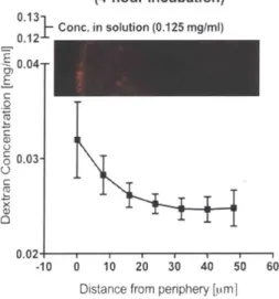
In a pilot study to model the diffusion of large molecules such as growth factors, a diffusible 10-kDa dextran probe was used to investigate gradients within SC- cell clusters. This study found that the probe formed a gradient within the first 50 Im from the periphery of SC-0 cell clusters that had been differentiated in spinner flask culture. (Fig. 4) This result is consistent with other findings in the literature for the diffusion of growth factors such as EGF.16
10-kDa Dextran Gradient in SC-P Clusters (4-hour Incubation) Conc. in solution (0.125 mglm 0.04 E C 0 0.03 C 0.02. -10 0 10 20 30 40 50 60
Distance from periphery [pim1
Figure 4. Dextran diffusion within SC-3 cell clusters taken from spinner flask suspension culture, after a 4-hour
incubation in 0.125 mg/ml 10-kDa Texas Red dextran. Clusters were fixed, cryosectioned, imaged by confocal microscopy, and then analyzed using mean gray value along concentric circles in ImageJ. Three representative clusters were used to generate the data.
Optimization of 3D Culture Conditions for Beta Cell Differentiation
In suspension culture in a spinner flask or bioreactor, convection helps with oxygenation and mass transport. However, there are inherent viability issues with static 3D cultures.
Therefore, it was necessary to identify conditions to maximize their viability.
The Disque Platform enables the formation of thin 3D cell discs with a defined thickness and diffusion distance for the controlled presentation of soluble cues, such as oxygen,
nutrients, and cytokines. Since the cell disc is suspended on a permeable membrane within a custom cell culture insert, diffusion can occur from both above and below. The thickness of the 3D cell disc in the Disque Platform can be precisely regulated by changing the seeding density. (Fig. 5A/B)
The reduced diffusion distance in thin cell discs is hypothesized to result in enhanced viability, minimized phenotypic heterogeneity and enrichment for the 0 cell phenotype. A core-shell structure forms in many spinner flask-cultured clusters, although it does not appear consistently in all batches, which may be a result of necrosis due to central hypoxia from 02 diffusion limits17, or perhaps from the development of a blastocoel-like structure during in vitro differentiation. In the Disque Platform, a thin cell disc approximately 50 im in thickness was shown to have high viability throughout the cell layer, while retaining expression of key P-cell markers. (Fig. 5C)
A.
C.
50~~5 pP10 m
40k cells/mm2 320 k cells/mm2 500 k cells/mmt
Representa veimages fromn 23 dics per seeding denwty, seeded f drone bdagca batcn
B.B
Thickness of Cell LayerSC-0
SC-400as Controlled by Seeding Density Cli
- 300- R2= 0.9886 .
0.2200-o
100-0 500000 1x10⁸ 2x10⁶
Seeding density (viable cells/mm2)
(n = 3 cell discs per seeding density)
SC-0 cluster immunohistochemistry image courtesy
of Melton Lab.
Figure 5. (A) Seeding density precisely controls the final thickness of the 3D cell disc on the Disque Platform, and (B)
this can be fit to a linear model with a coefficient of determination of R2
= 0.99. (C) Cell clusters cultured in suspension
(spinner flask) sometimes exhibit a core-shell structure. This structure is not observed on the Disque Platform. Immunostaining of representative cryosectioned cell discs and clusters shows live (fluorescein diacetate), dead cells (propidium iodide), nuclei (DAPI), Nkx-6.1, and C-peptide. Representative images of spinner flask clusters were taken
from n > 50 clusters across 3 biological batches, and representative images of cell discs were taken from n 3
differentiated cell discs.
Oxygen forms a gradient from the air-liquid interface to the surface of the cultured cells.15 Due to this fact, we hypothesized that a shorter media height would improve viability of the cell disc. Therefore, we set out to determine the optimum media height above the 3D cell discs. In a pilot study, it was found that reducing the media column height to 4.5 mm ensured sufficiently high cell disc viability, and the media did not excessively evaporate between media changes. (Fig. 6A) Future studies will determine whether reducing the media height further
could provide benefits beyond enhanced viability, such as enhanced Neurogenin3 expression due to repression of HIFla, as has been found in the example of air-liquid interface
differentiation of
1
cells.6'18Oxygen and other soluble factors also form a gradient from the periphery to the core of any 3D culture construct.15 In a second pilot study, it was found that setting the cell disc
thickness to 50 pm maximized the cell disc viability, however the effect of reducing disc thickness on improving yiability was not large in the range studied, likely because oxygen diffuses readily into tissue at all the thicknesses tested. (Fig. 6B)
We then hypothesized that limiting the disc thickness would also improve key differentiation outcomes, such as the expression of key endocrine progenitor markers. In a third pilot study carried out twice in separate biological batches, minimizing the cell disc thickness to 50 pm (and thus the maximum diffusion distance to 25 Im) maximized Pdx1 and Nkx6.1 co-expression in Disque Culture. This may be due to gradients of soluble cues that contribute toward driving the differentiation. (Fig. 6C)
A. B. C. m 1% 100% 0 3 13 so%-medIa e -0- DifferentiationA colum.t0sm he 49 -E& Differantiation heigh 4091 *
R=. 50%4A R2= 0 (for both trials)
A) 0 Y = -3.358*X + 2 102.2 -R1) 40%- 0 Diereniation A 0 Differentiaton B 20%- (Oreoe
0= N 20%
0 5 10 15 600 pm 400pm 200pm O pm 600pm 400 pm 200 pm O pm
Media Height Above Cell Disc [mm] Estimated Cell Disc Thickness Estimated Cell Disc Thickness (for a 50-pm cell disc)
Figure 6. Correlation between diffusion-related cell culture parameters, viability, and differentiation outcomes. Each
experiment was carried out on a different biological batch of differentiating cells. (A) Correlation between media height above the cell disc and viability. A linear model provides a coefficient of determination equal to 0.98. (B) Estimated disc thickness and the percentage of viable cells in the disc. (C) Correlation between the estimated diffusion distance (half of the thickness) and the percentage of cells that became pancreatic endocrine progenitors after 5-day differentiations from two biological batches (co-expression of Pdx-1 and Nkx-6.1). A one-phase exponential fit gives R2 = 0.80 for both differentiations.
Cell Discs Form Cell-Cell Junctions Criticalfor Islet Function
Cell-cell junctions play a role in contact-dependent signaling in development and mediate glucose-dependent insulin secretion in mature islets.19 We hypothesized that differentiating pancreatic progenitor cells in direct contact will lead to the formation of a 3D
micro-tissue with cell-cell junctions. The preserved 3D architecture of the Disque Platform is a key requirement for 1-cell differentiation and disease modeling, since dispersed
1
cells quickly lose their function in vitro. It was found that dispersed pancreatic progenitor clusters initially grown in suspension culture can successfully re-form a differentiating micro-tissue within the Disque Platform. (Fig. 7) In addition, confocal imaging can be performed directly on 50-pm cell discs without the need for sectioning.Figure 7. Representative image of a cell disc on the Disque Platform cultured on a thin layer of laminin and collagen
IV, taken at the pancreatic endocrine progenitor stage after 5 days in culture. Taken by confocal microscopy through the disc without sectioning. Background corrected by comparison to unstained controls. Adherens junctions (E-cadherin) indicate the initiation of cell-cell contacts. Tight junctions (ZO-1) indicate the potential for polarity and the gating of ions and solutes between cells. Gap junctions (Connexin-36, not shown) play a role in the synchronization of insulin secretion through the exchange of current-carrying ions between cells.
The Disque Platform Supports High Viability During Multi-Week Culture
We hypothesized that the optimized cell culture parameters would promote high viability despite moving from suspension culture (spinner flask) to static 3D culture (Disque Platform). Indeed, the Disque Platform (with optimized culture parameters) achieves
comparable high viability to Spinner Flask culture during pancreatic progenitor differentiation to endocrine progenitors. (Fig. 8A) The ability to recapitulate suspension culture in a 3D static culture format suggests its utility for studying 0-cell differentiation. After optimizing the 3D culture conditions, the Disque Platform maintains the high viability required for multi-week differentiations. (Fig. 8B)
A.
B.
100%- 100% _a 90% 1--2 go %-wE .!0%O 50% - 80%-A Disque Platform E 0 70% * Spinner Flask N N 0% 60% -% Viable Cells S4d1 S4C S5C S6d7 at S4-C Differentiation Day(total = 18 days in culture)
Figure 8. The percentage of viable cells in the 3D cell disc versus same-batch spinner flask clusters during
differentiation from pancreatic progenitors, assessed by Zombie UV dye exclusion and flow cytometry. (A) Viability at the endocrine progenitor stage (S4-Complete, S4-C) after a 5-day differentiation from pancreatic progenitors (Stage4 dayl, S4dl), shown as an average of 5 experimental repeats with n > 3 differentiations per repeat. (B)
Viability during the course of an 18-day differentiation from pancreatic progenitors (S4dl) to SC-0 cells (Stage6 day7,
The Disque Platform Replicates Key Outcomes of Spinner Flask Differentiation
In the Disque Platform, Nkx-6.1 expression levels increased synchronously with the spinner flask. Nkx-6.1 is a critical transcription factor that promotes insulin gene expression. This is a baseline result that suggests the platform's potential utility to predict the response of Spinner Flask-cultured cell clusters to conditions and signals. (Fig. 9)
Nkx-6.1 Induction:
Differentiation from Pancreatic Progenitors (S4dl -> S4-C) 50% Z I Disque Platform 4 Spinner Flask EcLt30%, 20% 0%
Differentiation Timeline in Stage 4
Figure 9. The percentage of viable cells that differentiated into Nkx-6.1-positive endocrine progenitors in the Disque Platform, and the same biological match in spinner flask culture. Five-day differentiation from pancreatic progenitors (Stage4 dayl, S4dl) to endocrine progenitors (S4-Complete, S4-C), n = 4 Disque Platform differentiations per time point seeded from a single batch of pancreatic progenitors. Assessed by antibody staining and flow cytometry.
The Disque Platform with optimized baseline culture parameters is able to achieve comparable differentiation outcomes to spinner flask cultures for stem cell-derived pancreatic progenitors. This result confirms that the cell discs on the Disque Platform recapitulate key outcomes of spinner flask-cultured clusters in suspension. (Fig. 10A) In addition, the Disque Platform can produce C-peptide-expressing -like cells if allowed to differentiate for longer periods of time. (Fig. 10B) See the Supplemental Data for additional information on expression of key differentiation markers in both culture systems.
A.
Pdx1 and Nkx6.1 ExpressionB.
Chromogranin-A and C-Peptide Expression5-Day Differentiations from Pancreatic Progenitors 12-Day Differentiation from Pancreatic Progenitors
n.s. I cc 100% A Disque Platform 100 c
j
* Spinner Flask 80% Boo% 60% A n.s. n.s. E 600 u40%- AA * AA4% 20% a 20% 0 " i 0% x xPdxl+ Pdxl+ Pdxl+ Pdx+ X+ Nkx6.1+ Nkx6.1+ Nkx6.1+ ChgA+ ChgA+ C-peptlde+Figure 10. Key transcription factor expression after differentiation on the Disque Platform. (A) Six nonsimultaneous experimental repeats of side-by-side 5-day differentiations from pancreatic progenitors (S4d1 to S4-C, six experiments with n > 4 Disque Platform differentiations per experiment). Each experimental repeat was started from
a matched batch pancreatic progenitor cells. Shows the percentage of cells expressing each marker on the final day of each experiment. (B) The Disque Platform generated 6.8% 1-like cells after a 12-day differentiation from
pancreatic progenitors (S4d1 to S6d1, n = 3 differentiations seeded from a single batch of pancreatic progenitors), defined as cells which co-express Pdxl, Nkx6.1, Chromogranin-A, and C-peptide/proinsulin.
The Disque Platform Shows More Promise for Screening Versus Simpler Culture Methods
Although the Disque Platform is relatively straightforward to set up, it is still more complex and costly compared to traditional screening assays such as 96-well or 384-well planar culture or monolayer culture on Matrigel. Therefore, we set out to determine whether a
simpler culture system could effectively measure the effects of microenvironmental
manipulations on 1-cell differentiation. We hypothesized that a thick cell layer with cell-cell contact would promote a more effective differentiation. We also hypothesized that placing the cell layer on a cell culture insert surrounded by media on the top and bottom would enable improved differentiation outcomes. Finally, we hypothesized that the Disque Platform would more effectively sense the effects of changes in chemical and physical cues, and that this would be reflected by the ability to detect significant changes in differentiation outcomes.
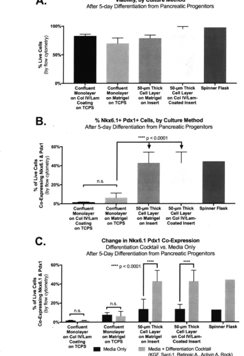
Various cell culture systems were compared to evaluate their ability to differentiate endocrine cells from pancreatic progenitors over the course of five days. A differentiation cocktail consisting of cytokines and growth factors was added to the media, and each cell culture system was analyzed to determine whether these chemical signals could produce a detectable change in differentiation outcomes. Although all the cell culture systems exhibited high viability (Fig. 11A), the percentages of Nkx6.1 Pdx1 co-positive cells varied significantly. Monolayer culture generally had poor differentiation outcomes, whereas culture on the Disque Platform led to expression levels similar to those obtained in the matched spinner flask. (Fig.
11B) Moreover, the multi-layer cell culture systems were able to reflect changes soluble cues
that the monolayer systems could not. Also, a chemically-defined Matrigel-free system is sufficient to produce this effect. (Fig. 11C) Future investigations will determine whether a collagen IV/laminin coating is required, and whether a monolayer on a cell culture insert could
produce an effective differentiation. Overall, this data suggests that a system such as the Disque Platform is better able to predict changes in -cell differentiation based on the addition of soluble cues compared to the other methods tested. This data also suggests that the
presence of a multilayer cell aggregate is a pre-requisite for endocrine differentiation.
A.
Viablity, by Culture MethodAfter 5-day Diferentiation from Pancreatic Progenitors
Confluent Conuent
Monolayer Monolayer
on CollV/Lam on Matrigel
Coating on TCPS
on TCPS
50-pm Thick 50-m Thick Spinner Flask
Cell Layer Cell Layer on Matrigel on Col
IVILem-on Insert Coated Insert
% Nkx6.1+ Pdxl+ Cells, by Culture Method
After 5-day Differentiation from Pancreatic Progenitors
*p < O.01
T
n.s.i 100%_ s0% .% B. Go%-S40 20%-2 o 0%-Monolayer on Col IV/Lam Coating on TCPS Confluent Monolayer on Matrigel on TCPS50jm Thick 50.pm Thick Spinner Flask Cell Layer Cell Layer
on Matrigel on Col
IViLam-on Insert Coated Insert
C.
60%' "a 40% 3. pn.S. 20% * ns Change In Nkx6.1 Pdxl Co-Expression Differentiation Cocktail vs. Media Only After 5-Day Differentiation from Pancreatic Progenitors***p < 0.0W01
n.s.
50-pm Thick 50-jam ThIck Spinner Flask
Cell Layer Cell Layer
on Matrigel on Col IVLam-on Insert Coated Insert
O Media + Derentiation cocktail
(KGF, Sant-1, Retinoic A., Activin A, Rock)
Figure 11. Culture method comparison. The Disque Platform is comprised of a 50-pm thick cell layer on a collagen IV and laminin-coated custom cell culture insert. (A) All cell culture systems supported high viability, as determined
by Zombie UV dye exclusion by flow cytometry. (B) Nkx6.1 Pdx1 co-expression by culture method after a 5-day
differentiation from pancreatic progenitors. Cells in a monolayer on either tissue culture polystyrene (TCPS) or Matrigel showed poor differentiation outcomes compared to culture of a thick cell layer on a custom cell culture insert. (C) The effect of a differentiation cocktail on differentiation outcomes within each cell culture platform. (N = 5 Disque Platform differentiations per condition from a single batch of pancreatic progenitors.)
Page 14 of 31
-~--
-~-on umnt onU6n11
Monolayer Monolayer on Col IV/Lam on Matrigel
Coating on TCPS
on TCPS M Media Only
The Disque Platform: A Springboard for the Study of Islet Differentiation
Together, these data support the ability of the Disque Platform to investigate specific interventions to enhance the -cell differentiation from pancreatic progenitors, for disease modeling, drug screening, and transplantation. Due to its innovative format, the Disque Platform is uniquely suited for medium- to high-throughput studies: all steps have been designed in a format amenable to automation. Furthermore, its ability to support long-term multi-week viability raises the possibility of using the Disque Platform to sustain native islets in preparation for transplantation. We believe that the Disque 3D Islet Culture Platform can serve as a springboard for
P
cell and islet study within the diabetes community.Specific Aim 2: Identifying Microenvironments to Enhance Beta Cell Differentiation
"To utilize the 3D culture system as a screening tool to interrogate various microenvironments (chemical and mechanical cues) and culture conditions to enhance the 6 cell differentiation efficiency and yield."
Mechanical and ECM Microenvironments and Beta Cell Differentiation
The Disque Platform is distinct from monolayer or spinner-flask suspension culture, as the 3D cell disc may be cultured directly on ECM or may have ECM components incorporated, allowing for the investigation of mechanical cues critical for P-cell development. The simplicity and automation-friendliness of our platform show greater promise for ECM composition and general screening purposes in comparison to 3D culture with matrigel, alginate encapsulation, and other common 3D culture methods2 0. The Disque Platform is well suited for the
manipulation of the developmental niche, including tissue architecture (stiffness and ECM composition), paracrine and juxtacrine signaling, morphogen gradients, oxygen tension, and co-culture with other islet, endothelial, nerve, or immune cells, among other factors.
A key feature of the islet microenvironment is tissue stiffness. The pancreas is known to be a soft organ, with an elastic modulus in the range of ~1-2 kPa.2 ,22 Cells sense and respond to
the stiffness of their surroundings by mechanotransduction, and their mechanical
microenvironment serves as a physical cue that plays a role in driving differentiation or
de-23,2
differentiation. In addition, cells have a memory of past physical environments that conditions their future fate even after changing to a new physical environment.2s
In the case of islet differentiation, a more efficient pancreatic progenitor differentiation has been achieved within alginate gel capsules of -4-7 kPa versus those of -22-73 kPa (elastic modulus). (However, all of the capsules tested in that case were harder than native pancreas.) The harder capsules were found to upregulate the sonic hedgehog pathway as measured by
26
downstream expression of the transcription factor GL1l, which suppressed PDX1 expression. Sonic hedgehog (Shh) is excluded from pancreatic tissue in several species, and elevated SHH expression is known to impair pancreas organogenesis while promoting liver bud formation. It has been proposed that Shh plays different roles at different stages of development, restricting pancreatic growth early on yet supporting 1-cell differentiation and function during islet
We therefore hypothesized that softer substrates in the range of 1-2 kPa would help to maintain Pdx1 expression during the differentiation from PDX1-expressing pancreatic
progenitors to NKX6.1-expressing endocrine progenitors. In addition, we reasoned that SHH pathway activation may affect cells differently when they have already acquired PDX1
expression. We also reckoned that softer substrates would inhibit SHH activation, and that the molecule KAAD-cyclopamine (CYC), which inhibits the SHH pathway through the receptor smoothened, would accentuate any SHH inhibition provided by a softer substrate, and this would be seen through lower GLI1 expression.
We identified a commercially-available, chemically-defined, transparent hydrogel with tunable mechanical properties called PuraMatrixm. PuraMatrixT M is an aqueous solution of the 16-residue peptide RADA16-1, which contains four repeats of the amino acid sequence RADA (arginine-alanine-aspartate-alanine). When exposed to millimolar quantities of monovalent cations, it self-assembles into a transparent hydrogel made up of 0 sheets with a fibril size on the order of ~10 nm and pore diameter of ~5-200 nm, analogous to native ECM.2 8,29 When used in concentrations from 0.1% to 3%, PuraMatrixm has tunable mechanical properties in the range of ~0.2-10 kPa (shear storage modulus)3 0
-3 2 and a Poisson's ratio of 0.47 similar to other hydrogels.33
We thus reasoned that it would be possible to mimic the islet mechanical microenvironment by controlling the concentration of RADA16 in solution.
RADA16-l Oligopeptide Gels Mimic Pancreatic Stiffness
Rheological testing confirmed that the storage modulus of PuraMatrix gels increased in a concentration-dependent manner, and that concentrations of PuraMatrix between 0.2% and 0.3% mimicked the stiffness of native healthy pancreatic tissue. PuraMatrix exhibited a mean
modulus of elasticity of 1.3 0.3 kPa at 0.2%, 1.7 0.5 kPa at 0.25%, 2.5 0.8 kPa at 0.3%, 2.8 0.5 kPa at 0.4%, and 6.3 2.0 kPa at 0.5%. (Fig. 12)
Upcoming studies will determine whether softer gels can aid in Pdx1 retention and whether gel stiffness influences the acquisition of Nkx6.1 expression. In addition, future studies will evaluate whether changes in the differentiation due to the modulation of stiffness occur through the sonic hedgehog pathway, and whether these effects could be mimicked or amplified through the use of a sonic hedgehog pathway inhibitor such as KAAD-cyclopamine (CYC).
A.
Il
11IQ
al I. 1IS
Ift 2 4.kU * )GPsU
6 3I~a 44 OPo Ti~i~a
I 0 1 rum rimI
I
I
0 -to & 6 11Iii
I
II
0.35% PINVW Ii 0*1 3.1 1 0 - ow g~ggaiUiI33SUUU .0 o i~* 0.1 i I, runor
InI
I
II
~ ~pow lowI
I
I
Figure 12. Modulating the mechanical microenvironment for 0-cell differentiation with an oligopeptide hydrogel
on the Disque Platform. (A) Changing the concentration of PuraMatrixM in solution controlled the stiffness of the
hydrogel (n > 3 gels per condition, except n = 2 gels for 0.5% PuraMatrix). (B) Modulating the concentration of
PuraMatrixTM
allowed for matching the pancreatic stiffness. Reference data adapted from Sugimoto 2014", Shi
2015, and the Technical Datasheet for INEOS Styrolution 1300/1301 polystyrene for medical labware . (C) Storage modulus (G') greater than loss modulus (G") indicates gel formation, and parallel G' vs. G" across a wide frequency range indicates gel stability. A frequency-dependent complex viscosity (shear thinning) indicated that the gels behave more like a fluid at higher shear rates.
DISCUSSION
Performing the differentiation process in a multi-well format on the Disque Platform enables the preservation of 3D tissue architecture and the investigation of variables that play a
role in cell specification, and in particular, the enrichment of pancreatic -cells in vitro, in a higher-throughput manner. In addition, the Disque Platform enables the detection of changes
mG.
B.
ZXI
I-I
I
r4
&.
~QgQI
/
/
'I 0.20% Pu.r LI owI
I
i
I I. b 1I
I
I
U 9 o,-il il iI
I
4010 110 0.40% Puradt Psij"ei
I,I
2
I.V
I
I
i
ite Ir gi 5 .' i - ow1
i6~ I12
A, 0401 044 "MMO&I 1 1 a ll I Ise 1 1 Wn.9 !N1;11?V#w111 QQQQ 0in differentiation outcomes in response to soluble cues, which simpler culture systems could not detect.
With the characterization and optimization of the Disque Platform, we now have a powerful tool in hand to address interesting and critical questions regarding -cell biology, and to ascertain 3D microenvironments that best support maturation of pancreatic progenitor cells
into functional insulin-producing P cells.
The Disque Platform can easily accommodate 3D culture of a different cell type, and it is also amenable to use as a co-culture system. The preserved 3D architecture is an important aspect for the Disque Platform to be used in diabetes disease modeling as well as drug screening purposes. Finally, thin cell discs hold promise as a novel transplantation strategy
requiring further testing to transition from in vitro cultures to in vivo implementations. Despite its advantages for studying $-cell differentiation, the Disque Platform in its current form does have limitations. First, it requires 800,000 cells per well to form a 50-Im thick cell disc, which means that a reliable high-volume cell source must be available. We are fortunate to collaborate with Melton Lab at Harvard, which has extensive experience in scaling
up differentiation from stem cells. Efforts are underway to optimize culture conditions to enable moving from a 24- to a 96-well format for high-throughput screening. Second, setting up cell discs within the Disque Platform requires significantly more time and practice than a 2D culture system. Nevertheless, all the steps can be performed in sequence using common
laboratory techniques and equipment, and are automation-friendly for scale-up to higher-throughput screening.
FUTURE WORK
Simultaneous Differentiation of Beta, Alpha, and Delta Cells
Current differentiation protocols are designed to differentiate one islet cell type at a time, however all islet cell types develop together in the context of in vivo human development.
Both glucagon-producing alpha cells and somatostatin-producing delta cells work together with beta cells to control blood sugar in a paracrine feedback system. Wang et al. demonstrated in 2017 that human embryonic stem cell organoids within soft Matrigel-Collagen type I hybrid gels can differentiate into insulin-expressing cells alongside somatostatin and glucagon-expressing cells, indicating that simultaneous differentiation in vitro is possible.8 Unfortunately, Matrigel is not suitable for the culture of cells destined for human transplantation due to its animal origin, and the authors were not able to demonstrate effective differentiation in Collagen type I alone. Moreover, Collagen I is not the only component of islet extracellular matrix, whose basement membranes are mainly composed of Collagen IV and laminin.3s,3
> We hypothesize that
reproducing key islet physical and biochemical cues on the Disque Platform using chemically-defined xeno-free materials, will enable the simultaneous differentiation of functional islet endocrine cell types. Its multi-well format and simple, automation-friendly configuration facilitates the study of a wide variety of microenvironmental cues in a medium- to high-throughput manner.
Oxidative Sterss and Beta Cell Differentiation
It is well-known that in vitro cell culture can generate large numbers of reactive oxygen species.3739 In addition,
P
cells have lower levels of the cellular antioxidant enzymes catalase and glutathione peroxidase than any other tissue tested40'41, while overexpression and addition of cellular antioxidants have been shown to protectP
cells from oxidative stress.4244Furthermore, oxidative stress reduces Nkx-6.1, Pdx-1, MafA, and MafB expression, transcription factors that play key roles in @-cell differentiation and maturation.45~4 (Pdx-1 binds to the
promoter region of the insulin gene. Nkx-6.1 promotes insulin gene expression in Pdx-1
expressing cells.49'50 MafA binds to the promoter region of the insulin gene. MafB, which is also required for
P-cell
maturation, binds within the insulin and glucagon transcriptional control regions of endocrine cells as well as stimulates MAFA transcription.51)We hypothesize that the addition of antioxidants to the culture medium will improve the expression of insulin-promoting transcription factors that are sensitive to oxidative stress. In addition, we hypothesize that increasing serum levels in the culture medium will improve the expression of insulin-promoting transcription factors that are sensitive to oxidative stress due to antioxidants present in non-heat-deactivated serum.
Glucose and Beta Cell Differentiation
It is well known that supra-physiological glucose levels in culture media causes mitochondria to produce large numbers of reactive oxygen intermediates. We hypothesize that reducing glucose levels in the culture medium will improve the expression of insulin-promoting transcription factors that are sensitive to glucose-induced oxidative stress.
Investigating Developmental Pathways and Cell-Cell Signaling
The cell-cell contact provided by our platform enables the probing of juxtacrine signaling pathways critical for @-cell differentiation, such as the Notch signaling pathway. Similarly, cell junctions re-formed in our platform allow us to investigate the role of cell junctions in
cadherin-mediated 0-cell communication and islet structure cohesiveness. In future studies of multiple islet cell type co-culture, the Disque Platform's precise control over diffusion gradients can be
52
applied to activate and explore paracrine signaling between a- and 0-cells in insulin secretion Studies of human and rodent foetal islets reveal the role of the Notch pathway in patterning and cell specification during pancreas and (-cell development.s3-s' Notch activation during trunk/tip specification leads to pro-ductal or pro-endocrine cell types. Subsequent Notch inhibition of trunk progenitors during endocrine/ductal specification leads to an endocrine fate and subsequent transient Neurogenin-3 (Ngn3) expression, which is a requirement for
endocrine development. Specifically, Notch activation up-regulates Hesi, and Hesl inhibits Ngn3 expression. Therefore, Notch inhibition during endocrine/ductal specification is a requirement for Ngn3 expression and endocrine specification. Then, expression of MafA and other factors leads to the 0-cell phenotype. To that end, pilot studies have begun to test cocktails of Notch inhibitors and activators for (-cell differentiation (Supplemental Figure 3).
We hypothesize that Notch inhibition during the differentiation from pancreatic
progenitors to pancreatic endocrine progenitors will promote Ngn3 expression and subsequent endocrine (but not ductal) fate through repression of Hesi.
Generation of Transplantable Material to Cure TiD In Vivo
It is thought that over half of transplanted islets or -cells die within the first few weeks following transplantation.58-60 A major contributor to impaired function and necrosis is acute
ischemia. Even mild hypoxia is severely detrimental to function, with a 50% reduction in insulin release at 27 mmHg oxygen tension and a 98% reduction at a P0 2 of 5 mmHg.61 To complicate
matters, macroencapsulation devices used in islet transplantation can have thicknesses exceeding oxygen diffusion limits.15 Such devices also lead to delayed glucose sensing and
insulin release, reducing their ability to provide complete glycemic control.
The
B-cell
discs grown on the Disque Platform represent an attractive transplantation option due to their reduced thickness, which we hypothesize would result in better viability andinsulin dynamics, while reducing the number of cells required to achieve a therapeutic effect. We hypothesize that transplantable cell discs generated on the Disque Platform can restore normoglycemia in an immunodeficient diabetic mouse model of T1D. Specifically, a large cell disc with 5 million HUES8 pancreatic endocrine progenitor (S4-C) cells will restore
normoglycemia after transplantation into an immunocompromised diabetic mouse model (by differentiating in vivo into insulin-secreting cells).
Disease Modeling of Beta Cells in T1D: Redox Imbalance and Beta Cell Death
In Type 1 Diabetes, necrosis is thought to be a major contributor to
P
cell death.62 Twophenomena contribute to necrosis: insulitis and auto-immune attack. Insulitis is characterized by the presence of activated macrophages and cytokine-stimulated capillary endothelial cells in islets, which release toxic amounts of nitric oxide. Infiltrating immune cells release reactive oxygen intermediates as well as inflammatory cytokines such as IL-1B, TNFa, and IFNy.
Together, these phenomena cause damage in several compartments, including to key enzymes present in mitochondria, and to nuclear DNA. Single-strand DNA breaks lead to PARP-mediated repair, which requires NAD+. High levels of DNA damage from this inflammatory environment therefore lead to depletion of NAD+ pools, depletion of cellular ATP, and necrotic
1
cell death. A loss of more than 65% of1
cell mass ultimately leads to a loss of glycemic control.2Attempts to prevent
1
cell death with the PARP inhibitor nicotinamide have shown mitigated results in a series clinical trials on patients with Type 1 Diabetes risk factors and newly diagnosed patients. Although PARP inhibition helped preserve baseline C-peptide (insulin secretory capacity) versus untreated controls, there was no difference insulin injectionrequirement. In addition, PARP inhibition impedes DNA repair.
Recent studies have shown that oral nicotinamide riboside (NR) supplementation raises blood and tissue NAD+ levels in vivo.6
' Therefore, we propose the following hypotheses:
* Hypothesis 1 (in vitro): Replenishment of cellular NAD+ (by media supplementation with NR) will prevent PARP-mediated necrotic death of SC-B cells (from damage by ROI, NO, and inflammatory cytokines), by preventing the depletion of ATP stores.
" Hypothesis 2 (in vivo): Direct replenishment of cellular NAD+ by oral NR
supplementation will protect against
1
cell death while allowing PARP to initiate DNA repair.METHODS
Manufacturing Custom Cell Culture Inserts with Reducing Washer
Custom reducing washers were cut from 1/16-in. (1.6-mm) thick cast poly(methyl methacrylate) (PMMA) sheets (McMaster-Carr) by laser machining with a 10-pim CO2 laser (Universal Laser Systems PLS6MW). The washers were then soaked in 70% ethanol and shaken for 15 minutes, rinsed briefly in 10% bleach, and then rinsed in sterile PBS. Then they were sprayed with 70% ethanol, which was allowed to evaporate, and finally they were washed twice
in sterile PBS and stored in a sterile container. Washers were placed on the permeable
membrane within a cell culture insert before cell seeding. The cell culture insert had a polyester (PETE) membrane and a 3-pim pore size (Corning 3472). At this stage, the cell culture insert could optionally be coated with a gel or ECM coating within the reducing washer.
Gel Preparation and ECM Coating
PuraMatrix TM gels and Reduced Growth Factor Matrigel* (Corning 354230) were fabricated within cell culture inserts on a polyester membrane with a pore size of 3 Im, according to the manufacturers' protocols. Where applicable, a thin coating of collagen IV and laminin (each at 100 jig/mL) was applied to the surface of the gel, incubated for 2 h, and then washed.
Cell Seeding: Forming a Thin Cell Disc within the Disque Platform
Pancreatic progenitor cell clusters in their medium were obtained from Melton Lab at Harvard, and transported from Melton Lab at Harvard to Karp Lab at Brigham and Women's hospital via ground transportation (30-45 min) at ambient temperature in a sterile tube or bottle. There was no evidence that such handling affected the differentiation outcome. (Fig.
10A and Supp. Fig. 4)
The clusters were dispersed into a single cell solution in TrypLE Express, pre-warmed to 37 *C (8-12 min). Cells were then concentrated by centrifugation at 200xg for 4 minutes to obtain the required number of cells per well in a seeding volume of 20 jil. At this stage, ECM or gel could optionally be integrated into the cells. For a 50-jim thick cell disc, single cells were seeded within the washer onto the cell culture insert at a density of 110,000 viable cells / mm 2. The cell disc was then formed by centrifugation at 200xg for 4 minutes. Pre-warmed media containing 10 jiM rho-kinase inhibitor Y-27632 (R&D Systems) was then slowly added to the bottom and top of the cell culture insert, taking care not to disrupt the cell disc by adding media along the side of the well or onto the reducing washer. Consistent results have been obtained with 80 Il of media above and 600-900 Id of media below the cell culture insert.
Cell discs were incubated at 37 "C, with media changes every 48 hours. Media
composition and growth factor formulas followed the f cell differentiation protocol published by Melton Lab.s
Cell Disc Removalfor Downstream Analysis
Cell discs were removed from cell culture inserts for further analysis. First the cell culture insert was removed from its well and any excess media was carefully discarded. Then,
the insert was placed flat onto a clean surface and the membrane was separated from the bottom of the cell culture insert by simultaneously applying a downward force on the washer with tweezers and pulling upward on the insert's outer edge. The washer could then be
removed from atop the membrane, and the cell disc could be sectioned or dispersed into single cells for downstream analysis. Effective single cell dispersal was achieved by submerging the cells still on their permeable membrane support in pre-warmed 37 *C TrypLE Express for 8-12 minutes.
Cryosectioning and IHC
Cell discs were either stained directly or sectioned and then stained. For sectioning, cell discs (still on their membrane) or whole cell clusters were embedded in optimal cutting
temperature (OCT) compound and flash frozen above liquid nitrogen. They were then sectioned at a thickness of 10 pm on a cryostat at -20 *C and transferred to glass slides. Samples were then blocked for 1 h at R.T. in 1% Triton X100 + 5% donkey serum + 1% BSA in PBS. Then they were incubated in primary antibody solution in 5% donkey serum + 1% BSA in PBS for 1 h at R.T. or overnight at 4 *C. Samples were stained with secondary antibody solution for 30 min, and mounted in ProLong Gold antifade reagent with DAPI. Finally, samples were imaged on a Zeiss LSM 800 with Airyscan confocal system on a Zeiss Axio Observer Z1 Inverted Microscope.
96-Well Multi-Color Flow Cytometry
Cell discs were removed from each well, as described above, transferred to
microcentrifuge tubes, and submerged in pre-warmed 37 *C TrypLE, to disperse them into a single cell solution (8-12 min). A P200 air displacement pipette was used to mechanically disrupt the cell disc by pipetting solution down onto the permeable membrane support. Single cells were then stained with 1:200 Zombie UV dye in PBS for 10 minutes. The cells were then fixed in 4% paraformaldehyde for 12 minutes and stored in PBS at 4 'C until staining.
Fixed single cells were transferred to 96-well ultra-low attachment U-bottom plates for staining (Corning Costar 7007). Blocking and permeabilization was performed with 5% donkey serum + 0.1% saponin in PBS (blocking solution) for 30 minutes at 4 *C. Cells were incubated in primary antibodies in blocking solution for 60-90 minutes at R.T., then washed, and incubated in secondary antibodies in blocking solution for 30 minutes at 4 *C. Washing between steps was performed in 5% donkey serum in PBS, and following each centrifugation the supernatant from all wells was decanted by inverting the plate.
Stained cells in 96-well plates were run in high-throughput mode on a BDTM LSR II Flow Cytometer with a high-throughput sample (HTS) attachment. Compensation was applied based on unstained and single-color controls, as well as fluorescence minus one controls in instances where more than 6 fluorophores were present in the same sample. Population and gating analysis was performed in accordance with validated methods provided by Melton Lab.
Measurement of Gel Modulus
A Discovery HR-3 hybrid rheometer (TA Instruments, P/N 533003.901, S/N 5333-0209) was set up for parallel plate rheometry and programmed to record storage modulus (G') and loss modulus (G") in response to oscillatory shearing of the gel surface. A 20-mm round flat
plate geometry was zeroed to a gap height equal to the thickness of the permeable membrane gel backing on top of a layer of paraffin wax (which was meant to prevent slippage during testing). Each gel was hydrated within a moat of PBS and conditioned at 37 *C on the
temperature-controlled Peltier plate prior to measurement (15 minutes for PuraMatrixTM and 120 minutes for Matrigel*). In order to reduce sample-to-sample measurement error due to relaxation or flowing of the gel, the geometry was programmed to first lower onto the sample to achieve a force of 2.75 g, followed by a 30-s relaxation, then 2.50 g followed by another 30-s
relaxation, then 2.25 g followed by a 60-s relaxation. Oscillatory frequency sweep tests were performed at 1% shear strain from 0.01 Hz to 10 Hz with five points recorded per frequency decade. Storage modulus (G') was related to the Young's modulus (E) by the formula
E=2G'(1+v). The Poisson's ratio (v) for an 8-residue self-assembling oligopeptide matrix similar to PuraMatrix TM has been reported as 0.47.
ACKNOWLEDGEMENTS
This work was supported by a grant from the NIH NIDDK (UC4DK104165), and is part of a collaboration between Melton Lab (Harvard), Karp Lab (BWH), and Parker Lab (Wyss Institute).
Thank you to Prof. Douglas Melton and Quinn Peterson, PhD at Melton Lab for providing key insights and feedback. Thank you also to Elise Engquist and Yi Yu at the Beta Cell Foundry at Melton Lab for providing pancreatic progenitor and SC- cell clusters.
Thank you to the Koch Institute Flow Cytometry Core at MIT for the use of their flow cytometry analysis facility, which receives support from NCI Grant P30-CA14051.
Thank you to the Confocal Microscopy Core Facility at Brigham and Women's Hospital for the use of their microscope.
Thank you to the Harvard MRSEC (DMR-1420570) for the use of their rheometry equipment. Thank you to the Neurogenomics Laboratory led by Clemens R. Scherzer, M.D. at Brigham and
Women's Hospital, for the use of their qRT-PCR equipment.
STUDENT'S INVOLVEMENT IN THE RESEARCH
This project has been a collaborative effort between Karp Lab (BWH), Melton Lab (HSCI), and Parker Lab (Wyss Institute), funded by the NIH under RFA-DK-13-016 UC4. Post-doctoral researcher Quinn Peterson and Prof. Douglas Melton from Melton Lab provided valuable expertise on the subjects of beta cell biology and differentiation, and have generated embryonic stem-cell derived pancreatic progenitor cells for our platform. Parker Lab is
developing an islet-on-a-chip for the evaluation of beta cell physiology, disease modeling, and drug testing, that will house stem-cell-derived beta cells developed by Melton Lab and Karp Lab.
Our three-laboratory team represents a larger effort within the Consortium on Human Islet Biomimetics (CHIB) and the NIH Human Islet Research Network (HIRN). I have participated in the leadership of this project at Karp Lab since December of 2014, becoming the project leader at Karp Lab in late 2015. During that time, four post-doctoral researchers and three undergraduate researchers have contributed to the project at Karp Lab. Other than myself, the