HAL Id: tel-01097264
https://tel.archives-ouvertes.fr/tel-01097264v2
Submitted on 12 May 2016HAL is a multi-disciplinary open access archive for the deposit and dissemination of sci-entific research documents, whether they are pub-lished or not. The documents may come from teaching and research institutions in France or abroad, or from public or private research centers.
L’archive ouverte pluridisciplinaire HAL, est destinée au dépôt et à la diffusion de documents scientifiques de niveau recherche, publiés ou non, émanant des établissements d’enseignement et de recherche français ou étrangers, des laboratoires publics ou privés.
Structural insight into photobleaching mechanisms of
reversible photoswitchable fluorescent proteins
Chenxi Duan
To cite this version:
Chenxi Duan. Structural insight into photobleaching mechanisms of reversible photoswitchable fluorescent proteins. Biomolecules [q-bio.BM]. Université de Grenoble, 2014. English. �NNT : 2014GRENV034�. �tel-01097264v2�
Université Joseph Fourier / Université Pierre Mendès France / Université Stendhal / Université de Savoie / Grenoble INP
THÈSE
Pour obtenir le grade de
DOCTEUR DE L’UNIVERSITÉ DE GRENOBLE
Spécialité : Biologie Structurale et Nanobiologie Arrêté ministériel : 7 août 2006
Présentée par
Chenxi DUAN
Thèse dirigée par Martin BYRDIN codirigée par Dominique BOURGEOIS
préparée au sein de l’Institut de Biologie Structurale École Doctorale Chimie et Science du vivant
Etude structurale des mécanismes de
photoblanchiment des protéines
fluorescentes photocommutables
Thèse soutenue publiquement le 5 Décembre 2014, devant le jury composé de :
Mme Marie ERARD
Enseignant-Chercheur, Laboratoire de Chimie Physique, Paris, Rapporteur Mr Christian CAMBILLAU
Directeur de recherche, Laboratoire d’Architecture et Fonction des Molécules Biologiques, Marseille, Rapporteur
Mr Stefan JAKOBS
Professeur, Max-Planck-Institut, Göttingen, Germany, Examinateur Mr Jacques DEROUARD
Professeur, Laboratoire interdisciplinaire de Physique, Grenoble, Président Mr Martin BYRDIN
Ingénieur-Chercheur, Institut de Biologie Structurale, Grenoble, Directeur de thèse
Mr Dominique BOURGEOIS
Directeur de recherche, Institut de Biologie Structurale, Grenoble, Co-directeur de thèse
Remerciement / Acknowledgements
Je n’arrive pas à exprimer mes sentiments complexes maintenant. Le temps passe si vite, au moment de ma soutenance, ça fait déjà quatre ans dans l’équipe PIXEL. En revanche, mon souvenir de la première journée dans cette équipe est plus clair que celle d’hier.
Je voudrais bien remercier particulièrement mes directeurs de thèse : Martin BYRDIN et Dominique BOURGEOIS. Vous m’avez appris les connaissances en physique, optique et spectroscopie petit à petit. Etant un biologiste de base, ce n’est pas une tâche facile. Je vous remercie d’avoir une patience importante envers moi surtout quand j’ai fait des bêtises.
Je tiens à remercier à Virgile ADAM, chercheur dans l’équipe. Avec lui, j’ai acquis énormément de connaissances sur la cristallographie, les protéines fluorescentes et la microscopie, qui me permettent d’avancer mes projets en douceur.
Romain BERARDOZZI, doctorant en 2ème année. Grace à lui, je me suis familiarisé avec notre nouveau bâtiment IBS puisque j’étais en Allemagne lors qu’on déménageait vers ce nouveau bâtiment. Je te souhaite une bonne continuation et un bon avenir.
Un grand merci à Delphine ARCIZET, l’ancienne post-doc d’équipe qui était ma tutrice de stage. Une personne avec pleine de charme et gentillesse. Merci beaucoup de ton accompagnement au début de mes recherches.
Aline REGIS FARO, l’ancienne doctorante de l’équipe qui m’a passé le bâton. Elle donnait toujours un coup de main lorsque j’ai rencontré des difficultés.
Merci à tous nos collaborateurs : Cécile MORLOT, Thierry VERNET, Laure ROUX, Xavier Henry, Florence GERIN, Isabelle DEMACHY, Jacqueline RIDARD, Bernard LEVY, Mikolaj FELIKS, Martin FIELD, Sylvie KIEFFER-JAQUINOD, et David VON STETTEN, je ne pourrais pas avancer mes projets sans votre aide.
A cause de ma santé, j’ai subi une période difficile depuis le début 2014. Merci à tous les personnels administratifs qui m’ont supporté et m’ont aidé, avec votre immense gentillesse je suis capable de finir ma thèse.
Je souhaite remercier aussi les membres de mon comité de suivi de thèse : Frank FIESCHI, Marc JAMIN et Eve DE ROSNY. Nous avons une réunion par an pour discuter l’avancement de thèse. A chaque fois, j’ai pu recevoir des conseils importants.
Je voudrais exprimer ma reconnaissance envers tout le monde avec qui j’ai travaillé: Christophe GUERIN, Jérémie GAILLARD, Laurent GUILLON, Laurent BLANCHOIN, Martin WEIK, Virginia GUILLON, Mudalige S. GUNEWARDENE, Sergiy AVILOV,
Mariam EL KHATIB, Nicolas COQUELLE, Jaques-PHILLIPPE COLLETIER, Damien CLAVEL, Maxime JACQ, Christian FRANK et Markus SAUER et tout le monde à l’IBS.
Enfin et surtout, le plus grand merci à ma femme Lin WU, qui m’accompagne tout au long de ma thèse, me supporte et m’écoute. Sans toi, je ne pourrais pas finir ma thèse. Je t’aime !
Table of contents
CHAPTER 1-Introduction………...1
1.1 Fluorescent proteins (FPs)……….……….……...3
1.2 PhotoTransformable Fluorescent Proteins (PTFPs)…………..….…..…….6
1.2.1 PhotoActivatable Fluorescent Proteins (PAFPs)………..….…………....6
1.2.2 PhotoConvertible Fluorescent Proteins (PCFPs)………...………...7
1.2.3 Reversibly Switchable Fluorescent Proteins (RSFPs)………...…………...8
1.3 IrisFP: A combination of RSFP and PCFP ..….…....…….……..…...10
1.4 Photobleaching and photofatigue.………...…..….12
1.5 Goal of thesis ...……….………...….14
CHAPTER 2-Articles……….………...….17
Abstract of article 1………..….……....…19
Abstract of article 2………..….…....…67
Abstract of article 3………..……….…89
CHAPTER 3-Conclusion and Perspectives………...…….117
1 Introduction
1
CHAPTER 1
Introduction
1 Introduction
1 Introduction
3
1.1
Fluorescent proteins (FPs)
Around the 1970s, wtGFP was firstly studied by a Japanese scientist named Osamu Shimomura (Shimomura et al., 1962). The protein was purified from a marine Hydrozoa species: Aequorea victoria, a jelly fish found off the West coast of North America. However, wtGFP didn’t attract attention of biologists until 1992, when Douglas Prasher cloned and sequenced its gene although at that time few amino acids were missing (Prasher et al., 1992). Two years after, Martin Chalfie expressed the gene in E. coli and C. elegans cells (Chalfie et al., 1994) and successfully observed green fluorescence upon illumination with UV light. However, wtGFP has some drawbacks such as its major excitation peak located in the UV region (395-397nm) (Chattoraj et al., 1996) (Fig1. A), and poor folding at 37 °C (Tsien, 1998). Roger Y. Tsien began to engineer wtGFP by mutagenesis to produce GFP derivatives. The first important improvement was a single point mutation (GFP-S65T) (Heim et al., 1995). This mutation largely improved the spectral characteristics of GFP by shifting the major excitation peak to 488 nm (Fig1. B), and increased fluorescence brightness and photostability (Cubitt et al., 1995). Although organic dyes are reported to be more stable against photobleaching, FPs are still largely used as fluorescent markers thanks to their advantages such as labeling specificity, non-invasive labeling and one-to-one labeling that facilitates quantitative counting.
Fig1. Absorption spectra of GFP (A) (Tsien, 1998), and EGFP (B ).
Tsien and collaborators continued to make various GFP mutants aiming at designing proteins with better brightness and different colors, such as yellow, cyan, blue and red, which cover almost the whole range of visible colors (Fig2.). Another important mutant of GFP,
1 Introduction
4
Enhanced GFP (EGFP) developed by Thastrup and Falkow’s lab has two mutations compared to wtGFP: F64L and S65T (Cormack et al., 1996). The F64L mutation increased largely the folding efficiency at 37 °C, which, combined with the improvements of S65T, makes EGFP more practical to use as compared to wtGFP.
Fig2. FP variants that emit from blue fluorescence to red fluorescence. (Image from internet)
The Remington group firstly published the crystal structure of GFP-S65T (Ormö et al., 1996) and subsequently the wtGFP crystal structure was published by the Phillips group (Yang et al., 1996). GFP has a beta barrel structure with a length of 42 Å and a diameter of 24 Å. It is composed of 230 amino acids that form eleven beta strands connected by short polypeptide turns. There is one alpha helix going through the beta barrel with a chromophore 4-(p-hydroxybenzylidene)-5-imidazolinone (p-HBI) formed by the three central amino acids of this helix and situated in the center of the beta barrel. It is believed that this structure, fully conserved within all FPs, largely protects the chromophore from solvent interactions. However, within this thesis, our studies show that this strong protection might be overestimated. In wtGFP, the chromophore is formed by the three amino acids Ser65–Tyr66– Gly67. After protein folding, these three amino acids undergo a complex maturation reaction following a multi-step process including cyclization, oxidation and dehydration (Zhang et al., 2006) (Fig3.). In the first step, cyclization of Tyr and Gly initializes the maturation of the chromophore, and then with the presence of molecular oxygen a hydrogen peroxide is produced by the oxidation reaction. A hydroxylated cyclic imine is formed in this step. At last the final product, a double bonded imidazolinone ring, is formed and a matured chromophore that is fully conjugated is ready to fluoresce (Zhang et al., 2006). The maturation of the chromophore is an autocatalytic process and it doesn’t need any external factor except molecular oxygen.
1 Introduction
5
Fig3. Maturation of the chromophore. (Zhang et al., 2006)
The resolved structures allowed researchers to visualize the chromophore that is responsible for fluorescence as well as its environment and interactions with neighboring residues. With the help of these crystal structures, engineering of new GFP variants by directed mutagenesis was accelerated and rationalized.
We are far from completely understanding all residues’ roles in the FPs, rational engineering of FPs is usually very difficult and brings unexpected consequences. Thus, random mutagenesis is a standard method used in developing FP mutants as well.
Martin Chalfie, Osamu Shimomura and Roger Y. Tsien were honored the 2008 Nobel Prize in Chemistry for their discovery and development of the green fluorescent protein (Fig4.).
Fig4. The Nobel Prize in Chemistry 2008: Osamu Shimomura, Martin Chalfie, Roger Y Tsien
1 Introduction
6
1.2 PhotoTransformable Fluorescent Proteins (PTFPs)
A subfamily of fluorescent proteins called PhotoTransformable FPs (PTFPs) has the unique optical property that their fluorescent state can be quantitatively modified by actinic illumination. Most of the PTFPs were found in Anthozoan corals unlike wtGFP that is found in Hydrozoan Jellyfish. However, engineering of Hydrozoan FPs can also result in phototransformability. Thanks to their subtle manipulation by light, PTFPs are more and more in the focus of research and in fact, they revolutionized the field of fluorescent microscopy. PTFPs can be classified in three groups: PhotoActivatable Fluorescent Proteins (PAFPs) that can be activated from a non-fluorescent state to a fluorescent state irreversibly by proper light; PhotoConvertible Fluorescent Proteins (PCFPs) that exhibit a nonreversible photoconversion from one fluorescent emission state to another, normally from green to red; and finally, Reversibly Switchable Fluorescent Proteins (RSFPs) that are able to be reversibly switched between nonfluorescent state (off-state) and fluorescent state (on-state) many times.
1.2.1 PhotoActivatable Fluorescent Proteins (PAFPs)
A single site mutation T203H of wtGFP (Patterson and Lippincott-Schwartz, 2002) generated PhotoActivatable Green Fluorescent Protein (PA-GFP). Its chromophore is initially found in its protonated, non-fluorescent state. The T203H substitution made the side chain of Glu222 (GFP numbering) rotate away from His203. Thus it occupies a slightly different position than that in wtGFP and stabilizes the protonated chromophore that hence cannot fluoresce under 488-nm illumination (Henderson et al., 2009). By illumination with high energy violet light, the highly conserved Glu222 undergoes an oxidative decarboxylation (Kolbe mechanism) followed by a reorganization of the H-bond network, stabilizing the deprotonated chromophore. The fluorescence brightness under excitation by 488-nm light is thus increased 100 times after photoactivation (Patterson and Lippincott-Schwartz, 2002) (Fig5.). Other PAFPs such as PA-mRFP1 (Gurskaya et al., 2006), PA-mCherry1 (York et al., 2011), PA-TagRFP (Subach et al., 2010a) can also be activated by UV light to emit red fluorescence.
1 Introduction
7 Fig5. Photoactivation of PA-GFP.
1.2.2 PhotoConvertible Fluorescent Proteins (PCFPs)
Green-to-Red photoconvertible fluorescent proteins were discovered by chance. Once, Ando et al left a sample of Kaede protein on the lab bench without any protection from sunlight. The next day, they noticed that the exposed sample emitted red fluorescence instead of green fluorescence (Ando et al., 2002). Further studies revealed that this green to red conversion mechanism is due to the cleavage of the protein backbone, between the amide nitrogen and Cα atoms of His62 (Mizuno et al., 2003) (Fig6.). This cleavage resulted from the absorption of UV light in the protonated state of the chromophore and produced an extension of the electron conjugated system of the chromophore, which induced a red-shifted fluorescence emission. Other proteins such as EosFP (Wiedenmann et al., 2004), mEosFP (Wiedenmann et al., 2004), mEosFP2 (McKinney et al., 2009), Dendra (Gurskaya et al., 2006), Dendra2 (Evrogen) all belong to this protein type. PCFPs are mostly used in PALM (PhotoActivated Localization Microscopy) technique thanks to their good brightness in the green form that is observed at the ensemble level and in the red form that is observed at the single molecule level.
1 Introduction
8
1.2.3 Reversibly Switchable Fluorescent Proteins (RSFPs)
The phenomenon of reversible photoswitching was first observed in wtGFP and its yellow variants (Dickson et al., 1997). However, their switching contrast (ratio of emitted fluorescence in the on and off states) was limited. The first well studied RSFP was Dronpa (Mizuno et al., 2008) due to its good photoswitching behavior. Thanks to the unique ability of repetitive switching, RSFPs open up a number of new possibilities including photochromic FRET (Giordano et al., 2002), and super-resolution microscopy such as two-color PALM (Shroff et al., 2007), RESOLFT (REversible Saturable OpticaL Fluorescence Transitions) (Hofmann et al., 2005) and SSIM (Saturated Structured Illumination Microscopy) (Rego et al., 2012) approaches. With years of development, by using site-directed mutagenesis or random mutagenesis, the RSFP family has largely expanded. In particular, Anthozoan RSFPs and their variants are widely used such as mGOS family (Chang et al., 2012), rsTagRFP (Subach et al., 2010b) and the Dronpa family: Dronpa(Ando et al., 2004), Dronpa2 (Ando et al., 2007), Dronpa3 (Ando et al., 2007), bsDronpa (Andresen et al., 2008), rsFastLime (Stiel et al., 2007) and Padron (Andresen et al., 2008). In recent years, by protein engineering of Hydrozoan EGFP, Mut2Q, EYQ1, EYQ2 (Bizzarri et al., 2010), Dreiklang (Brakemann et al., 2011) , modBFP (Jablonski et al., 2013), rsEGFP (Grotjohann et al., 2011) and rsEGFP2 (Grotjohann et al., 2012) were introduced and reported to have a very good performance in photoswitching.
RSFPs can be subdivided in three types: negative RSFPs, positive RSFPs, and decoupled RSFP.
Crystallographic structures suggested that in negative and positive RSFPs, photoswitching is the consequence of cis-trans isomerization of the chromophore, whereas spectroscopic studies showed that light induced protonation/deprotonation were also involved in the photoswitching.
In negative RSFPs such as Dronpa and its variants, in the native state of the proteins, the chromophores are in their on state. The excitation light makes the proteins fluoresce and in the meantime, on-to-off photoswitching competes with the fluorescence. Generally, the chromophore is changed from the cis conformation to the trans conformation and the protein absorption peak switches from blue to UV, which means off switched proteins are protonated. Fluorescence emission can decrease to a very low level. However, the fluorescence emission level can be brought back when the protein is illuminated by UV light (Fig7.).
1 Introduction
9 Fig7. Photoswitching of Dronpa.
On the contrary to the negative RSFPs, positive RSFPs are typically non-fluorescent in their native state. Chromophores are in their off state and upon excitation, they are switched from the off state to the on state and fluorescence emission level keeps increasing. Padron and asFP595 belong to this group (Fig8.).
Fig8. Photoswitching of Padron.
In 2011, the Jakobs group reported a RSFP with a different photoswitching mechanism: Dreiklang. Dreiklang is neither a negative nor a positive RSFP. Based on crystallography studies, it is proposed that a water molecule close to the imidazolinone ring can be covalently attached or detached, upon photoswitching. This hydration and dehydration is triggered by light that is not absorbed by the on or off states, hence decoupled from excitation (Fig9.).
1 Introduction
10
In recent years, researchers aimed to develop switching speed and contrast of RSFPs to adapt them to advanced imaging technics. For example, rsEGFP2 is a fast switcher with high switching contrast, which allows it to be a good fluorescent marker for RESOLFT. Thanks to its fast switching speed, it significantly accelerates the scanning speed of RESOLFT.
1.3 IrisFP : A combination of RSFP and PCFP
IrisFP is derived from EosFP, a PCFP that is able to be converted from green to red emission by 405 nm light. Mutation of F173S together with a silent mutation F191L generated IrisFP, the first FP reported to combine the properties of RSFPs and PCFPs at the same time. IrisFP is able to photoswitch efficiently in its green state. Upon UV illumination, it can be converted into a red form in which it is also able to switch (Fig10.).
Fig10. Photoswitching and photoconversion of IrisFP. *Red-trans structure is not available
Based on the crystallographic structures of IrisFP-green-on (PDB entry 2VVH), IrisFP-green-off (PDB entry 2VVI) and IrisFP-red-on (PDB entry 2VVJ), we can see that this protein undergoes a cis-trans isomerization of the chromophore during photoswitching in green form. Like in EosFP, absorption of UV light can lead to protein backbone cleavage and convert IrisFP to its red form. Although the structure of IrisFP in its red-off state could not be obtained, it is reasonable to think that in the red state the chromophore isomerizes as well.
1 Introduction
11
The reason why the F173S mutation can turn EosFP into a photoswitchable protein is that Phe173 in EosFP occupies a large space. Upon substitution by the smaller serine residue, a considerable space is freed, which becomes occupied by a labile water molecule w2166 that makes an H-bond to both Ser173 and Thr59. Moreover, Met159 is rotated towards Ser173, which results in another new water molecule w2188 filling the cavity. This water molecule is H-bonded to Ser142, lowering the bond energy of this residue to the chromophore (Moeyaert, 2010).
When illuminated by 488nm light (absorption peak in green-on form corresponding to deprotonated chromophore) we can see the decrease of both fluorescence at 512 nm and absorption at 488 nm, meanwhile the absorption peak at 390 nm corresponding to the protonated chromophore increases. When the off switched protein is illuminated by UV light (405 nm), the absorption of the protonated chromophore decreases and that of the deprotonated chromophore increases together with fluorescence emission. In the red form, the situation is similar, except that red fluorescence emission is at 580 nm and that 551 nm light is responsible for on-to-off switching and 450 nm light is responsible for off-to-on switching (Fig11.). Monomeric variants exist that combine properties of RSFPs and PCFPs such as Denra2-M159A, Dendra2-F173S (Adam et al., 2011), mIrisFP (Adam et al., 2011), pcDronpa2 (Moeyaert et al., 2014) and NijiFP (Adam et al., 2011).
Fig11. Absorption and emission spectra of IrisFP displayed by solide line and dashed lines,
respectively. (A) Irradiation by 488 nm light, decreases absorption at 488 nm as well as fluorescence at 516 nm and increases absorption at 390 nm. (B) Irradiation by 532 nm light, decreases absorption at 551 nm and fluorescence at 580 nm and increases absorption at 440 nm. Image from (Adam et al., 2008)
1 Introduction
12
1.4
Photobleaching and photofatigue
Photobleaching (Fig12.) is the permanent loss of fluorescence emission capacity, in contrast to photoblinking that is reversible and photophysically more complex. We don’t discuss the photoblinking in this thesis.
Fig12. Photobleaching of EGFP. Fluorescence decays upon excitation of 473 nm light
(~100W/cm²).
In fluorescence microscopy, photobleaching is always a problematic issue since it can introduce difficulties for data accuracy and analysis. For example, in time-lapse fluorescence microscopy, a series of images are recorded as a function of time. Photobleaching of fluorescent markers makes the time of observation of live cells limited. Most published studies about photobleaching deal with organic dyes (Zheng et al., 2014). Oxygen is generally considered as the main factor that is responsible for photobleaching of these fluorophores (Christ et al., 2001; Song et al., 1995; Zondervan et al., 2004). It is reported that fluorophores in their singlet excited state can hardly react with oxygen due to the short lifetime (several ns) of these states (Gollnick et al., 1992). Because of the considerably longer lifetime of the triplet state, it is usually considered to be the departure point of the harmful reactions that can induce photobleaching (Song et al., 1996). Typically, electron transfer or energy transfer from a fluorophore in the triplet state to molecular oxygen (whose ground state is triplet state), can return the chromophore to its ground state and generate superoxide radical or singlet oxygen that are very reactive (Zheng et al., 2014). The reactive oxygen species (ROS) can easily
1 Introduction
13
attack and destroy the fluorophore by introducing irreversible chemical modifications, typically oxidation. The generated ROS also have the possibility to diffuse and react with closeby molecules, which causes phototoxicity. Thus people often add antifading reagents (acting thanks to their reducing property), or oxygen scavenging enzyme (Cordes et al., 2009; Penttilä et al., 1996), or both of them (Rasnik et al., 2006) to the cell culture medium to reduce photobleaching effects. Some studies showed that the photobleaching rate non-linearly increases as a function of excitation power intensity, indicating multi-photon absorption by the chromophore (Hoogenboom et al., 2005).
However, in case of FPs, the photobleaching mechanism is more complicated. Unlike in the organic dyes that are fully exposed to the solvent environment, the chromophore of FPs is buried in the center of the beta barrel and has close interactions with the surrounding residues. The beta barrel limits the access of oxygen molecules. A study demonstrated that EGFP can generate singlet oxygen but with lower efficiency than that of the isolated HBDI chromophore exposed to the solvent, suggesting that the beta barrel provides shielding of the chromophore (Jimenez-Banzo et al., 2008). Therefore, less oxygen could produce less phototoxicity. However, photobleaching in FPs is facilitated due to the complex interaction of the chromophore with its surrounding residues. The generated ROS are not only able to attack the chromophore itself but also other residues inside the FP molecule. Another study showed that, for example, in RFP, photobleaching is caused by chromophore photoreduction (Vegh et al., 2014). Photobleaching also depends on the cell environment (Malkani and Schmid, 2011). Photobleaching can be used at advantage in biological applications as well. The most widely used technics are FRAP (Fluorescence Recovery After Photobleaching) (White and Stelzer, 1999), FLIP (Fluorescence Loss In Photobleaching) (Wustner et al., 2012) that allow investigators to measure molecule mobility in vivo. Photobleaching can also be applied in counting protein molecules by stepwise photobleaching, which allows researchers to quantify molecule number in a cluster or determine the protein oligomerization sate (Groulx et al., 2011; Zijlstra et al., 2012). PALM is based on the principle of repeated single molecules activation, localization and photobleaching.
Different FPs display diverse photobleaching kinetics that result from different chromophore photostability and environments. Usually the fluorescence curve doesn’t show a mono-exponential decay suggesting that multiple photophysical processes may occur.
Within RSFPs, upon excitation, fluorescence emission competes with other photophysical processes, in particular, on- or off–switching and photobleaching. For example, under illumination, negative RSFPs quickly lose their fluorescence due to a combination of
1 Introduction
14
off-switching and photobleaching. In one photoswitching cycle, due to the much lower
photobleaching quantum yield (typically 10-5) than photoswitching quantum yield (10-2-10-3), most of the chromophores are switched to their off state and are still capable to fluoresce when switched on again. However, upon a large number of photoswitching cycles, the photobleached chromophores accumulate. Overall, at the ensemble level, the amplitude of fluorescence of each cycle decreases and the achievable number of photoswitching cycles is thus limited, a process known as photofatigue (Fig13.).
Fig13. Photofatigue of IrisFP. The maximum of fluorescence intensity at each cycle decreases
because of photobleaching.
1.5
Goal of thesis
Due to their unique property of reversible switchability, RSFPs have become a very powerful tool in the field of bio-imaging, reversible data storage, viscosity measurement, optogenetics (Fig14.)
1 Introduction
15 Fig14. Applications of RSFPs (Duan et al., 2014).
Amongst these technics, super-resolution imaging such as RESOLFT, Photochromic SOFI (pcSOFI) (Dedecker et al., 2012), Nonlinear Structured Illumination Microscopy (NSIM) (Gustafsson, 2005) essentially depend on a large number of switching cycles of RSFP molecules. Thus a high resistance against photofatigue is required. For instance, in RESOLFT, a confocal system is employed. At each position of the scanner, a donut shaped dump light is applied to switch off all the molecules in the peripheral area leaving only molecules in the beam center active. A readout beam is then applied to detect the active labeled molecules and their signals are recorded. At the end, a reset beam is employed to restore molecules back to their on state, then the detector head moves to the next point. To obtain a high spatial resolution, the scanning step is required to be very small. Thus, a given molecule should be able to undergo several dozens of switching cycles (typically ~100 to achieve a spatial resolution of 1/10 of the PSF width in both X and Y dimensions).
As mentioned before, the photobleaching mechanism of FPs is not clearly understood. And this also applies to the photobleaching mechanisms within RSFPs. Random mutagenesis and large scale screening are usually used to generate better RSFPs mutants in terms of brightness, fluorescence quantum yield, photoswitching speed, photoswitching contrast.
1 Introduction
16
However, screening of mutants with enhanced photoresistance would be more delicate because it is notably time consuming. Thus, it would be strongly desirable to design better performing mutants in a rational manner. Although our understanding of brightness or fluorescence quantum yield is increasing, we still have very limited knowledge about photobleaching. Therefore, the understanding of the photobleaching mechanisms of RSFPs is very important. However, there was no previous study of photobleaching of RSFPs before this thesis. Thanks to our experience in combined kinetic X-ray crystallography and optical spectroscopy (absorption, fluorescence and Raman spectroscopy) and good knowledge of PTFPs, and in collaborating with researchers specialized in QM/MM and molecular dynamics modeling, we have acquired the tools to explore the photobleaching mechanisms of RSFPs. We have chosen IrisFP as our model FP to investigate the photobleaching mechanism in Anthozoan RSFPs since its photophysical properties have been extensively studied in our team, including its crystallographic structures in green-cis, green-trans, red-cis (Adam et al., 2008) and blinked states (Adam et al., 2009). Based on our understanding of the photobleaching mechanism of IrisFP from these studies, we rationally designed an IrisFP mutant with increased photostability that could be more suitable to the above-mentioned techniques.
2 Articles
17
CHAPTER 2
Articles
Article 1:
Duan, C., Adam, V., Byrdin, M., Ridard, J., Kieffer-Jaquinod, S., Morlot, C., Arcizet, D., Demachy, I., Bourgeois, D., 2013. Structural evidence for a two-regime photobleaching mechanism in a reversibly switchable fluorescent protein. J Am Chem Soc 135, 15841-15850.Article 2:
Duan, C., Byrdin, M., El Khatib, M., Henry, X., Adam, V., Bourgeois, D., 2014b. Rational Design of Enhanced Photoresistance in a Photoswitchable Fluorescent Protein. Methods and Applications in Fluorescence, In press.Article 3:
Duan, C., Adam, V., Byrdin, M., Bourgeois, D., 2014a. Structural basis of photoswitching in fluorescent proteins. Methods Mol Biol 1148, 177-202.2 Articles
2 Articles
19
Abstract of article 1
The first photobleached structure of IrisFP that we observed was from a crystal irradiated by X-rays at cryotemperature (100K). It displayed a decarboxylation of Glu212 and a distorted chromophore. However, this condition is far from the condition that is found in a microscopy experiment. We were interested to know if these structural changes also happen under real experimental conditions. Thus, we tried to carry out experiments at room temperature using the PALM microscope that was set up in our lab. Using the PALM setup, we were able to photobleach protein crystals under PALM conditions. X-ray crystallography then showed that an oxygen-independent photobleaching occurred where dramatic structural changes happened (characterized by light-induced decarboxylation of the strictly conserved Glutamate 212 coupled with chromophore conformational changes and H-bond network rearrangement, which destroy the π conjugated system of the chromophore). To try to figure out the chemical nature of the photobleached chromophore, molecular dynamics simulations were performed by our collaborators at Physical Chemistry Laboratory at University of Paris 11. Suggested by the simulation, under high-intensity illumination, photobleaching is a redox-based process, where the methylene bridge of the chromophore was reduced resulting in the destruction of the π conjugated system. Due to the high laser intensity, the crystals were often damaged. In order to preserve crystal quality, we largely decreased the laser intensity. To our surprise, although crystals survived photobleaching, they had even worse diffraction quality. We found that, in contrast to the high-intensity illumination, under low-intensity illumination, an oxygen-dependent photobleaching occurred, which introduces only few structural changes. The photobleached structure shows an intact chromophore and surrounding H-bond network. The decarboxylation of Glu212 doesn’t take place. Crystallographic data showed positive electron densities close to Met159 and Cys171. To interpret this data, we collaborated with Sylvie Kieffer-Jaquinod at Laboratoire de Biologie à Grande Echelle at CEA. Confirmed by mass spectrometry, Met159 and Cys171 are found sulfoxided. The neighboring Met159 is supposed to be responsible for the photobleaching, because when sulfoxided, it forms a strong H-bond with the chromophore, trapping it in a protonated state that is non-fluorescent.
2 Articles
Structural Evidence for a Two-Regime Photobleaching Mechanism in
a Reversibly Switchable Fluorescent Protein
Chenxi Duan,
†,‡,§,∥Virgile Adam,
†,‡,§,∥Martin Byrdin,
†,‡,§,∥Jacqueline Ridard,
⊥Sylvie Kieffer-Jaquinod,
#,$Cécile Morlot,
†,‡,§Delphine Arcizet,
†,‡,§,∥Isabelle Demachy,
⊥and Dominique Bourgeois*
,†,‡,§,∥†Université Grenoble Alpes, Institut de Biologie Structurale (IBS), F-38027 Grenoble, France ‡CNRS, IBS, F-38027 Grenoble, France
§CEA, DSV, IBS, F-38027 Grenoble, France
∥Laboratoire de Physiologie Cellulaire et Végétale, IRTSV, CNRS/CEA/INRA/Université Grenoble Alpes, Grenoble, 38054, France ⊥Laboratoire de Chimie Physique, UMR 8000, CNRS, Université Paris Sud 11, 91405 Orsay, France
#Biologie à Grande Echelle, IRTSV, CEA, Grenoble, France $
INSERM, U1038, Grenoble, France *S Supporting Information
ABSTRACT: Photobleaching, the irreversible photodestruc-tion of a chromophore, severely limits the use of fluorescent proteins (FPs) in optical microscopy. Yet, the mechanisms that govern photobleaching remain poorly understood. In Rever-sibly Switchable Fluorescent Proteins (RSFPs), a class of FPs that can be repeatedly photoswitched between nonfluorescent and fluorescent states, photobleaching limits the achievable number of switching cycles, a process known as photofatigue. We investigated the photofatigue mechanisms in the protein IrisFP using combined X-ray crystallography, optical in
crystallo spectroscopy, mass spectrometry and modeling approaches. At laser-light intensities typical of conventional wide-field fluorescence microscopy, an oxygen-dependent photobleaching pathway was evidenced. Structural modifications induced by singlet-oxygen production within the chromophore pocket revealed the oxidation of two sulfur-containing residues, Met159 and Cys171, locking the chromophore in a nonfluorescent protonated state. At laser-light intensities typical of localization-based nanoscopy (>0.1 kW/cm2), a completely different, oxygen-independent photobleaching pathway was found to take place. The
conserved Glu212 underwent decarboxylation concomitantly with an extensive rearrangement of the H-bond network around the chromophore, and an sp2-to-sp3hybridization change of the carbon atom bridging the chromophore cyclic moieties was
observed. This two-regime photobleaching mechanism is likely to be a common feature in RSFPs from Anthozoan species, which typically share high structural and sequence identity with IrisFP. In addition, our results suggest that, when such FPs are used, the illumination conditions employed in localization-based super-resolution microscopy might generate less cytotoxicity than those of standard wide-field microscopy at constant absorbed light-dose. Finally, our data will facilitate the rational design of FPs displaying enhanced photoresistance.
■
INTRODUCTIONRecent progresses influorescence bioimaging techniques have benefited from the rapid development of a large palette of fluorescent proteins (FPs).1
However, the limited photo-stability of FPs has remained a major impediment to their successful use in many approaches including single-molecule, FRET, time-lapse or super-resolution microscopies. FPs typically can only emit ∼105 photons before their chromo-phores fall victim to irreversible photodestruction.2 The detailed photophysical mechanisms leading to photobleaching in FPs remain largely unknown, although some structural insight has been obtained in the case of KillerRed, a highly phototoxic FP.3−5Successful efforts to obtain more photostable
variants have mainly relied on directed evolution ap-proaches.2,6−8
In reversibly switchable fluorescent proteins (RSFPs),9 photobleaching manifests itself in a process referred to as “photofatigue”. RSFPs can be repeatedly photoswitched between a fluorescent (on) and a nonfluorescent (off) state by illumination with visible light of appropriate wavelengths. Photoswitching capabilities are central to a growing number of advanced techniques including a variety of super-resolution modalities,10−14 photochromic FRET,15optical lock-in detec-tion,16 frequency-domain imaging,17 optogenetic
manipula-Received: July 5, 2013 Published: September 23, 2013
Article
pubs.acs.org/JACS
tion18 and bio data-storage.19,20 However, because photo-bleaching competes with photoswitching, a progressive decrease in fluorescence intensity at each on−off cycle (that is, photofatigue) is observed in ensemble experiments, and at the single molecule level, the number of achievable on−off cycles is limited. Thus, photofatigue fundamentally limits the achievable resolution in nanoscopy approaches such as optically linear fluorescence transition microscopy (RESOLFT)10 and nonlinear structured illumination microscopy (NSIM),14or the contrast enhancement capability in frequency-domain based microscopy schemes.16,17 Efforts to develop fatigue-resistant RSFPs have been made recently, again essentially based on directed-evolution approaches.20−22
Several parameters exert an influence on the photostability of fluorescent proteins. Although the compact FP β-barrel partially shields the chromophore against molecular oxygen, a number of experimental,23−25 and theoretical26,27 investigations con-cluded that O2plays an important role in photobleaching of FPs. However, the exact mechanism by which oxygen-dependent chromophore photodestruction occurs has not been directly visualized. Light-induced redox chemistry has also been described to cause a variety of chromophore phototransformations28 and the photostability of GFP and some derivatives has been shown to depend on the presence of redox-active components in the environment.29,30 Decarbox-ylation of the strictly conserved Glu212 (IrisFP numbering, corresponding to Glu222 in GFP) has been described in several cases and is notably involved in photoactivation31−33 or the formation of super-red species.34Such decarboxylation was also observed in experimental conditions not routinely used in fluorescence microscopy, such as at cryogenic temperature5,35
or under X-ray irradiation.36,37 The possible involvement of Glu212 decarboxylation in photobleaching under commonly used fluorescence microscopy schemes, nevertheless, has not been documented. In general, it has been repeatedly reported that the photostability of fluorescent proteins may strongly depend on illumination conditions.2,38−41
Here, we set out to study the photofatigue mechanism of the biphotochromic RSFP named IrisFP,42 under experimental conditions typical of wide-field diffraction-limited and super-resolution microscopy. Our previous investigations had revealed the structural and spectroscopic signature of IrisFP in its green and red on-states (chromophore in cis conformation), in the corresponding off-states (chromophore in trans conformation), and in a reversible dark state (displaying a transiently distorted chromophore geometry consistent with a radical species protonated at atom Cα of the methylene bridge37,43). In the present work, employing a combination of kinetic X-ray crystallography, in crystallo UV−vis absorbance, fluorescence and Raman spectroscopies, mass spectrometry, and molecular dynamics simulations, we discovered two distinct photobleaching mechanisms in IrisFP. The first mechanism is oxygen-independent, whereas the second is oxygen-dependent. A switch from thefirst to the second mechanism was observed as the illumination power density was decreased from levels typical of localization-based nanoscopy to those of standard wide-field microscopy.
■
EXPERIMENTAL PROCEDURESExperimental procedures are described in details in the Supporting Information.
■
RESULTSPhotofatigue at High Illumination Intensity. In green IrisFP, illumination by 488-nm (cyan) light isomerizes the fluorescent cis anionic chromophore to a trans nonfluorescent neutral configuration (quantum yield = 3.2 × 10⁻³). Back-switching to thefluorescent cis state is efficiently achieved with 405-nm (violet) light (quantum yield =0.15).42We thought of studying the photofatigue of green IrisFP in crystallo under experimental conditions typical of super-resolution PALM microscopy, that is, under relatively high excitation laser power density (∼0.1 kW/cm2 at 488 nm, see the Supporting Information, Figure S1A). After 10 min of alternating illumination at 488 and 405 nm, corresponding to about 260 switching cycles, thefluorescence intensity was largely reduced (Figure 1). The decay of the fluorescence envelope appears
biphasic and can befitted with a biexponential model in which ∼75% of the molecules undergo rapid bleaching and ∼25% undergo ∼10 times slower bleaching (see discussion in the Supporting Information). Absorbance by the anionic chromo-phores largely decreased without any increase of the neutral species, suggesting that most molecules were irreversibly bleached (Figure 2A). Similar behavior was observed in solution (Figure S2, Supporting Information).
Experimental difference electron density maps between fatigued and nonfatigued parts of a single crystal (Figure 2C and Table S1, Supporting Information) revealed a complex set of structural modifications confined in the chromophore pocket (Figure S5A, Supporting Information). Strong negative electron density at Glu212 suggested decarboxylation of this residue, as unambiguously confirmed by mass spectrometry analysis (Table S2, Supporting Information). The hydrogen-bond network around the chromophore was largely perturbed, with three water molecules being dislocated and Arg66 and His194 adopting a conformation similar to that found in the trans state of the chromophore. The phenolate group of the chromophore appeared largely disordered, together with the hydroxyl group of Ser142 normally H-bonded to the chromophore phenolate. The imidazolinone ring, however, remained planar. These crystallographic data suggest that the chromophore is no longer
Figure 1.Photofatiguefluorescence decay of crystalline IrisFP under high-intensity illumination by 488-nm (∼100 W/cm2, continuous) and
405-nm (∼1 W/cm2, on for 0.3 s every 2.3 s) laser light. Fluorescence
was recorded at 512 nm. The crystal was submitted to 260 switching cycles during 10 min of illumination, which resulted in the loss of ∼75% of its initial fluorescence emission. The inset shows an enlarged view of the decay over thefirst 10 cycles. The fluorescence envelope can befitted with a biexponential decay model (red trace).
Journal of the American Chemical Society Article
dx.doi.org/10.1021/ja406860e| J. Am. Chem. Soc. 2013, 135, 15841−15850 15842
properly held in place by the protein matrix and may have lost its electron conjugation.
To gain further insight into the chemical modifications of the chromophore, we performed in crystallo Raman spectroscopy of IrisFP crystals illuminated in the same manner. Upon photobleaching, the spectra (Figure 2B) showed considerable modifications (see discussion in the Supporting Information), notably a complete disappearance of the bands at 1503 cm−1 and 1545 cm−1 (anionic chromophore) and 1564 cm−1 and 1604 cm−1 (neutral chromophore). The strongly resonance-enhanced band at 1545 cm−1has been assigned to a mode that combines stretching of the CαC5 exocyclic double bond and deformation of the imidazolinone moiety of the chromophore44 (for chromophore atoms nomenclature, see Scheme S1 in the Supporting Information). In the neutral state of the chromophore, this band is shifted to 1564 cm−1, consistent with a more localized electron density in this state. The loss of these bands, together with the vanished UV−vis absorbance, strongly suggests that the Cα carbon atom of the chromophore methylene bridge converts from an sp2-hybridized to an sp3
-hybridized configuration, possibly as a result of intramolecular electron and proton transfer.43Based on thesefindings, as well as on further molecular dynamics simulations (see below) a model of photodamaged IrisFP was refined (Figure 2D).
Overall, these data suggest that under high-intensity illumination IrisFP suffers from a redox-based photofatigue mechanism, which is associated to decarboxylation of Glu212.
Photofatigue at Low Illumination Intensity. Submitting IrisFP crystals to repeated high-intensity laser illumination is a harsh procedure that often compromises their diffraction quality. In an attempt to better preserve the samples, we photofatigued IrisFP crystals at∼10 times lower power density (∼10 W/cm2 at 488 nm, see the Supporting Information, Figure S1B). After 100 min of alternating cyan and violet illumination (460 switching cycles),fluorescence emission was again largely reduced (Figure 3). However, the fluorescence photofatigue decay profile (Figure 3, red curve) significantly differed from that under high illumination, although a biexponential model was still required to achieve a satisfactory fit (see discussion in the Supporting Information).
To our surprise, the diffraction quality of the crystals was not improved. Instead, it was generally even more degraded. Furthermore, crystallographic analysis revealed neither any substantial structural changes of Glu212 nor any significant distortion or disorder of the chromophore (Figure 4C and Table S1, Supporting Information). Instead, additional positive electron density close to the sulfur atoms of both Met159 and Cys171 was noticed in the difference electron density maps (Figure 4C and Figure S5B, Supporting Information). Absorption spectra of crystals fatigued in this way showed a decreased anionic band but an increased neutral band (Figure 4A). A titration experiment revealed that the pKa of the
photofatigued chromophore increased to ∼12 (Figure S8, Supporting Information) instead of 5.7 under native con-ditions,45suggesting that the chromophore has been trapped in
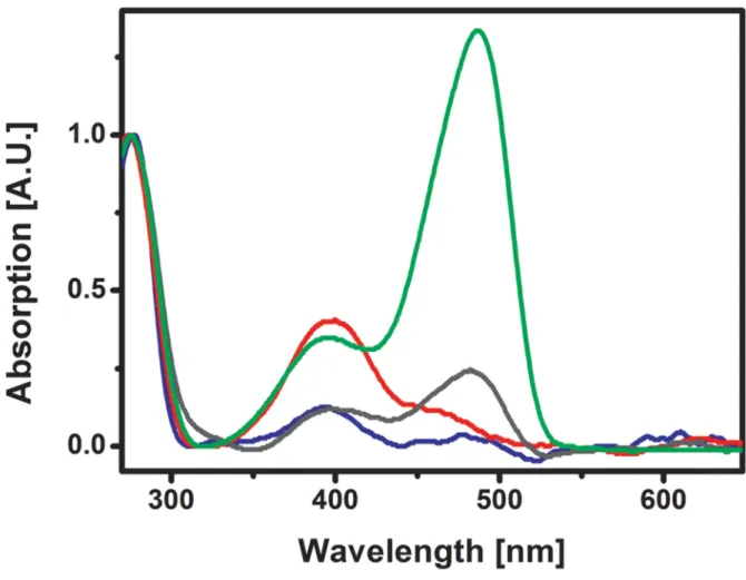
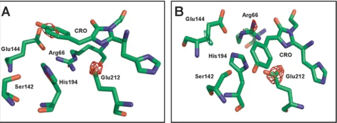
Figure 2.(A) Absorption spectra of crystalline IrisFP before (green) and after (blue) high-intensity photobleaching. Both anionic and neutral bands are largely decreased. (B) Raman spectra of crystalline IrisFP before (green) and after (blue) high-intensity photobleaching. The complete disappearance of the 1503, 1545, 1604, and 1564 cm−1 bands (inset, arrows) suggests the breakage of the chromophore methylene bridge π-conjugation (C) NCS-averaged experimental difference electron density map Fobs,bleached− Fobs,nativeupon high-intensity photobleaching, overlaid on
the crystallographic structure of intact IrisFP (PDB model 2VVH). The chromophore (orange) and the important surrounding residues (gray) in the chromophore pocket are shown. Positive electron density is shown in green (+5σ) and negative electron density is shown in red (−5 σ). (D) Refined model of high-intensity photobleached IrisFP. The chromophore phenolate moiety is represented in dim color to highlight the disorder observed in the electron difference density map. H-bonds are represented with dashed lines, and water molecules as red balls.
Journal of the American Chemical Society Article
dx.doi.org/10.1021/ja406860e| J. Am. Chem. Soc. 2013, 135, 15841−15850 15843
a nonfluorescent protonated state. Raman spectrometry of a partially fatigued crystal confirmed this finding, displaying an increase of the vibrational bands associated with the neutral chromophore (1564 cm−1, 1604 cm−1) at the expense of those
associated with the anionic chromophore (1503 cm−1, 1545 cm−1) (Figure 4B), but without signs of a rupturedπ-system. Finally, peptide analysis of a photofatigued sample by mass spectrometry revealed a substantial increase in the level of oxidation of a number of fragments containing methionine, cysteine, and tryptophan residues (Figure S9, Supporting Information). In particular, fragments that contained Met159 had∼320% higher mono-oxidation levels than those of intact IrisFP. Oxidation levels of fragments containing Cys171 were also raised significantly (see Figure S9 caption in the Supporting Information). Conversely, the decarboxylation level of Glu212 remained unaltered as compared to nonbleached IrisFP (Table S2, Supporting Information). These data suggest that, under low-intensity illumination, photofatigue of IrisFP results in sulfoxidation of Met159 and Cys171, followed by oxidation of other residues more remote from the chromophore pocket. Comforted by the mass spectrometry results, we modeled the two positive features near Met159 and Cys171 in the difference electron density map as oxygen atoms covalently bonded to the sulfur atoms of these residues, respectively (Figure 4D). A new water molecule was also modeled next to Met159, above the chromophore hydroxybenzylidene ring. The sulfoxidized Met159 was found to form a tight H-bond (2.8 Å) between the newly added oxygen atom and the presumably protonated chromophore phenol moiety. This tight H-bond is consistent with the extremely high pKa measured for the fatigued chromophore.
Interestingly, the level of Met159 and Cys171 sulfoxidation
Figure 3.Photofatiguefluorescence decay of crystalline IrisFP under low-intensity illumination by 488-nm (∼10 W/cm2, continuous) and
405-nm (∼0.01 W/cm2, on for 4 s every 12 s) laser light. Fluorescence
was recorded at 512 nm. The crystal was submitted to 460 switching cycles during 100 min of illumination, which resulted in the loss of ∼90% of its initial fluorescence emission. The inset shows an enlarged view of the decay over thefirst 10 cycles. The fluorescence envelope can befitted with a biexponential decay model (red trace).
Figure 4.(A) Absorption spectra of crystalline IrisFP before (green) and after (red) low-intensity photobleaching. The intensity of the anionic band at 488 nm decreased while that of the neutral band at 390 nm increased. (B) Raman spectra of crystalline IrisFP before (green) and after (red) partial low-intensity photobleaching. The decrease of the 1503 cm−1and 1545 cm−1bands and the increase of the 1604 cm−1and 1564 cm−1bands (inset, arrows) is consistent with a conversion from the anionic to a neutral state of the chromophore without the loss ofπ-conjugation (C) NCS-averaged electron difference density map Fobs− Fcalcupon low-intensity photobleaching, overlaid on the crystallographic structure of intact IrisFP
(PDB model 2VVH), as in Figure 2C. Positive electron density is shown in green (+5.3σ) and negative electron density is shown in red (−5.3 σ, no feature visible). (D) Refined model of low-intensity photobleaching IrisFP. H-bonds are represented with dashed lines, and water molecules as red balls.
Journal of the American Chemical Society Article
dx.doi.org/10.1021/ja406860e| J. Am. Chem. Soc. 2013, 135, 15841−15850 15844
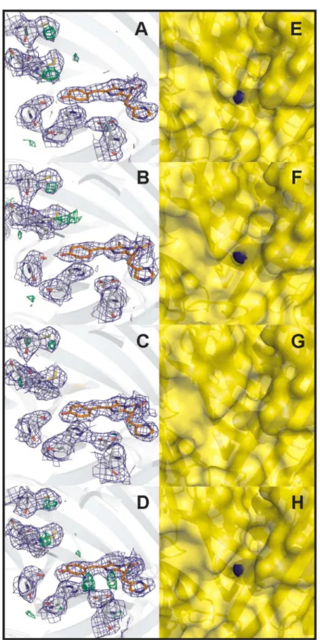
observed in the electron density maps strongly varied between the four IrisFP monomers. Whereas significant electron densities were observed in monomers A, B, and D, no sign of sulfoxidation could be detected in monomer C (Figure S6A−D, Supporting Information). This difference could be explained by a variable diffusion capacity of oxygen molecules into the chromophore pocket via pores in the IrisFPβ-barrel, probably due to crystal packing effects. In monomers A, B, and D, inspection of the IrisFP static structure showed a pore between the residues Glu140 and Ile196, in the close vicinity of the chromophore phenolate (Figure S6E−H, Supporting Informa-tion) at a location previously identified in other FPs such as His148Asp-YFP46or the Arthropoda TurboGFP.47This pore is occluded in monomer C.
Overall, the data suggest that under low-intensity illumina-tion IrisFP suffers from an oxidaillumina-tion-based photofatigue mechanism, which is initiated by the production of singlet-oxygen within the chromophore pocket.
Further Insight into IrisFP Photobleaching under High-Intensity Illumination. The novelty of the decarbox-ylation-based photofatigue mechanism of IrisFP, its complex structural signature and its potential relevance for high-resolution microscopy prompted us to investigate this mechanism in more details. Notably, we asked the four following questions: (i) Which wavelength (488 or 405 nm) is primarily responsible for photodestruction? (ii) What is the temporal order of the structural events leading to the observed photofatigued structure? (iii) What is the chemical nature of the photobleached chromophore? (iv) Is a two-consecutive-photon absorption process involved?
Question i can be readily answered in the case of low-intensity photobleaching, as the chromophore clearly adopts a cis configuration in this bleached state, strongly suggesting that photobleaching results from the absorption of a 488-nm photon. The question is more delicate to answer in the case of high-intensity photobleaching, as two arguments could favor the hypothesis that photodestruction originates from violet light illumination. First, it has been reported that decarbox-ylation reactions occurring via a Photo-Kolbe mechanism are favored in the UV range.48Second, the conformational switch of the His194-Arg66 pair observed in our structure (Figure 2C and 2D) seems atfirst glance consistent with the chromophore being photobleached in its trans configuration by violet light. However, close inspection of the difference electron density map of Figure 2C shows that residue Ile157 maintains a conformation typical of the cis chromophoric state.42 This suggests that the His194-Arg66 switch could be a consequence
of photobleaching by cyan light in the cis configuration of the chromophore. To test this hypothesis, crystals of green IrisFP were illuminated at 100 K at 488 nm for a prolonged time. At this temperature, chromophore isomerization is prevented, likely due to the lack of sufficient conformational flexibility of the IrisFP chromophore pocket, and thus no switching by cis− trans isomerization can occur. Despite a deterioration of the crystalline order resulting from this harsh sample treatment, difference electron density maps clearly showed that 488 nm light is able to induce extensive Glu212 decarboxylation, similarly to 405 nm light (Figure S4, Supporting Information). The conformational switch of the His194-Arg66 pair subsequent to Glu212 decarboxylation in the cis chromophoric state was then confirmed by molecular dynamics simulations (see below). These arguments, together with the fact that the sample was exposed to ∼600 times more cyan than violet photons, favor the hypothesis that photodestruction upon repeated switching in IrisFP predominantly results from light absorption at 488 nm by the cis chromophore.
We next attempted to disentangle the order of the structural events leading to chromophore destruction (question ii) by collecting a high-quality structure of IrisFP en-route to photobleaching. Knowing that X-rays efficiently induce IrisFP photobleaching through Glu212 decarboxylation,37 we rea-soned that collecting a pair of high-resolution crystallographic structures at 100K at different X-ray doses might uncover structural differences representative of early events along the photobleaching pathway. The results, presented in Figure S7 (Supporting Information) (see also Tables S1 and S3, Supporting Information), reveal decarboxylation of Glu212 as well as the disappearance of several water molecules participating in the hydrogen bond network surrounding the chromophore. However, instead of the disorder of the phenolate moiety observed in the photofatigued structure, the chromophore exhibits a clear distortion with an upward tilt of the chromophore phenolate and a downward bend of the methylene bridge. This distortion is also consistent with sp3 -hybridization of the Cαcarbon atom (Figure 2D). Furthermore, His194 and Arg66 are not significantly displaced as compared to their native conformation. If we admit that photobleaching pathways induced by high-intensity visible and X-ray light are both initiated by redox processes leading to Glu212 decarboxylation, it is reasonable to associate these structural changes with an early intermediate state along the high-intensity photofatigue pathway.
To further investigate whether the X-ray bleached cryo-structure is a plausible intermediate state along the
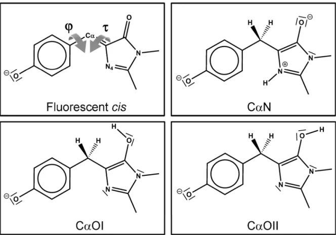
high-Figure 5. (A) Lewis representation of the proposed doubly reduced/protonated chromophore CαN photobleached under high-intensity illumination conditions. This structure was used to model the bleached pBlredand Blredstates. (B) MD simulations: time-evolution of characteristic
distances between atoms involved in the H-bond network around the IrisFP chromophore pocket, from the pBlred to the Blred states. Blue:
His194(NE2)-Glu144. Red: His194(ND1)-Arg66. Green: His194(NE2)-Ser142.
Journal of the American Chemical Society Article
dx.doi.org/10.1021/ja406860e| J. Am. Chem. Soc. 2013, 135, 15841−15850 15845
intensity photofatigue pathway, we conducted molecular dynamics simulations using software specifically tailored for fluorescent proteins simulations.49
These simulations also allowed us to examine the chemical nature of the fatigued chromophore (question iii). Starting from the putative intermediate state with the chromophore adopting various chemical structures, we analyzed whether or not the IrisFP conformation evolved toward the experimentally observed fatigued structure. In order to ensure the sp3-character of the Cα atom revealed by Raman spectroscopy while excluding an unstable radical state of the fatigued chromophore, the latter must be considered as formally reduced by two hydrogen atoms. Among all available choices tested (Figure S10, Table S4, Supporting Information), the reduced structure protonated at positions Cα and N4 (referred to as CαN) was found to be the only one compatible with our experimental data (Figure 5A). Protonation at other positions either altered the planar character of the imidazolinone ring or weakened the hydrogen bond between that ring and Arg91 (see further discussion in the Supporting Information). Using structure CαN in our MD simulations, we noticed that the conformational switch of His194 and Arg66 to their positions observed in the fatigued structure occurred within a nanosecond (Figure 5B and Figure S14, Supporting Information). Furthermore, largefluctuations of the chromophore torsions around the Cα atom were observed afterward (the 80% confidence interval of the dihedral τ angle ranged from −5° to 55°), reflecting the interplay between the intramolecular energy landscape of the reduced chromophore CαN (Figure S11, Supporting Information) and the decreased constraints due to the protein environment. These fluctuations are consistent with the phenolate disorder observed experimentally.
Finally, in order to investigate whether the high-intensity bleaching mechanism involves a two-consecutive-photon absorption process (question iv), we measured the initial photofatigue rate as a function of the illumination power density in the absence of oxygen (Figure S3, Supporting Information). The results confirm that O2 is indeed not
required in this mechanism. The best linear fit to a double-logarithmic plot of the data showed a slope of 1.8± 0.3 (Figure S3, Supporting Information), indicating a mechanism mainly involving two photons with a possible weak residual from a one-photon contribution.
■
DISCUSSIONOur study of the photofatigue mechanism of the reversibly switchable protein IrisFP reveals a two-regime photodestruc-tion pathway. At illuminaphotodestruc-tion intensities of ∼100 W/cm2, photobleaching of IrisFP involves decarboxylation of Glu212 probably via electron transfer to the chromophore in a photo-Kolbe reaction.31,48We propose that this leads to a prebleached dark state, represented by the cryo-trapped structure of Figure S7 (Supporting Information), in which the chromophore is already reduced and protonated at the Cα atom and at the N4 nitrogen atom. In this state, denoted pBlred, the chromophore has lost its electron conjugation and adopts a tilted geometry. (Note that pBlred differs from the blinked radical state DH•
described earlier by us.43) Subsequently, while the chemical structure of the chromophore does not evolve further, the hydrogen bond network surrounding it collapses, and the His194-Arg66 pairflips to a conformation resembling that of IrisFP in its switched-off state. The phenolate moiety of the chromophore loses its anchoring to the protein matrix, resulting in large fluctuations of the τ and φ dihedral angles. A complete loss of absorbance and fluorescence is thus observed in this bleached state, referred to as Blred, and the presence of oxygen is not required.
At illumination intensities ten times lower (∼10 W/cm2), a completely different scenario takes place. The chromophore reacts in its triplet state T1with molecular oxygen to produce
singlet oxygen 1O2. We propose that singlet oxygen then
rapidly reacts with the nearby Met159 and a water molecule to produce sulfoxidized-Met159 and hydrogen peroxide H2O2.50
H2O2in turn attacks Cys171 to give sulfoxidized Cys171 and a
hydroxyl molecule.50Because the S−O bond in a sulfoxidized methionine is highly polarized, with partial negative charge on
Figure 6.Proposed photophysical scheme for a two-regime photobleaching in IrisFP. Yellow arrows represent chemical steps involving electron/ proton transfer or oxidation reactions. Blue arrows represent entry/exit of oxygen species.
Journal of the American Chemical Society Article
dx.doi.org/10.1021/ja406860e| J. Am. Chem. Soc. 2013, 135, 15841−15850 15846