Mini-Review
Fetal Diagn Ther 2010;28:129–139 DOI: 10.1159/000313331
Retinoid Pathway and Congenital
Diaphragmatic Hernia: Hypothesis from the
Analysis of Chromosomal Abnormalities
Carole Goumy
a
Laetitia Gouas
a
Geoffroy Marceau
b
Karen Coste
b
Lauren Veronese
a
Denis Gallot
a
Vincent Sapin
b
Philippe Vago
a
Andrei Tchirkov
aa
University Clermont 1, UFR Médecine, Histologie Embryologie Cytogénétique, and CHU Clermont-Ferrand, Cytogénétique Médicale, and b
GReD, CNRS UMR6247, Clermont Université, INSERM U931, UFR Médecine, Clermont-Ferrand , France
Twelve retinoid-related genes have been proposed as po-tential candidates. Among them, COUP-TFII, FOG2 and GATA4 have already been well studied, especially in animal models. We propose other candidates such as STRA6, LRAT, CRBP1, CRBP2 and CRABP1 are directly implicated in retinoic acid metabolism. Conclusion: The identification of CDH-re-lated genes and pathways affecting a normal diaphragm will contribute to the understanding of the pathophysiology of this severe embryopathy and might help to facilitate prena-tal management and devise more individual treatment strat-egies. Further studies are necessary to screen large cohorts of patients with CDH for microimbalances or de novo muta-tions in these candidate genes. Moreover, functional analy-ses are needed to establish their exact role in CDH etiology.
Copyright © 2010 S. Karger AG, Basel
Background/Objectives
Congenital diaphragmatic hernia (CDH) is a severe birth defect with an estimated prevalence of 1 in 3,000 [1–4] . Posterolateral defects, named Bochdalek hernias, account for approximately 95% of CDH, with more than 80% of cases being left-sided. The defect in diaphragm development leads to the herniation of abdominal viscera
Key Words
Congenital diaphragmatic hernia ⴢ Retinoic acid ⴢ
Chromosome loci
Abstract
Background/Objectives: Although there is strong evidence implicating genetic factors in congenital diaphragmatic her-nia (CDH) pathogenesis, few causal genes have been identi-fied. Many studies suggest that early disruption of the reti-noid signaling pathway during gestation may contribute to CDH etiology. Chromosome abnormalities are detected in 10–20% of CDH cases. Chromosomal regions that are in-volved in balanced translocations or are recurrently deleted or duplicated in patients with CDH are of particular interest to researchers because they are more likely to harbor genes that cause or predispose one to the development of CDH. The aim of this review was to select chromosome loci which have been shown to be associated with CDH and to investi-gate if these loci contain candidate genes involved in the retinoic signaling pathway. Data Sources: We have re-exam-ined the known CDH-critical chromosomal loci and searched in available databases, such as the UCSC Genome Browser and OMIM, to see whether candidate genes related to the retinoid pathway were present within these loci. Results:
Received: September 4, 2009
Accepted after revision: October 15, 2009 Published online: May 26, 2010
Dr. Carole Goumy Cytogénétique Médicale © 2010 S. Karger AG, Basel
into the chest cavity during the early stages of lung devel-opment. Newborns with CDH often have severe respira-tory distress resulting from pulmonary hypoplasia. CDH occurs as an isolated birth defect (isolated CDH) or is as-sociated with additional malformations (non-isolated CDH), such as cardiovascular defects, abnormalities of the CNS and urogenital anomalies.
The development of the human diaphragm occurs be-tween the 4th and 12th week of gestation. The primor-dial diaphragm development arises from four different structures: septum transversum, pleuroperitoneal folds, dorsal mesentery and elements from the thoracic body wall [5, 6] . Several theories have been proposed to explain primary embryologic events leading to CDH, including failure of closure of the pleuroperitoneal canals, defective myoblast formation or abnormal phrenic nerve innerva-tion [7–9] . In animal models, CDH arises from a malfor-mation of the amuscular mesenchymal substratum of the pleuroperitoneal folds before pleuroperitoneal canal clo-sure [10, 11] .
Some individuals with non-isolated CDH have pat-terns of anomalies that are strongly suggestive of a spe-cific genetic syndrome ( table 1 ) [12–17] . The same rare mutation in WT1 has been reported in three cases of CDH with clinical features of Denys-Drash syndrome [18–20] . This gene encodes a zinc-finger transcription factor expressed in the septum transversum and in the pleural and abdominal mesothelial tissues that form the diaphragm. Homozygous null mouse embryos for WT1 develop diaphragmatic hernia [21] . Thus, WT1 appeared as a good candidate for CDH in humans, even though no mutation was found in a screening study of 27 children [22] . Interestingly, this gene is located on chromosome
11p13, a region recurrently deleted in individuals with CDH [23] .
Chromosomal abnormalities were identified in ap-proximately 10–20% of CDH cases, the rate being higher in cases with associated malformations [24–28] . The ex-istence of several chromosome ‘hot spots’ suggests the presence of genes that cause or predispose one to the de-velopment of CDH in these regions. Trisomy 18, more rarely trisomies 13 and 21, and structural chromosome abnormalities, such as the presence of a supernumerary derivative chromosome 22, have been described in asso-ciation with CDH. CDH is also frequently present in the Pallister-Killian syndrome associated with a tetrasomy 12p [29–31] . Other chromosomal defects involving al-most all the chromosome pairs have been described [25, 32] .
In the majority of published cases, chromosome ab-normalities were identified using R- or G-banded analy-sis and FISH. Recently, high resolution techniques such as array-based comparative genomic hybridization (aCGH) have revealed various small recurrent chromo-some abnormalities in CDH patients and allowed a more precise breakpoint characterization facilitating the iden-tification of CDH-related genes [33–38] . Several studies have suggested that 15q24–26 and 8p23.1 are critical for normal development of the diaphragm since recurrent deletions within these regions were associated with CDH [33–35, 39–42] . CDH has also been reported in several cases of monosomy 4p16pter associated with Wolf-Hirschhorn syndrome [43–45] . Other candidate regions such as 1q41–q42, 6p22–p25, or 22q11 have also been de-scribed [23, 36, 38] . Balanced reciprocal translocations were also described in CDH patients [23, 25] . These
trans-Table 1. M onogenic syndromes in which CDH commonly occurs
S yndrome OMIM Gene Chromosomal location
Simpson-Golabi-Behmel 312870 GPC3 (glypican-3) Xq26.1
Denys-Drash 194080 WT1 (Wilms’ tumor 1) 11p13
Donnai-Barrow 222448 LRP2 (low-density lipoprotein-related protein 2) 2q31.1
Spondylocostal dysostosis 277300 DLL3 (delta-like-3)* 19q13.2
Matthew-Wood 601186 STRA6 (stimulated by retinoic acid gene 6 homolog) 15q24.1
Craniofrontal dysplasia 304110 EFNB1 (ephrin B1) Xq12
Cornelia de Lange 122470 NIPBL (nipped-B-like) 5p13.1
Marfan 154700 FBN1 (fibrillin 1) 15q21.1
Ehlers-Danlos types IV and VII 130050 130060
COL3A1 (collagen type III) COL1A1 and COL1A2
2q31
17q21–q22, 7q22.1 * Most commonly mutated gene.
locations might cause CDH by disrupting or inactivating specific genes, and the characterization of breakpoints in such cases may be a valuable approach to identify candi-date genes [46] .
The development of the diaphragm strongly depends on the role of proteins associated with the metabolism and binding of retinoids [47, 48] . The nitrofen rat model has particularly highlighted the importance of retinoic acid (RA) in the diaphragm development, but this model has also provided limited insights into understanding the genetic basis of CDH [9] . Retinoids play a central role in many biological processes, particularly during
embryo-genesis and lung development [49–53] . The RA signaling pathway is complex, but recent studies in several species have increased our understanding of the role of RA as a signaling molecule during vertebrate development [54, 55] .
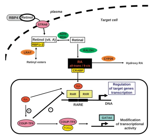
Figure 1 represents a schematic overview of the RA signaling pathway. Numerous studies have revealed the role of a retinoid signaling pathway disruption in the pathogenesis of CDH [47, 48, 56] . In rodents, the first ev-idence linking retinoids with CDH comes from the ob-servation that 25–40% of the offspring of rat dams that were fed a diet deficient in vitamin A developed CDH [57,
Retinol (vit. A) Retinal
RALDH ADH/ RDH RBP1+ 2 plasma RA
all-trans / 9-cis Hydroxy RA
DNA RARE Regulation of target genes transcription RAR RXR RA RA + COUP-TFII GATA4 Modification of transcriptional activity -FOG2 COUP-TFII CYP26 LRAT Retinyl esters Target cell STRA6 RBP4 Retinol CRABP
Fig. 1. A conceptual model describing RA synthesis and signaling
in target cells’ retinol transported in the plasma bound RBP4 (plasma retinol-binding protein). STRA6 binds RBP4, removes the retinol from RBP4 and transports it across the plasma mem-brane, where it can be metabolized. Within the target cell, retinol binds to CRBP (gene names RBP1 and RBP2) that regulates the cellular metabolism of retinol by presenting it either to alcohol or retinol dehydrogenases (ADHs/RDHs) for conversion to retinal or to LRAT (lecithin:retinol acyltransferase) that esterifies the retinol to retinyl esters. Retinal is then oxidized to RA by retinal dehydrogenase (RALDH). Cellular retinoic-acid-binding protein (CRABP) assists RA entry into the nucleus and RA exerts its
bio-logical effects through binding to the nuclear receptors RAR and RXR. Excess of RA is catabolized in the cytoplasm by the CYP26 class of P450 enzymes. In the nuclei, RA binds to RAR and RXR, which themselves heterodimerize and bind to a short DNA se-quence: the RA-response element (RARE). This binding activates the transcription of many target genes. COUP-TFII can act as a repressor of this pathway by directly sequestering RXR, thereby preventing the formation of RAR/RXR heterodimer and inhibit-ing gene transcription. COUP-TFII interacts with FOG2 that modulates the transcriptional activity of GATA4 proteins, which is a transcription factor playing an important role in early em-bryogenesis.
58] . The number of affected pups decreased when vita-min A was reintroduced into the diet in mid-gestation [59] . A proportion of RAR double mutants in mice lack-ing both ␣ - and  -subtypes exhibited a posterolateral di-aphragmatic defect which is similar to that seen in hu-mans [60] . In utero exposure to the nitrofen herbicide, which inhibits the enzymatic activity of RALDH2 – a key molecule responsible for the conversion of retinal in RA – was shown to cause CDH and primary lung defects [61] . Recently, Clugston et al. [62] have shown that a blockage of RAR signaling with the pan-RAR antagonist BMS493 induced a very high degree of CDH with a marked left-right sidedness that depended on the timing of drug delivery. In humans, preliminary evidence that retinoids may play a role in CDH development comes from a small study in which retinol and retinol-binding-protein plasma levels were found to be decreased by around 50% in newborns with CDH compared with healthy newborns [63] . Recently, the retinoid hypothesis has been reinforced by the identification of mutations in STRA6 (stimulated by RA 6), a membrane receptor for the retinol-binding protein that mediates cellular uptake of vitamin A, in a pleiotropic malformation syndrome in-cluding CDH [15] .
The aim of this review was to select chromosome loci which have been shown to be associated with CDH and to investigate if these loci contain candidate genes in-volved in the retinoic signaling pathway. This overview of chromosomal hot spots and associated candidate genes could have future diagnostic and therapeutic interests in terms of clinical management of CDH.
Data Sources
This work was performed thanks to the collaboration between a cytogenetic laboratory and a research team working on the developmental implications of the active derivatives of retinoids for mammalian species over the last few years.
We first identified chromosomal loci recurrently af-fected in the CDH context using an extensive review of the literature with the PubMed database (http://www. ncbi.nlm.nih.gov/pubmed). Both Lurie [32] and Enns et al. [25] have published useful reviews of chromosomal anomalies associated with CDH. Using these reviews as a foundation, we have compiled an updated list of the CDH-associated chromosomal anomalies. Then we searched in available databases such as UCSC Genome Browser (http://genome.ucsc.edu) and OMIM (http://
www.ncbi.nlm.nih.gov/omim) to see whether candidate genes related to the retinoid pathway were present within the selected CDH-critical chromosomal loci.
More than twenty RA metabolic pathway genes are currently known. These genes are involved in vitamin A binding and transport ( RBP4, transthyretin, STRA6, CRBP1-2, CRABP1-2 ), storage ( LRAT , diacylglycerol
ac-yltransferase), intracellular RA synthesis [ ADH3-4,
de-hydrogenase/reductase ( SDR family) members 4 and 9,
retinol dehydrogenase 4, epimerase, RALDH1-4 , aldo-keto reductase family 1, members B1 and B10 ] and RA degradation (CYP26A1, B1 and C1) . In addition, six genes encoding nuclear receptors are involved in the RA signal transduction (3 RARs and 3 RXRs ) and many oth-er genes such as GATA4, FOG2 and COUP-TFII act as regulators of this pathway. Among all these genes, we identified 12 candidates in the selected chromosome re-gions.
Results
CDH-Associated Chromosomal Hot Spots and Candidate Genes Involved in the RA Pathway or RA Regulated
Chromosome 1
1q41: DISP1 [MIM 607502]
There is emerging evidence that loss of one or more genes in the 1q41–q42 region predisposes one to CDH [36, 41, 64–67] . Combining cytogenetic results from all published CDH cases, the smallest region of overlap is approximately 1.2 Mb. The DISP1 (dispatched 1) gene could be the prime candidate gene in this region due to its interaction with Sonic hedgehog (Shh), a crucial pro-tein for the patterning of the early respiratory system in the mouse embryo [67–69] .
In the nitrofen rat model of CDH and in the hypoplas-tic lung of human fetuses with CDH, it has been shown that Shh was downregulated [70] . It has been shown that COUP-TFII, a repressor of the retinoid pathway ( fig. 1 ), is a target gene of Shh [71] . Therefore, it is tempting to speculate that a deregulation of the Shh pathway, through an alteration of DISP1, might disrupt the RA pathway by deregulating COUP-TFII and lead to CDH.
Recently, HLX [MIM 142995] has been proposed as a new candidate gene in this region because sequence vari-ants have been identified in patients with isolated CDH [72] .
Chromosome 3
3q23: CRBP1/RBP1 [MIM 180260] and CRBP2/RBP2 [MIM 180280]
Wolstenholme et al. [73] reported an association of blepharophimosis sequence and CDH in a child with a del(3)(q21q23). Dillon et al. [74] also reported CDH and del(3)(q22) in two patients. Lurie [32] suggested that 3q22 may harbor a gene that when deleted could lead to CDH. RBP1 (retinol binding protein 1) and RBP2 (retinol bind-ing protein 2) map at the distal end of the 3q23 band flanking 3q22. RBP1 and RBP2 encode cellular RBPs (CRBPs) involved in the intracellular movement of reti-nol [75] ( fig. 1 ). These genes are part of the retireti-nol signal-ing pathway and have been shown to play a role in vita-min A homeostasis and lung maturation in mice [10, 76] .
Chromosome 4
4q32.1: LRAT [MIM 604863]
CDH has been described in four individuals with 4q31 deletion and four individuals with duplication of this re-gion [23] . The LRAT gene maps to 4q32.1 flanking 4q31. The protein encoded by this gene is a microsomal enzyme that catalyzes the esterification of retinol into retinyl es-ters, an essential reaction for the retinoid homeostasis [77, 78] ( fig. 1 ).
Recently, Nakazawa et al. [79] studied the effects of nitrofen on the retinoid-signaling pathway in hypoplastic lungs. They demonstrated that LRAT was downregulat-ed, causing a shift of retinol from storage to conversion in RA. This suggests that nitrofen disturbs retinoid sig-naling at an early stage of this pathway rather than by blocking RALDH as mentioned by Mey et al. [61] . Kim et al. [80] also confirmed recently that retinyl ester forma-tion by LRAT is a key regulator of retinoid homeostasis in mouse embryogenesis and that, in contrast, the path-way of RA synthesis does not contribute significantly to the regulation of retinoid homeostasis during mamma-lian development. Interestingly, RA receptors and GATA transcription factors activate the transcription of the hu-man LRAT gene [81] .
Chromosome 6
6q23.3: ALDH8A1/RALDH4 [MIM606467]
Two reports describe a del(6)(q23) associated with CDH [32] . Lurie [32] suggested that the distal part of 6q may contain a locus whose deletion leads to CDH. Fur-thermore, Howe et al. [43] showed a balanced t(6; 8) (q24;q23) translocation in a CDH fetus. One candidate gene in this region could be RALDH4 (retinal dehydroge-nase 4) encoding a protein belonging to the aldehyde
de-hydrogenases family of proteins. This protein plays a role in the in vivo pathway of 9-cis-RA biosynthesis ( fig. 1 ). This enzyme converts 9-cis-retinal into the retinoid X receptor ligand 9-cis-RA, and has approximately 40-fold higher activity with 9-cis-retinal than with all-trans-ret-inal.
Chromosome 8
8p23.1: GATA4 [MIM 600576]
The human GATA4 gene is located on 8p23.1 where microdeletions are recurrent abnormalities in patients with CDH [32, 37, 82, 83] . Faivre et al. [42] suggested that cases with a combination of CDH and cardiac defect should be analyzed for the presence of an 8p23.1 deletion. GATA4 is a zinc-finger transcription factor expressed in mesenchymal cells of the developing diaphragm, lung and heart. The expression and activity of GATA4 are in-fluenced by retinoids [84, 85] . Jay et al. [86] described a novel mouse model of CDH based on heterozygosity of a GATA4 deletion mutation. This GATA4 +/ ⌬ ex2 mouse
de-veloped midline diaphragmatic hernia, dilated distal air-ways, thickened pulmonary mesenchyme and cardiac malformations.
Recently, somatic mutations have been detected in GATA4 and other ‘cardiac’ transcription factors in the hearts of patients who died of congenital heart disease [87] . Thus, somatic mutations that arise during cardio-genesis may be a novel molecular cause of congenital heart disease and it is conceivable that somatic mutations in GATA4 might contribute to the pathogenesis of CDH.
8q23.1: FOG2/ZFPM2 [MIM 603693]
Three patients with CDH and 8q deletions have been reported in the review by Holder et al. [23] . Temple et al. [88] described two CDH patients with a balanced trans-location involving the 8q22.3 region and CDH. Howe et al. [43] also showed a balanced t(6; 8)(q24;q23) in a patient with CDH. FOG2 (Friend of GATA2 ) is located at the proximal region of the 8q23.1 band flanking the 8q22.3 band. We also suggest that the truncation of FOG2 or a positional effect affecting the transcription regulatory of this gene could be responsible for a CDH in these three patients. FOG2 is a multi-zinc-finger transcriptional pro-tein that binds to members of the family of transcription factors as GATA4. FOG2 is expressed in mesodermal tis-sues, including pulmonary mesenchyme, mesothelium and pleuroperitoneal fold tissue [89] . It has been demon-strated that this protein can activate or downregulate ex-pression of GATA-target genes via the formation of a het-erodimer with transcription factors of the GATA family
(GATA4, GATA5 and GATA6), suggesting different modulation depending on the cell and promoter context.
Huggins et al. [90] demonstrated that FOG-2 can serve as a corepressor protein for both COUP-TFII and GATA4 proteins. Jay et al. [86] suggested that a concerted action of FOG2 and GATA4 is required to regulate mesenchy-mal cell function in the developing diaphragm and lung. In a screen of fetal mice carrying chemically induced ge-netic mutations, Ackerman et al. [91] found that a muta-tion in the gene FOG2 causes abnormal diaphragm devel-opment and pulmonary hypoplasia. Based on this result, the authors identified a de novo R112X heterozygous mu-tation in an infant who died shortly after birth with dia-phragmatic defect and severe pulmonary hypoplasia. More recently, Bleyl et al. [92] have identified two novel sequence alterations in FOG2 in two patients with iso-lated CDH, reinforcing the hypothesis that FOG2 is crit-ical for normal development of the diaphragm.
Chromosome 12
CDH is one of the most frequent abnormalities de-scribed in tetrasomy 12p cases, also known as Pallister-Killian syndrome. Likewise, Tonks et al. [28] described a t(3; 12)(q21.1;p13.3) balanced translocation associated with CDH. We identified two candidate genes related to retinoids on the chromosome 12p13.
12p13.31: RBP5/CRBPIII [MIM 611866]
RBP5 (retinol binding protein 5) is a new family mem-ber of RBPs and is predominantly expressed in the liver [93] . RBP5 binds all-trans retinol with a specific interac-tion similar to that observed in the retinol-RBP1 com-plex. RBP5 is a direct target of PPAR- ␥ , a member of the peroxisome proliferator-activated receptor (PPAR, MIM 601487) subfamily of nuclear receptors. PPARs form het-erodimers with retinoid X receptors (RXRs) and these heterodimers regulate transcription of various genes [94] . Alteration of 9-cis RA generation could modify the PPAR/RXR activation and, therefore, RBP5 expression. It is not known to date whether the RBP5 gene is ex-pressed and whether it plays a role during embryogenesis.
12p13.1: RAIG1/RAI3 [MIM 604138]
The RAIG1 (retinoic acid-inducible gene 1) gene en-codes a member of the type 3 G protein-coupling receptor family, characterized by the ‘7-transmembrane domain motif’ signature [95] . This G-protein-coupled receptor could be involved in modulating differentiation and maintaining homeostasis of epithelial cells. The compa-rable expression level in fetal lung and kidney with adult
tissues suggests a possible role in embryonic development and maturation of these organs. RAIG1 expression is in-duced by all-trans-RA via its receptors [96] . The encoded protein may be involved in the interaction between RA and G protein cellular signaling pathways.
Chromosome 15
15q24.1: STRA6 [MIM 610745]
Sharp et al. [97] described a patient with a de novo 15q24 microdeletion associated with diaphragmatic her-nia. Aviram-Goldring et al. [98] studied a family in which two fetuses had CDH associated with an apparently bal-anced t(5; 15)(p15.3;q24), also present in the mother and in a normal child, suggesting that the CDH in these fe-tuses may have been caused by a cryptic imbalance at one of the breakpoints during meiosis. More recently, Van Esch et al. [99] described a 15q24 microdeletion of 3.1 Mb including the STRA6 gene in a patient with severe mental retardation, facial dysmorphisms and CDH. STRA6 en-codes a specific receptor for RBP4-retinol located on cell membranes in the target tissues ( fig. 1 ). It removes the retinol from RBP4 and mediates retinol uptake by cells [100–102] . The transcription of STRA6 is directly regu-lated by RA levels. During embryogenesis, STRA6 is ex-pressed in respiratory mesenchyme and in respiratory/ bronchial epithelium [103] . Consistent with various roles of vitamin A and the wide tissue expression pattern of STRA6 , mutations in STRA6 are associated with severe pathological phenotypes in humans. CDH is an impor-tant component of the phenotype observed in cases of STRA6 mutations [15, 104] .
15q24: CRABP1 [MIM 180230]
CRABP1 (cellular retinoic acid binding protein 1) be-longs to a superfamily of lipid-binding proteins that are thought to act by maintaining tolerable concentrations of intracellular RA as modulators of RA catabolism and as intracellular transporters for RA from the cytoplasm to nuclear receptors [105–107] . CRAPB1 is supposed to play an important role in RA-mediated differentiation and proliferation processes.
15q26.1–q26.2: COUP-TFII/NR2F2 [MIM 107773] A minimally recurrently deleted region has been iden-tified using FISH and aCGH on chromosome 15q26.1– q26.2 in patients with non-isolated CDH [32–34, 38, 40, 108] .
COUP-TFII encodes a transcriptional factor from the steroid/thyroid hormone receptor superfamily. This gene is a nuclear orphan receptor expressed during embryonic
development in a variety of tissues, including mesoder-mal derivatives in the diaphragm, lung and heart [109] . Homozygous tissue-specific deletion of COUP-TFII in mice causes posterolateral CDH similar to the Boch-dalek-type CDH seen in humans [110] .
COUP-TFII appears to be a good candidate in the 15q26 region because (i) its expression is regulated by ret-inoids and (ii) COUP-TFII regulates gene transcription by influencing RAR/RXR heterodimerization [111, 112] . COUP-TFII is able to sequester RXR in a functionally in-active complex and to reduce the available nuclear con-centrations of RXR. Thus, COUP-TFII can act as a re-pressor of the retinoid pathway by preventing RAR/RXR heterodimer formation and inhibiting target gene tran-scription [112] . In the nitrofen rat model, the repression of retinoid signaling pathway by upregulation of COUP-TFII may cause hypoplastic lung [18] . This process may be a negative feedback system that precisely balances the transcription of relevant genes during diaphragm devel-opment. COUP-TFII has been shown to interact physi-cally with FOG2, implying that these two factors may co-operate during diaphragm morphogenesis [90] ( fig. 1 ).
Recently, Clugston et al. [113] reinforced this hypoth-esis showing that 15q26 contains a cluster of genes, in-cluding COUP-TFII, which are expressed in the develop-ing rodent diaphragm.
Taken together, these data suggest that COUP-TFII is likely to play a key role in diaphragm development, even though no mutations were found in 73 CDH samples ed by Scott et al. [38] and in more than 100 samples test-ed by Slavotinek et al. [41] .
Chromosome X
Xp22.3: TBL1X [MIM 300196]
The Xp22.3 region is frequently affected in CDH [23] . This region carries the TBL1X (transducin  -like 1X) gene which encodes a protein that plays an essential role in transcriptional activation mediated by nuclear recep-tors [114] . TBL1X is found as a subunit in corepressor SMRT (silencing mediator for retinoid and thyroid recep-tors) complex along with histone deacetylase 3 protein, which is known to modulate the nuclear retinoid signal-ing pathway [115] .
Discussion
A major challenge of CDH research is to characterize genes and signaling pathways that are critical for early mesenchymal cell function during morphogenesis of the
diaphragm. Although several genes have been clearly shown to underlie abnormal diaphragm development in mice, few CDH-related mutations have been identified in the corresponding genes in humans. In this review, we focused on genes involved in retinoid metabolism or reg-ulated by retinoids, which are located within chromo-somal regions recurrently affected in CDH patients.
The analysis of chromosomal aberrations may help in the mapping of disease loci and isolation of disease genes by positional cloning strategy [46, 116] . The principal pit-fall in this chromosomal approach is that the localization of breakpoints may not be accurate since it is generally based on standard karyotyping. Recent aCGH technolo-gy provides a more precise characterization of chromo-somal abnormalities, which helps to define the minimal-ly affected region in patients with CDH and identify can-didate genes within this region [38, 40, 41] . With this, de novo microdeletions in the regions 1q41–q42, 4p16.3, 8p23.1 and 15q26.1–q26.2 have been reported. These de-leted chromosomal regions may be assumed to contain genes necessary for normal diaphragm development and these genes can subsequently be selected for sequencing in CDH patients. Among the retinoid-related genes in-cluded in these regions, COUP-TFII and FOG2 were se-quenced in CDH patients. To date, no mutation could be identified in COUP-TFII [38, 41] . A de novo mutation and sequence alterations in FOG2 were found in three pa-tients, reinforcing the hypothesis that FOG2 is critical in diaphragmatic and lung development in humans [91, 92] .
The STRA6 gene located at chromosome 15q24.1 is also a promising candidate since CDH is an important component of the polymalformative syndrome observed in cases with STRA6 mutations. Recently, Isken et al. [117] have shown that STRA6 is essential in maintaining em-bryonic RA homeostasis and that STRA6-dependent transfer of retinol from RBP4 depends on LRAT. LRAT activity is required, like those of STRA6 and RBP4 pro-tein, for uptake of appropriate amounts of retinol into cells. As a result, LRAT is also likely to play a role in the development of CDH in individuals with 4q32.1–q31 re-arrangements.
Other CDH-associated chromosomal hot spots such as 2q37, 6p25 and 22q11 do not contain genes related to the retinoid pathway. These regions might carry genes involved in pathways regulating differentiation of mesen-chymal cells or cell migration, which are important for diaphragm development. For example, the COL6A3 gene [MIM 120250] is located on chromosome 2q37, a region frequently deleted in CDH. Impaired formation of the extracellular matrix, caused by disruptions in either
col-lagen or elastic fibers, can lead to developmental defects in a wide range of organs including the diaphragm. Of note, CDH has been linked to several subtypes of Ehlers-Danlos syndrome caused by mutations in genes belong-ing to the collagen family ( table 1 ), which is expressed during embryonic development in several organs [17] . In the same way, the 6p25 region repeatedly deleted in CDH patients contains the FOXF2 gene (forkhead box F2), one of the human homologues of the Drosophila
melanogas-ter transcription factor forkhead, which could be a good
candidate [23, 38] . FOXF2 is expressed in lung and pla-centa and was shown to activate transcription of several lung-specific genes [118, 119] .
Systematic screening for mutations in CDH patients has been reported only for WT1 , COUP-TFII and FOG2 .
The same approach could be used to search for mutations in related genes from the RA metabolic and molecular signaling pathway. In addition, a simultaneous analysis of several loci by quantitative multiplex PCR of short flu-orescent fragments in a cohort of CDH patients may be used to estimate the frequency of microdeletions or mi-croduplications of candidate genes, which may help to establish their role in CDH etiology. The identification of CDH-related genes and pathways affecting normal dia-phragm and lung development will contribute to the un-derstanding of the pathophysiology of this severe embry-opathy. Given the substantial mortality and morbidity associated with this developmental abnormality, advanc-es in this area are critical.
References
1 Butler N, Claireaux AE: Congenital dia-phragmatic hernia as a cause of perinatal mortality. Lancet 1962; 1: 659–663.
2 David TJ, Illingworth CA: Diaphragmatic hernia in the south-west of England. J Med Genet 1976; 13: 253–262.
3 Philip N, Gambarelli D, Guys JM, Cam-boulives J, Ayme S: Epidemiological study of congenital diaphragmatic defects with spe-cial reference to aetiology. Eur J Pediatr 1991; 150: 726–729.
4 Torfs CP, Curry CJ, Bateson TF, Honore LH: A population-based study of congenital dia-phragmatic hernia. Teratology 1992; 46: 555– 565.
5 Greer JJ, Cote D, Allan DW, Zhang W, Babi-uk RP, Ly L, Lemke RP, Bagnall K: Structure of the primordial diaphragm and defects as-sociated with nitrofen-induced CDH. J Appl Physiol 2000; 89: 2123–2129.
6 Rottier R, Tibboel D: Fetal lung and phragm development in congenital dia-phragmatic hernia. Semin Perinatol 2005; 29: 86–93.
7 Iritani I: Experimental study on embryogen-esis of congenital diaphragmatic hernia. Anat Embryol (Berl) 1984; 169: 133–139. 8 Thebaud B, Tibboel D, Rambaud C, Mercier
JC, Bourbon JR, Dinh-Xuan AT, Archer SL: Vitamin A decreases the incidence and se-verity of nitrofen-induced congenital dia-phragmatic hernia in rats. Am J Physiol 1999; 277:L423–L429.
9 Noble BR, Babiuk RP, Clugston RD, Under-hill TM, Sun H, Kawaguchi R, Walfish PG, Blomhoff R, Gundersen TE, Greer JJ: Mech-anisms of action of the congenital diaphrag-matic hernia-inducing teratogen nitrofen. Am J Physiol Lung Cell Mol Physiol 2007; 293:L1079–L1087.
10 Babiuk RP, Greer JJ: Diaphragm defects oc-cur in a CDH hernia model independently of myogenesis and lung formation. Am J Physi-ol Lung Cell MPhysi-ol PhysiPhysi-ol 2002; 283:L1310– L1314.
11 Babiuk RP, Zhang W, Clugston R, Allan DW, Greer JJ: Embryological origins and develop-ment of the rat diaphragm. J Comp Neurol 2003; 455: 477–487.
12 Slavotinek AM: The genetics of
congeni-tal diaphragmatic hernia. Semin Perinatol 2005; 29: 77–85.
13 Slavotinek AM: Single gene disorders associ-ated with congenital diaphragmatic hernia. Am J Med Genet C Semin Med Genet 2007; 145C:172–183.
14 Kantarci S, Al-Gazali L, Hill RS, Donnai D, Black GC, Bieth E, Chassaing N, Lacombe D, Devriendt K, Teebi A, Loscertales M, Robson C, Liu T, MacLaughlin DT, Noonan KM, Russell MK, Walsh CA, Donahoe PK, Pober BR: Mutations in lRP2, which encodes the multiligand receptor megalin, cause Don-nai-Barrow and facio-oculo-acoustico-renal syndromes. Nat Genet 2007; 39: 957–959. 15 Pasutto F, Sticht H, Hammersen G,
Gilles-sen-Kaesbach G, Fitzpatrick DR, Nurnberg G, Brasch F, Schirmer-Zimmermann H, Tol-mie JL, Chitayat D, Houge G, Fernandez-Martinez L, Keating S, Mortier G, Henne-kam RC, von der Wense A, Slavotinek A, Meinecke P, Bitoun P, Becker C, Nurnberg P, Reis A, Rauch A: Mutations in STRA6 cause a broad spectrum of malformations includ-ing anophthalmia, congenital heart defects, diaphragmatic hernia, alveolar capillary dysplasia, lung hypoplasia, and mental retar-dation. Am J Hum Genet 2007; 80: 550–560.
16 Iglesias JL, Renard T: Diaphragmatic hernia in an 8-year-old with Ehlers-Danlos syn-drome. Pediatr Surg Int 1998; 13: 553–555. 17 Lin IC, Ko SF, Shieh CS, Huang CF, Chien SJ,
Liang CD: Recurrent congenital diaphrag-matic hernia in Ehlers-Danlos syndrome. Cardiovasc Intervent Radiol 2006; 29: 920– 923.
18 Doi T, Sugimoto K, Puri P: Up-regulation of COUP-TFII gene expression in the nitrofen-induced hypoplastic lung. J Pediatr Surg 2009; 44: 321–324.
19 Cho HY, Lee BS, Kang CH, Kim WH, Ha IS, Cheong HI, Choi Y: Hydrothorax in a patient with Denys-Drash syndrome associated with a diaphragmatic defect. Pediatr Nephrol 2006; 21: 1909–1912.
20 Antonius T, van Bon B, Eggink A, van der Burgt I, Noordam K, van Heijst A: Denys-Drash syndrome and congenital diaphrag-matic hernia: another case with the 1097G 1 A(Arg366His) mutation. Am J Med Genet A 2008; 146A:496–499.
21 Kreidberg JA, Sariola H, Loring JM, Maeda M, Pelletier J, Housman D, Jaenisch R: WT-1 is required for early kidney development. Cell 1993; 74: 679–691.
22 Nordenskjold A, Tapper-Persson M, Anvret M: No evidence of WT1 gene mutations in children with congenital diaphragmatic her-nia. J Pediatr Surg 1996; 31: 925–927. 23 Holder AM, Klaassens M, Tibboel D, de
Klein A, Lee B, Scott DA: Genetic factors in congenital diaphragmatic hernia. Am J Hum Genet 2007; 80: 825–845.
24 Bollmann R, Kalache K, Mau H, Chaoui R, Tennstedt C: Associated malformations and chromosomal defects in congenital dia-phragmatic hernia. Fetal Diagn Ther 1995; 10: 52–59.
25 Enns GM, Cox VA, Goldstein RB, Gibbs DL, Harrison MR, Golabi M: Congenital dia-phragmatic defects and associated syn-dromes, malformations, and chromosome anomalies: a retrospective study of 60 pa-tients and literature review. Am J Med Genet 1998; 79: 215–225.
26 Geary MP, Chitty LS, Morrison JJ, Wright V, Pierro A, Rodeck CH: Perinatal outcome and prognostic factors in prenatally diagnosed congenital diaphragmatic hernia. Ultra-sound Obstet Gynecol 1998; 12: 107–111. 27 Witters I, Legius E, Moerman P, Deprest J,
Van Schoubroeck D, Timmerman D, Van Assche FA, Fryns JP: Associated malforma-tions and chromosomal anomalies in 42 cas-es of prenatally diagnosed diaphragmatic hernia. Am J Med Genet 2001; 103: 278–282. 28 Tonks A, Wyldes M, Somerset DA, Dent K,
Abhyankar A, Bagchi I, Lander A, Roberts E, Kilby MD: Congenital malformations of the diaphragm: findings of the West Midlands Congenital Anomaly Register 1995 to 2000. Prenat Diagn 2004; 24: 596–604.
29 Bergoffen J, Punnett H, Campbell TJ, Ross AJ 3rd, Ruchelli E, Zackai EH: Diaphragmat-ic hernia in tetrasomy 12p mosaDiaphragmat-icism. J Pe-diatr 1993; 122: 603–606.
30 Rodriguez JI, Garcia I, Alvarez J, Delicado A, Palacios J: Lethal Pallister-Killian syn-drome: phenotypic similarity with Fryns syndrome. Am J Med Genet 1994; 53: 176– 181.
31 Takakuwa K, Hataya I, Arakawa M, Tamura M, Sekizuka N, Tanaka K: A case of mosaic tetrasomy 12p (Pallister-Killian syndrome) diagnosed prenatally: comparison of chro-mosome analyses of various cells obtained from the patient. Am J Perinatol 1997; 14: 641–643.
32 Lurie IW: Where to look for the genes related to diaphragmatic hernia? Genet Couns 2003; 14: 75–93.
33 Biggio JR Jr, Descartes MD, Carroll AJ, Holt RL: Congenital diaphragmatic hernia: Is 15q26.1–26.2 a candidate locus? Am J Med Genet A 2004; 126A:183–185.
34 Slavotinek A, Lee SS, Davis R, Shrit A, Leppig KA, Rhim J, Jasnosz K, Albertson D, Pinkel D: Fryns syndrome phenotype caused by chromosome microdeletions at 15q26.2 and 8p23.1. J Med Genet 2005; 42: 730–736. 35 Lopez I, Bafalliu JA, Bernabe MC, Garcia F,
Costa M, Guillen-Navarro E: Prenatal diag-nosis of de novo deletions of 8p23.1 or 15q26.1 in two fetuses with diaphragmatic hernia and congenital heart defects. Prenat Diagn 2006; 26: 577–580.
36 Kantarci S, Casavant D, Prada C, Russell M, Byrne J, Haug LW, Jennings R, Manning S, Blaise F, Boyd TK, Fryns JP, Holmes LB, Do-nahoe PK, Lee C, Kimonis V, Pober BR: Findings from aCGH in patients with con-genital diaphragmatic hernia (CDH): a pos-sible locus for Fryns syndrome. Am J Med Genet A 2006; 140: 17–23.
37 Shimokawa O, Miyake N, Yoshimura T, So-sonkina N, Harada N, Mizuguchi T, Kondoh S, Kishino T, Ohta T, Remco V, Takashima T, Kinoshita A, Yoshiura K, Niikawa N, Matsu-moto N: Molecular characterization of del(8) (p23.1p23.1) in a case of congenital diaphrag-matic hernia. Am J Med Genet A 2005; 136: 49–51.
38 Scott DA, Klaassens M, Holder AM, Lally KP, Fernandes CJ, Galjaard RJ, Tibboel D, de Klein A, Lee B: Genome-wide oligonucle-otide-based array comparative genome hy-bridization analysis of non-isolated congeni-tal diaphragmatic hernia. Hum Mol Genet 2007; 16: 424–430.
39 Schlembach D, Zenker M, Trautmann U, Ul-mer R, Beinder E: Deletion 15q24–26 in pre-natally detected diaphragmatic hernia: in-creasing evidence of a candidate region for diaphragmatic development. Prenat Diagn 2001; 21: 289–292.
40 Klaassens M, van Dooren M, Eussen HJ, Douben H, den Dekker AT, Lee C, Donahoe PK, Galjaard RJ, Goemaere N, de Krijger RR, Wouters C, Wauters J, Oostra BA, Tibboel D, de Klein A: Congenital diaphragmatic her-nia and chromosome 15q26: determination of a candidate region by use of fluorescent in situ hybridization and array-based compara-tive genomic hybridization. Am J Hum Ge-net 2005; 76: 877–882.
41 Slavotinek AM, Moshrefi A, Davis R, Leeth E, Schaeffer GB, Burchard GE, Shaw GM, James B, Ptacek L, Pennacchio LA: Array comparative genomic hybridization in pa-tients with congenital diaphragmatic hernia: mapping of four CDH-critical regions and sequencing of candidate genes at 15q26.1– 15q26.2. Eur J Hum Genet 2006; 14: 999– 1008.
42 Faivre L, Morichon-Delvallez N, Viot G, Narcy F, Loison S, Mandelbrot L, Aubry MC, Raclin V, Edery P, Munnich A, Vekemans M: Prenatal diagnosis of an 8p23.1 deletion in a fetus with a diaphragmatic hernia and re-view of the literature. Prenat Diagn 1998; 18: 1055–1060.
43 Howe DT, Kilby MD, Sirry H, Barker GM, Roberts E, Davison EV, McHugo J, Whittle MJ: Structural chromosome anomalies in congenital diaphragmatic hernia. Prenat Di-agn 1996; 16: 1003–1009.
44 Tachdjian G, Fondacci C, Tapia S, Huten Y, Blot P, Nessmann C: The Wolf-Hirschhorn syndrome in fetuses. Clin Genet 1992; 42: 281–287.
45 van Dooren MF, Brooks AS, Hoogeboom AJ, van den Hoonaard TL, de Klein JE, Wouters CH, Tibboel D: Early diagnosis of Wolf-Hirschhorn syndrome triggered by a life-threatening event: congenital diaphragmatic hernia. Am J Med Genet A 2004; 127A:194– 196.
46 Bache I, Hjorth M, Bugge M, Holstebroe S, Hilden J, Schmidt L, Brondum-Nielsen K, Bruun-Petersen G, Jensen PK, Lundsteen C, Niebuhr E, Rasmussen K, Tommerup N: Sys-tematic re-examination of carriers of bal-anced reciprocal translocations: a strategy to search for candidate regions for common and complex diseases. Eur J Hum Genet 2006; 14: 410–417.
47 Greer JJ, Babiuk RP, Thebaud B: Etiology of congenital diaphragmatic hernia: the reti-noid hypothesis. Pediatr Res 2003; 53: 726– 730.
48 Gallot D, Marceau G, Coste K, Hadden H, Robert-Gnansia E, Laurichesse H, Deche-lotte PJ, Labbe A, Dastugue B, Lemery D, Sapin V: Congenital diaphragmatic hernia: a retinoid-signaling pathway disruption dur-ing lung development? Birth Defects Res A Clin Mol Teratol 2005; 73: 523–531.
49 Morriss-Kay GM, Sokolova N: Embryonic development and pattern formation. FASEB J 1996; 10: 961–968.
50 Massaro GD, Massaro D: Retinoic acid treat-ment abrogates elastase-induced pulmonary emphysema in rats. Nat Med 1997; 3: 675– 677.
51 Chytil F: Retinoids in lung development. FASEB J 1996; 10: 986–992.
52 Kumar VH, Lakshminrusimha S, El Abiad MT, Chess PR, Ryan RM: Growth factors in lung development. Adv Clin Chem 2005; 40: 261–316.
53 Marceau G, Gallot D, Lemery D, Sapin V: Metabolism of retinol during mammalian placental and embryonic development. Vi-tam Horm 2007; 75: 97–115.
54 Duester G: Retinoic acid synthesis and sig-naling during early organogenesis. Cell 2008; 134: 921–931.
55 Niederreither K, Dolle P: Retinoic acid in de-velopment: towards an integrated view. Nat Rev Genet 2008; 9: 541–553.
56 Montedonico S, Nakazawa N, Puri P: Con-genital diaphragmatic hernia and retinoids: searching for an etiology. Pediatr Surg Int 2008; 24: 755–761.
57 Anderson EG: Translocations in maize in-volving the short arm of chromosome I. Ge-netics 1941; 26: 452–459.
58 Anderson DP: The cancer problem from the standpoint of the practicing physician in the small community. Minn Med 1949; 32: 65– 69.
59 Wilson JG, Roth CB, Warkany J: An analysis of the syndrome of malformations induced by maternal vitamin A deficiency. Effects of restoration of vitamin A at various times during gestation. Am J Anat 1953; 92: 189– 217.
60 Mendelsohn C, Lohnes D, Decimo D, Lufkin T, LeMeur M, Chambon P, Mark M: Func-tion of the retinoic acid receptors (RARs) during development (II). Multiple abnor-malities at various stages of organogenesis in RAR double mutants. Development 1994; 120: 2749–2771.
61 Mey J, Babiuk RP, Clugston R, Zhang W, Greer JJ: Retinal dehydrogenase-2 is inhib-ited by compounds that induce congenital diaphragmatic hernias in rodents. Am J Pathol 2003; 162: 673–679.
62 Clugston RD, Zhang W, Alvarez S, De Lera AR, Greer JJ: Understanding abnormal reti-noid signaling as a causative mechanism in congenital diaphragmatic hernia. Am J Respir Cell Mol Biol 2010; 42: 276–285. 63 Major D, Cadenas M, Fournier L, Leclerc S,
Lefebvre M, Cloutier R: Retinol status of newborn infants with congenital diaphrag-matic hernia. Pediatr Surg Int 1998; 13: 547– 549.
64 Rogers JC, Rogers SW: Comparison of the ef-fects of N6-methyldeoxyadenosine and N5-methyldeoxycytosine on transcription from nuclear gene promoters in barley. Plant J 1995; 7: 221–233.
65 Smith SA, Martin KE, Dodd KL, Young ID: Severe microphthalmia, diaphragmatic her-nia and Fallot’s tetralogy associated with a chromosome 1; 15 translocation. Clin Dys-morphol 1994; 3: 287–291.
66 Youssoufian H, Chance P, Tuck-Muller CM, Jabs EW: Association of a new chromosomal deletion [del(1)(q32q42)] with diaphragmat-ic hernia: assignment of a human ferritin gene. Hum Genet 1988; 78: 267–270.
67 Shaffer LG, Theisen A, Bejjani BA, Ballif BC, Aylsworth AS, Lim C, McDonald M, Ellison JW, Kostiner D, Saitta S, Shaikh T: The dis-covery of microdeletion syndromes in the post-genomic era: review of the methodolo-gy and characterization of a new 1q41q42 microdeletion syndrome. Genet Med 2007; 9: 607–616.
68 Kawakami T, Kawcak T, Li YJ, Zhang W, Hu Y, Chuang PT: Mouse dispatched mutants fail to distribute hedgehog proteins and are defective in hedgehog signaling. Develop-ment 2002; 129: 5753–5765.
69 Tian H, Tenzen T, McMahon AP: Dose de-pendency of Disp1 and genetic interaction between Disp1 and other hedgehog signaling components in the mouse. Development 2004; 131: 4021–4033.
70 Unger S, Copland I, Tibboel D, Post M: Down-regulation of sonic hedgehog expres-sion in pulmonary hypoplasia is associated with congenital diaphragmatic hernia. Am J Pathol 2003; 162: 547–555.
71 Krishnan V, Pereira FA, Qiu Y, Chen CH, Beachy PA, Tsai SY, Tsai MJ: Mediation of Sonic hedgehog-induced expression of COUP-TFII by a protein phosphatase. Sci-ence 1997; 278: 1947–1950.
72 Slavotinek AM, Moshrefi A, Lopez Jiminez N, Chao R, Mendell A, Shaw GM, Pennac-chio LA, Bates MD: Sequence variants in the HLX gene at chromosome 1q41–1q42 in pa-tients with diaphragmatic hernia. Clin Gen-et 2009; 75: 429–439.
73 Wolstenholme J, Brown J, Masters KG, Wright C, English CJ: Blepharophimosis se-quence and diaphragmatic hernia associated with interstitial deletion of chromosome 3 (46,xy,del(3)(q21q23)). J Med Genet 1994; 31: 647–648.
74 Dillon E, Renwick M, Wright C: Congenital diaphragmatic herniation: antenatal detec-tion and outcome. Br J Radiol 2000; 73: 360– 365.
75 Napoli JL: Interactions of retinoid binding proteins and enzymes in retinoid metabo-lism. Biochim Biophys Acta 1999; 1440: 139– 162.
76 Ghyselinck NB, Bavik C, Sapin V, Mark M, Bonnier D, Hindelang C, Dierich A, Nilsson CB, Hakansson H, Sauvant P, Azais-Braesco V, Frasson M, Picaud S, Chambon P: Cellular retinol-binding protein 1 is essential for vi-tamin A homeostasis. EMBO J 1999; 18: 4903–4914.
77 Liu L, Gudas LJ: Disruption of the lecithin: retinol acyltransferase gene makes mice more susceptible to vitamin A deficiency. J Biol Chem 2005; 280: 40226–40234. 78 O’Byrne SM, Wongsiriroj N, Libien J, Vogel
S, Goldberg IJ, Baehr W, Palczewski K, Bla-ner WS: Retinoid absorption and storage is impaired in mice lacking lecithin: retinol ac-yltransferase (LRAT). J Biol Chem 2005; 280: 35647–35657.
79 Nakazawa N, Takayasu H, Montedonico S, Puri P: Altered regulation of retinoic acid synthesis in nitrofen-induced hypoplastic lung. Pediatr Surg Int 2007; 23: 391–396. 80 Kim YK, Wassef L, Hamberger L, Piantedosi
R, Palczewski K, Blaner WS, Quadro L: Reti-nyl ester formation by lecithin: retinol acyl-transferase is a key regulator of retinoid ho-meostasis in mouse embryogenesis. J Biol Chem 2008; 283: 5611–5621.
81 Cai K, Gudas LJ: Retinoic acid receptors and GATA transcription factors activate the transcription of the human lecithin:retinol acyltransferase gene. Int J Biochem Cell Biol 2009; 41: 546–553.
82 Barber JC, Maloney V, Hollox EJ, Stuke- Sontheimer A, du Bois G, Daumiller E, Klein-Vogler U, Dufke A, Armour JA, Liehr T: Duplications and copy number variants of 8p23.1 are cytogenetically indistinguishable but distinct at the molecular level. Eur J Hum Genet 2005; 13: 1131–1136.
83 Pecile V, Petroni MG, Fertz MC, Filippi G: Deficiency of distal 8p – report of two cases and review of the literature. Clin Genet 1990; 37: 271–278.
84 Kostetskii I, Jiang Y, Kostetskaia E, Yuan S, Evans T, Zile M: Retinoid signaling required for normal heart development regulates GATA-4 in a pathway distinct from cardio-myocyte differentiation. Dev Biol 1999; 206: 206–218.
85 Clabby ML, Robison TA, Quigley HF, Wil-son DB, Kelly DP: Retinoid X receptor alpha represses GATA-4-mediated transcription via a retinoid-dependent interaction with the cardiac-enriched repressor FOG-2. J Biol Chem 2003; 278: 5760–5767.
86 Jay PY, Bielinska M, Erlich JM, Mannisto S, Pu WT, Heikinheimo M, Wilson DB: Im-paired mesenchymal cell function in GATA4 mutant mice leads to diaphragmatic hernias and primary lung defects. Dev Biol 2007; 301: 602–614.
87 Reamon-Buettner SM, Spanel-Borowski K, Borlak J: Bridging the gap between anatomy and molecular genetics for an improved un-derstanding of congenital heart disease. Ann Anat 2006; 188: 213–220.
88 Temple IK, Barber JC, James RS, Burge D: Diaphragmatic herniae and translocations involving 8q22 in two patients. J Med Genet 1994; 31: 735–737.
89 Ackerman KG, Wang J, Luo L, Fujiwara Y, Orkin SH, Beier DR: GATA4 is necessary for normal pulmonary lobar development. Am J Respir Cell Mol Biol 2007; 36: 391–397. 90 Huggins GS, Bacani CJ, Boltax J, Aikawa R,
Leiden JM: Friend of GATA 2 physically in-teracts with chicken ovalbumin upstream promoter-TF2 (COUP-TF2) and COUP-TF3 and represses COUP-TF2-dependent activa-tion of the atrial natriuretic factor promoter. J Biol Chem 2001; 276: 28029–28036. 91 Ackerman KG, Herron BJ, Vargas SO, Huang
H, Tevosian SG, Kochilas L, Rao C, Pober BR, Babiuk RP, Epstein JA, Greer JJ, Beier DR: Fog2 is required for normal diaphragm and lung development in mice and humans. PLoS Genet 2005; 1: 58–65.
92 Bleyl SB, Moshrefi A, Shaw GM, Saijoh Y, Schoenwolf GC, Pennacchio LA, Slavotinek AM: Candidate genes for congenital dia-phragmatic hernia from animal models: Se-quencing of FOG2 and PDGFRalpha reveals rare variants in diaphragmatic hernia pa-tients. Eur J Hum Genet 2007; 15: 950–958. 93 Folli C, Calderone V, Ottonello S, Bolchi A,
Zanotti G, Stoppini M, Berni R: Identifica-tion, retinoid binding, and x-ray analysis of a human retinol-binding protein. Proc Natl Acad Sci USA 2001; 98: 3710–3715.
94 Zizola CF, Schwartz GJ, Vogel S: Cellular ret-inol-binding protein type III is a PPARga-mma target gene and plays a role in lipid me-tabolism. Am J Physiol Endocrinol Metab 2008; 295:E1358–E1368.
95 Cheng Y, Lotan R: Molecular cloning and characterization of a novel retinoic acid-in-ducible gene that encodes a putative G pro-tein-coupled receptor. J Biol Chem 1998; 273: 35008–35015.
96 Tao Q, Cheng Y, Clifford J, Lotan R: Charac-terization of the murine orphanG-protein-coupled receptor gene Rai3 and its regula-tion by retinoic acid. Genomics 2004; 83: 270–280.
97 Sharp AJ, Selzer RR, Veltman JA, Gimelli S, Gimelli G, Striano P, Coppola A, Regan R, Price SM, Knoers NV, Eis PS, Brunner HG, Hennekam RC, Knight SJ, de Vries BB, Zuf-fardi O, Eichler EE: Characterization of a recurrent 15q24 microdeletion syndrome. Hum Mol Genet 2007; 16: 567–572. 98 Aviram-Goldring A, Daniely M, Frydman
M, Shneyour Y, Cohen H, Barkai G: Con-genital diaphragmatic hernia in a family seg regating a reciprocal translocation t(5; 15)(p15.3;q24). Am J Med Genet 2000; 90: 120–122.
99 Van Esch H, Backx L, Pijkels E, Fryns JP: Congenital diaphragmatic hernia is part of the new 15q24 microdeletion syndrome. Eur J Med Genet 2009; 52: 153–156. 100 Blaner WS: STRA6, a cell-surface receptor
for retinol-binding protein: the plot thick-ens. Cell Metab 2007; 5: 164–166.
101 Kawaguchi R, Yu J, Honda J, Hu J, Whiteleg-ge J, Ping P, Wiita P, Bok D, Sun H: A mem-brane receptor for retinol binding protein mediates cellular uptake of vitamin A. Sci-ence 2007; 315: 820–825.
102 Kawaguchi R, Yu J, Wiita P, Ter-Stepanian M, Sun H: Mapping the membrane topolo-gy and extracellular ligand binding do-mains of the retinol binding protein recep-tor. Biochemistry 2008; 47: 5387–5395. 103 Bouillet P, Sapin V, Chazaud C, Messaddeq
N, Decimo D, Dolle P, Chambon P: Devel-opmental expression pattern of STRA6, a retinoic acid-responsive gene encoding a new type of membrane protein. Mech Dev 1997; 63: 173–186.
104 Golzio C, Martinovic-Bouriel J, Thomas S, Mougou-Zrelli S, Grattagliano-Bessieres B, Bonniere M, Delahaye S, Munnich A, En-cha-Razavi F, Lyonnet S, Vekemans M, At-tie-Bitach T, Etchevers HC: Matthew-Wood syndrome is caused by truncating muta-tions in the retinol-binding protein recep-tor gene STRA6. Am J Hum Genet 2007; 80: 1179–1187.
105 Mansfield SG, Cammer S, Alexander SC, Muehleisen DP, Gray RS, Tropsha A, Bol-lenbacher WE: Molecular cloning and characterization of an invertebrate cellular retinoic acid binding protein. Proc Natl Acad Sci USA 1998; 95: 6825–6830. 106 Gorry P, Lufkin T, Dierich A,
Rochette-Eg-ly C, Decimo D, Dolle P, Mark M, Durand B, Chambon P: The cellular retinoic acid binding protein I is dispensable. Proc Natl Acad Sci U S A 1994; 91: 9032–9036. 107 Flagiello D, Apiou F, Gibaud A, Poupon
MF, Dutrillaux B, Malfoy B: Assignment of the genes for cellular retinoic acid binding protein 1 (CRABP1) and 2 (CRABP2) to hu-man chromosome band 15q24 and 1q21.3, respectively, by in situ hybridization. Cyto-genet Cell Genet 1997; 76: 17–18.
108 Castiglia L, Fichera M, Romano C, Galesi O, Grillo L, Sturnio M, Failla P: Narrowing the candidate region for congenital dia-phragmatic hernia in chromosome 15q26: contradictory results. Am J Hum Genet 2005; 77: 892–894; author reply 894–895. 109 Pereira FA, Tsai MJ, Tsai SY: COUP-TF
or-phan nuclear receptors in development and differentiation. Cell Mol Life Sci 2000; 57: 1388–1398.
110 You LR, Takamoto N, Yu CT, Tanaka T, Ko-dama T, Demayo FJ, Tsai SY, Tsai MJ: Mouse lacking COUP-TFII as an animal model of Bochdalek-type congenital dia-phragmatic hernia. Proc Natl Acad Sci USA 2005; 102: 16351–16356.
111 Qiu Y, Krishnan V, Pereira FA, Tsai SY, Tsai MJ: Chicken ovalbumin upstream promot-er-transcription factors and their regula-tion. J Steroid Biochem Mol Biol 1996; 56: 81–85.
112 Tsai SY, Tsai MJ: Chick ovalbumin upstream promoter-transcription factors (COUP-TFs): coming of age. Endocr Rev 1997; 18: 229–240.
113 Clugston RD, Zhang W, Greer JJ: Gene ex-pression in the developing diaphragm: sig-nificance for congenital diaphragmatic hernia. Am J Physiol Lung Cell Mol Physiol 2008; 294:L665–L675.
114 Perissi V, Aggarwal A, Glass CK, Rose DW, Rosenfeld MG: A corepressor/coactivator exchange complex required for transcrip-tional activation by nuclear receptors and other regulated transcription factors. Cell 2004; 116: 511–526.
115 Yoon HG, Choi Y, Cole PA, Wong J: Read-ing and function of a histone code involved in targeting corepressor complexes for re-pression. Mol Cell Biol 2005; 25: 324–335. 116 Collins FS: Positional cloning: let’s not call
it reverse anymore. Nat Genet 1992; 1: 3–6. 117 Isken A, Golczak M, Oberhauser V,
Hun-zelmann S, Driever W, Imanishi Y, Palcze-wski K, von Lintig J: RBP4 disrupts vitamin A uptake homeostasis in a STRA6-deficient animal model for Matthew-Wood syn-drome. Cell Metab 2008; 7: 258–268. 118 Pierrou S, Hellqvist M, Samuelsson L,
En-erback S, Carlsson P: Cloning and charac-terization of seven human forkhead pro-teins: binding site specificity and DNA bending. EMBO J 1994; 13: 5002–5012. 119 Aitola M, Carlsson P, Mahlapuu M,
Ener-back S, Pelto-Huikko M: Forkhead tran-scription factor FoxF2 is expressed in me-sodermal tissues involved in epithelio-mesenchymal interactions. Dev Dyn 2000; 218: 136–149.