HAL Id: hal-02555759
https://hal.sorbonne-universite.fr/hal-02555759
Submitted on 27 Apr 2020
HAL is a multi-disciplinary open access
archive for the deposit and dissemination of
sci-entific research documents, whether they are
pub-lished or not. The documents may come from
teaching and research institutions in France or
abroad, or from public or private research centers.
L’archive ouverte pluridisciplinaire HAL, est
destinée au dépôt et à la diffusion de documents
scientifiques de niveau recherche, publiés ou non,
émanant des établissements d’enseignement et de
recherche français ou étrangers, des laboratoires
publics ou privés.
Germline mutation of MDM4, a major p53 regulator, in
a familial syndrome of defective telomere maintenance
Eléonore Toufektchan, Vincent Lejour, Romane Durand, Neelam Giri, Irena
Draskovic, Boris Bardot, Pierre Laplante, Sara Jaber, Blanche Alter,
José-Arturo Londoño-Vallejo, et al.
To cite this version:
Eléonore Toufektchan, Vincent Lejour, Romane Durand, Neelam Giri, Irena Draskovic, et al..
Germline mutation of MDM4, a major p53 regulator, in a familial syndrome of defective telomere
maintenance. Science Advances , American Association for the Advancement of Science (AAAS),
1970, 6 (15), pp.eaay3511. �10.1126/sciadv.aay3511�. �hal-02555759�
G E N E T I C S
Germline mutation of MDM4, a major p53
regulator, in a familial syndrome of defective
telomere maintenance
Eléonore Toufektchan1,2,3,4*, Vincent Lejour1,2,3,4*, Romane Durand1,2,3,4*, Neelam Giri5,
Irena Draskovic2,3,4,6, Boris Bardot1,2,3,4, Pierre Laplante1,2,3,4, Sara Jaber1,2,3,4, Blanche P. Alter5,
José-Arturo Londono-Vallejo2,3,4,6, Sharon A. Savage5, Franck Toledo1,2,3,4†
Dyskeratosis congenita is a cancer-prone inherited bone marrow failure syndrome caused by telomere dysfunction. A mouse model recently suggested that p53 regulates telomere metabolism, but the clinical relevance of this finding remained uncertain. Here, a germline missense mutation of MDM4, a negative regulator of p53, was found in a family with features suggestive of dyskeratosis congenita, e.g., bone marrow hypocellularity, short telomeres, tongue squamous cell carcinoma, and acute myeloid leukemia. Using a mouse model, we show that this mutation (p.T454M) leads to increased p53 activity, decreased telomere length, and bone marrow failure. Variations in p53 activity markedly altered the phenotype of Mdm4 mutant mice, suggesting an explanation for the variable expressivity of disease symptoms in the family. Our data indicate that a germline activation of the p53 pathway may cause telomere dysfunction and point to polymorphisms affecting this pathway as potential genetic modifiers of telomere biology and bone marrow function.
INTRODUCTION
TP53 is the gene most frequently mutated in human tumors (1),
and germ line–inactivating p53 mutations cause the Li-Fraumeni syndrome of cancer predisposition (2). In addition, accelerated tumorigenesis has been associated with polymorphisms increasing the expression of MDM2 or MDM4, the essential p53 inhibitors (3, 4). Alterations of the p53/MDM2/MDM4 regulatory node are, thus, mainly known to promote cancer. Unexpectedly, however, we recently found that mice expressing p5331, a hyperactive mutant
p53 lacking its C terminus, recapitulated the complete phenotype of patients with dyskeratosis congenita (DC) (5).
DC is a telomere biology disorder characterized by the muco-cutaneous triad of abnormal skin pigmentation, nail dystrophy, and oral leukoplakia; patients are also at very high risk of bone marrow failure, pulmonary fibrosis, and cancer, especially head and neck squamous cell carcinoma (HNSCC) and acute myeloid leukemia (AML) (6). Patients with DC are known to exhibit disease diversity in terms of age of onset, symptoms, and severity due to the mode of inheritance and causative gene (7, 8). DC is caused by germline mutations in genes encoding key components of telomere biology: the telomerase holoenzyme (DKC1, TERC, TERT, NOP10, and NHP2), the shelterin telomere protection complex (ACD, TINF2, and POT1), telomere capping proteins (CTC1 and STN1), and other proteins interacting with these cellular processes (RTEL1, NAF1, WRAP53, and PARN) (6). Twenty to 30% of affected individuals remain un-explained at the molecular level.
Our finding that p5331/31 mice were remarkable models of DC
was initially unexpected for two reasons. First, an increased p53 activity was not expected to cause telomere dysfunction, given the
well-accepted notion that p53 acts as “the guardian of the genome”. However, p53 is now known to down-regulate the expression of many genes involved in genome maintenance (5, 9, 10), and this might actually contribute to its toolkit to prevent tumor formation (11). Second, telomere biology diseases are usually difficult to model in mice because of differences in telomere length and telomerase expression between mice and humans. Mice that lack telomerase exhibited short telomeres only after three or four generations (G3/G4) of intracrosses (12, 13). However, mice with a telomerase haploinsufficiency and a deficient shelterin complex exhibited telo-mere dysfunction and DC features in a single generation (G1) (14). Because DC features were observed in G1 p5331/31 mice, we supposed
that p53 might exert pleiotropic effects on telomere maintenance. Consistent with this, we found that murine p53 down-regulates several genes implicated in telomere biology (5, 9). Because some of these genes were also down-regulated by p53 in human cells (5, 9), our data suggested that an activating p53 mutation might cause features of DC in humans. However, this conclusion remained speculative in the absence of any clinical evidence.
Here, we report the identification of a germline missense mutation in MDM4, encoding an essential and specific negative regulator of p53, in a family presenting some DC-like phenotypic traits. We used a mouse model to demonstrate that this mutation leads to p53 activation, short telomeres, and bone marrow failure. Together, our results provide compelling evidence that a germline mutation affect-ing a specific p53 regulator may cause DC-like features in both humans and mice.
RESULTS
A MDM4 mutation in a family with DC-like features
Family NCI-226 first enrolled in the National Cancer Institute (NCI) inherited bone marrow failure syndrome (IBMFS) cohort in 2008 (Fig. 1A and table S1). At the time, the proband (226-1) was 17 years of age and had a history of neutropenia, bone marrow hypocellularity,
1Genetics of Tumor Suppression, Institut Curie, Paris, France. 2CNRS UMR 3244, Paris,
France. 3Sorbonne Université, Paris, France. 4PSL Research University, Paris, France. 5Clinical Genetics Branch, Division of Cancer Epidemiology and Genetics, National
Cancer Institute, Rockville, MD, USA. 6Telomeres and Cancer, Institut Curie, Paris, France.
*These authors contributed equally to this work. †Corresponding author. Email: franck.toledo@curie.fr
Copyright © 2020 The Authors, some rights reserved; exclusive licensee American Association for the Advancement of Science. No claim to original U.S. Government Works. Distributed under a Creative Commons Attribution License 4.0 (CC BY). on April 27, 2020 http://advances.sciencemag.org/ Downloaded from
S C I E N C E A D V A N C E S
|
R E S E A R C H A R T I C L E
vague gastrointestinal symptoms, and chronic pain. His mother (226-4) also had intermittent neutropenia and a hypocellular bone marrow. Notably, his maternal aunt (226-7) had a history of mela-noma and died at age 52 because of AML. The maternal aunt’s
daughter (proband’s cousin, 226-8) had HNSCC at age 27 years, intermittent neutropenia, and bone marrow hypocellularity, while her son (proband’s cousin, 226-9) was diagnosed with metastatic HNSCC at 42 years of age. The proband’s father (226-3) was healthy
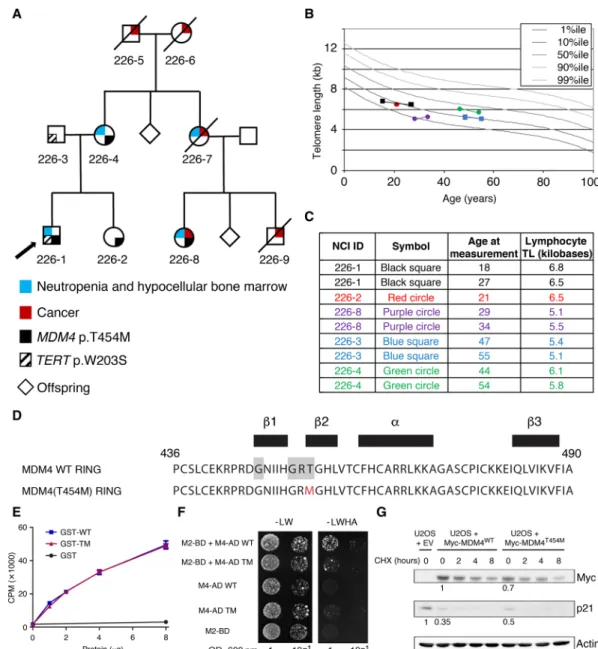
Fig. 1. A germline MDM4 p.T454M mutation identified in a family with bone marrow hypocellularity and short telomeres. (A) Pedigree of family NCI-226. Arrow
indicates proband. Cancer histories include oral squamous cell carcinoma for 226-8 at age 27 years and for 226-9 at age 42 years, and melanoma at 51 years and AML at 52 years for 226-7 (see table S1 for further details). 226-5 had lung cancer at age 69 years. 226-6 had non-Hodgkin lymphoma at age 91 years. In addition, four siblings of 226-6 had cancer: one with breast, two with lung, and one with ovary or uterus (not specified). Sequencing of 226-5, 226-6, 226-7, and 226-9 was not possible because of lack of available DNA. (B and C) Lymphocyte telomere lengths (TL) of study participants. Total lymphocyte telomere lengths are shown and were measured by flow cytometry with in situ hybridization. (B) Graphical depiction of telomere length in relation to age. Four individuals had telomeres measured twice. Legend is in (C). Percentiles (%ile) are based on 400 healthy individuals (50). (C) Age at measurement(s) and telomere length in kilobases. (D) Sequence of the MDM4 RING domain (residues 436 to 490) with secondary structure residues indicated (black boxes). The P-loop motif is highlighted in gray, and the mutated residue in red. (E) The mutant RING domain retains ATP-binding capacity. Wild-type (WT) and mutant (TM) glutathione S-transferase (GST)–RING proteins, or GST alone, were incubated with 10 nM ATP and 5 Ci ATP-32P for 10 min at
room temperature, filtered through nitrocellulose, and counted by liquid scintillation CPM, counts per minute. Results from two independent experiments. (F) The mutant MDM4 RING domain has an altered capacity to dimerize with the MDM2 RING. Two-hybrid assays were carried out as described (47). -LW, minus leucine and tryptophan; -LWHA, minus leucine, tryptophan, histidine and adenine; OD, optical density. Growth on the -LWHA medium indicates protein interaction, readily observed between MDM2 (M2-BD) and WT MDM4 (M4-AD WT) but faintly visible between MDM2 and MDM4T454M (M4-AD TM). (G) Impact of the mutation in transfected human cells. U2OS
cells were transfected with an empty vector (EV) or an expression plasmid encoding a Myc-tagged MDM4 (WT or T454M) protein and then treated or not with cyclohex-imide (CHX) to inhibit protein synthesis, and protein extracts were immunoblotted with antibodies against Myc, p21, or actin. Bands were normalized to actin, and a value of 1 was assigned to cells transfected with the WT MDM4 expression plasmid (for Myc) or with the empty vector (for p21).
on April 27, 2020
http://advances.sciencemag.org/
with the exception of hemochromatosis. An IBMFS was suspected on the basis of the family history of cancer and neutropenia. Chro-mosome breakage for Fanconi anemia was normal, while lymphocyte telomeres were between the 1st and 10th percentiles in the proband and maternal cousin (226-8) (Fig. 1, B and C). The proband was tested for mutations in known DC-causing genes, and a TERT variant (p.W203S) was identified. Unexpectedly, however, the variant was found to be inherited from his father. TERT p.W203S is not present in gnomAD, but it is predicted to be tolerated by MetaSVM (15).
Since the TERT variant did not track with disease inheritance, whole-exome sequencing (WES) was performed to search for a causal gene. The whole-exome data were filtered by maternal autosomal inheritance and revealed three genes with heterozygous missense mutations potentially deleterious according to bioinformatics pre-dictions: MDM4, KRT76, and REM1 (table S2). Given the limited knowledge of the function of KRT76 and REM1, and our prior knowledge of a DC-like phenotype in p5331/31 mice, we chose to
focus on the mutation affecting MDM4 because it encodes a major negative regulator of p53. Although the T454M mutation does not affect the p53 interaction domain of MDM4, it might affect p53 regulation because it affects the MDM4 RING domain: Residue 454 is both part of a P-loop motif thought to confer adenosine triphosphate (ATP)–binding capacity (16) and part of a strand important for MDM2-MDM4 heterodimerization (Fig. 1D) (17). The mutant RING domain had fully retained its capacity to bind ATP specifically (Fig. 1E and fig. S1A) but exhibited an altered capacity to interact with the MDM2 RING domain in a yeast two-hybrid assay (Fig. 1F). We next used transfection experiments to evaluate the consequences of this mutation on the full-length protein in human cells. We transfected U2OS cells—known to have a functional but attenuated p53 pathway due to MDM2 overexpression (18)—with either an empty vector or an expression plasmid encoding a Myc-tagged MDM4WT or MDM4T454M protein. Compared with cells transfected with the empty vector, cells transfected with a MDM4WT or a
MDM4T454M expression plasmid exhibited decreased p21 levels, indicating MDM4-mediated p53 inhibition in both cases (Fig. 1G). However, the decrease in p21 levels was less pronounced in cells expressing MDM4T454M than in cells expressing MDM4WT (Fig. 1G)
despite similar transfection efficiencies (fig. S1B). The lower ex-pression levels of the MDM4T454M protein likely contributed to its
decreased capacity to inhibit p53 (Fig. 1G). In this experimental setting, the treatment with cycloheximide did not reveal any significant difference in stability between the mutant and wild-type (WT) MDM4 proteins (Fig. 1G and quantification in fig. S1C), raising the possibility that the observed lower MDM4T454M protein levels might result from differences in mRNA translation efficiency. Together, these preliminary results argued for an impact of the mutation on MDM4 function, leading to p53 activation.
Targeting of an Mdm4T454M mutation in the mouse
The MDM4 RING domain is remarkably conserved throughout evolution, e.g., with 91% identity between the RING domains of human MDM4 and mouse Mdm4 (19). Thus, we decided to create a mouse model to precisely evaluate the physiological impact of the human mutation. We used homologous recombination in embryonic stem (ES) cells to target the p.T454M mutation at the Mdm4 locus (Fig. 2A). Targeted recombinants were identified by long-range polymerase chain reaction (PCR) (Fig. 2B), confirmed by DNA se-quencing (Fig. 2C), and the structure of the recombinant allele was
further analyzed by Southern blots with probes located 5′ and 3′ of the targeted mutation (Fig. 2D). Recombinant ES clones were then microinjected into blastocysts to generate chimeric mice, and chi-meras were mated with PGK-Cre mice to excise the Neo gene. PCR was used to verify transmission through the germ line of the Mdm4T454M (noted below Mdm4TM) mutation and to genotype the mouse colony
and mouse embryonic fibroblasts (MEFs) (Fig. 2E). We first isolated RNAs from Mdm4TM/TM MEFs and sequenced the entire Mdm4
coding sequence: The Mdm4TM sequence was identical to the WT Mdm4 sequence except for the introduced missense mutation (not shown). Furthermore, like its human counterpart, the Mdm4 gene encodes two major transcripts: Mdm4-FL, encoding the full-length oncoprotein that inhibits p53, and Mdm4-S, encoding a shorter, extremely unstable protein (20, 21). We observed, in unstressed cells as well as in cells treated with Nutlin [a molecule that activates p53 by preventing Mdm2-p53 interactions (22) without altering Mdm4- p53 interactions (23, 24)], that the Mdm4TM mutation affected neither Mdm4-FL nor Mdm4-S mRNA levels (Fig. 2F). In Western blots, however, Mdm4-FL was the only detectable isoform, and it was expressed at lower levels in the mutant MEFs (Fig. 2G).
Mdm4TM/TM fibroblasts exhibit increased p53 activity
and short telomeres
Mdm4TM/TM MEFs contained higher mRNA levels for the p53 targets p21(Cdkn1a) and Mdm2, indicating increased p53 activity (Fig. 3A).
Consistent with this, Mdm4TM/TM MEFs exhibited increased p21
and Mdm2 protein levels (Fig. 3B and fig. S2). Moreover, Mdm4TM/TM MEFs prematurely ceased to proliferate when submitted to a 3T3 protocol (Fig. 3C), which also suggests an increased p53 activity. The mean telomere length was decreased by 11% in Mdm4TM/TM
MEFs, and a subset of very short telomeres was observed in these cells, hence demonstrating a direct link between the Mdm4TM mutation,
p53 activation, and altered telomere biology (Fig. 3D). In p5331/31
MEFs, subtle but significant decreases in expression were previously observed for several genes involved in telomere biology, and in particular, small variations in Rtel1 gene expression were found to have marked effects on the survival of p5331/31 mice (5, 9). Similarly, Mdm4TM/TM MEFs exhibited subtle but significant decreases in
expression for Rtel1 and several other genes contributing to telo-mere biology (Fig. 3E). We previously showed that p53 activation correlates with an increased binding of the E2F4 repressor at the
Rtel1 promoter (9). Hence, the decreased Rtel1 mRNA levels in Mdm4TM/TM MEFs most likely resulted from increased p53 signal-ing. Consistent with this, a further increase in p53 activity, induced by Nutlin, led to further decreases in Rtel1 mRNA and protein levels, in both WT and Mdm4TM/TM cells (fig. S3A). Recently, in apparent
contradiction with our finding that p53 activation can cause telo-mere shortening (5), p53 was proposed to prevent telotelo-mere DNA degradation by inducing subtelomeric transcripts, including telomere repeat-containing RNA (TERRA) (25, 26), which suggested a com-plex, possibly context-dependent impact of p53 on telomeres (27). This led us to compare TERRA transcripts in WT and Mdm4TM/TM
cells. Consistent with an earlier report (26), p53 activation led to increased TERRA at the mouse Xq subtelomeric region in WT cells (fig. S3B). However, Mdm4TM/TM cells failed to induce TERRA in response to stress (fig. S3B). Together, our data suggest that the telomere shortening observed in Mdm4TM/TM cells results from a p53-dependent decrease in expression of several telomere-related genes and, notably, Rtel1, a gene mutated in several families with
on April 27, 2020
http://advances.sciencemag.org/
S C I E N C E A D V A N C E S
|
R E S E A R C H A R T I C L E
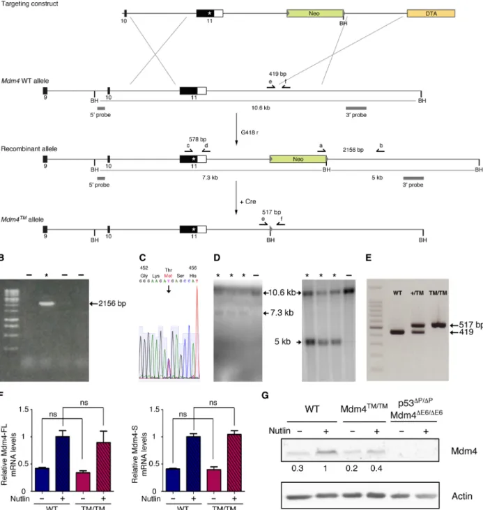
Fig. 2. Generation of a mouse model with a targeted Mdm4T454M mutation. (A) Targeting strategy. Homologous recombination in ES cells was used to target the T454M mutation at the Mdm4 locus. For the Mdm4 WT allele, exons 9 to 11 are shown [black boxes, coding sequences; white box, 3′ untranslated region (3′UTR)] and Bam HI (BH) restriction sites. Above, the targeting construct contains the following: (i) a 2.9-kb-long 5′ homology region encompassing exon 10, intron 10, and exon 11 sequences upstream the mutation; (ii) the mutation (asterisk) within exon 11; (iii) a 2.6-kb-long fragment encompassing the 3′ end of the gene and sequences imme-diately downstream; (iv) a neomycin selection gene (Neo) flanked by loxP sequences (gray arrowheads) and an additional BH site; (v) a 2.1-kb-long 3′ homology region containing sequences downstream Mdm4; and (vi) the Diphtheria toxin a gene (DTA) for targeting enrichment. (B to D) screening of G418-resistant ES clones as described in (A), with asterisks (*) indicating positive recombinants: (B) PCR with primers a and b; (C) sequencing after PCR with primers c and d: the sequence for codons 452 to 456 demonstrates heterozygosity at codon 454; (D) Southern blot of Bam HI–digested DNA with the 5′ (left) or 3′ (right) probe. (E) Examples of fibroblast genotyping by PCR with primers e and f. (F) The Mdm4T454M mutation does not alter Mdm4 mRNA levels. Mdm4-FL (left) and Mdm4-S (right) mRNAs were extracted from WT and Mdm4TM/TM
MEFs before or after treatment for 24 hours with 10 M Nutlin, quantified using real-time PCR, and normalized to control mRNAs, and then the value in Nutlin-treated WT MEFs was assigned a value of 1. Results from five independent experiments and >4 MEFs per genotype. ns, not significant in a Student’s t test. (G) Decreased Mdm4 protein levels in Mdm4TM/TM MEFs. Protein extracts, prepared from MEFs treated as in (F), were immunoblotted with antibodies against Mdm4 or actin. Bands were
normalized to actin, and then the values in Nutlin-treated WT cells were assigned a value of 1. p53P/P Mdm4E6/E6 MEFs do not express a full-length Mdm4 protein (20):
They were loaded to unambiguously identify the Mdm4(-FL) band in the other lanes.
on April 27, 2020
http://advances.sciencemag.org/
DC (6). In addition, although evidence that altered TERRA levels can cause DC is currently lacking, we cannot exclude that an altered regulation of TERRA expression might contribute to telomere defects in Mdm4TM/TM cells.
The perinatal death of Mdm4TM/TM mice is rescued
by p53 loss
Mdm4TM/TM mice were born in Mendelian proportions from Mdm4+/TM
intercrosses (Fig. 4A) but were smaller than their littermates and died within 0 to 30 min after birth, with signs of severe respiratory distress (Fig. 4, B and C). Consistent with this, Mdm4TM/TM pups at
postnatal day 0 (P0) appeared hypoxic (Fig. 4C), and their lungs were very small and dysfunctional (Fig. 4D). Thus, Mdm4TM/TM pups most
likely died from neonatal respiratory failure. Tissues from Mdm4TM/TM
pups exhibited increased p21 mRNA levels, suggesting an increase in p53 activity in these animals (fig. S4). We next used flow–FISH (fluorescence in situ hybridization) with a telomere-specific probe to evaluate the impact of the mutation on telomere length in vivo. Lung cells from Mdm4TM/TM pups (and control G3 Terc−/− mice)
exhibited a 25% decrease in mean telomere length compared with cells from WT or Mdm4+/TM littermates, indicating altered telomere
biology in G1 homozygous mutants (Fig. 4E). Notably, p53 loss or haploinsufficiency rescued the perinatal lethality of Mdm4TM/TM
pups, illustrating that the premature death of Mdm4TM/TM mice
likely resulted from increased p53 activity (Fig. 4F). However, p53−/−
and Mdm4TM/TM p53−/− mice exhibited similar survival curves, with
a fraction of the mice (respectively 4 of 12 and 1 of 6) succumbing to thymic lymphoma in less than 180 days. In contrast, after 180 days, all the p53+/− mice remained alive, whereas most Mdm4TM/TM p53+/−
mice had died. Mdm4TM/TM p53+/− mice were smaller than their
littermates (Fig. 4G) and exhibited hyperpigmentation of the foot-pads (Fig. 4H), and 120-day-old Mdm4TM/TM p53+/− mice exhibited
abnormal hemograms (Fig. 4I). Furthermore, the Mdm4TM/TM p53+/−
mice that died 60 to 160 days after birth exhibited bone marrow hypocellularity (Fig. 4J), indicating bone marrow failure as the likely cause for their premature death.
Mdm4+/TM mice are hypersensitive to increases in
p53 activity
Although Mdm4TM/TM MEFs and mice were useful to demonstrate
that the Mdm4T454M mutation leads to p53 activation and short
telomeres, a detailed analysis of Mdm4+/TM mice appeared more
relevant to model the NCI-226 family, in which all affected relatives were heterozygous carriers of the MDM4T454M mutation. Unlike Mdm4TM/TM mice, most Mdm4+/TM animals remained alive 6 months
after birth and had no apparent phenotype, similarly to WT mice (Fig. 5A). This was consistent with our analyses in fibroblasts be-cause Mdm4+/TM MEFs behaved like WT cells in a 3T3 proliferation
assay (Fig. 3C). However, p53 target genes appeared to be transactivated slightly more efficiently in Mdm4+/TM than in WT cells (Fig. 3A),
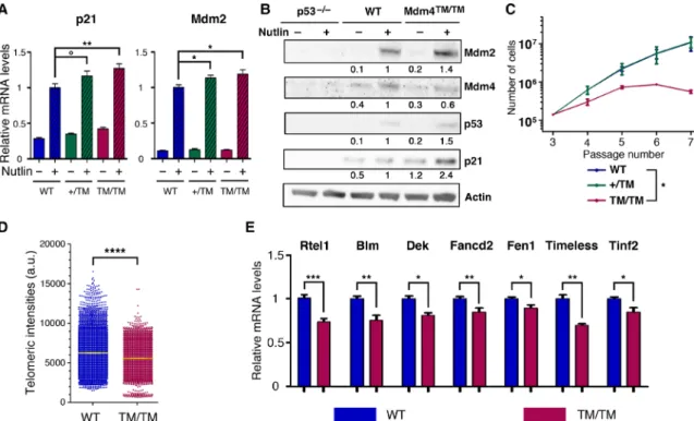
Fig. 3. Mdm4TM/TM fibroblasts exhibit increased p53 activity and short telomeres. (A) Quantification of p21 and Mdm2 mRNAs extracted from WT, Mdm4+/TM, and Mdm4TM/TM MEFs, treated or not for 24 hours with 10 M Nutlin. mRNA levels were quantified using real-time PCR and normalized to control mRNAs, and then the value
in Nutlin-treated WT MEFs was assigned a value of 1. Results from 10 independent experiments. (B) Protein extracts, prepared from p53−/−, WT, and Mdm4TM/TM MEFs
treated as in (A), were immunoblotted with antibodies against Mdm2, Mdm4, p53, p21, or actin. Bands were normalized to actin, and then the values in Nutlin-treated WT MEFs were assigned a value of 1. (C) Proliferation of MEFs in a 3T3 protocol. Each point is the average value of three independent MEFs. (D) Decreased telomere length in Mdm4TM/TM MEFs, as measured by quantitative FISH with a telomeric probe. Results from two MEFs per genotype, and 68 to 75 metaphases per MEF [means + 95%
confi-dence interval (CI) are shown in yellow]. a.u., arbitrary units. (E) Telomere-related genes down-regulated in Mdm4TM/TM MEFs. mRNAs were extracted from unstressed WT
and Mdm4TM//TM MEFs, quantified using real-time PCR, and normalized to control mRNAs, and the value in WT MEFs was assigned a value of 1. Results from >3
indepen-dent experiments and two MEFs per genotype. In relevant panels: °P = 0.08, *P < 0.05, **P < 0.01, ***P < 0.001, and ****P < 0.0001 by Stuindepen-dent’s t (A, C at passage 7, and E) or Mann-Whitney (D) statistical tests.
on April 27, 2020
http://advances.sciencemag.org/
S C I E N C E A D V A N C E S
|
R E S E A R C H A R T I C L E
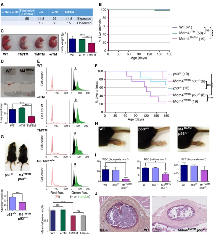
Fig. 4. Mdm4TM/TM mice die perinatally but can be rescued by p53 loss or haploinsufficiency. (A) Mendelian distribution of the offspring from 8 Mdm4+/TM intercrosses.
(B) Mdm4TM/TM mice die at birth. Cohort sizes are in parentheses. (C) Mdm4TM/TM neonates are smaller than their littermates and appear hypoxic. (D) Lungs from Mdm4TM/TM
P0 pups are hypoplastic and sink in phosphate-buffered saline owing to a lack of air inflation. (E) Flow-FISH analysis of P0 lung cells with a telomere-specific peptide nucleic acid (PNA) probe. Top: Representative results from a WT, a Mdm4+/TM, a Mdm4TM/TM, and a G3 Terc−/− mouse are shown. Right: Green fluorescence (fluo.) with black
histograms for cells without the probe (measuring cellular autofluorescence) and green histograms for cells with the probe. The shift in fluorescence intensity is smaller in Mdm4TM/TM and Terc−/− cells (c or d < a or b), indicating reduced telomere length. Left: Propidium iodide (PI) fluorescence histograms are superposed for cells with or
without the probe. Below: Statistical analysis of green fluorescence shifts (see Materials and Methods). Means + 95% CI are shown; data are from two to three mice and >3800 cells per genotype. (F) Impact of decreased p53 activity on Mdm4TM/TM animals. Cohort sizes are in parentheses. (G) Examples of littermates with indicated
genotypes. (H) Hind legs of mice with indicated genotypes. (I) Mdm4TM/TM p53+/− mice exhibit abnormal hemograms. Counts for white blood cells (WBC), red blood cells
(RBC), and platelets (PLT) for age-matched (120 days old) animals are shown. (J) Hematoxylin and eosin staining of sternum sections from WT and Mdm4TM/TM p53+/− mice.
In relevant panels: ns, not significant; *P < 0.05, ***P < 0.001, and ****P < 0.0001 by Mantel-Cox (B and F), Student’s t (C, D, G, and I), or Mann-Whitney (E) statistical tests. Photo credits: E.T. and R.D., Institut Curie (C, G, and H); R.D., Institut Curie (D).
on April 27, 2020
http://advances.sciencemag.org/
and 30% of Mdm4+/TM mice exhibited a slight hyperpigmentation of the footpads, suggesting a subtle increase in p53 activity (Fig. 5B). We reasoned that a further, subtle increase in p53 activity might affect the survival of Mdm4+/TM mice. We tested this hypothesis by
mating Mdm4+/TM animals with p53+/31 mice. p53+/31 mice were
previously found to exhibit a slight increase in p53 activity and to remain alive for over a year (5). Notably, unlike Mdm4+/TM or p53+/31
heterozygous mice, Mdm4+/TM p53+/31 compound heterozygotes died in less than 3 months (Fig. 5A) and exhibited many features associ-ated with strong p53 activation. Mdm4+/TM p53+/31 mice exhibited
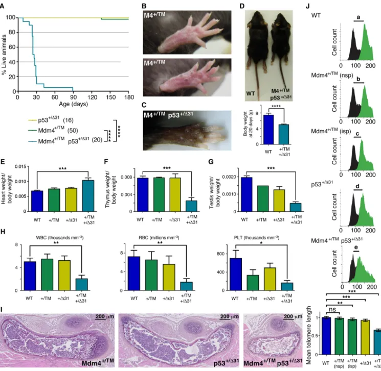
Fig. 5. Mdm4+/TM mice are hypersensitive to increased p53 activity. (A) Impact of increased p53 activity on Mdm4+/TM animals. Cohort sizes are in parentheses.
(B) Footpads from Mdm4+/TM mice appear normal (top) or exhibit a subtle increase in pigmentation (bottom). (C) Mdm4+/TM p53+/31 mice exhibit strong skin
hyperpigmen-tation. (D) Mdm4+/TM p53+/31 mice are smaller than age-matched WT mice. (E to G) Mdm4+/TM p53+/31 mice exhibit heart hypertrophy (E) as well as thymic (F) and testicular
(G) hypoplasia. (H) Mdm4+/TM p53+/31 mice exhibit abnormal hemograms. Counts for white blood cells, red blood cells, and platelets for five age-matched (P18) animals
per genotype are shown. (I) Hematoxylin and eosin staining of sternum sections from mice of the indicated genotypes. (J) Flow-FISH analysis of P18 bone marrow cells with a telomere-specific PNA probe. Top: Representative results for a WT, a Mdm4+/TM with normal skin pigmentation (nsp), a Mdm4+/TM with increased footpad skin
pigmentation (isp), a p53+/31, and a Mdm4+/TM p53+/31 mouse are shown; black histograms, cells without the probe; green histograms, cells with the probe. The smallest
shift in fluorescence intensity (e) was observed with Mdm4+/TM p53+/31 cells. Bottom: Statistical analysis of green fluorescence shifts. Means + 95% CI are shown; data are
from >1500 cells per genotype. In relevant panels: ns, not significant; *P < 0.05, **P < 0.01, ***P < 0.001, and ****P < 0.0001 by Mantel-Cox (A), Student’s t (D and E to H), or Mann-Whitney (J) statistical tests. Photo credits: R.D. and P.L., Institut Curie (B); E.T. and R.D., Institut Curie (C and D).
on April 27, 2020
http://advances.sciencemag.org/
S C I E N C E A D V A N C E S
|
R E S E A R C H A R T I C L E
intense skin hyperpigmentation (Fig. 5C), were much smaller than their littermates (Fig. 5D), and exhibited heart hypertrophy (Fig. 5E) and thymic hypoplasia (Fig. 5F) and the males had testicular hypo-plasia (Fig. 5G). Bone marrow failure was the likely cause for the premature death of Mdm4+/TM p53+/31 mice, as indicated by abnormal hemograms of 18-day-old (P18) compound heterozygotes (Fig. 5H) and bone marrow hypocellularity in the sternum sections of mori-bund Mdm4+/TM p53+/31 animals (Fig. 5I). We next used flow-FISH
to analyze telomere length in the bone marrow cells of P18 WT,
Mdm4+/TM, p53+/31, and Mdm4+/TM p53+/31 mice. We found no
significant difference between telomere lengths in cells from five WT and three Mdm4+/TM mice with normal skin pigmentation, whereas
cells from two Mdm4+/TM mice with increased skin pigmentation (or from p53+/31 mice) exhibited marginal (5 to 7%) decreases in
mean telomere length. Notably, in G1 Mdm4+/TM p53+/31 cells, the average telomere length was decreased by 34% (Fig. 5J). Together, these results demonstrate that Mdm4+/TM mice are hypersensitive to subtle increases in p53 activity. Consistent with this, Mdm4+/TM p53+/31 MEFs also exhibited increased p53 signaling and accelerated proliferation arrest in a 3T3 protocol (fig. S5). In sum, the compar-ison between Mdm4TM/TM and Mdm4TM/TM p53+/− mice, or between Mdm4+/TM and Mdm4+/TM p53+/31 animals, indicated that subtle
variations in p53 signaling had marked effects on the phenotypic consequences of the Mdm4T454M mutation (table S3).
Polymorphisms that may affect p53 signaling differ among DC-like family members
The carriers of the MDM4T454M mutation exhibited considerable
heterogeneity in their phenotypes (Fig. 1 and table S1). The data from our mouse model suggested that variations in p53 activity might account for the variable expressivity and penetrance of clinical features among the NCI-226 MDM4+/T454M relatives. Hence, we
analyzed nine known common polymorphisms reported to affect p53 activity and tumorigenesis (four at the TP53 locus, two at the
MDM2 locus, and three at the MDM4 locus) (3,4,28–32). Among
the four MDM4+/T454M relatives, the proband (NCI-226-1) is more
difficult to interpret because the potential contribution of the TERT p.W203S variant to his phenotype cannot be ruled out (even though it appears unlikely according to in silico predictions). The MDM4 allele encoding the mutant protein (p.T454M) appears associated with the C allele of single-nucleotide polymorphism (SNP) rs4245739, the G allele of SNP rs11801299, and the G allele of SNP rs1380576 (Fig. 6A). These three MDM4 variant alleles are associated with increased p53 activity (4,32) and might, thus, synergize with the
MDM4T454M mutation in this family.
The proband’s affected cousin (226-8) exhibited a very early onset of disease, with lymphocyte telomere length within or below the first percentile of age-matched control participants and tongue squamous cell carcinoma at age 27 (Fig. 1 and table S1). The WT MDM4 allele of 226-8 carried the rs4245739 C, the rs11801299 G, and the rs1380576 G variants associated with increased p53 activity. This suggests a potential disease-modifying effect of these MDM4 SNPs. In contrast, the proband’s mother (226-4) was much less severely affected, with telomere length between the 10th and 50th percentiles (Fig. 1). Although we cannot rule out that disease anticipation might con-tribute to her milder phenotype, note that her WT MDM4 allele carried variants that might correlate with decreased p53 activity and could antagonize the MDM4T454M mutation (rs4245739 A, rs11801299
A, and rs1380576 C; Fig. 6A). Family members 226-4 and 226-8
shared the same genotypes for all the other tested variants, except for TP53 rs1042522, a SNP first reported to affect apoptotic or cell cycle arrest responses (28), but with a clinical effect that now appears controversial (33). The proband’s sister (226-2), with a B cell defi-ciency and telomere lengths around the 10th percentile, also appeared less affected than 226-8. All the tested variants at the MDM2 and
MDM4 loci were identical between 226-2 and 226-8. However,
unlike 226-8, 226-2 exhibited a TP53 allele with an A1A1 haplotype for variants rs17878362 and rs17880560 that might decrease p53 activity (30) and antagonize the effects of the MDM4T454M mutation
(Fig. 6A).
We had primary fibroblasts available for two of these family members, 226-4 and 226-8, allowing us to directly assess the func-tional effect of the MDM4T454M variant in these cells. These
fibro-blasts were grown in parallel with primary fibrofibro-blasts from patients with DC carrying either a TINF2K280E mutation or a TERTP704S
mutation, and mRNA levels for p21 and RTEL1 were quantified. In agreement with the notion that a MDM4T454M heterozygous
muta-tion activates p53 signaling in NCI-226 family members, fibroblasts from both 226-4 and 226-8 exhibited increased p21 mRNA levels compared with TINF2 or TERT mutant cells (Fig. 6B). However, cells from 226-4 only exhibited a 2-fold increase in p21 levels, whereas a 12-fold increase was observed for cells from 226-8, consistent with the notion that SNPs affecting the p53 pathway might counteract (for 226-4) or strengthen (for 226-8) the effect of the MDM4T454M mutation. Furthermore, we previously showed that RTEL1 mRNA levels are down-regulated upon p53 activation in human cells (5). RTEL1 mRNA levels appeared normal in cells from 226-4 but were markedly decreased in cells from 226-8, raising the possibility that a threshold in p53 activation might be required to affect RTEL1 ex-pression (Fig. 6B).
DISCUSSION
Although MDM4 is primarily known for its clinical relevance in cancer biology, our study shows that a germline missense MDM4 mutation may cause features suggestive of DC. In humans, the MDM4 (p.T454M) mutation was identified in this family with neutropenia, bone mar-row hypocellularity, early-onset tongue SCC, AML, and telomeres between the 1st and 10th percentiles in the younger generation. In mice, the same Mdm4 mutation notably correlated with increased p53 activity, short telomeres, and bone marrow failure. In both human transfected cells and MEFs, the mutant protein was expressed at lower levels than its WT counterpart, likely contributing to in-creased p53 activity. Together, these results demonstrate the impor-tance of the MDM4/p53 regulatory axis on telomere biology and DC-like features in both species. Notably, p5331/31 mice were
previously found to phenocopy DC (5), but whether this finding was relevant to human disease had remained controversial. When a mutation in PARN was found to cause DC (34), it first appeared con-sistent with the p5331 mouse model because PARN, the polyadenylate-
specific ribonuclease, had been proposed to regulate p53 mRNA stability (35). However, whether PARN regulates the stability of mRNAs is now contested (36). Rather, PARN would regulate the levels of over 200 microRNAs, of which only a few might repress p53 mRNA translation (37). Furthermore, PARN regulates TERC, the telomerase RNA component (38), and TERC overexpression increased telomere length in PARN-deficient cells (39). Thus, whether a germline mutation that specifically activates p53 can cause DC-like features
on April 27, 2020
http://advances.sciencemag.org/
remained to be demonstrated in humans, and our report provides compelling evidence for this, because unlike PARN, MDM4 is a very specific regulator of p53.
A germline antiterminating MDM2 mutation was recently identified in a patient with a Werner-like syndrome of premature aging. Although multiple mechanisms might contribute to the clinical features in that report, a premature cellular senescence resulting from p53 hyperactivation was proposed to play a major role in his segmental progeroid phenotype (40). In that regard, our finding that increased p53 activity correlates with short telomeres appears rele-vant because telomere attrition is a primary hallmark of aging, well known to trigger cellular senescence (41). Furthermore, germline
TP53 frameshift mutations were recently reported in two patients
diagnosed with pure red blood cell aplasia and hypogammaglobu-linemia, resembling but not entirely consistent with Diamond Blackfan anemia (DBA) (42). In addition to the pure red cell aplasia diagnostic of DBA, those patients were found to exhibit relatively short telo-meres (although not as short as telotelo-meres from patients with DC), which may also seem consistent with our results. Our finding of an
MDM4 missense mutation in a DC-like family, together with recent
reports linking an antiterminating MDM2 mutation to a Werner-
like phenotype and TP53 frameshift mutations to DBA-like features, indicates that the clinical impact of germline mutations affecting the p53/MDM2/MDM4 regulatory network is just emerging. An inherited hyperactivation of the p53 pathway—via a germline TP53,
MDM2, or MDM4 mutation—may thus cause either DBA, Werner-
like, or DC-like features, but additional work will be required to determine whether mutations in any of these three genes can cause any of these three syndromes. Likewise, several mouse models have implicated p53 deregulation in features of other developmental syndromes including the CHARGE, Treacher-Collins, Waardenburg, or DiGeorge syndrome (43), and it will be important to know whether germline mutations in TP53, MDM2, or MDM4 may cause these additional syndromes in humans.
Heterozygous Mdm4+/TM mice appeared normal but were
hyper-sensitive to variations in p53 activity, and, perhaps most notably,
Mdm4+/TM p53+/31 compound heterozygous mice rapidly died from
bone marrow failure. Thus, the p5331 mutation acted as a strong
genetic modifier of the Mdm4TM mutation. It is tempting to
specu-late that similarly, among the NCI-226 family members heterozygous for the MDM4T454M allele, differences in the severity of phenotypic
traits (e.g., lymphocyte telomere length and bone marrow cellularity)
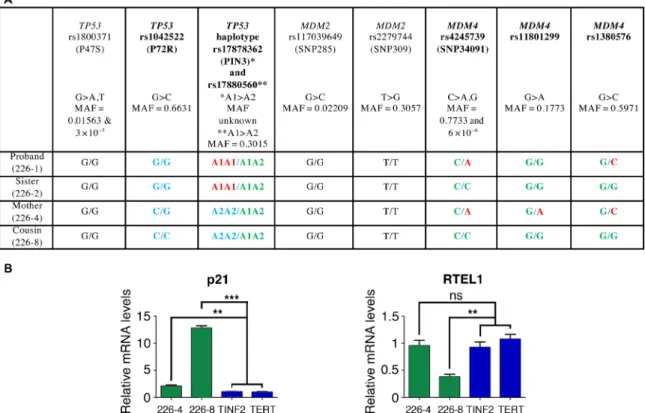
Fig. 6. TP53 or MDM4 polymorphisms might alter the impact of the MDM4T454M mutation among NCI-226 family members. (A) Genotyping of polymorphisms that may affect the p53 pathway. The SNPs rs1800371 and rs1042522 modify the p53 protein sequence (28,29), whereas rs17878362 and rs17880560 are singlets (A1) or doublets (A2) of G-rich sequences in noncoding regions of TP53 that affect p53 expression (30). SNPs rs117039649 and rs2279744, in the MDM2 promoter, affect MDM2 mRNA levels (3,31). Three SNPs are at the MDM4 locus: rs4245739 in the 3′UTR region affects MDM4 protein levels (4), whereas rs11801299 and rs1380576 were associated with an increased risk of developing retinoblastoma (32), a cancer type with frequent MDM4 alterations (51). Polymorphisms that differ among family members are in bold, with the allele (or haplotype) associated with increased p53 activity in green (because it may synergize with the effects of the MDM4T454M mutation). Alleles
(or haplotypes) for which there is evidence of decreased p53 activity, or for which the effect is uncertain, are highlighted in red or blue, respectively. Please note that the clinical effects of the TP53 rs1042522 SNP have recently been contested (33), so that all alleles for this SNP were labeled in blue. MAF, minor allele frequency reported for all gnomAD populations combined. https://gnomad.broadinstitute.org (52). (B) Comparative analysis of primary fibroblasts from family members 226-4 and 226-8. p21 and RTEL1 mRNAs, extracted from cells from relatives NCI 226-4 and NCI 226-8 or two unrelated patients with DC carrying a TINF2 or a TERT mutation, were quantified using real-time PCR, normalized to control mRNAs, and then expressed relative to the mean values in TINF2 and TERT mutant cells. ns, not significant, **P < 0.01 and ***P < 0.001 in a Student’s t test.
on April 27, 2020
http://advances.sciencemag.org/
S C I E N C E A D V A N C E S
|
R E S E A R C H A R T I C L E
may result, in part, from modifiers affecting the p53 pathway and synergize or antagonize with the effects of the MDM4T454M
muta-tion. To search for potentially relevant modifiers, we looked at nine polymorphisms at the TP53, MDM2, and MDM4 loci that were previously reported to affect p53 activity. Notably, we found that the family member most severely affected (226-8, the proband’s cousin) carried a TP53 haplotype, as well as SNPs on the WT MDM4 allele, that might synergize with the effects of the MDM4T454M
mutation. Conversely, a TP53 haplotype for the proband’s sister (226-2), or SNPs at the WT MDM4 locus for the proband’s mother (226-4), might antagonize the impact of MDM4T454M allele. Consistent
with this, primary fibroblasts from 226-4 and 226-8 exhibited in-creased p53 activity, but p53 activation was much stronger in cells from 226-8. Our data, thus, appear consistent with the existence of genetic modifiers at the TP53 and MDM4 loci that may affect DC-like phenotypic traits among family members carrying the MDM4 (p.T454M) mutation. However, this remains speculative given the small number of individuals that could be analyzed. Furthermore, nonexonic variants affecting other genes might also contribute to DC-like traits (44). Last, the TP53 and MDM4 polymorphisms considered here were previously evaluated for their potential impact on tumorigenic processes, rather than DC-like traits such as telo-mere length or bone marrow hypocellularity. Our data suggest that polymorphisms at the TP53 and MDM4 (and possibly MDM2) loci should be evaluated for their potential impact on bone marrow function and telomere biology.
MATERIALS AND METHODS
Patient and family
The individuals in this study are participants in an Institutional Review Board–approved longitudinal cohort study at the NCI entitled “Etiologic Investigation of Cancer Susceptibility in Inherited Bone Marrow Failure Syndromes” (www.marrowfailure.cancer.gov, ClinicalTrials.gov NCT00027274) (7). Patients and their family members enrolled in 2008 and completed detailed family history and medical history questionnaires. Detailed medical record review and thorough clinical evaluations of the proband, his sister, parents, and maternal cousin were conducted at the National Institutes of Health (NIH) Clinical Center. Telomere length was measured by flow cytometry with in situ hybridization (flow-FISH) (45) in leukocytes of all patients and family members reported. DNA was extracted from whole blood using standard methods. DNA was not available from 226-7 or 226-9 (Fig. 1). Given the time frame of participant enrollment, Sanger sequencing of DKC1, TINF2, TERT, TERC, and
WRAP53 was performed first, followed by exome sequencing.
Whole-exome sequencing
WES of blood-derived DNA for family NCI-226 was performed at the NCI’s Cancer Genomics Research Laboratory as previously described (46). Exome enrichment was performed with NimbleGen’s SeqCap EZ Human Exome Library v3.0 + UTR (Roche NimbleGen Inc., Madison, WI, USA), targeting 96 Mb of exonic sequence and the flanking untranslated regions (UTRs) on an Illumina HiSeq. Annotation of each exome variant locus was performed using a custom software pipeline. WES variants of interest were identified if they met the following criteria: heterozygous in the proband, his mother, and maternal cousin; nonsynonymous; had a minor allele frequency <0.1% in the Exome Aggregation Consortium databases;
and occurred <5 times in our in house database of 4091 individuals. Variants of interest were validated to rule out false-positive findings using an Ion 316 chip on the Ion PGM Sequencer (Life Technologies, Carlsbad, CA, USA).
ATP-binding assays
Primers flanking the MDM4 RING domain were used to amplify RING sequences, and PCR products were cloned (or cloned and mutagenized) in the pGST-parallel2 plasmid. Glutathione S-transferase (GST) fusion proteins were expressed in BL21 (DE3) cells. After induction for 16 hours at 20°C with 0.2 mM IPTG (isopropyl--d-
thiogalactopyranoside), soluble proteins were extracted by sonica-tion in lysis buffer [50 mM tris (pH 7.0), 300 mM LiSO4, 1 mM
dithiothreitol (DTT), 0.5 mM phenylmethylsulfonyl fluoride (PMSF), 0.2% NP-40, complete Protease inhibitors (Roche) 1×]. The soluble protein fraction was incubated with Glutathione Sepharose beads (Pharmacia) at 4°C for 2 hours, and the bound proteins were washed with 50 mM tris (pH 7.0), 300 mM LiSO4, and 1 mM DTT and then
eluted with an elution buffer [50 mM tris-HCl (pH 7.5), 300 mM NaCl, 1 mM DTT, and 15 mM glutathione]. WT and mutant GST-RING proteins (0, 1, 2, 4, or 8 g) or GST alone (0 or 8 g) was incubated with 10 nM ATP and 5 Ci ATP-32P for 10 min at room
temperature, filtered through nitrocellulose, and counted by liquid scintillation. Alternatively, 7 g of either WT or mutant GST-RING proteins was incubated with 5 Ci ATP-32P for 10 min at room temperature and increasing amounts (0, 0.02, 2, 20, and 200 M) of ATP or guanosine triphosphate (GTP), filtered through nitrocellulose, and counted by liquid scintillation.
Yeast two-hybrid assays
The yeast two-hybrid assays were performed as described (47). Briefly, MDM4 and MDM2 RING open reading frames were cloned in plasmids derived from the two-hybrid vectors pGADT7 (Gal4-activating domain) and pGBKT7 (Gal4-binding domain) creating N-terminal fusions and transformed in yeast haploid strains Y187 and AH109 (Clontech). Interactions were scored, after mating and diploid selection on dropout medium without leucine and tryptophan, as growth on dropout medium without leucine, tryptophan, histidine, and adenine.
Transfection assays
U2OS cells (106) were transfected by using Lipofectamine 2000 (Invitrogen) with pCDNA3.1 (6 g), or 5 × 10⁶ cells were transfected
with 30 g of pCDNA3.1-MycTag-MDM4WT or pCDNA3.1-Myc-Tag-MDM4TM. Twenty-four hours after transfection, cells were treated
with cycloheximide (50 g/ml; Sigma-Aldrich, C4859), then scratched in phosphate-buffered saline (PBS) after 2, 4, or 8 hours, pelleted, and snap frozen in liquid nitrogen before protein or RNA extraction with standard protocols.
Mdm4T454M targeting construct
The targeting vector was generated by recombineering from the RP23-365M5 BAC (bacterial artificial chromosome) clone (CHORI BACPAC Resources) containing mouse Mdm4 and downstream sequences of C57Bl6/J origin. A loxP-flanked neomycin cassette (Neo) and a diphtheria toxin a gene (DTA) were inserted downstream of the Mdm4 gene, respectively, for positive and negative selections, and a single-nucleotide mutation encoding the missense mutation T454M (TM) was targeted in the exon 11 of Mdm4. The targeting construct was fully sequenced before use.
on April 27, 2020
http://advances.sciencemag.org/
Targeting in ES cells and mouse genotyping
CK-35 ES cells were electroporated with the targeting construct linearized with Not I. Recombinant clones were identified by long-range PCR, confirmed by Southern blot, PCR, and DNA sequencing (primer sequences in table S4). Two independent recombinant clones were injected into blastocysts to generate chimeras, and germline transmission was verified by genotyping their offspring. Reverse transcription PCR (RT-PCR) of RNAs from Mdm4TM/TM MEFs
showed that the mutant complementary DNA (cDNA) differed from an Mdm4 WT sequence only by the engineered missense mutation. The genotyping of p53+/−, p53+/∆31, and G3 Terc−/− mice was performed as previously described (5, 12). All experiments were performed according to Institutional Animal Care and Use Com-mittee regulations.
Cells and cell culture reagents
MEFs isolated from 13.5-day embryos were cultured in a 5% CO2
and 3% O2 incubator, in Dulbecco’s modified Eagle’s medium
GlutaMAX (Gibco), with 15% fetal bovine serum (Biowest), 100 M 2-mercaptoethanol (Millipore), 0.01 mM Non-Essential Amino Acids, and penicillin/streptavidin (Gibco) for five or fewer passages, except for 3T3 experiments, performed in a 5% CO2 incubator for seven
passages. Cells were treated for 24 hours with 10 M Nutlin 3a (Sigma- Aldrich) (22) or 15 M cisplatin (Sigma-Aldrich). Primary human fibroblasts at low passage (p.2 for TINF2K280E, p.3 for NCI-226-4 and NCI-226-8, and p.4 for TERTP704S) were thawed and cultured in
fibroblast basal medium (Lonza) with 20% fetal calf serum, l-glutamin,
10 mM Hepes, penicillin/streptavidin, and gentamicin before quan-titative PCR (qPCR) analysis.
Quantitative RT-PCR
Total RNA, extracted using NucleoSpin RNA II (Macherey-Nagel), was reverse transcribed using SuperScript IV (Invitrogen), with, for TERRA quantification, a (CCCTAA)4 oligo as described (48). Real- time
qPCRs were performed with primer sequences as described (5, 9, 48) on a QuantStudio using Power SYBR Green (Applied Biosystems).
Western blots
Protein detection by immunoblotting was performed using antibodies against Mdm2 (4B2), Mdm4 (M0445; Sigma-Aldrich), p53 (AF1355, R&D Systems), actin (A2066; Sigma-Aldrich), p21 (F5; Santa Cruz Biotechnology), Myc-Tag (SAB2702192; Sigma-Aldrich), and Rtel1 (from J.-A.L.-V.). Chemiluminescence revelation was achieved with SuperSignal West Dura (Perbio). Bands of interest were quantified by using ImageJ and normalized with actin.
Telomeric quantitative FISH
Cells were treated with colcemide (0.5 g/ml) for 1.5 hours, submitted to hypotonic shock, fixed in an (3:1) ethanol/acetic acid solution, and dropped onto glass slides. Quantitative FISH was then carried out as described (5) with a TelC-Cy3 peptide nucleic acid (PNA) probe (Panagene). Images were acquired using a Zeiss Axioplan 2, and telomeric signals were quantified with iVision (Chromaphor).
Telomeric flow-FISH
Flow-FISH with mouse cells was performed as described (45). For each animal, either the lungs were collected or the bone marrow from two tibias and two femurs was collected and red blood cells were lysed; then, 2 × 10⁶ cells were fixed in 500 l of PNA hybridization
buffer [70% deionized formamide, 20 mM tris (pH 7.4), and 0.1% Blocking reagent; Roche] and stored at −20°C. Either nothing (control) or 5 l of probe stock solution was added to cells [probe stock solution: 10 M TelC-FAM PNA probe (Panagene), 70% form-amide, and 20 mM tris (pH 7.4)], and samples were denatured for 10 min at 80°C before hybridization for 2 hours at room tempera-ture. After three washes, cells were resuspended in PBS 1×, 0.1% bovine serum albumin, ribonuclease A (1000 U/ml), and propidium iodide (12.5 g/ml) and analyzed with an LSR II fluorescence-activated cell sorter. WT and G3 Terc−/− mice were included in all flow-FISH
experiments, respectively, as controls of normal and short telomeres. For fluorescence shift analyses, the green histograms (correspond-ing to cells with the telomeric probe) were sliced into 18 windows of equal width and numbered 0 to 17 according to their distance from the median value in cells without the probe, and the number of cells in each window was quantified with ImageJ. The data from two to five mice per genotype were typically used to calculate mean telo-mere lengths, expressed relative to the mean in WT cells.
Anatomopathology and hemograms
Organs were fixed in formol 4% for 24 hours and then ethanol 70% and embedded in paraffin wax. Serial sections were stained with hematoxylin and eosin using standard procedures (49). For hemo-grams, 100 l of blood from each animal was recovered retro-orbitally in a 10-l citrate-concentrated solution (S5770; Sigma-Aldrich) and analyzed using an MS9 machine (Melet Schloesing Laboratoires).
Human polymorphisms affecting the p53 pathway
DNA extracted from Epstein-Barr virus–transformed lymphocytes of NCI-226 family members was amplified with primers flanking nucleotide polymorphisms of interest (primer sequences in table S5), and then PCR products were analyzed by Sanger DNA sequencing.
Statistical analyses
Analyses with Student’s t, Mann-Whitney, or Mantel-Cox statistical tests were performed by using GraphPad Prism, and values of P < 0.05 were considered significant.
SUPPLEMENTARY MATERIALS
Supplementary material for this article is available at http://advances.sciencemag.org/cgi/ content/full/6/15/eaay3511/DC1
View/request a protocol for this paper from Bio-protocol.
REFERENCES AND NOTES
1. J. M. Nigro, S. J. Baker, A. C. Preisinger, J. M. Jessup, R. Hosteller, K. Cleary, S. H. Signer, N. Davidson, S. Baylin, P. Devilee, T. Glover, F. S. Collins, A. Weslon, R. Modali, C. C. Harris, B. Vogelstein, Mutations in the p53 gene occur in diverse human tumour types.
Nature 342, 705–708 (1989).
2. D. Malkin, F. P. Li, L. C. Strong, J. F. Fraumeni, C. E. Nelson, D. H. Kim, J. Kassel, M. A. Gryka, F. Z. Bischoff, M. A. Tainsky, Germ line p53 mutations in a familial syndrome of breast cancer, sarcomas, and other neoplasms. Science 250, 1233–1238 (1990).
3. G. L. Bond, W. Hu, E. E. Bond, H. Robins, S. G. Lutzker, N. C. Arva, J. Bargonetti, F. Bartel, H. Taubert, P. Wuerl, K. Onel, L. Yip, S.-J. Hwang, L. C. Strong, G. Lozano, A. J. Levine, A single nucleotide polymorphism in the MDM2 promoter attenuates the p53 tumor suppressor pathway and accelerates tumor formation in humans. Cell 119, 591–602 (2004). 4. J. Wynendaele, A. Böhnke, E. Leucci, S. J. Nielsen, I. Lambertz, S. Hammer, N. Sbrzesny,
D. Kubitza, A. Wolf, E. Gradhand, K. Balschun, I. Braicu, J. Sehouli, S. Darb-Esfahani, C. Denkert, C. Thomssen, S. Hauptmann, A. Lund, J.-C. Marine, F. Bartel, An illegitimate microRNA target site within the 3’ UTR of MDM4 affects ovarian cancer progression and chemosensitivity. Cancer Res. 70, 9641–9649 (2010).
5. I. Simeonova, S. Jaber, I. Draskovic, B. Bardot, M. Fang, R. Bouarich-Bourimi, V. Lejour, L. Charbonnier, C. Soudais, J.-C. Bourdon, M. Huerre, A. Londono-Vallejo, F. Toledo,
on April 27, 2020
http://advances.sciencemag.org/
S C I E N C E A D V A N C E S
|
R E S E A R C H A R T I C L E
Mutant mice lacking the p53 C-terminal domain model telomere syndromes. Cell Rep. 3, 2046–2058 (2013).6. S. A. Savage, Beginning at the ends: Telomeres and human disease. F1000Res. 7, 524 (2018). 7. B. P. Alter, N. Giri, S. A. Savage, P. S. Rosenberg, Cancer in the National Cancer Institute
inherited bone marrow failure syndrome cohort after fifteen years of follow-up.
Haematologica 103, 30–39 (2018).
8. S. C. Ward, S. A. Savage, N. Giri, B. P. Alter, P. S. Rosenberg, D. C. Pichard, E. W. Cowen, Beyond the triad: Inheritance, mucocutaneous phenotype, and mortality in a cohort of patients with dyskeratosis congenita. J. Am. Acad. Dermatol. 78, 804–806 (2018). 9. S. Jaber, E. Toufektchan, V. Lejour, B. Bardot, F. Toledo, p53 downregulates the Fanconi
anaemia DNA repair pathway. Nat. Commun. 7, 11091 (2016).
10. D. Filipescu, M. Naughtin, K. Podsypanina, V. Lejour, L. Wilson, Z. A. Gurard-Levin, G. A. Orsi, I. Simeonova, E. Toufektchan, L. D. Attardi, F. Toledo, G. Almouzni, Essential role for centromeric factors following p53 loss and oncogenic transformation. Genes Dev. 31, 463–480 (2017).
11. E. Toufektchan, F. Toledo, The guardian of the genome revisited: p53 downregulates genes required for telomere maintenance, DNA repair, and centromere structure.
Cancers 10, E135 (2018).
12. M. A. Blasco, H. W. Lee, M. P. Hande, E. Samper, P. M. Lansdorp, R. A. DePinho, C. W. Greider, Telomere shortening and tumor formation by mouse cells lacking telomerase RNA. Cell 91, 25–34 (1997).
13. E. Aix, Ó. Gutiérrez-Gutiérrez, C. Sánchez-Ferrer, T. Aguado, I. Flores, Postnatal telomere dysfunction induces cardiomyocyte cell-cycle arrest through p21 activation. J. Cell Biol.
213, 571–583 (2016).
14. D. Hockemeyer, W. Palm, R. C. Wang, S. S. Couto, T. de Lange, Engineered telomere degradation models dyskeratosis congenita. Genes Dev. 22, 1773–1785 (2008). 15. S. Kim, J.-H. Jhong, J. Lee, J.-Y. Koo, Meta-analytic support vector machine for integrating
multiple omics data. BioData Min. 10, 2 (2017).
16. C. Priest, C. Prives, M. V. Poyurovsky, Deconstructing nucleotide binding activity of the Mdm2 RING domain. Nucleic Acids Res. 38, 7587–7598 (2010).
17. K. Linke, P. D. Mace, C. A. Smith, D. L. Vaux, J. Silke, C. L. Day, Structure of the MDM2/ MDMX RING domain heterodimer reveals dimerization is required for their ubiquitylation in trans. Cell Death Differ. 15, 841–848 (2008).
18. J. E. Landers, S. L. Cassel, D. L. George, Translational enhancement of mdm2 oncogene expression in human tumor cells containing a stabilized wild-type p53 protein.
Cancer Res. 57, 3562–3568 (1997).
19. B. X. Tan, H. P. Liew, J. S. Chua, F. J. Ghadessy, Y. S. Tan, D. P. Lane, C. R. Coffill, Anatomy of Mdm2 and Mdm4 in evolution. J. Mol. Cell Biol. 9, 3–15 (2017).
20. B. Bardot, R. Bouarich-Bourimi, J. Leemput, V. Lejour, A. Hamon, L. Plancke, A. G. Jochemsen, I. Simeonova, M. Fang, F. Toledo, Mice engineered for an obligatory Mdm4 exon skipping express higher levels of the Mdm4-S isoform but exhibit increased p53 activity. Oncogene 34, 2943–2948 (2015).
21. M. Dewaele, T. Tabaglio, K. Willekens, M. Bezzi, S. X. Teo, D. H. P. Low, C. M. Koh, F. Rambow, M. Fiers, A. Rogiers, E. Radaelli, M. Al-Haddawi, S. Y. Tan, E. Hermans, F. Amant, H. Yan, M. Lakshmanan, R. C. Koumar, S. T. Lim, F. A. Derheimer, R. M. Campbell, Z. Bonday, V. Tergaonkar, M. Shackleton, C. Blattner, J.-C. Marine, E. Guccione, Antisense oligonucleotide-mediated MDM4 exon 6 skipping impairs tumor growth. J. Clin. Invest.
126, 68–84 (2016).
22. L. T. Vassilev, B. T. Vu, B. Graves, D. Carvajal, F. Podlaski, Z. Filipovic, N. Kong, U. Kammlott, C. Lukacs, C. Klein, N. Fotouhi, E. A. Liu, In vivo activation of the p53 pathway by small-molecule antagonists of MDM2. Science 303, 844–848 (2004).
23. B. Hu, D. M. Gilkes, B. Farooqi, S. M. Sebti, J. Chen, MDMX overexpression prevents p53 activation by the MDM2 inhibitor Nutlin. J. Biol. Chem. 281, 33030–33035 (2006). 24. M. Wade, E. T. Wong, M. Tang, J. M. Stommel, G. M. Wahl, Hdmx modulates the outcome
of p53 activation in human tumor cells. J. Biol. Chem. 281, 33036–33044 (2006). 25. S. Tutton, G. A. Azzam, N. Stong, O. Vladimirova, A. Wiedmer, J. A. Monteith, K. Beishline,
Z. Wang, Z. Deng, H. Riethman, S. B. McMahon, M. Murphy, P. M. Lieberman, Subtelomeric p53 binding prevents accumulation of DNA damage at human telomeres.
EMBO J. 35, 193–207 (2016).
26. S. Tutton, Z. Deng, N. Gulve, O. Vladimirova, K. Beishline, A. Wiedmer, M. Murphy, P. M. Lieberman, Elevated telomere dysfunction in cells containing the African-centric Pro47Ser cancer-risk variant of TP53. Oncotarget 10, 3581–3591 (2019).
27. F. Toledo, p53: A two-faced regulator of telomere metabolism? (comment on DOI 10.1002/bies.201600078). Bioessays 38, 938 (2016).
28. M. C. Marin, C. A. Jost, L. A. Brooks, M. S. Irwin, J. O’Nions, J. A. Tidy, N. James, J. M. McGregor, C. A. Harwood, I. G. Yulug, K. H. Vousden, M. J. Allday, B. Gusterson, S. Ikawa, P. W. Hinds, T. Crook, W. G. Kaelin, A common polymorphism acts as an intragenic modifier of mutant p53 behaviour. Nat. Genet. 25, 47–54 (2000). 29. X. Li, P. Dumont, A. Della Pietra, C. Shetler, M. E. Murphy, The codon 47 polymorphism
in p53 is functionally significant. J. Biol. Chem. 280, 24245–24251 (2005). 30. C. Sagne, V. Marcel, M. Bota, G. Martel-Planche, A. Nobrega, E. I. Palmero, L. Perriaud,
M. Boniol, S. Vagner, D. G. Cox, C. S. Chan, J.-L. Mergny, M. Olivier, P. Ashton-Prolla, J. Hall,
P. Hainaut, M. I. Achatz, Age at cancer onset in germline TP53 mutation carriers: Association with polymorphisms in predicted G-quadruplex structures. Carcinogenesis
35, 807–815 (2014).
31. S. Knappskog, M. Bjørnslett, L. M. Myklebust, P. E. A. Huijts, M. P. Vreeswijk, H. Edvardsen, Y. Guo, X. Zhang, M. Yang, S. K. Ylisaukko-Oja, P. Alhopuro, J. Arola, R. A. E. M. Tollenaar, C. J. van Asperen, C. Seynaeve, V. Staalesen, R. Chrisanthar, E. Løkkevik, H. B. Salvesen, D. G. Evans, W. G. Newman, D. Lin, L. A. Aaltonen, A.-L. Børresen-Dale, G. S. Tell, C. Stoltenberg, P. Romundstad, K. Hveem, J. R. Lillehaug, L. Vatten, P. Devilee, A. Dørum, P. E. Lønning, The MDM2 promoter SNP285C/309G haplotype diminishes Sp1 transcription factor binding and reduces risk for breast and ovarian cancer in Caucasians.
Cancer Cell. 19, 273–282 (2011).
32. F. Yu, Z. Jiang, A. Song, Association of rs11801299 and rs1380576 polymorphisms at MDM4 with risk, clinicopathological features and prognosis in patients
with retinoblastoma. Cancer Epidemiol. 58, 153–159 (2019).
33. J. B. Kodal, S. Vedel-Krogh, C. J. Kobylecki, B. G. Nordestgaard, S. E. Bojesen, TP53 Arg72Pro, mortality after cancer, and all-cause mortality in 105,200 individuals. Sci. Rep. 7, 336 (2017).
34. H. Tummala, A. Walne, L. Collopy, S. Cardoso, J. de la Fuente, S. Lawson, J. Powell, N. Cooper, A. Foster, S. Mohammed, V. Plagnol, T. Vulliamy, I. Dokal, Poly(A)-specific ribonuclease deficiency impacts telomere biology and causes dyskeratosis congenita.
J. Clin. Invest. 125, 2151–2160 (2015).
35. E. Devany, X. Zhang, J. Y. Park, B. Tian, F. E. Kleiman, Positive and negative feedback loops in the p53 and mRNA 3′ processing pathways. Proc. Natl. Acad. Sci. U.S.A. 110, 3351–3356 (2013).
36. A. Son, J.-E. Park, V. N. Kim, PARN and TOE1 Constitute a 3′ end maturation module for nuclear non-coding RNAs. Cell Rep. 23, 888–898 (2018).
37. S. Shukla, G. A. Bjerke, D. Muhlrad, R. Yi, R. Parker, The RNase PARN controls the levels of specific miRNAs that contribute to p53 regulation. Mol. Cell 73, 1204–1216.e4 (2019).
38. D. H. Moon, M. Segal, B. Boyraz, E. Guinan, I. Hofmann, P. Cahan, A. K. Tai, S. Agarwal, Poly(A)-specific ribonuclease (PARN) mediates 3′-end maturation of the telomerase RNA component. Nat. Genet. 47, 1482–1488 (2015).
39. B. Boyraz, D. H. Moon, M. Segal, M. Z. Muosieyiri, A. Aykanat, A. K. Tai, P. Cahan, S. Agarwal, Posttranscriptional manipulation of TERC reverses molecular hallmarks of telomere disease. J. Clin. Invest. 126, 3377–3382 (2016).
40. D. Lessel, D. Wu, C. Trujillo, T. Ramezani, I. Lessel, M. K. Alwasiyah, B. Saha, F. M. Hisama, K. Rading, I. Goebel, P. Schütz, G. Speit, J. Högel, H. Thiele, G. Nürnberg, P. Nürnberg, M. Hammerschmidt, Y. Zhu, D. R. Tong, C. Katz, G. M. Martin, J. Oshima, C. Prives, C. Kubisch, Dysfunction of the MDM2/p53 axis is linked to premature aging. J. Clin. Invest.
127, 3598–3608 (2017).
41. C. López-Otín, M. A. Blasco, L. Partridge, M. Serrano, G. Kroemer, The hallmarks of aging.
Cell 153, 1194–1217 (2013).
42. T. Toki, K. Yoshida, R. Wang, S. Nakamura, T. Maekawa, K. Goi, M. C. Katoh, S. Mizuno, F. Sugiyama, R. Kanezaki, T. Uechi, Y. Nakajima, Y. Sato, Y. Okuno, A. Sato-Otsubo, Y. Shiozawa, K. Kataoka, Y. Shiraishi, M. Sanada, K. Chiba, H. Tanaka, K. Terui, T. Sato, T. Kamio, H. Sakaguchi, S. Ohga, M. Kuramitsu, I. Hamaguchi, A. Ohara, H. Kanno, S. Miyano, S. Kojima, A. Ishiguro, K. Sugita, N. Kenmochi, S. Takahashi, K. Eto, S. Ogawa, E. Ito, De Novo mutations activating germline TP53 in an inherited bone-marrow-failure syndrome. Am. J. Hum. Genet. 103, 440–447 (2018).
43. J. L. Van Nostrand, L. D. Attardi, Guilty as CHARGED: p53’s expanding role in disease.
Cell Cycle 13, 3798–3807 (2014).
44. E. G. Arias-Salgado, E. Galvez, L. Planas-Cerezales, L. Pintado-Berninches, E. Vallespin, P. Martinez, J. Carrillo, L. Iarriccio, A. Ruiz-Llobet, A. Catalá, I. Badell-Serra, L. I. Gonzalez-Granado, A. Martín-Nalda, M. Martínez-Gallo, A. Galera-Miñarro, C. Rodríguez-Vigil, M. Bastos-Oreiro, G. P. de Nanclares, V. Leiro-Fernández, M.-L. Uria, C. Diaz-Heredia, C. Valenzuela, S. Martín, B. López-Muñiz, P. Lapunzina, J. Sevilla, M. Molina-Molina, R. Perona, L. Sastre, Genetic analyses of aplastic anemia and idiopathic pulmonary fibrosis patients with short telomeres, possible implication of DNA-repair genes.
Orphanet J. Rare Dis. 14, 82 (2019).
45. G. M. Baerlocher, I. Vulto, G. de Jong, P. M. Lansdorp, Flow cytometry and FISH to measure the average length of telomeres (flow FISH). Nat. Protoc. 1, 2365–2376 (2006).
46. B. J. Ballew, M. Yeager, K. Jacobs, N. Giri, J. Boland, L. Burdett, B. P. Alter, S. A. Savage, Germline mutations of regulator of telomere elongation helicase 1, RTEL1, in Dyskeratosis congenita. Hum. Genet. 132, 473–480 (2013).
47. C. Adam, R. Guérois, A. Citarella, L. Verardi, F. Adolphe, C. Béneut, V. Sommermeyer, C. Ramus, J. Govin, Y. Couté, V. Borde, The PHD finger protein Spp1 has distinct functions in the Set1 and the meiotic DSB formation complexes. PLOS Genet. 14, e1007223 (2018). 48. I. López de Silanes, O. Graña, M. L. De Bonis, O. Dominguez, D. G. Pisano, M. A. Blasco,
Identification of TERRA locus unveils a telomere protection role through association to nearly all chromosomes. Nat. Commun. 5, 4723 (2014).
49. E. Prophet, B. Mills, J. Arrington, L. Sobin, Laboratory Methods in Histotechnology (AFIP, 1992).
on April 27, 2020
http://advances.sciencemag.org/