HAL Id: inserm-02701538
https://www.hal.inserm.fr/inserm-02701538
Submitted on 1 Jun 2020
HAL is a multi-disciplinary open access archive for the deposit and dissemination of sci-entific research documents, whether they are pub-lished or not. The documents may come from teaching and research institutions in France or abroad, or from public or private research centers.
L’archive ouverte pluridisciplinaire HAL, est destinée au dépôt et à la diffusion de documents scientifiques de niveau recherche, publiés ou non, émanant des établissements d’enseignement et de recherche français ou étrangers, des laboratoires publics ou privés.
organization of dorsal horn neuron populations and their
contribution to cutaneous mechanical allodynia
Cedric Peirs, Radhouane Dallel, Andrew Todd
To cite this version:
Cedric Peirs, Radhouane Dallel, Andrew Todd. Recent advances in our understanding of the organi-zation of dorsal horn neuron populations and their contribution to cutaneous mechanical allodynia. Journal of Neural Transmission, Springer Verlag, 2020, 127 (4), pp.505-525. �10.1007/s00702-020-02159-1�. �inserm-02701538�
1 23
Journal of Neural TransmissionTranslational Neuroscience, Neurology and Preclinical Neurological Studies, Psychiatry and Preclinical Psychiatric Studies
ISSN 0300-9564 J Neural Transm
DOI 10.1007/s00702-020-02159-1
Recent advances in our understanding of
the organization of dorsal horn neuron
populations and their contribution to
cutaneous mechanical allodynia
Cedric Peirs, Radhouane Dallel &
Andrew J. Todd
1 23
Commons Attribution license which allows
users to read, copy, distribute and make
derivative works, as long as the author of
the original work is cited. You may
self-archive this article on your own website, an
institutional repository or funder’s repository
and make it publicly available immediately.
https://doi.org/10.1007/s00702-020-02159-1
NEUROLOGY AND PRECLINICAL NEUROLOGICAL STUDIES - REVIEW ARTICLE
Recent advances in our understanding of the organization of dorsal
horn neuron populations and their contribution to cutaneous
mechanical allodynia
Cedric Peirs1,2 · Radhouane Dallel1,2 · Andrew J. Todd2
Received: 26 November 2019 / Accepted: 10 February 2020 © The Author(s) 2020
Abstract
The dorsal horns of the spinal cord and the trigeminal nuclei in the brainstem contain neuron populations that are critical to process sensory information. Neurons in these areas are highly heterogeneous in their morphology, molecular phenotype and intrinsic properties, making it difficult to identify functionally distinct cell populations, and to determine how these are engaged in pathophysiological conditions. There is a growing consensus concerning the classification of neuron popula-tions, based on transcriptomic and transductomic analyses of the dorsal horn. These approaches have led to the discovery of several molecularly defined cell types that have been implicated in cutaneous mechanical allodynia, a highly prevalent and difficult-to-treat symptom of chronic pain, in which touch becomes painful. The main objective of this review is to provide a contemporary view of dorsal horn neuronal populations, and describe recent advances in our understanding of on how they participate in cutaneous mechanical allodynia.
Keywords Dorsal horn · Neurons · Cutaneous mechanical allodynia · Chronic pain
Populations of dorsal horn neurons
Neuronal composition of the dorsal horn
The spinal and medullary dorsal horns (DH) are the first central relay for somatosensory inputs innervating the extra-cephalic and trigeminal areas, respectively. Neuronal size and density vary along the dorso-ventral axis of the spinal DH (and the medio-lateral axis of the medullary DH), result-ing in six parallel layers that are consistently found across mammalian species (Rexed 1952; Ribeiro-da-Silva and De Koninck 2008). Lamina I (a.k.a. the marginal layer) and
lamina II (a.k.a. the substantia gelatinosa) form the super-ficial DH and appear translucent in living tissue, due to the low level of myelination in this area. The remaining DH laminae include the nucleus proprius (laminae III–IV), the neck (lamina V) and the base (lamina VI) of the DH. Nissl staining of the DH shows more numerous and smaller cells in laminae I–II compared to deeper laminae. Lamina I is a thin layer that includes cell bodies of both small and large neurons. It is distinct from lamina II, which contains densely packed neurons with small cell bodies. Lamina II is subdi-vided into two bands of approximatively equal size. In mice, the inner part of lamina II (IIi) can be further subdivided into
a dorsal (IIid) and ventral (IIiv) zone (Abraira et al. 2017). The border between lamina III and IV is usually set by the heterogeneity in neuronal size of lamina IV neurons, com-pared to the smaller cells of lamina III. Lamina V is marked by the presence of numerous myelinated afferents that form a reticulated area. Lamina VI, which only exists in the cervical and lumbosacral enlargements, is characterized by smaller and more regularly arranged cells than lamina V.
The great majority of spinal DH neurons have axonal arborizations that remain within the spinal cord and are thus considered interneurons (Bice and Beal 1997a). Many of them are intrasegmental, but some propriospinal
Electronic supplementary material The online version of this article (https ://doi.org/10.1007/s0070 2-020-02159 -1) contains supplementary material, which is available to authorized users. * Cedric Peirs
cedric.peirs@inserm.fr
1 Université Clermont Auvergne, CHU Clermont-Ferrand, Inserm, Neuro-Dol, Clermont-Ferrand F-63000, France 2 Institute of Neuroscience and Psychology, College
of Medical, Veterinary and Life Sciences, University of Glasgow, Glasgow G12 8QQ, UK
interneurons can also send axons to other spinal segments (Bice and Beal 1997b; Gutierrez-Mecinas et al. 2018). DH interneurons make up to 95% of the total number of neurons in lamina I, virtually all neurons in lamina II (Bice and Beal
1997a; Spike et al. 2003) and around 98% of those in lamina III (Abraira et al. 2017). DH neurons display remarkable heterogeneity in morphology, electrophysiological prop-erties and transcriptomic profiles (Gatto et al. 2019; Todd
2017), reflecting the complex role of the DH in integrating and modulating, rather than simply relaying, somatosensory inputs before they reach supraspinal regions. The different laminae of the DH are differentially enriched in neuropep-tides and proteins (Abraira et al. 2017), suggesting the exist-ence of populations of neurons that are organized in layers through the dorso-ventral axis to receive and process soma-tosensory information. Accordingly, the DH contains some neurons that are functionally highly specialized to process specific modalities or manifestations of pain (Koch et al.
2018). They also display complex cross-modality interac-tions (e.g., for inhibition or exacerbation of pain by touch) suggesting convergence rather than specificity in chronic pain circuits.
Excitatory versus inhibitory neurons
DH neurons can be classified into two major groups, based on their main neurotransmitter. In the DH, virtually all excit-atory neurons are glutamatergic and express the vesicular glutamate transporter 2 (VGLUT2) (Oliveira et al. 2003; Todd et al. 2003). Inhibitory neurons release γ-aminobutyric acid (GABA) and/or glycine, although most glycinergic neu-rons in the superficial laminae are also thought to release GABA (Todd and Sullivan 1990). Interestingly, mice in which EGFP expression is driven by the glutamic acid decar-boxylase 67 (GAD67) or the glycine transporter 2 (GLYT2) promoter revealed that GABAergic inhibitory neurons are preferentially expressed in laminae II–III, whereas those expressing glycine are located mostly in laminae III–V and lamina I (Zeilhofer et al. 2012b). Inhibitory neurons express-ing GABA account for 30% of neurons in lamina I, 24% in lamina II and 38% in lamina III in the mouse DH, and similar proportions have been seen in the rat (Polgar et al.
2003, 2013a; Todd and Sullivan 1990). Importantly, analysis of the expression of transcription factors, mainly homeodo-main (HD) and basic helix-loop-helix (bHLH), has revealed key elements that are involved in determining whether DH neuron display excitatory or inhibitory phenotypes (Lai et al. 2016). Some of these genes continue to be expressed in the adult, such as the transcription factor paired box gene 2 (PAX2), the gastrulation brain homeobox 1 (GBX1), the T-cell leukemia homeobox protein 3 (TLX3) and the LIM homeobox transcription factor 1 beta (LMX1B). This makes them suitable markers for the identification of inhibitory
(PAX2, GBX1) and excitatory (TLX3, LMX1B) DH neu-rons (Del Barrio et al. 2013). Using such markers, it was recently shown in rats that 36–53% neurons in laminae I–III, and 54–58% of neurons in laminae IV–V are inhibitory neu-rons that coexpress PAX2 and γ-aminobutyric acid (GABA) (Larsson 2017).
Lamina I
For over a century since the work of Ramón y Cajal (1909), several attempts have been made to classify neuronal popu-lations using cell morphology, with the hope that cellular shape would relate to specific functions in sensory process-ing. Early Golgi staining of the DH revealed neurons in lam-ina I with pyramidal, multipolar and fusiform morphologies (Lima et al. 1993; Lima and Coimbra 1986; Zhang et al.
1996) (Fig. 1, Online Resource 1 and 4). Pyramidal cells have triangular perikarya in any viewing plane, with den-drites that typically remain in lamina I in the medio-lateral axis. Multipolar cells have round perikarya with dendrites emerging in various directions, whereas fusiform cells have typical elongated perikarya in the rostro-caudal axis with elongated longitudinal bipolar dendrites, but round cell bod-ies in transverse sections. These studbod-ies, however, did not distinguish interneurons from projection neurons, meaning that most of these morphological classes are likely to include both types, and are therefore distributed among distinct functional populations.
Interestingly, there is a correlation between lamina I cell morphology and their respective pattern of action poten-tial discharge. Upon current injection during patch-clamp recording, lamina I neurons can be identified as tonic— which fire slowly but continuously, phasic (a.k.a. adapting or initial bursting)—which fire with a high frequency burst of variable duration, delayed—which fire with a marked delay to the first spike, and single spike—which fire a single action potential even upon strong depolarization (Prescott and De Koninck 2002). Fusiform neurons are typically tonic, pyram-idal ones are phasic, and multipolar cells are either delayed or single spiking. Importantly, these firing patterns seem to be related to specific functions within the DH, such as tonic and delayed cells that predominantly act as integrators, whereas phasic and single spike cells serve as coincidence detectors (Prescott and De Koninck 2002).
Projection neurons Among lamina I neurons, a subset com-prises projection cells that send axons through the antero-lateral tracts (ALT) in the contraantero-lateral spinal cord, to the lateral parabrachial nucleus (LPb), the caudal ventrolateral medullary reticular formation (CVLM), the periaqueductal gray (PAG) and the thalamus (Al-Khater and Todd 2009; Spike et al. 2003). Very little is known about the molecu-lar phenotype of ALT lamina I neurons, as most if not all
molecular markers identified in lamina I are also found in deeper laminae (Koch et al. 2018). Early work identified several neuropeptides in lamina I neurons following col-chicine treatment to increase the concentration of peptides in the cell bodies (Willis and Coggeshall 1978). It is how-ever unclear whether these are expressed in physiologi-cal conditions, as colchicine can alter mRNA expression, with potential de novo expression of peptides (Cortes et al.
1990). It has been estimated that 80% and 90% of ALT lamina I neurons express the neurokinin 1 receptor (NK1R) in rats and mice, respectively (Al Khater et al. 2008; Cam-eron et al. 2015; Spike et al. 2003; Todd et al. 2000). As suggested above, these include pyramidal, multipolar and fusiform types (Almarestani et al. 2007; Brown et al. 1995; Polgar et al. 2008; Spike et al. 2003; Zhang et al. 1996). NK1R + ALT lamina I neurons are generally larger than surrounding interneurons (Al Ghamdi et al. 2009), making the large size of NK1R + neurons a relatively good indicator for identifying projection neurons in lamina I. Around 20% of NK1R + lamina I neurons express the gamma isoform of protein kinase C (PKCγ) (Polgar et al. 1999a), and these typically have a fusiform morphology (Polgar et al. 1999a). However, the NK1R is not restricted to projection neurons, but is also expressed at a lower level in some interneurons throughout the DH (Polgar et al. 2013a; Todd et al. 1998). PKCγ is expressed by the majority of trigeminothalamic neurons in lamina I of the medullary DH (Li et al. 2001). There is a small population of giant multipolar neurons that are densely coated with inhibitory and excitatory synapses. These cells, which generally lack the NK1R, account for about 3% of ALT lamina I neurons in the rat (Polgar et al.
2008; Puskar et al. 2001). A small fraction of
trigeminotha-lamic neurons in lamina I express dynorphin (DYN) (Li et al. 1999). Interestingly, ALT lamina I projection neurons seem to display different firing patterns compared to other neurons in this area (Ruscheweyh and Sandkuhler 2002), with sustained rhythmic discharge with either constant inter-spike intervals, or bursts of high-frequency action potentials (Grudt and Perl 2002; Luz et al. 2014, 2019; Ruscheweyh et al. 2004).
Interneurons As with projection neurons, knowledge about the phenotype of lamina I interneurons is limited (Koch et al. 2018). Lamina I contains scattered neurons belonging to larger populations that extend into deeper laminae. These include neurons expressing calretinin (CR) (Gutierrez-Mecinas et al. 2019c), substance P (SP) (Gutierrez-Mecinas et al. 2018), cholecystokinin (CCK) (Gutierrez-Mecinas et al. 2019b) or parvalbumin (PV) (Laing et al. 1994), which are often not quantified, or make up a very small fraction of neurons in this area. Excitatory neurons that express prepro-tachykinin (PPTB), the precursor for neurokinin B (NKB), make up ~ 11% of all lamina I neurons in the rat (Polgar et al. 2006), although these cells are not seen in the mouse (Gutierrez-Mecinas et al. 2016). Additionally, PKCγ + neu-rons are exclusively excitatory in lamina I and account for 8% of all neurons in this area, including the small fraction of ALT neurons mentioned above (Peirs et al. 2014; Polgar et al. 1999a). Lamina I DYN + cells do not express PKCγ and are presumably mostly interneurons (Marvizon et al.
2009), aside from the few trigeminothalamic neurons men-tioned above. They make up about 17% of lamina I neurons in the rat (Sardella et al. 2011a). The DYN + neuron popu-lation, however, is heterogeneous, as about 50% of lamina
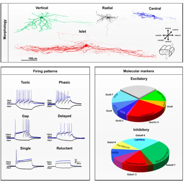
Fig. 1 Spinal cord dorsal horn lamination. a Confocal image of a transverse section of mouse lumbar spinal cord immunostained with antibodies directed against NEUN to mark all neurons (blue), and against PAX2 to reveal only inhibitory neurons (white). Dotted lines
represent boundaries of the six dorsal horn laminae. LSN, lateral spi-nal nucleus. Scale = 100 µm. b Schematic of the location and trajecto-ries of projection neurons within the dorsal horn. ALT antero-lateral tracts, PSDC post-synaptic dorsal columns.
I DYN + neurons express GABA and account for about 32% of lamina I inhibitory neurons in this species (Sardella et al. 2011a). The remaining lamina I DYN + neurons are presumably excitatory neurons that do not express NK1R (Marvizon et al. 2009). Interestingly, in the mouse (but not in the rat) the excitatory DYN + population is located almost exclusively in regions of the DH that receive innervation from glabrous skin (Boyle et al. 2017). Entirely included within the lamina I DYN + inhibitory population are neu-rons expressing galanin (GAL) (Sardella et al. 2011a) which are virtually all GABAergic (Simmons et al. 1995). A dis-tinct population of inhibitory neurons in lamina I express neuropeptide Y (NPY) and these make up around a quarter of the inhibitory neurons in this lamina (Polgar et al. 2011). These are largely distinct from neurons that belong to the DYN + population (Boyle et al. 2017). Additionally, neu-rons expressing the neuronal nitric oxide synthase (NNOS) are virtually all GABAergic in lamina I. In the rat, these are distinct from DYN + and NPY + neurons, and account for 17% of inhibitory neurons in this area (Laing et al. 1994; Sardella et al. 2011b). A subset of small DH lamina I cells express the gastrin-releasing peptide receptor (GRPR), but not NK1R (Sun et al. 2009), and these seem to mediate itch but not pain (Sun and Chen 2007). Because of their small cell bodies, these are likely interneurons, and different from lamina I spinothalamic neurons identified in cats that prefer-entially mediate itch (Andrew and Craig 2001).
Lamina II interneurons
Most investigations of DH neurons have been performed in lamina II (Merighi 2018), the core of the gate control theory, which postulated an interaction between nocicep-tive and non-nocicepnocicep-tive inputs at the spinal level (Melzack and Wall 1965). In this proposal, inhibitory interneurons in lamina II are activated by non-nociceptive sensory neurons to reduce pain. However, when DH inhibition is diminished after injury, innocuous mechanical stimulation of the skin no longer reduces DH nociceptive activity, but rather engages nociceptive circuits through a dorsally directed polysynaptic pathway, leading to cutaneous mechanical allodynia (CMA) (Braz et al. 2014). In lamina II, the broadly accepted mor-phological classification of Grudt and Perl (2002) identi-fied vertical, radial, central and islet cells (Fig. 2, Online Resource 2 and 4). Importantly, the original description of these morphologies was specific to neurons in this area, and obtained in sagittal slices, which include most of their den-dritic arbors (Lu and Perl 2005; Punnakkal et al. 2014; Todd and Lewis 1986). Lamina II vertical, radial, central and islet cells, which will be briefly described below, are thus quite distinct from other DH neurons described across the dorso-ventral axis, and yet these terms are often assigned to neu-rons outside lamina II, or to neuneu-rons observed in transverse
slices (Koch et al. 2018). Nevertheless, the following sec-tion will classify lamina II neurons based on the Grudt and Perl scheme, including current knowledge of their molecular and electrophysiological profiles. Indeed, as in lamina I, DH lamina II neurons can be distinguished depending on their pattern of action potential discharge. However, in addition to be tonic, phasic, delayed or single spike, lamina II neu-rons also include gap (a.k.a. irregular)—which have a delay between two spikes that is greater than 1.5 times the delay between two previous or two following spikes, and reluctant (which do not spike upon depolarization current) neurons (Balachandar and Prescott 2018).
Vertical cells Lamina II vertical cells, previously described as “stalked neurons”, have extensive dendrites running ven-trally in a cone shape through the dorso-ventral axis, when viewed in sagittal slices (Gobel 1978; Maxwell et al. 2007). Vertical cells are thought to represent a population of excita-tory DH neurons with cell bodies located mostly in the outer part of lamina II, and include cells that send axons directly to lamina I projection neurons (Boyle et al. 2019; Cordero-Erausquin et al. 2009; Lu et al. 2013; Lu and Perl 2005). It has been recently suggested that lamina II interneurons expressing DYN include vertical cells (Duan et al. 2014) and belong to the excitatory component of the otherwise mixed lamina II DYN + population (Huang et al. 2018). A subset of excitatory neurons expressing CR may also include verti-cal cells (Smith et al. 2015, 2016). Although it was reported that cells expressing green fluorescent protein under the control of the promotor for gastrin-releasing peptide (GRP), included vertical cells (Sun et al. 2017), these had dorsally directed dendrites and therefore do not fit the Grudt and Perl criteria of vertical cells. Vertical cells have tonic or delayed firing patterns and invariably show sustained action poten-tials during a depolarizing step, with regular or irregular interspike intervals (Grudt and Perl 2002). These particular firing properties have been reported in the putative verti-cal cells described above, including delayed firing lamina II excitatory DYN + (Huang et al. 2018) and CR + neurons (Smith et al. 2015), but are rarely seen in GRP + neurons (Dickie et al. 2018).
Radial cells Lamina II radial cells have relatively short dendrites radiating in all directions, as seen in both trans-verse and sagittal sections. However, the term has also been applied to “stellate cells” described in humans, which are multipolar neurons with straight dendrites that cover a very large elliptic area, extending up to 500 µm into laminae I and III (Schoenen 1982). Radial cells described by Grudt and Perl are, however, quite distinct, with small and highly branched dendrites that do not extend more than few tens of micrometers in the dorso-ventral or medio-lateral axis, and less than 200 µm in the rostro-caudal plane. Axons of radial
cells are generally located in lamina II, with some running in the dorsolateral fasciculus to target the lateral spinal nucleus (LSN) (Dickie et al. 2018). It has recently been reported
that excitatory neurons in lamina II expressing substance P (SP) (Dickie et al. 2018), and some of those expressing PKCγ (Abraira et al. 2017; Alba-Delgado et al. 2015) or
Fig. 2 Morphological, electrophysiological and neurochemical fea-tures of interneurons in laminae I–II of the mouse dorsal horn. Top
panel Morphological features of lamina II dorsal horn neurons
observed in sagittal slices. Confocal images of neurons filled with neurobiotin showing vertical (green), radial (gray), central (blue) or islet (red) morphologies. Scale = 100 µm. Lower left panel Electro-physiological whole-cell patch-clamp recording of dorsal horn neu-rons. Firing pattern of dorsal horn neurons can be tonic, phasic, with a gap between spikes, or with one or no action potential upon depo-larizing current injection. Traces in black display membrane poten-tial at -20pA or at rehobase respectively. Superimposed blue traces are representative firing patterns observed at suprathreshold current injection. Value for hyperpolarizing and depolarizing currents are indicated. Lower right panel The proportions of excitatory and inhibi-tory interneurons in this region that belong to different neurochemical populations. The relationship to the different transcriptomic
popula-tions identified by Haring et al. (2018) is also shown. Note that the
NKB neurokinin B, NTS neurotensin, CCK cholecystokinin, SP
sub-stance P, NPFF neuropeptide FF and GRP–GFP cells form largely non-overlapping populations of excitatory interneurons, although there is some overlap between NKB/NTS and CCK/SP popula-tions [reproduced from Gutierrez-Mecinas et al. (2019a)]. The GRP– GFP cells are defined as those that express GFP in the BAC trans-genic GRP::eGFP line. For inhibitory interneurons, there is overlap between the galanin/dynorphin (GAL/DYN) population and the neu-ronal nitric oxide synthase (NNOS) population, and this is shown in purple. Similarly, the GAL/DYN population overlaps with the NPY population, and this is shown in brown. There is limited over-lap between NPY cells and both NNOS and parvalbumin (PV) cells, although this is not shown on the pie chart. Reproduced from Boyle et al. (2017)
CR (Smith et al. 2015, 2016), correspond to the radial cells defined by Grudt and Perl. Radial cells all have delayed firing patterns, with irregular interspike intervals or high-frequency bursts (Grudt and Perl 2002). Accordingly, the large majority of CR + (Smith et al. 2015) and SP + (Dickie et al. 2018) neurons, and most PKCγ + (Abraira et al. 2017) neurons, have delayed firing, except in the medullary DH where radial PKCγ + neurons are never delayed (Alba-Del-gado et al. 2015).
Central and islet cells Lamina II central and islet cells have characteristic dendrites that are elongated in the sag-ittal plane and can extend, for islet cells, to considerable distances (> 400 µm in the rat) in the rostro-caudal axis. Because of this particular spatial orientation, identification of these cells is difficult or impossible in transverse sec-tions, particularly for islet cells (Gobel 1975; Maxwell et al.
2007; Smith et al. 2015). Central cells can have either tonic or phasic firing patterns (Grudt and Perl 2002). A specific population of excitatory interneurons, originally described as “transient central cells” (Lu and Perl 2005), are thought to receive input from PKCγ + neurons and to project directly to vertical cells (Lu et al. 2013). It has been reported that lamina II excitatory GRP + neurons (Albisetti et al. 2019; Dickie et al. 2018) and subpopulations of excitatory neurons in inner lamina II that express PKCγ (Alba-Delgado et al.
2015) or CR (Smith et al. 2015, 2016) show central mor-phology. Accordingly, the majority of GRP + (Dickie et al.
2018; Pagani et al. 2019) and a third of PKCγ + (Abraira et al. 2017) neurons are phasic, with an initial burst of one or more action potentials, except for trigeminal central PKCγ + neurons, which are mostly tonic (Alba-Delgado et al. 2015). Some excitatory CR + neurons, which include cells with central morphology, also show phasic firing prop-erties in addition to their characteristic delayed first action potential (Smith et al. 2015). Interestingly, the majority of lamina II excitatory GRP + , CR + or PKCγ + neurons that are not delayed and/or phasic fall into the class of single spiking or reluctant spiking neurons (Abraira et al. 2017; Dickie et al. 2018; Smith et al. 2015), and this may reflect an extreme form of adaptation that characterizes the phasic fir-ing pattern. Surprisfir-ingly, in the spinal DH, excitatory lam-ina II GRP + , CR + or PKCγ + neurons are rarely or never tonic, indicating that tonic central cells, which account for about a quarter of recorded central cells (Grudt and Perl
2002), might represent a distinct functional class of inhibi-tory cells. Lamina II inhibiinhibi-tory neurons expressing neuro-peptide Y (NPY) (Iwagaki et al. 2016), parvalbumin (PV) (Abraira et al. 2017), or CR (Smith et al. 2015) are nearly all tonic, but only the PV + population includes cells with central morphology.
Islet cells are invariably inhibitory interneurons and use either GABA, glycine or both neurotransmitters (Heinke
et al. 2004; Maxwell et al. 2007; Todd and McKen-zie 1989; Todd and Sullivan 1990; Yasaka et al. 2010). Interestingly, the islet cell population does not include NPY + inhibitory neurons (Iwagaki et al. 2016). Islet cells include inhibitory neurons that express PV (Abraira et al.
2017; Boyle et al. 2019) or CR (Smith et al. 2015, 2016), the latter population accounting for up to 15% of the entire CR + population, and including a group of inhibi-tory neurons that express preprotachykinin A (PPTA), the precursor for SP (Gutierrez-Mecinas et al. 2019c; Smith et al. 2015, 2016). PV + inhibitory neurons innervate PKCγ + neurons (Petitjean et al. 2015) and also provide axo-axonic synapses onto myelinated LTMR afferent terminals (Boyle et al. 2019). The target of the axons of CR + inhibitory neurons is unknown, but as islet cells they most likely reside in the same layer as their cell bodies (Abraira et al. 2017; Smith et al. 2016). Notably, all islet cells have tonic firing properties (Grudt and Perl 2002). However, it is important to note that inhibitory neurons are not always islet cells and do not always show tonic fir-ing. For example, inhibitory cells that express galanin and dynorphin (GAL/DYN), NNOS or NPY, are morphologi-cally and electrophysiologimorphologi-cally heterogeneous (Ganley et al. 2015; Iwagaki et al. 2016, 2013).
Non‑overlapping populations of lamina II neurons Inter-estingly, lamina II neurons can be classified based on non-overlapping expression of different neurochemical markers, regardless of their morphology or intrinsic firing properties (Fig. 2). Among the excitatory neurons in laminae I–II, six largely non-overlapping populations can be defined based on expression of neurotensin (NTS), SP (also referred to as Tachykinin 1 (TAC1) or PPTA), NKB (also referred to as TAC2 or PPTB), GRP, cholecystokinin (CCK) or neuropep-tide FF (NPFF). In this scheme, the GRP cells are defined by the expression of enhanced green fluorescent protein (EGFP) in a bacterial artificial chromosome (BAC) trans-genic mouse line (GRP::EGFP). Although all of the EGFP cells in this mouse line possess the mRNA for GRP, they seem to represent a distinct subset among the GRP + neu-rons (Gutierrez-Mecinas et al. 2019a). These neurochemi-cally defined populations account for 9% (NTS), 24% (SP), 14% (NKB), 15% (GRP), 7% (CCK) and 6% (NPFF) of all excitatory laminae I–II neurons in the mouse (Gutier-rez-Mecinas et al. 2016, 2017, 2019a, b; Mar et al. 2012), accounting for about 75% of all excitatory neurons in this area. The remaining 25% of lamina II excitatory neurons are likely to include vertical cells, which are rarely found within these six populations of cells. Although vertical cells have been described among certain neurochemical classes, such as those expressing somatostatin (SOM) (Duan et al. 2014), enkephalin (ENK) (Francois et al. 2017) or CR (Gutierrez-Mecinas et al. 2019c), these classes are relatively broad and
overlap extensively with each other, as well as with the six classes defined above. For example, many excitatory neu-rons in laminae I–II contain CR, and these seem to corre-spond to those that express SP, NKB, as well as many of the GRP-EGFP cells, but not those that express NPFF, CCK or NTS (Gutierrez-Mecinas et al. 2019c; Haring et al. 2018).
For inhibitory interneurons in this region, a similar approach has revealed five largely non-overlapping popu-lations that express GAL/DYN, NPY, NNOS, PV or CR. These populations account for nearly all laminae I–II inhibi-tory neurons, making up to 24% (GAL/DYN), 33% (NPY), 17% (NNOS), 11% (PV) and 27% (CR) of the inhibitory cells in this area (Boyle et al. 2017; Gutierrez-Mecinas et al. 2016, 2017, 2019c). Importantly however, in contrast to NPY + lamina II neurons, which are nearly all inhibitory (Rowan et al. 1993), neurons that express DYN (Sardella et al. 2011a), NNOS (Sardella et al. 2011b), PV (Abraira et al. 2017; Laing et al. 1994) or CR (Gutierrez-Mecinas et al. 2019c; Smith et al. 2015) include a relatively large proportion of excitatory cells. Neurons that express the basic helix-loop-helix domain containing, class B, 5 (BHLHB5) during development have been implicated in the inhibition of itch, and these include the vast majority of the GAL/ DYN + and NNOS + populations (Kardon et al. 2014). However, it was recently shown that in contrast to the GAL/ DYN + population which suppresses pruritogen-evoked itch, NNOS + neurons are likely to have an anti-nociceptive role (Huang et al. 2018).
How this classification of excitatory and inhibitory interneurons fits with specific functions in chronic pain pro-cessing is under investigation (Todd 2017).
Laminae III–VI
Laminae III–IV are usually considered together, not from a functional perspective, but because neurons in this area have dendritic trees that generally extend within these laminae, and because cells within them give rise to several ascending tracts (Brown 1982). Deep DH neurons include both projec-tion neurons and interneurons with several morphological patterns and axons that typically run in the ventral direction (Willis and Coggeshall 1978) (Fig. 1, Online Resource 3 and 4). Dendritic trees usually form conical arborizations in laminae III–IV, but are better described as flattened disks in laminae V–VI. They extend in both rostro-caudal and dorso-ventral directions in lamina III, but are mostly restricted to the transverse plane in deeper laminae (Scheibel and Schei-bel 1968). A subset of lamina III neurons are antenna-type cells with long dorsally directed dendrites, some of which express NK1R (Fernandes et al. 2018; Naim et al. 1997). In the rat, these NK1R + cells can be identified as projection cells belonging to the ALT (Marshall et al. 1996; Naim et al.
1997; Todd et al. 2000). However, other cells with similar
morphology that lack the NK1R appear to be interneurons (Polgar et al. 2007). Within the deep DH, there are several neurons that synapse directly onto ventral horn motor neu-rons. It is important to note that these pre-motor neurons are located through the whole DH, but are highly concentrated in the lumbar laminae IV–VI. These neurons were recently identified as excitatory and inhibitory interneurons that express the transcription factor TCFAP2β and the nuclear and chromatin organization factors SATB1/2 during devel-opment, and are located predominantly in medial lamina V (Levine et al. 2014). The firing patterns of lamina III–IV neurons are similar to those in lamina II, and include tonic, phasic, delayed, single, gap and reluctant spiking (Abraira et al. 2017). In contrast, cells in the deepest part of the DH have mostly tonic or phasic firing patterns, with higher fir-ing frequency than in superficial laminae (Ruscheweyh and Sandkuhler 2002). Of note, medullary DH neurons located in lamina V also include neurons with delayed firing (Moris-set and Nagy 1998).
Projection neurons The deep DH contains several classes of projection neurons. These give rise to various tracts, but most of them send projections through the spinocervical tract (SCT), the ALT, or the dorsal columns (Brown 1982). Laminae III–IV SCT neurons send axons through the most medial and superficial parts of the ipsilateral dorsal funicu-lus (Brown 1982). These cells usually have elongated den-drites that form a cylinder in the dorso-ventral axis. How-ever, it is unclear whether the SCT exists in humans, as this tract might be rudimentary in primates (Truex et al. 1970).
Lamina III–IV cells include ALT projection neurons that send axons to the thalamus, CVLM and LPb area. The proportion of spinothalamic DH neurons in these laminae is highly dependent on the spinal segment, and these are numerous in the cervical and thoracic spinal cord, but much less frequent in the lumbosacral region (Al Khater et al.
2008; Burstein et al. 1990; Davidson et al. 2010). In con-trast to lamina I spinothalamic cells described above, most of those in the deep DH are located in laminae V–VI and have very large cell bodies and dendritic trees spreading across multiple laminae (Willis et al. 1979). They display several morphologies with dendrites oriented dorsally up to lamina I, or radiating as far as the lateral funiculus or the border of laminae VII and X. There are also a few neurons of the spinoreticular (SRT) and spinomesencephalic (SMT) tracts in the lateral part of laminae V–VI with multipolar morphologies and long straight dendrites (Kevetter et al.
1982; Menetrey et al. 1982). Axons of lamina III ALT neu-rons typically cross the ventral commissure near their cell body and run mostly through the ventrolateral quadrant, and also through the dorsolateral funiculus in cats, rats and mon-keys (Sengul and Watson 2015). Two major sources of local synaptic input to lamina III ALT cells have been identified,
originating from DYN + excitatory and NPY + inhibitory neurons (Baseer et al. 2012; Naim et al. 1997; Polgar et al.
1999b). In the rat, the great majority of these lamina III ALT projection neurons show strong NK1R expression (Ding et al. 1995; Marshall et al. 1996; Todd et al. 2000). A simi-lar population of ALT neurons is also present in the mouse, but in this species, few of them express NK1R (Cameron et al. 2015).
Laminae III–V include neurons that belong to the post-synaptic dorsal column (PSDC) system, which send axons to the ipsilateral gracile or cuneate nucleus through the dor-sal columns (Brown 1982). They are located in the medial part of the DH with extensive dendritic trees that radiate in various axes, but that are mainly restricted to the transverse plane (Brown and Fyffe 1981). The molecular identity of PSDC cells is unclear, but some of these cells transiently express the transcription factor ZIC2 (Paixão et al. 2019). Interestingly, these cells do not appear to express the NK1R (Polgar et al. 1999b, 2007).
Additionally, most neurons of the spinocerebellar tract are located in the ventral horn, mainly in the Clarke’s columns, but a few of them have also been identified in the DH lamina V, particularly in the thoracic and the rostral half of the lum-bar spinal cord (Matsushita and Hosoya 1979). Interestingly, in contrast to spinocerebellar neurons from Clarke’s column, which selectively express lial derived neurotrophic factor (GDNF) and send axons through the contralateral spinal cord (Hantman and Jessell 2010), DH spinocerebellar neu-rons run through the ipsilateral lateral funiculus (Edgley and Gallimore 1988). These cells have recently been identified as deep DH excitatory neurons that express the basic helix-loop-helix (bHLH) transcription factor, ATOH1 (Yuengert et al. 2015). These cells are critical to perform motor tasks, but are dispensable for nocifensive pain behavior.
Several of these projection neurons have been quantified in the deep DH, but the relative proportion of each of these cell classes within this region is not available. The molecular phenotype and firing patterns of these projection neurons are also unclear, but they may belong to excitatory popula-tions of the deep DH including those expressing LMX1B, the ladybird homeobox I (LBX1), the ROR alpha nuclear orphan receptor (RORα), RORβ, the V-maf musculoaponeu-rotic fibrosarcoma oncogene homolog A (MAFA), MAFB or CMAF (Del Barrio et al. 2013), and may feature in recent DH single-cell transcriptomic studies (Haring et al. 2018; Sathyamurthy et al. 2018; Zeisel et al. 2018).
Excitatory interneurons As mentioned above, the deep DH contains both projection neurons and interneurons. The morphology of deep DH interneurons is highly heterogene-ous and has been described for several populations of neu-rochemically defined cells. Some deep DH neurons were suggested to have morphologies similar to vertical, radial or
central cells described by Grudt and Perl in lamina II, such as excitatory neurons in lamina III/IV expressing RORα (Bourane et al. 2015b) or those transiently expressing VGLUT3 (Peirs et al. 2015). Once again, while it is likely that those cells do belong to real morphological classes, they seem to be distinct from lamina II neurons, with different morphology and neurochemistry. The neurochemical popu-lations identified in this region are also not exclusive to the deep DH. About 60% of RORα + neurons also express CCK (Bourane et al. 2015b), and RORα + neurons also partially overlap with the excitatory subset of neurons that express RORβ (Del Barrio et al. 2013). As indicated earlier, a few scattered excitatory neurons in lamina III express NK1R or PKCγ and are included in the major cell populations of the deep DH that express SOM (Duan et al. 2014), CCK (Gutierrez-Mecinas et al. 2019b), RORα (Bourane et al.
2015b) or VGLUT3 during development (Peirs et al. 2015). SOM + neurons are predominantly excitatory neurons with phasic, delayed and single spiking patterns, and account for 16–17% of lamina III neurons (Duan et al. 2014; Gutier-rez-Mecinas et al. 2016). CCK + excitatory neurons, which again partially overlap with the excitatory RORα + and RORβ + cells, account for a third to a quarter of excitatory neurons in lamina III (Gutierrez-Mecinas et al. 2019b) and have phasic or tonic firing patterns (Abraira et al. 2017). VGLUT3 + neurons are subdivided into at least two popula-tions, one with tonic or phasic firing patterns (Peirs et al.
2015), and a second located more dorsally that predomi-nantly displays delayed firing (Cheng et al. 2017).
Inhibitory interneurons The deep DH contains large popu-lations of inhibitory neurons such as those expressing the receptor tyrosine kinase Ret (RET) (Cui et al. 2016) and some that express Rorβ (Koch et al. 2017). Based on the morphological classification described in lamina II by Grudt and Perl, it has been suggested that these neurons have morphologies similar to vertical, radial and islet cells for the RET + population, and vertical and central cells for the RORβ + population, with the exception of RORβ + neu-rons of laminae V–VI that resemble islet cells. Similar to the excitatory neurons described above, the morphologies of these cells are, however, most likely unrelated to those assigned to lamina II neurons, although they may reflect specific functions of neurons of the deep DH. In contrast to inhibitory neurons in lamina II described above which mostly have tonic firing patterns, those in lamina III include tonic, phasic, delayed, gap and reluctant firing (Abraira et al.
2017). Tonic firing is, however, still predominant in the deep DH, in both GABAergic and glycinergic neurons (Punnak-kal et al. 2014). Similar to the excitatory neuron populations of the deep DH described above, inhibitory neurons in this area are highly heterogeneous. RET + inhibitory neurons make up about one-third of all inhibitory neurons of the deep
DH and these cells express other inhibitory markers such as GLYT2, GAD1/2 and PV, but not NNOS or DYN (Cui et al.
2016). As for excitatory neurons of the deep DH, scattered inhibitory neurons in this area express markers described in superficial laminae, such as NPY, DYN/GAL, NNOS, PV or CR (Gutierrez-Mecinas et al. 2019c; Polgar et al. 2013a, b; Sardella et al. 2011a). However, in contrast to the superficial laminae, inhibitory neurons in lamina III do not apparently coexpress GAL and DYN (Sardella et al. 2011a). In fact, DYN + neurons make up less than 1% of lamina III neurons and include excitatory neurons in the medial part of the DH in this area (Boyle et al. 2017). GAL + neurons in lamina III coexpress NNOS and account for 5% of inhibitory neurons in this area (Tiong et al. 2011). The firing pattern of these cells is unknown. PV + inhibitory neurons make up about 8% of all lamina III neurons (Abraira et al. 2017). Because about 40% of lamina III neurons express GABA, we can estimate that the PV + inhibitory population represents 20% of inhibitory neurons in this area (Polgar et al. 2013a). PV + inhibitory neurons have rostro-caudally elongated dendrites, resembling lamina II islet cells (Abraira et al.
2017; Boyle et al. 2019; Hughes et al. 2012) and generally show tonic firing (Abraira et al. 2017; Boyle et al. 2019; Hughes et al. 2012). CR + inhibitory neurons are very sparse in lamina III, but more numerous in laminae IV–VI. Most of them express PAX2 (Peirs, unpublished observation) but not much is known about their relative number or firing patterns. Similar to lamina II, NPY + inhibitory neurons in lamina III do not express DYN/GAL, PV or NNOS (Iwagaki et al. 2016). NPY + inhibitory neurons account for up to 25% of inhibitory neurons in lamina III (Boyle et al. 2017) and are morphologically heterogeneous, but never display elongated dendrites like islet cells (Iwagaki et al. 2016). Lamina III NPY + inhibitory neurons are mostly tonic fir-ing neurons, but also include phasic and sfir-ingle spikfir-ing cells (Iwagaki et al. 2016).
Non‑overlapping populations of deep dorsal horn neu‑ rons Interestingly, a recent study used the Allen Brain Insti-tute Spinal Cord Atlas to identify seven classes of excitatory and four classes of inhibitory DH neurons in lamina IIi–III that largely do not overlap, and that account for 70–82% of all neurons in this area (Abraira et al. 2017). This clas-sification includes excitatory neurons expressing cerebel-lin-2 (CBLN2), CCK, the serotonin receptor 6 (5HTR6), the insulin-like growth factor binding protein 5 (IGFBP5), the neurogenic differentiation factor-4 (NEUROD4), PV or PKCγ, and inhibitory neurons expressing cadherin-3 (CDH3), the Kv channel interacting protein-2 (KCNIP2), RORβ or PV. Of note, this classification of DH neurons par-tially matches previously described cell populations, such as deep excitatory neurons expressing CCK that have recently been implicated in touch processing (Abraira et al. 2017;
Liu et al. 2018), or deep inhibitory neurons expressing PV that contribute to feedforward inhibition of peripheral sensory neurons and DH interneurons (Boyle et al. 2019; Petitjean et al. 2015).
Classification of dorsal horn neuron populations: pitfalls and future directions
The DH clearly displays a high degree of heterogeneity in neuronal morphologies, neurochemistry and intrinsic prop-erties. Studies described above that performed morpho-metric analysis of neurochemicaly identified DH neurons have shown that a morphological class is often shared by several cell subpopulations, including by projection neu-rons and interneuneu-rons, or excitatory/inhibitory interneuneu-rons classes, thus making it difficult to determine how morphol-ogy or neurochemistry relates to function, and what criteria define a functionally distinct population of cells. A good example of such complexity can be seen for one of the DH populations first implicated in CMA: PKCγ-expressing cells. These are nearly exclusively excitatory interneurons (Pol-gar et al. 1999a) and make up 30% of neurons in the inner part of lamina II, with very scattered cells in laminae III and I (Mermet-Joret et al. 2017; Peirs et al. 2014). Such a restricted cellular phenotype and location within the DH make these cells good candidates to define a functionally distinct population of DH neurons, and these neurons have indeed been the subject of numerous investigations (Artola et al. 2020). However, morphological (Peirs et al. 2014), electrophysiological (Alba-Delgado et al. 2015) and tran-scriptomic (Haring et al. 2018) analysis of PKCγ + neurons revealed significant heterogeneity in these cells, exposing at least two subgroups of PKCγ + neurons, with potentially distinct functions. In fact, PKCγ + neurons can be distrib-uted among the non-overlapping populations of DH neurons described above. Cells in laminae II–III that possess PKCγ frequently coexpress NTS (Polgar et al. 1999a), CCK (Gut-ierrez-Mecinas et al. 2019b) or NKB (Gutierrez-Mecinas et al. 2016), although the NKB + cells generally show very low levels of the kinase. It has recently been reported that most (~ 85%) of the cells with strong PKCγ immunoreactiv-ity express either NTS or CCK (Gutierrez-Mecinas et al.
2019b; Polgar et al. 1999a), and it will be of interest to deter-mine whether these groups correspond to the subclasses of PKCγ neurons that were previously identified. Furthermore, as mentioned above, a significant proportion of ALT lamina I neurons express PKCγ, suggesting again that expression of the kinase alone is not sufficient to define a unique neuronal population.
It is also important to note that regardless of the loca-tion within the DH, there are several neurons described in the above studies that could not be assigned to any of the morphological classes that have been proposed, once again
making it difficult to build a bridge between DH cell mor-phology, molecular phenotype and function. Remarkably, however, analysis of laminae IIi–IV DH neurons has recently
demonstrated that linear discriminant morphometric analy-sis of DH neurons, including cell body size, neurite length, spine density and neurite complexity using Sholl-based metrics and branching index measurements, creates linear classifiers that recognize interneuron subtypes with up to 88% accuracy (Abraira et al. 2017). Of note, however, it is important to emphasize that morphological features identi-fied in healthy DH neurons may no longer apply after injury. For example, the population of PKCγ + neurons described in Abraira et al. (2017) have a dendritic tree that severely shrinks after peripheral inflammation (Alba-Delgado et al.
2018).
Importantly, recent clustering from single cell transcrip-tomic analysis of the spinal cord has revealed about 15 popu-lations of DH excitatory and 15 popupopu-lations of DH inhibi-tory neurons (Haring et al. 2018; Sathyamurthy et al. 2018; Zeisel et al. 2018). Notably, most clusters are defined by coexpression of more than one marker, which most likely better reflects the DH complexity compared to previous DH neuronal classifications. RNA-seq data have already pro-vided promising anatomical information, with some of these populations corresponding particularly well to the previously described neurochemical classification. For example, among inhibitory interneurons, the GAL/DYN + , NPY + , CR + and PV + populations identified in laminae I–II seem to include cells in the GABA clusters 1–3, 5–7, 8–9 and 14 of Haring et al. (2018), respectively (Fig. 2). Among excitatory neu-rons in this region, the CCK + , NTS + , NKB + , NPFF + and SP + cells include those in the GLUT clusters 2–3, 4, 5–7, 9 and 10–11 of Haring et al. (2018), respectively (Fig. 2). Most importantly, the transcriptomic data also reveal the cel-lular content of DH neuronal populations and already con-firms previous observations, for example identifying neurons of the DE-4 cluster of Sathyamurthy et al (2018) that express PKCγ, but not the delta opioid receptor (DOR) (Wang et al.
2018). However, the clusters defined by these studies are often extensive in their laminar spread (Haring et al. 2018). For example the Glut10–11 clusters, which include excita-tory SP + cells, extend throughout all DH laminae. Since Dickie et al. (2018) identified a distinctive population of SP + neurons in lamina II, it will be important to determine the extent to which the transcriptomic clusters contain func-tionally distinct subsets of DH neurons, and whether this fits current knowledge of DH neuronal populations (Koch et al.
2018). Functional analysis of DH neurons in pain process-ing has also begun usprocess-ing transcriptomic data, by combin-ing detection of the immediate-early gene Arc or cFos, with markers of identified cell types using immunohistochemistry or in situ hybridization multiplexing, in pathophysiological conditions (Haring et al. 2018; Sathyamurthy et al. 2018).
Most importantly, these transcriptomic analyses provide putative markers for cell clusters located in the superficial, but also deep DH, and may reveal the as-yet unknown chem-ical phenotype of several superficial neurons and most of deep DH neurons. Additionally, it has recently been shown that ion channel density distributions, and particularly densi-ties of KV1- and A-type potassium conductances, can predict
neuronal firing pattern in the DH (Balachandar and Prescott
2018). It will be thus very interesting to see if transcriptomic expression of potassium conductance-related channels from the newly identified cell clusters can predict the firing pat-tern properties of previously recorded DH neurons, in physi-ological conditions but also during persistent pain states.
Of note however, future investigations will have to test whether this new classification of DH neurons can be applied to different dermatomes and to different species, including humans. For example, in mice, expression of the neuronal calcium-binding protein NECAB1 is used to label the cluster DE-5 in Sathyamurthy et al. (2018), and is strongly expressed in the Glut5 cluster in Haring et al. (2018). However, NECAB1 is expressed almost exclusively by excitatory neurons in the superficial laminae, excluding the PKCγ + population, in mice, but by half of inhibitory neurons expressing the SOM receptor SST2R and half of excitatory PKCγ + neurons in rats (Zhang et al. 2014, 2016). On the other hand, NECAB2 labels similar populations of neurons in the inner part of lamina II in mice, rats and humans (Zhang et al. 2014, 2016), but is expressed by sev-eral clusters defined by Haring et al. (2018) and Sathyamur-thy et al. (2018). There are also important differences in gene expression in distinct spinal segments and DH territories innervated by hairy and glabrous skin, which may be missed in current transcriptomic clustering of DH neurons. Clusters of Haring et al. (2018), and most likely Sathyamurthy et al. (2018) as well, are widely distributed in the medio-lateral axis and were defined by cells originating from several somatotopic territories. For example, excitatory DYN + neu-rons are included in the DE-15 (Sathyamurthy et al. 2018) and Glut14 (Haring et al. 2018) clusters. However, excita-tory DYN + neurons are largely restricted to mid-lumbar DH (corresponding to glabrous skin territory) (Huang et al.
2018), and transcriptomic clusters do not seem to reflect these differences. Finally, it will be critical to determine whether these molecular markers remain unchanged after injury, as mRNA expression undergoes important regula-tions in persistent pain states (Tansley et al. 2018; Uttam et al. 2018).
Current advances in dorsal horn neurons
associated with cutaneous mechanical
allodynia
Cutaneous mechanical allodynia
CMA is a highly debilitating symptom of chronic pain reported by about 45% of patients, for whom normally innocuous stimulation of the skin is perceived as painful (Bouhassira et al. 2005). A similar prevalence of CMA is found in patients with pain syndromes that are associated with nervous system or somatic lesions, including nerve trauma or inflammatory arthropathies (Bouhassira et al.
2005). CMA can be generated following a gentle movement (dynamic CMA) or a blunt pressure (static CMA) applied to the surface of the skin. Prevalence of static CMA does not differ between patients with or without neuropathic char-acteristics, whereas dynamic CMA preferentially affects patients with neuropathic pain (Bouhassira et al. 2005). The large range of terms (burning, stabbing, electric shocks…) and conditions associated with this disease illustrates well that CMA is multidimensional and related to highly hetero-geneous symptoms, mechanisms and neural circuits (Bou-hassira et al. 2008; Peirs and Seal 2016).
The dorsal horn: a critical entry point for cutaneous mechanical allodynia
Understanding of chronic pain circuits, including those responsible for CMA, is currently limited, but yet critical for the development of an efficient evidence-based therapeu-tic approach (Freeman et al. 2014). Nevertheless, neuronal circuits underlying CMA have been the subject of numer-ous investigations, with a large contribution from preclinical animal studies (Braz et al. 2014; Lechner 2017; Peirs and Seal 2016). During the last decade, there has been particular interest in understanding CMA neural circuits within the DH. Indeed, selective loss of a large proportion of DH neu-rons, through conditional deletion of the testicular orphan nuclear receptor 4, results in near complete absence of supraspinal integrated pain behaviors (Wang et al. 2013), revealing the critical role of the DH in chronic pain process-ing. Frustratingly however, an accurate description of DH neuronal circuits engaged during CMA remains difficult to achieve. Part of the reason for this relates to the fact that many studies have investigated relatively large neuronal populations that may be somewhat heterogeneous. Thus, it is often difficult to identify which subpopulation may actu-ally participate in CMA, where within the DH and how. Furthermore, evidence suggests that the phenotype of DH neurons engaged during CMA is closely correlated with the
pain etiology, changing our view of the neuronal circuitry to specific microcircuits underlying the symptom.
Dorsal horn neurons associated with persistent cutaneous mechanical allodynia
Persistent neuropathic pain
Neuropathic pain affects 7–10% of the general population and results from a lesion affecting peripheral or central neu-rons (Colloca et al. 2017). Etiology for chronic neuropathic pain is multifactorial, and this has led to the development of several animal models (Coderre and Laferriere 2019; Kumar et al. 2018), potentially associated with different neuronal circuits. Neuronal populations engaged in the DH during neuropathic pain were historically revealed using immu-nohistochemistry directed against immediate-early genes (a.k.a. primary response genes) such as cFos, on DH slices. Analysis of these so-called “activity markers” in the DH after sciatic nerve lesion reveals cells located in the most superficial DH, through the injured segment ipsilateral to the nerve lesion, but also some scattered cells in deeper laminae from III to V. Interestingly, the distribution of cFos + cells within the DH changes over time, with numerous cells located in laminae I–II immediately after the nerve lesion, but which spread to deeper laminae from 2 days to 4 weeks after the injury (Chi et al. 1993).
In is important to note that prolonged general anesthe-sia may affect the expression of these activity markers. Indeed, the DH neuronal activity of non-anesthetized ani-mals includes ongoing activity related to the nerve trauma itself, but also activity induced by mechanical, and some-times chemical or thermal, unpredicted stimulations of the limbs and body that are invariably generated by the animal motor behaviors. Thus, in deeply anesthetized neuropathic mice (e.g., in the absence of peripheral stimulus), expres-sion of the activity marker pERK1/2 or cFos is only slightly increased in superficial DH compared to sham animals (Liu et al. 2018; Peirs et al. 2015), suggesting that DH neu-ronal plasticity, rather than ongoing activity, remains for an extended period of time after nerve lesion. Importantly, such plastic changes seem to occur only in the injured, but not adjacent, segment in rats (Lu et al. 2013). The molecular identity of these activated cells is currently unknown.
Neuropathic static cutaneous mechanical allodynia As mentioned above, peripheral neuropathy can induce static and/or dynamic CMA in 46% and 41% of patients, respec-tively (Bouhassira et al. 2005). The facts that: (1) disruption of DH GABAergic (Peirs et al. 2016) or glycinergic (Mirau-court et al. 2009) inhibition induces static or dynamic CMA, respectively, (2) morphine dose dependently blocks static but not dynamic CMA, and (3) static and dynamic CMA
seem to be preferentially signaled by Aδ and Aβ afferents, respectively (Field et al. 1999), suggest that different DH circuits underlie these symptoms.
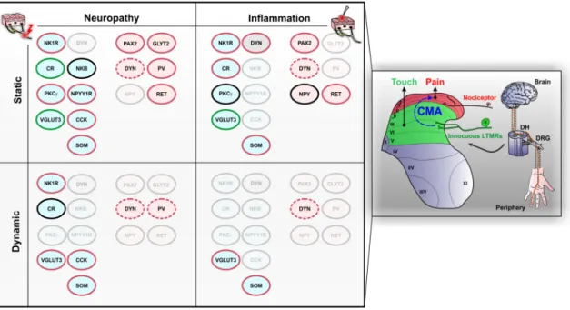
In anesthetized neuropathic mice, static mechanical stimulation of the skin using innocuous von Frey filaments recruits cells in the DH that extend from lamina I to VI (Peirs et al. 2015). Using cFos as an activity marker, it was reported that about 70% of DH neurons activated during neuropathic static CMA are PAX2-negative, suggesting that about two-thirds of this neuronal circuit is excitatory (Peirs et al. 2015). Co-immunolabeling of cFos with neuronal markers previously described reveals activity in excitatory neurons that transiently express VGLUT3 in lamina III, and PKCγ and CR neurons in lamina II. These cells account for 24% of all cFos + cells in laminae I–III. Because in these conditions, 29% of laminae I–III cFos + cells are inhibitory, we can estimate that VGLUT3 + , PKCγ + and CR + neurons account for 34% of all excitatory neurons expressing cFos during neuropathic static CMA in this area. Of note, selec-tive intersectional ablation of DH VGLUT3 + or CR + neu-rons does not affect neuropathic static CMA (Cheng et al.
2017; Duan et al. 2014), suggesting that these cells are engaged but not required in these conditions. Selective inhibition of DH PKCγ + neurons in neuropathic conditions has not been described yet. Nonetheless, pharmacological inhibition (Petitjean et al. 2015) or virally mediated down-regulation (Zou et al. 2011) of the DH PKCγ kinase strongly reduces static mechanical allodynia in rats and mice after sciatic nerve injury, suggesting that this population is neces-sary for the expression of neuropathic static CMA.
Other excitatory DH neurons also participate in neuro-pathic static CMA. Selective intersectional ablation of DH neurons expressing SOM significantly reduces static CMA following sciatic nerve injury (Duan et al. 2014). However, SOM is expressed by many excitatory neurons in laminae I–II, including most of those belonging to each of the six neurochemically defined populations shown in Fig. 2, sug-gesting that this ablation strategy is likely to have affected a large proportion of superficial DH excitatory interneurons belonging to different functional populations (Gutierrez-Mecinas et al. 2019a; Todd 2017). Selective ablation of DH excitatory neurons expressing the NPY receptor NPYY1R, using intrathecal delivery of NPY-saporin, significantly reduces neuropathic static CMA in rats (Nelson et al. 2019). Interestingly, such intervention also reduces cold hypersen-sitivity induced by nerve injury, which is reported by 28% of patients with neuropathic pain (Bouhassira et al. 2005). However, it is unclear which of the different types of neu-rons that have been ablated is responsible for the behavioral effect, as NPYY1R is expressed by numerous excitatory neu-rons in the superficial DH including SOM + and CR + (but not PKCγ +) neurons (Nelson et al. 2019). Similarly, virally mediated ablation of the large population of DH CCK + cells
nearly abolishes static CMA after peripheral nerve injury in mice (Liu et al. 2018). Saporin-mediated ablation of DH NK1R + neurons also strongly reduces neuropathic static CMA after ligation of the L5 and L6 spinal nerves in rats (Nichols et al. 1999). Because 80% of projection neurons in lamina I express NK1R in rats, this major spinal output of the ALT is likely to have been substantially affected by the saporin treatment. However, as indicated earlier, other cells mostly located in laminae III and IV also express NK1R, and may be included among those ablated with substance P-saporin. It is thus unclear which of the NK1R + cells are responsible for the behavioral effect observed after cellu-lar ablation. Of note, selective intersectional ablation of TAC2 + DH neurons (i.e., those that express neurokinin B) did not affect neuropathic CMA in mice (Duan et al. 2014).
It is commonly accepted that chronic neuropathic pain is associated with reduced neuronal inhibition in the DH (Gradwell et al. 2019; Zeilhofer et al. 2012a), and that restor-ing spinal inhibition, usrestor-ing GABAergic cell transplants for example (Braz et al. 2012), has a therapeutic potential to relieve neuropathic CMA. Accordingly, selective activa-tion of DH PV + (Petitjean et al. 2015) or GLYT2 + (Fos-ter et al. 2015) inhibitory neurons, using virally delivered designer receptors exclusively activated by designer drugs (DREADDs), strongly reduces static CMA following sciatic nerve injury in mice. Similarly, selective activation of deep DH inhibitory interneurons that express RET, using virally delivered excitatory DREADDs, significantly reduces static CMA induced by L4 spinal nerve ligation in mice, whereas ablation of the cells enhances it even more (Cui et al. 2016). In contrast, selective intersectional ablation of DH neu-rons expressing DYN does not aggravate static CMA after spared nerve injury in mice (Duan et al. 2014). However, it is important to note that DYN + ablated mice spontane-ously develop static CMA, and it is thus possible that nerve injury affects these cells, leading to neuropathic static CMA. Similarly, selective ablation of DH PV + inhibitory neurons using virally delivered saporin leads to static CMA in mice, and as indicated above, DH PV + neurons are involved in neuropathic static CMA (Petitjean et al. 2015).
Neuropathic dynamic cutaneous mechanical allodynia Less is known about DH circuits underlying neuropathic dynamic CMA. In anesthetized mice with nerve injury, innocuous stimulation of the skin with a paintbrush evokes intense activity in the whole DH, including in neurons expressing CCK in laminae III–IV and NK1R in lamina I (Liu et al.
2018). As for static CMA, virally mediated ablation of DH CCK + cells nearly abolishes dynamic CMA after periph-eral nerve injury in mice (Liu et al. 2018). Interestingly, selective intersectional ablation of DH VGLUT3 + neurons also strongly reduces neuropathic dynamic CMA in mice (Cheng et al. 2017), indicating that while these cells are
dis-pensable for static neuropathic CMA, they are required for the dynamic form of neuropathic CMA. Selective intersec-tional ablation of DH neurons expressing SOM significantly reduces dynamic CMA following sciatic nerve injury in mice (Duan et al. 2014) but, as indicated above, it is unclear which subpopulation(s) of SOM + cells is/are responsible for this effect. As for neuropathic static CMA, selective intersectional ablation of DH CR + neurons does not affect dynamic CMA after sciatic nerve injury, indicating that CR neurons are not required for any form of CMA in neuro-pathic conditions (Duan et al. 2014).
Very little is known about DH inhibitory neurons that participate in neuropathic dynamic CMA. Similar to neuro-pathic static CMA, selective intersectional ablation of DH neurons expressing DYN does not affect dynamic CMA after spared nerve injury in mice (Duan et al. 2014). However, it is important to note that DYN + ablated mice also spontane-ously develop dynamic CMA, and activity from these cells could be affected by nerve lesions, leading to neuropathic dynamic CMA. Similarly, selective inhibition or ablation of DH PV + inhibitory neurons using respectively virally deliv-ered tetanus toxin light chain or saporin, leads to dynamic CMA and cFos activity in the DH induced by brush stimula-tion in mice, suggesting that DH PV + neurons might also be involved in neuropathic dynamic CMA (Boyle et al. 2019; Petitjean et al. 2015).
Persistent inflammatory pain
Inflammatory pain can be associated with a number of diseases and results from the local release of inflamma-tory signaling molecules from immune cells, including cytokines, growth factors and prostaglandins. Symptoms associated with inflammatory pain include redness, warmth and swelling of the affected area, but also manifestations that can overlap with those of neuropathic pain, such as CMA (Vardeh et al. 2016). Importantly, inflammatory mediators such as nitric oxide and tumor necrosis factor-α (TNF-α) can induce nerve damage and also elements of neuropathic pain. There are thus several common mechanisms associ-ated with neuropathic and inflammatory pain (Xu and Yaksh
2011), but also major differences, such as pharmacological responsiveness to non-steroidal anti-inflammatory drugs (NSAIDs) and opiates (Lynch and Watson 2006), suggesting that mechanisms, and by extension neuronal circuits engaged during CMA associated with inflammatory pain, might differ from those associated with neuropathic CMA.
Several animal models have been developed to study spe-cific pathologies associated with inflammatory pain, such as multiple sclerosis (MS) (Procaccini et al. 2015) or migraine (Chou and Chen 2018; Dallel et al. 2018), but most common models of inflammatory pain involve injections of agents such as capsaicin, formalin, mustard oil, carrageenan or
complete Freund’s adjuvant into the skin, muscle, visceral organs or joints (Coderre and Laferriere 2019; Gregory et al.
2013). Virtually, all of these models are associated with immediate CMA, with some lasting longer due to chronic inflammation, such as the carrageenan and CFA pain models for which CMA can persist from days to several weeks. In rats, CFA-induced inflammation rapidly increases cFos and pERK expression in the DH, predominantly in laminae I–II, and these remain upregulated for at least 24–48 h following the injection (Geranton et al. 2008; Ji et al. 2002). Neu-rons that are active 48 h after CFA injection include lamina I NK1R + and DYN + neurons in the superficial DH, but also a few DYN + neurons of the deep DH (Ji et al. 2002). However, and as for nerve injury, results from these stud-ies were obtained from behaving animals and may include cells directly activated by the injury, but also cells activated by stimulation of the skin due to motor behavior. Indeed, the DH of anesthetized mice that had undergone prolonged anesthesia 24 h after carrageenan or CFA treatment (i.e. in the absence of peripheral stimulus) displays very scattered cells through laminae I–VI, suggesting again that DH neu-ronal plasticity, rather than ongoing activity, remains for an extended period of time following long-term tissue inflam-mation (Gao and Ji 2010; Peirs et al. 2015).
Inflammatory static cutaneous mechanical allodynia In anesthetized mice with carrageenan-induced persistent inflammation, static mechanical stimulation of the skin with innocuous von Frey filaments elicits cFos in cells located predominantly in the medial part of the DH lamina I and II, but also through the deeper layers, mostly in laminae IV–VI (Peirs et al. 2015). In another study, innocuous static stimulation of the skin (by a cotton tip) of anesthetized rats treated 24 h earlier with CFA, increased pERK + neurons in the DH lamina I and the outer part of lamina II (Gao and Ji 2010). Interestingly, 59% of cells activated by inflam-matory static CMA do not express the inhibitory marker PAX2, suggesting that fewer excitatory neurons, or more inhibitory neurons, are engaged during inflammatory static CMA compared to CMA in neuropathic conditions (Peirs et al. 2015). Co-immunolabeling of cFos with neuronal markers reveals activity in excitatory neurons that tran-siently express VGLUT3 in lamina III, and CR neurons in lamina II in mice (Peirs et al. 2015). These cells account for a third of cFos + cells in laminae I–III, which we can esti-mate to be about 56% of all excitatory neurons expressing cFos during inflammatory static CMA in this area. Simi-lar to neuropathic injury, selective intersectional ablation of VGLUT3 + neurons does not alter inflammatory static CMA induced by subcutaneous injection of CFA in mice (Cheng et al. 2017), suggesting again that VGLUT3 + neu-rons are engaged in, but not required for, any form of static CMA. Interestingly, neither pERK1/2 nor cFos has been