HAL Id: inserm-00521648
https://www.hal.inserm.fr/inserm-00521648
Submitted on 1 Oct 2010HAL is a multi-disciplinary open access archive for the deposit and dissemination of sci-entific research documents, whether they are pub-lished or not. The documents may come from teaching and research institutions in France or abroad, or from public or private research centers.
L’archive ouverte pluridisciplinaire HAL, est destinée au dépôt et à la diffusion de documents scientifiques de niveau recherche, publiés ou non, émanant des établissements d’enseignement et de recherche français ou étrangers, des laboratoires publics ou privés.
Paraffin-embedded tissue is less accurate than frozen
section analysis for determining VHL mutational status
in sporadic renal cell carcinoma.
Grégory Verhoest, Jean-Jacques Patard, Patricia Fergelot, Florence Jouan,
Salim Zerrouki, Stéphane Dréano, Stéphanie Mottier, Nathalie
Rioux-Leclercq, Marc Denis
To cite this version:
Grégory Verhoest, Jean-Jacques Patard, Patricia Fergelot, Florence Jouan, Salim Zerrouki, et al.. Paraffin-embedded tissue is less accurate than frozen section analysis for determining VHL mutational status in sporadic renal cell carcinoma.. Urologic Oncology: Seminars and Original Investigations, Elsevier, 2012, 30 (4), pp.469-75. �10.1016/j.urolonc.2010.07.005�. �inserm-00521648�
Paraffin-embedded tissue is less accurate than frozen
section analysis for determining VHL mutational status in
sporadic renal cell carcinoma.
Grégory VERHOEST ¹ ², Jean-Jacques PATARD¹ ², Patricia FERGELOT ¹ ³,
Florence JOUAN¹, Salim ZERROUKI³, Stéphane DREANO¹, Stéphanie
MOTTIER¹, Nathalie RIOUX-LECLERCQ¹
4, Marc G DENIS ¹ ³
¹ CNRS/UMR 6061 IFR 140, Rennes University, 35043 Rennes Cedex, France
² Department of Urology, Rennes University Hospital ³ Department of Biochemistry, Rennes University Hospital
4 Department of Pathology, Rennes University Hospital
Key words: VHL gene, renal cell carcinoma, paraffin, VEGF, angiogenesis
Correspondence:
Dr Gregory VERHOEST
Department of Urology – Rennes University Hospital
Henri Le Guilloux St
35033 RENNES CEDEX – FRANCE
gregory.verhoest@chu-rennes.fr
Abstract
Introduction: Literature controversies exist regarding the prognostic value of VHL
mutations. The objective was to compare paraffin-embedded and frozen section specimens for
VHL mutations detection and to evaluate the reliability of DNA analysis in formalin-fixed
tissues.
Methods: 76 patients with clear cell RCCs previously assessed for VHL status from frozen
samples were included. 73 tumour samples were known to be mutated for VHL. DNA was extracted and an electrophoresis was performed to determine DNA quality. The whole coding sequence was synthesized by double PCR amplification followed by sequencing. Sequencing results were compared to those previously determined from frozen samples.
Results: DNA could be extracted from the 76 paraffin samples. DNA quality was highly
degraded and significantly less amplified by PCR in 34.2%, resulting in no sequence available for analysis in 57.7% and discordance with frozen samples in 42.3% of the cases respectively.
VHL mutations were found in 52.1% of the whole paraffin samples whereas 98% were
mutated. 72% could be sequenced, resulting in 69.1% of VHL mutations in this subset. Only half of observed mutations were fully consistent with frozen analysis in the 3 exons.
Neomutations were found in 10.5% and 28.9% of known mutations in frozen samples were not detected in paraffin blocks. Only DNA quality significantly influenced PCR amplification and sequencing.
Conclusion: Tumoral DNA extraction and VHL mutation analysis can be performed from
FFPE tissue in RCC. But mutations identified tissues are not strictly concordant with those from frozen analysis and therefore results obtained from FFPE samples should be interpreted with care.
Introduction
Renal cell carcinoma (RCC) is one of the most lethal urological cancers. About 30% of patients do have metastases at presentation and 40% will subsequently develop distant tumor spreading1. Obviously, the occurrence of solid tumors results from accumulation of
genetic changes and von Hippel-Lindau (VHL) inactivation is the most frequent genetic event in sporadic RCC2.
The VHL gene is located on chromosomal region 3p25-26, and is composed of 3 exons3. Inactivation of the VHL tumor suppressor gene plays an important role in hereditary
and sporadic clear cell RCC4. The main consequence of VHL gene inactivation is over
expression of a transcription factor called hypoxia inducible factor (HIF), that activates genes involved in chronic or acute hypoxia, such as vascular endothelial growth factor (VEGF), platelet-derived growth factor (PDGF), or transforming growth factor (TGF)5. Significant
progress in understanding molecular pathways involved in RCC recently led to the development of novel therapies targeting VHL downstream products, with unprecedented response rates, improved progression free and overall survival in metastatic RCC6. However,
there is an increasing need for identifying new predictors for drug efficacy in the context of anti-angiogenic treatment in metastatic disease. Because VHL gene alteration is usually
considered as an early event in RCC carcinogenesis, its determination could also be of interest for predicting outcome in localized disease.
Controversies exist in the literature regarding relationship between VHL alterations and renal cancer aggressiveness. Some authors consider VHL mutations as carrying out a favourable prognosis7,8, while others found no association or even demonstrate a poorer VHL
altered associated prognostic 9-11. These studies are based on heterogeneous populations and
in frozen specimens13. There is no study available so far comparing the performance of
determining VHL status in sporadic RCC in frozen and in formalin-fixed paraffin-embedded (FFPE) tissue respectively.
Since 2003, we prospectively determined VHL status in clear cell RCC tumors operated at our institution based on frozen samples analysis14. We therefore decided in the
present study to compare paraffin-embedded and frozen section specimens for VHL mutations detection and furthermore to evaluate the reliability of DNA analysis in FFPE tissues.
Materials and methods
Data collection
This retrospective study included 76 patients operated for a sporadic clear cell RCC at the department of Urology of the Rennes University Hospital between 2002 and 2005, and for
whom VHL mutations had been characterised prospectively on frozen samples14. Among these
patients, 3 were free of mutation and considered as a control group. The study protocol was approved by the institutional ethics committee and informed consent for participating in this study was obtained in each case. Clinical parameters such as age, sex and type of surgery and information on DNA concentration and quality, general aspect of the paraffin-embedded block, proportion of tumor present on the block, length of formalin fixation, and type of mutation were collected in all cases.
Pathological analysis
FFPE sections were stained with hematoxylin and eosin-safran for light microscopy. The slides were reviewed by one pathologist (NRL). Only conventional clear cell carcinomas were considered for analysis. Macroscopic and histologic parameters which were analysed included tumor size (cm) and nuclear Fuhrman grade. Tumor stage was defined according to the 2002 TNM classification15.
DNA extraction
For each patient, the best PFFE block was selected by a single uropathologist (NRL) using the following criteria: predominance of tumor present on the block, tumor homogeneity, zones of high Fuhrman grade, absence of tumor necrosis, cystic zone or normal renal tissue.
Genomic DNA was extracted from eight 10 µm-slices from each sample and prepared as follow: paraffin was removed with xylene according to standard procedures, followed by a
proteic digestion with a proteinase K solution. DNA was then extracted using a specific kit and according to the manufacturer’s instructions (RecoverAll™ Total Nucleic Acid Isolation – cat 1975, AMBION®). The DNA concentration was measured at 260 nm.
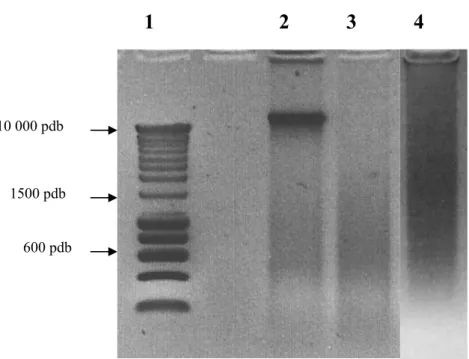
DNA quality was determined by electrophoresis on a 1% agarose gel running with 0.5X TBE buffer at room temperature for 30 min at 100V. Each sample was compared to a ladder (Smartladder®, Eurogentec) and classified according to their smear as “highly degraded” if only small DNA fragments were present, or “slightly degraded” if large DNA fragments were found (Figure 1).
VHL mutational analysis
DNA fragments encompassing the 3 exons of the VHL gene were amplified. In exon 1, the sequence located between codon 1 to 54 was not amplified because no mutation has ever been found either in our series, or in the literature. We used specific primers, as previously
described for frozen samples analysis14,16. A double amplification was performed using 100 ng
of the extracted DNA which was subjected to 35 cycles of PCR after an initial denaturation of 9 min at 95°C including: denaturing during 1 min at 95°C, annealing during 45 s at 58°C for exon 1 and 2, and 45 s at 57°C for exon 3, then extension during 45 s at 72°C.
Exon amplification was performed in 30µl containing: 500 mM KCl, 10 mM Tris-HCl (pH
8.3), 1.5 mM MgCl2, 250 µM dNTPs, 10 pmol of each primer and 1 U AmpliTaq Gold®
polymerase (Applied Biosystems). For exon 1 and 3, 3% DMSO was used because of their high content of guanine and cytosine. Electrophoresis was carried out on a 2% agarose gel in the same conditions than previously described to control the success of the amplification procedure.
For sequence analysis, purification of the amplified products was performed with ExoSAP-IT® (USB), according to the manufacturer’s instructions, to filter out small fragments. Amplification products detected on SSCP were purified using chromatography columns (Sephadex G50®, Amershan Bioscience). Sequencing used BigDye® Terminator mix V3. 1 (Applied Biosystems) and samples were re-amplified during 30 cycles of PCR: 30 s at 96°C, 15 s at 55°C at Tm, and 4 min at 60°C. Finally, the sequenced products were subjected to automated sequence analysis on an ABI PRISM® 3130 Genetic Analyser (Applied
Biosystems), and compared with the VHL sequence accession number AF0102383.
Mutations were identified by visual aspect of sequences by 2 persons and called when unequivocally present on the sense and/or the antisense strand.
Statistical analyses
χ² analysis was used for assessing differences in clinicopathologic parameters distribution. Associations between variables were assessed by χ² analysis. All analyses were conducted with SPSS 13.0.1 software (SPSS Inc, Chicago, Il, USA), and p-value significance was fixed at 0.05.
Results
Patients and tumors
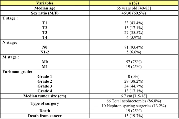
The population was composed of 46 men (60.5%) and 30 women (39.5%) with a median age
of 65 years [40-83].Tumors were organ confined in 46 cases (60.5%). Median tumor size was
6.7 cm [1.5 – 18]. Nodal invasion and distant metastases were present in 5 (6.6%) and 19 (25%) cases, respectively. Tumors were of high nuclear Furhman grade (grade 3 and 4) in 47 cases (61.8%). At the end of follow-up 15 patients (19.7%) died from renal cancer (Table 1).
DNA characteristics
Median DNA concentration was 222 ng/µl [30 – 693]. Even though the best quality block was selected by a single pathologist in all cases, imperfections potentially influencing DNA
extraction were noticed in half of the FFPE blocks: presence of necrosis, fat, oedema and fibrosis, haemorrhagic or cystic zones. Most of the samples were formalin-fixed during 24h (52.6%). DNA was considered as highly degraded on electrophoresis in 26 cases (34.2%) (Table 2). In this setting, no sequencing was possible in 57.7% of the cases (n= 15), and in 11 cases (42.3%) a sequencing discordance was found compared to frozen samples analysis (“extinction” of the mutation, other mutation, or multiple mutations present on the same exon or in different exons). Among the 76 samples, 72% could be sequenced, resulting in 69.1%
VHL mutation rate in this subset. Multiple mutations were found in 5 specimens. In frozen
analysis, 98% of the specimens were VHL mutated while a 52% mutation rate was identified in FFPE specimens. Among paraffin-embedded identified mutations, only 40.8% were fully consistent with frozen analysis in the 3 exons. Mutations not pre-existing in frozen samples were found in 10.5%, whereas 28.9% disappeared.
Regarding DNA quality and sequencing analysis, 26 samples presented a “highly degraded” DNA. Only 11 samples have been sequenced (42.3%). In this subset, no mutations was found
in 2 cases, mutations not pre-existing in frozen samples were found in 2 cases, and mutations had disappeared in 4 cases. Nevertheless, no mutations was fully consistent with frozen samples.
In the 50 “slightly degraded” DNA subgroup sample, the 3 exons of the VHL gene have been sequenced for 44 samples (88%). Thirty (68.2%) were strictly consistent with frozen samples. Mutations not pre-existing in frozen samples were found in 4 cases, and mutations had
disappeared in 2 cases.
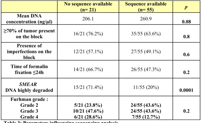
Parameters influencing sequencing analysis
We therefore tried to identify parameters influencing PCR amplification and sequencing analysis in FFPE tissue (Tables 3 & 4). Only quality of extracted DNA appeared as a statistically significant parameter (p= 0.0001). DNA concentration, presence of more than 70% of tumor on the block, imperfections on the block, duration of formalin-fixation or nuclear Furhman grade did not influence significantly sequencing analysis and occurrence of discordance.
Similarly, we tried to identify parameters influencing quality of extracted DNA. Neither duration of formalin-fixation, imperfection on the block, anteriority of the sample, nor percentage of tumor influenced the quality of extracted DNA.
For making sure that such discordances were not due to block selection or presence of
different mutated clones, we identified 10 samples with highly degraded DNA for which PCR amplification or sequencing analyses had not been possible, or where new mutations had appeared or disappeared during sequencing analysis. We subsequently repeated analysis in both new matched paraffin blocks and in frozen specimens. The same mutation was always identified in frozen specimen. In FFPE tissues, PCR amplification rates were comparable but
70% could not be sequenced as compared with 30% in initial samples. Only one result was retrieved within the 2 subsets: no mutation found whereas a mutation was identified in frozen analysis. In 2 cases, the mutation was not found whereas it had been initially identified.
Discussion
VHL is considered as an early “gatekeeper” tumor suppressor gene, involved in cell
cycle regulation, regulation of hypoxia inducible genes and proper fibronectin assembly in extracellular matrix2. It is presumed that further genetic alterations are needed for progression
of preneoplastic lesions. Nevertheless, restoration of pVHL function in VHL-deficient renal carcinoma cells can suppress tumor growth both in vivo and in vitro17. However, in 10 to 20%
sporadic ccRCCs, no alteration in the VHL alleles is detected, suggesting that other genes or pathways are involved in renal carcinogenesis18.
Gene abnormalities in hereditary Von Hippel-Lindau disease have been extensively analyzed. However few studies have focused on VHL alterations in sporadic ccRCC. These studies have been performed on frozen section tissues or on paraffin-embedded material and conflicting results have been reported. For example, 42 to 71% mutation rates have been described in frozen samples 7,9,13,19, while results seem to be inferior in FFPE tissues ranging from 20 to
61%10,12,20,21. Currently, frozen section tissue is considered as the benchmark for DNA
analysis22. Therefore, the objective of our study was to analyze the reliability of paraffin
derived technique, compared to results obtained from frozen samples in matched specimens. Indeed, no study had previously addressed the accuracy of VHL gene analysis on FFPE material compared to frozen tissue even though the issue of predicting anti-angiogenic drug response through VHL gene analysis has gained a recent interest20,23.
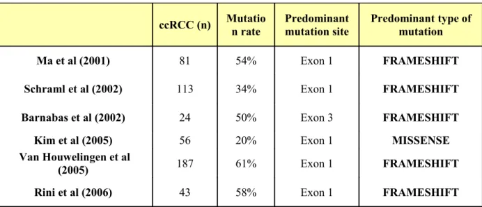
Overall, in terms of VHL mutation location our results are consistent with the current literature (Table 5). Similarly to us, the majority of the studies reported exon 1 as being the predominant mutation site. A group identified exon 2 as the more frequent mutation site but the methodology used in this small series is subject to criticisms since reverse sequencing was
identified on frozen section tissue, by amplifying and sequencing only one DNA strand. Another group reported VHL mutation predominance in exon 3 through a limited series of 67 tumors, and including only 24 ccRCCs25.
Our main finding was that out of the VHL mutated tumor population, only 52.1% mutations remained in FFPE tissue. New mutations not identified in frozen samples were found in 10.5%, whereas 28.9% disappeared. In other words, only half of the true VHL abnormalities were identified when using FFPE tissue. Furthermore, only half of these mutations were strictly concordant within the 3 exons.
When looking more accurately at the published data issued from the 2 types of tissue
analyses, it appears that many discrepancies exist. First, similarly to what we obtained in our frozen series14, no mutation has ever been identified before codon 54 in frozen samples9,13,19,26.
This region is known to interact with fibronectin and has been recently described as an important mediator for tumor invasion27. However, in FFPE tissues, similarly to the present
series, many authors reported mutations in this region9,10,21,25. Interestingly, as others9,10,21, we
found multiples mutations in some specimens, whereas it has never been described in any frozen series. Silent mutations have also been identified in FFPE samples in 11 to 13% of the cases11,12,21, whereas it has never been noticed in frozen samples. It is likely that these
uncommon mutations have been created by the technique. These artifacts can be ascribed to postmortem deamination of cytosine and adenine, resulting in uracil or hypoxanthine residues respectively28. Several authors have described this phenomenon in different tissues 29.
Williams et al reported up to 1 mutation artifact per 500 bases recorded30. It could also explain
why some investigators found VHL mutations in papillary 21, undifferentiated or chromophobe
carcinomas 11,25, and even in benign tumors like oncocytomas 11, while VHL abnormalities are
highly specific for clear cell histological subtype 31. No study on frozen section tissues has
along with the observation of mutational heterogeneity raises the question whether paraffin material is suitable for VHL analysis. Both from our study and from a comprehensive analysis of the literature, it appears that paraffin derived technique is inferior to frozen analysis for
VHL mutation analysis, resulting in quantitative and qualitative quality losses. In a
retrospective study of brain tissue, Ferrer et al. analyzed the effects of formalin fixation and time storage on DNA preservation, and compared with frozen specimens 32. Acceptable
results were obtained if DNA extraction was performed after a short time of fixation. Suboptimal and bad results (degraded DNA not allowing sequence analysis) occurred in FFPE tissues stored longer than 6 months. Moreover, the chance to obtain positive results was almost null in tissues that have been stored for years. Similar results were obtained with colorectal tissues. Though different artifacts have been used to optimize DNA extraction performance like higher-temperature heating under an alkaline condition 33 or longer
rehydratation step during DNA extraction 34. All this data taken together strongly suggests that
paraffin derived technique should not be considered as a standard for VHL gene analysis whether this evaluation should become important for predicting outcome following RCC treatment.
Beyond VHL gene analysis issues, this study suggests that results for DNA extraction and sequencing analysis from paraffin materials are less reliable than those taken from frozen samples. Therefore, caution is required when analyzing results of series using this material. However, when frozen tissue is not available, a very stringent selection of paraffin blocks with limited necrosis, fat, oedema, fibrosis, cystic or hemorrhagic zones is required. Additionally, selection of a homogeneous tumoral zone, exhibiting high nuclear grade and without any normal renal tissue is also of utmost importance. Ultimately, quality of the extracted DNA appears to be the major criterion for limiting errors associated with FFPE tissue analysis.
Conclusion
Tumoral DNA extraction and VHL mutation analysis can be performed from FFPE tissue in RCC. But mutations identified from FFPE tissues are not strictly concordant with those from frozen analysis. If available, frozen tissue analysis should be considered the gold standard. Otherwise paraffin-embedded tissue remains a great opportunity for DNA analysis in long term-follow-up series. Our study opens the gate for a critical analysis of the literature in this field.
References
1. Lam JS, Shvarts O, Leppert JT, Figlin RA and Belldegrun AS: Renal cell carcinoma
2005: new frontiers in staging, prognostication and targeted molecular therapy. J Urol. 173: 1853-62, 2005.
2. Kaelin WG, Jr.: The Von Hippel-Lindau Tumor Suppressor Gene and Kidney Cancer.
Clin Cancer Res. 10: 6290S-6295, 2004.
3. Latif F, Tory K, Gnarra J, et al.: Identification of the von Hippel-Lindau disease tumor suppressor gene. Science. 260: 1317-20, 1993.
4. Gnarra JR, Tory K, Weng Y, et al.: Mutations of the VHL tumour suppressor gene in
renal carcinoma. Nat Genet. 7: 85-90, 1994.
5. Patard J-J, Rioux-Leclercq N and Fergelot P: Understanding the Importance of Smart
Drugs in Renal Cell Carcinoma. European Urology. 49: 633-643, 2006.
6. Motzer RJ, Hutson TE, Tomczak P, et al.: Overall Survival and Updated Results for
Sunitinib Compared With Interferon Alfa in Patients With Metastatic Renal Cell Carcinoma. Journal of Clinical Oncology, 2009.
7. Yao M, Yoshida M, Kishida T, et al.: VHL tumor suppressor gene alterations
associated with good prognosis in sporadic clear-cell renal carcinoma. J Natl Cancer Inst. 94: 1569-75., 2002.
8. Lidgren A, Hedberg Y, Grankvist K, Rasmuson T, Vasko J and Ljungberg B: The
expression of hypoxia-inducible factor 1alpha is a favorable independent prognostic factor in renal cell carcinoma. Clin Cancer Res. 11: 1129-35, 2005.
9. Brauch H, Weirich G, Brieger J, et al.: VHL alterations in human clear cell renal cell
carcinoma: association with advanced tumor stage and a novel hot spot mutation. Cancer Res. 60: 1942-8, 2000.
10. Schraml P, Struckmann K, Hatz F, et al.: VHL mutations and their correlation with
tumour cell proliferation, microvessel density, and patient prognosis in clear cell renal cell carcinoma. J Pathol. 196: 186-93, 2002.
11. van Houwelingen KP, van Dijk BA, Hulsbergen-van de Kaa CA, et al.: Prevalence of
von Hippel-Lindau gene mutations in sporadic renal cell carcinoma: results from The Netherlands cohort study. BMC Cancer. 5: 57, 2005.
12. Kim JH, Jung CW, Cho YH, et al.: Somatic VHL alteration and its impact on
prognosis in patients with clear cell renal cell carcinoma. Oncol Rep. 13: 859-64, 2005.
13. Banks RE, Tirukonda P, Taylor C, et al.: Genetic and epigenetic analysis of von
Hippel-Lindau (VHL) gene alterations and relationship with clinical variables in sporadic renal cancer. Cancer Res. 66: 2000-11, 2006.
14. Patard JJ, Rioux-Leclercq N, Masson D, et al.: Absence of VHL gene alteration and
high VEGF expression are associated with tumour aggressiveness and poor survival of renal-cell carcinoma. Br J Cancer. 101: 1417-24, 2009.
15. Sobin LH and Wittekind C: TNM. Classification of malignant tumors. Sixth Edition.
UICC International Union Against Cancer, Ed Willey-Liss.: 193-195, 2003.
16. Patard JJ, Fergelot P, Karakiewicz PI, et al.: Low CAIX expression and absence of
VHL gene mutation are associated with tumor aggressiveness and poor survival of clear cell renal cell carcinoma. Int J Cancer. 123: 395-400, 2008.
17. Chen F, Kishida T, Duh FM, et al.: Suppression of growth of renal carcinoma cells by
the von Hippel-Lindau tumor suppressor gene. Cancer Res. 55: 4804-7, 1995.
18. Cohen HT and McGovern FJ: Renal-Cell Carcinoma. N Engl J Med. 353: 2477-2490,
19. Gallou C, Joly D, Mejean A, et al.: Mutations of the VHL gene in sporadic renal cell carcinoma: definition of a risk factor for VHL patients to develop an RCC. Hum Mutat. 13: 464-75, 1999.
20. Rini BI, Jaeger E, Weinberg V, et al.: Clinical response to therapy targeted at vascular endothelial growth factor in metastatic renal cell carcinoma: impact of patient
characteristics and Von Hippel-Lindau gene status. BJU Int, 2006.
21. Ma X, Yang K, Lindblad P, Egevad L and Hemminki K: VHL gene alterations in renal
cell carcinoma patients: novel hotspot or founder mutations and linkage disequilibrium. Oncogene. 20: 5393-400, 2001.
22. Hanage WP and Aanensen DM: Methods for data analysis. Methods Mol Biol. 551:
287-304, 2009.
23. Choueiri TK, Vaziri SA, Jaeger E, et al.: von Hippel-Lindau gene status and response
to vascular endothelial growth factor targeted therapy for metastatic clear cell renal cell carcinoma. J Urol. 180: 860-5; discussion 865-6, 2008.
24. Gimenez-Bachs JM, Salinas-Sanchez AS, Sanchez-Sanchez F, et al.: Determination of
vhl gene mutations in sporadic renal cell carcinoma. Eur Urol. 49: 1051-7, 2006.
25. Barnabas N, Amin MB, Pindolia K, Nanavati R, Amin MB and Worsham MJ:
Mutations in the von Hippel-Lindau (VHL) gene refine differential diagnostic criteria in renal cell carcinoma. J Surg Oncol. 80: 52-60, 2002.
26. Kondo K, Yao M, Yoshida M, et al.: Comprehensive mutational analysis of the VHL
gene in sporadic renal cell carcinoma: relationship to clinicopathological parameters. Genes Chromosomes Cancer. 34: 58-68, 2002.
27. Lolkema MP, Gervais ML, Snijckers CM, et al.: Tumor suppression by the von
Hippel-Lindau protein requires phosphorylation of the acidic domain. J Biol Chem. 280: 22205-11, 2005.
28. Hofreiter M, Jaenicke V, Serre D, Haeseler Av A and Paabo S: DNA sequences from
multiple amplifications reveal artifacts induced by cytosine deamination in ancient DNA. Nucleic Acids Res. 29: 4793-9, 2001.
29. Marchetti A, Felicioni L and Buttitta F: Assessing EGFR mutations. N Engl J Med.
354: 526-8; author reply 526-8, 2006.
30. Williams C, Ponten F, Moberg C, et al.: A high frequency of sequence alterations is
due to formalin fixation of archival specimens. Am J Pathol. 155: 1467-71, 1999.
31. Kenck C, Wilhelm M, Bugert P, Staehler G and Kovacs G: Mutation of the VHL gene
is associated exclusively with the development of non-papillary renal cell carcinomas. J Pathol. 179: 157-61, 1996.
32. Ferrer I, Armstrong J, Capellari S, et al.: Effects of formalin fixation, paraffin embedding, and time of storage on DNA preservation in brain tissue: a BrainNet Europe study. Brain Pathol. 17: 297-303, 2007.
33. Shi SR, Cote RJ, Wu L, et al.: DNA extraction from archival formalin-fixed,
paraffin-embedded tissue sections based on the antigen retrieval principle: heating under the influence of pH. J Histochem Cytochem. 50: 1005-1011, 2002.
34. Coura R, Prolla JC, Meurer L and Ashton-Prolla P: An alternative protocol for DNA
extraction from formalin fixed and paraffin wax embedded tissue. J Clin Pathol. 58: 894-895, 2005.
Variables n (%)
Median age 65 years old [40-83]
Sex ratio (M/F) 46/30 (60.5%) T stage : T1 T2 T3 T4 33 (43.4%) 13 (17.1%) 27 (35.5%) 4 (3.9%) N stage: N0 N1-2 71 (93.4%)5 (6.6%) M stage : M0 M1 57 (75%) 19 (25%) Furhman grade: Grade 1 Grade 2 Grade 3 Grade 4 0 (0%) 29 (38.2%) 34 (44.7%) 13 (17.1%)
Median tumor size (cm) 6.7 cm [1.5-18]
Type of surgery 10 Nephron sparing surgeries (13.2%)66 Total nephrectomies (86.8%)
Death 19 (25%)
Death from cancer 15 (19.7%)
Table 1: General characteristics of the studied population (n= 76).
Variables n (%)
Median DNA concentration 222 ng/µl [30 – 693]
Median tumor % on the FFPE block 70% [30-100]
Samples sequenced in the 3 exons 55/76 (72.4%)
DNA quality on the smear:
- Highly degraded
- slightly degraded 26/76 (34.2%)50/76 (65.8%) General aspect of the FFPE block :
- Excellent quality - Necrosis - Fat - Œdemea - Hemorragia - Fibrosis - Cystic 38/76 (50%) 25/76 (32.9%) 5/76 (6.6%) 1/76 (1.3%) 1/76 (1.3%) 3/76 (3.9%) 3/76 (3.9%) Formalin-fixed duration: 24h 48h 72h 96h 40/76 (52.6%) 13/76 (17.8%) 20/76 (26.3%) 3/76 (4.1%)
Mutations identified in FFPE tissue 38/73 (52%)
Mutations errors 29/76 (38.2%)
No sequence available (n= 21) Sequence available (n= 55) p Mean DNA concentration (ng/µl) 206.1 260.9 0.08 ≥70% of tumor present on the block 16/21 (76.2%) 35/55 (63.6%) 0.8 Presence of imperfections on the block 12/21 (57.1%) 27/55 (49.1%) 0.6 Time of formalin fixation ≤24h 14/21 (66.7%) 26/55 (47.3%) 0.2 SMEAR
DNA highly degraded 15/21 (71.4%) 11/55 (20%) 0.0001
Furhman grade : Grade 2 Grade 3 Grade 4 5/21 (23.8%) 10/21 (47.6%) 6/21 (28.6%) 24/55 (43.6%) 24/55 (43.6%) 7/55 (12.7%) 0.2 Table 3: Parameters influencing sequencing analysis.
Discordance (n= 28) with frozen samples (n= 30)Samples fully consistent p
Median DNA concentration (ng/µl) 266.1 248.1 0.5 ≥70% of tumor present on the block 20/28 (71.4%) 17/30 (56.7%) 0.4 Presence of imperfections on the block 13/28 (46.4%) 17/30 (56.7%) 0.5 Time of formalin fixation ≤24h 16/28 (57.1%) 14/30 (46.7%) 0.2 SMEAR
DNA highly degraded 14/28 (50%) 0/30 (0%) 0.0001
Furhman grade : Grade 2 Grade 3 Grade 4 10/28 (35.7%) 14/28 (50%) 4/28 (14.3%) 14/30 (46.7%) 11/30 (36.7%) 5/30 (16.7%) 0.8 Table 4: Parameters influencing sequencing errors.
ccRCC (n) Mutatio n rate Predominant mutation site Predominant type of mutation Ma et al (2001) 81 54% Exon 1 FRAMESHIFT
Schraml et al (2002) 113 34% Exon 1 FRAMESHIFT
Barnabas et al (2002) 24 50% Exon 3 FRAMESHIFT
Kim et al (2005) 56 20% Exon 1 MISSENSE
Van Houwelingen et al
(2005) 187 61% Exon 1 FRAMESHIFT
Rini et al (2006) 43 58% Exon 1 FRAMESHIFT
Table 5: VHL mutational analysis in FFPE histologic specimens, results from the literature.
Figure 1: DNA quality determined by electrophoresis on a 1% agarose gel. Each sample was compared to a ladder (1) and classified according to their smear as “highly
degraded” if only small DNA fragments were present (3), or “slightly degraded” if large DNA fragments were found (4). N°2 represents a frozen sample.
10 000 pdb
1500 pdb
600 pdb