HAL Id: hal-02990285
https://hal.archives-ouvertes.fr/hal-02990285
Submitted on 16 Nov 2020HAL is a multi-disciplinary open access archive for the deposit and dissemination of sci-entific research documents, whether they are pub-lished or not. The documents may come from teaching and research institutions in France or abroad, or from public or private research centers.
L’archive ouverte pluridisciplinaire HAL, est destinée au dépôt et à la diffusion de documents scientifiques de niveau recherche, publiés ou non, émanant des établissements d’enseignement et de recherche français ou étrangers, des laboratoires publics ou privés.
Maxence Lejars, Eliane Hajnsdorf
To cite this version:
Maxence Lejars, Eliane Hajnsdorf. The world of asRNAs in Gram-negative and Gram-positive bac-teria. Biochimica et Biophysica Acta - Gene Regulatory Mechanisms , Elsevier, 2020, 1863 (2), pp.194489. �10.1016/j.bbagrm.2020.194489�. �hal-02990285�
Antisense RNA world in bacteria
Maxence Lejars1and Eliane Hajnsdorf1*
1 UMR8261,CNRS, Université de Paris, Institut de Biologie Physico-Chimique, 75005 Paris, France maxence.lejars@ibpc.fr
Abstract
Bacteria exhibit an amazing diversity of mechanisms controlling gene expression to both maintain essential functions and modulate accessory functions in response to environmental cues. Over the years, it has become clear that bacterial regulation of gene expression is still far from fully understood. This review focuses on antisense RNAs (asRNAs), a class of RNA regulators defined by their location in cis and their perfect complementarity with their targets, as opposed to small RNAs (sRNAs) which act in trans with only short regions of complementarity. For a long time, only few functional asRNAs in bacteria were known and were almost exclusively found on mobile genetic elements (MGEs), thus, their importance among the other regulators was underestimated. However, the extensive application of global “omics” approaches has revealed the ubiquity of asRNAs in bacteria. This review aims, first, to describe the inherent ambiguity in the existence of asRNAs in bacteria, second, to highlight their diversity and their involvement in all aspects of bacterial life by presenting a list of 67 characterized asRNAs from both Gram-positive and Gram-negative bacteria.
Keywords
asRNAs, bacteria, gene regulation, physiological functions, Gram-negative, Gram-positive.
Abbreviations
Transcription factor (TF), antisense RNA (asRNA), messenger RNA (mRNA), ribosomal RNA (rRNA), open reading frame (ORF), untranslated region (UTR), small RNA (sRNA), nucleotides (nt), ribosome binding site (RBS), Ferric uptake regulation (Fur), horizontal gene transfer (HGT), mobile genetic elements (MGE).
Table of contents
1. Introduction...5
2. There is an ambiguity in the existence of asRNAs...6
2.1. Vestiges from the RNA world: the burden of asRNAs as regulatory entities ...6
2.2. Fast and flexible: conditional advantages of asRNAs ...7
3. asRNAs are involved in all aspects of bacterial life...8
3.1. Metal ion homeostasis ...9
3.2. Metabolism...10
3.3. Photosynthesis ...10
3.4. Accessory functions...11
4. What can we learn from the characterized asRNAs? ...12
4.1. Trends and exceptions in the asRNAs studied so far...12
4.2. Occurrence of asRNAs in mobile genetic elements...13
5. Conclusion and perspectives ...14
Legends and Tables ...16
1. Introduction
Bacterial genomes are composed of a core of essential genes and a large number of accessory genes, which together form an ensemble unique to each bacterium. Essential genes encode proteins required for the multiplication of the cell in all environments, primarily involved in replication, cell division, transcription, translation, catabolism, anabolism and quality control. On the other hand, accessory genes often encode factors required to perceive and respond to environmental cues. Most genes are subject to regulation at one or more of the steps of gene expression through an exquisite variety of mechanisms, thus, forming mixed regulatory circuits [1]. This arsenal of regulators enables the fine-tuning of constitutively expressed genes in order to maintain a precise cellular equilibrium but also to punctually induce gene expression in order to respond to environmental signals or stress. For decades, regulation at the level of transcription was thought to be the most important control of gene expression, mainly because of the discovery of an impressive number of protein transcriptional factors (TFs) able to regulate up to hundreds of genes [2]. However the discovery of numerous non-coding RNAs (ncRNAs) in all bacterial phyla, through advances in bioinformatics predictions and global experimental approaches, has revealed that RNAs are also involved in the control of all levels of gene expression [3, 4].
Global “omics” approaches greatly contributed to the expansion of the regulatory non-coding RNAs field. These studies were mostly focused on a class of small regulatory RNAs, ranging from tens to hundreds of nucleotides, defined as sRNAs. Located in trans relative to their RNA target, sRNAs were shown to control transcription negatively (Fig. 1A) or positively (Fig. 1B) but they predominately result in post-transcriptional regulation, which can also be either negative (Fig. 2A) or positive (Fig. 2B). On the contrary,
cis-localized antisense RNAs (asRNAs), ranging from tens to thousands of nucleotides, are derived from
the same DNA as their unique target and thus are always, partially or totally, complementary to the opposite transcript. They are involved in a wide variety of mechanisms at both the transcriptional and post-transcriptional levels. Primary transcription is affected when two convergent transcription apparatuses collide into each other or by attenuation when the two RNA molecules interact, forming an intermolecular double stranded RNA, which stabilizes the formation of a terminator structure (Fig. 1C). So far asRNAs were only shown to repress transcription of their target. They can also act post-transcriptionally, either negatively (Fig. 2C) or positively (Fig. 2D) by modulating the recruitment of ribosomes and/or ribonucleases (RNases) [5]. Ribonucleases can recognize specific RNA sequences and RNA structures. For example, RNase E cleaves AU-rich single-stranded sequence usually when they are close to a stem-loop structure, RNase III cleaves hairpins and double-stranded regions, while RNase II requires an unpaired 3′ terminus to perform the 3′ to 5′ degradation of the RNA. It has also been shown that asRNAs can protect transcripts from exonucleolytic degradation or RNase E cleavage or, on the contrary, promote degradation by recruitment of RNase III [6].
It is worth remembering that asRNAs were among the first regulatory RNAs studied, as early as 1969 [7, 8]. In the following decade, many asRNAs were discovered on mobile genetic elements (MGEs) such as prophages, plasmids and transposons. The R1 plasmid was the first source of CopA, asRNA to the repA mRNA, involved in controlling plasmid replication and Sok, asRNA to the hok mRNA, involved in post-segregational killing [9, 10]. The recent DNA sequence of R1 [11] (Fig. 3) revealed that R1 encode two other asRNAs identical to RNA-OUT, targeting the RNA-IN mRNA and FinP, targeting the traJ mRNA had been
previously studied on other MGEs [12, 13] where they control conjugation and transposition. Their function in R1 has not been verified. In plasmid R1, replication is negatively controlled at the translational level by binding of the asRNA CopA to the leader region of the repA mRNA (CopT). Rather than forming an extended sense-antisense duplex, a loop-loop contact, often called a “kissing complex,” between bulged loop residues in CopA and CopT (Fig. 3A), promotes the interaction between the two RNAs with a four-way junction structure and a side-by-side alignment which results in inhibition of the translation [14]. Thus, expression of the CopA asRNA fine-tunes the copy number of the R1 plasmid. Interestingly, the CopA/repA system of regulation is one of the best-characterized asRNA and is strongly conserved in many related plasmids in Gram-negative and even in Gram-positive bacteria as sRNA1 found on the pAM330 plasmid in
Corynebacterium glutamicum [15]. In addition, plasmid R1 also contains the hok gene encoding a toxic
protein whose translation is inhibited by the Sok asRNA. This asRNA interacts with a small upstream reading frame, mok, whose translation is coupled to that of hok (Fig. 3B). The pairing is most likely initiated with the formation of a linear-linear complex, which propagates to a stable complex [16]. Binding of the asRNA Sok inhibits mok translation and also that of hok mRNA by translational coupling and thus, prevents the expression of the toxin. This system was later classified as a type I toxin-antitoxin system and was shown to be common in bacteria [17]. Another historic example was found in the IS10 insertion sequence of the Tn10 transposon [12]. RNA-OUT is an asRNA regulating IS10 transposase expression by pairing with the RNA-IN transposase mRNA. The pairing occurs between the 5′ part of this asRNA and the 5′ translational initiation region of the mRNA (Fig. 3C). RNA-OUT consists of a stem-loop domain topped by a flexibly paired loop. The 5′ end of the target molecule, RNA-IN, is complementary to the top of the loop and to 35 nucleotides (nt) down one side of RNA-OUT. It was recently shown that IS10 antisense pairing is facilitated by the RNA chaperone Hfq [12]. asRNAs are, by definition, fully complementary to their RNA target and in most cases do not require RNA chaperones, unlike most sRNAs which do. Nonetheless some exceptions were found as in the case of the RNA chaperone Hfq facilitating the interaction of the asRNA RNA-OUT with its target RNA-IN or the RNA chaperone FinO essential for the ability of the FinP asRNA, to bind to its target mRNA traJ in the F plasmid of E. coli [13] (Fig. 3D). In the absence of FinO, the asRNA FinP is quickly degraded by RNase E; thus, TraJ is translated and the plasmid can be transferred by conjugation. These few early examples of asRNAs were considered for a long time as anecdotal but they were instrumental in the understanding of how asRNA functions in bacteria. Recently, improvements in global “omics” approaches and their massive use have led to the identification of numerous new asRNAs (reviewed in [5, 18]).
This review aims to depict the landscape of asRNAs in bacteria by presenting 67 experimentally validated and studied asRNAs. The first part will illustrate how asRNAs can be a burden for the cell or become advantageous according to the conditions and regulated system. The second part will report the ubiquity of these asRNAs in bacteria and their impact in all aspects of bacterial life. The third part will compare these asRNAs according to their presence in Gram-negative or Gram-positive bacteria, their localization relatively to their target, their coverage of the ribosome binding site (RBS) and their regulatory function. Finally, the striking occurrence of asRNAs mediated regulations in MGEs will be discussed.
2. There is an ambiguity in the existence of asRNAs
The idea that RNAs could have been the initial building block of life emerged almost 70 years ago. This hypothesis was further reinforced and put on a theoretical basis in the 1990s with the discovery of catalytic RNA activities in ribozymes [19]. Replicating ribozymes were shown to require an RNA template in order to produce a complementary antisense molecule [20]. It was suggested that this step provides selection, an essential feature of evolution, because RNAs which are unable to dissociate from their complementary strand, would lose their function and template properties and thus, were likely to be counter-selected [19]. In the RNA world, complementary RNAs may be considered as the ancestors of asRNAs because they were likely to counteract the role of their sister RNA molecule. One can imagine that complementary RNAs are much less common nowadays than in the RNA world, where the transcription apparatus did not exist, and RNAs would presumably have replicated via a double stranded intermediate [19, 20]. This RNA world has been replaced by the classic paradigm of life in which organic macromolecules are specialized to be more effective in different functions: DNA for information storage, RNA for information utilization and proteins for functionality. However, DNA, RNA and proteins can be involved in other functions for example RNAs are proposed to be the essential catalytic core in diverse complexes such as the ribosome [19] or RNase P [21]. Remarkably, mechanisms have evolved to prevent intermolecular double stranded RNA formation like the Rho and NusG proteins involved in transcription termination. In Salmonella enterica, the inactivation of NusG and Rho, allows the detection of transcripts covering the whole genome in both orientations [22]. Thus, excessive asRNA synthesis can be considered as spurious and a burden on the cell in most cases because synthesis of futile RNAs implies a waste of metabolic resources, nucleotide precursors and energy but more importantly because these asRNAs could impede gene expression.
2.2. Fast and flexible: Conditional advantages of asRNAs
The well-studied asRNAs described in the introduction reveal that paradoxically, despite many potential disadvantages, asRNAs can be valuable in specific systems such as the replication of MGEs (further discussed in the paragraph 4.2). The proximity of asRNAs with their target provides a first obvious advantage compared to other regulators. A second advantage is the ability of asRNAs to control gene expression directly and independently from other factors in most cases, with perhaps the exception of recruitment of an RNase. Modulation of transcription by sRNAs (Fig. 1A and 1B) was found possible in some cases but is always dependent on the termination factor Rho [23]. The regulation of transcription by asRNAs, always negative (so far), can happen co-transcriptionally either due to the interaction of the two RNA polymerases or of the two RNAs (Fig. 1C), thus providing a faster and potentially more efficient control than other regulators. The mechanism of post-transcriptional regulation by sRNAs and asRNAs is somewhat similar, as their interaction can hide or open the RBS of the mRNA (Fig. 2). Unlike sRNAs, the often long length of asRNAs can produce different modes of interaction with their target from an initial kissing complex to extended double stranded intermolecular duplexes, as in the case of the asRNA CopA (see Introduction). Thus, they can exert more flexibility in the modulation of translation and stability of the target. Furthermore, asRNAs promote recruitment of RNase III, which is rare in the case of sRNAs [6]. Looking at these features of asRNAs, they may be advantageous in complex genetic systems, which require correct sensing of multiple environmental cues, either to provide a fast and short-lived response or to fine-tune the expression of a specific gene.
On the other hand, perfect complementarity might be considered to be a burden because both molecules are likely to sequester each other. Nonetheless, the vision of static and stable intramolecular or intermolecular RNA structures in cells seem to be really far from reality. RNA conformation is governed by different parameters in the cell, such as molecular crowding, protein chaperones, metal-ion concentration and cellular solutes. Upon modification of the balance of one of these parameters, the RNA conformation can be strongly altered, particularly in the case of newly transcribed RNAs, as reviewed in [31]. Using structurally stable RNAs it was shown that a population of RNAs, identical in their sequence, is composed of different subpopulations differing in their conformation and forming together a so-called ensemble. Indeed, the abundance of each subpopulation (i.e. conformation) is relative to the stability of each conformation. Thus, the relative amount of each subpopulation changes over time and with intracellular parameters on a timescale ranging from microseconds to hours. The well-studied cases of CopA/CopT and
hok/Sok depict how asRNAs take advantage of this plasticity to provide an efficient regulatory mechanism
(see Introduction and Fig. 3) [10, 12-14]. Furthermore, asRNAs are also involved in the homeostasis of metal ions (described in the paragraph 3.1), thus are likely to be sensitive to small variations of metal concentrations and to efficiently fine-tune these regulatory systems.
Even though excessive synthesis of asRNAs is likely to be detrimental, the existence of specific asRNAs in modern life can also provide unexpected advantages. Recently, asRNAs were shown to promote R-loop formation due to their ability to bind to DNA during transcription of the opposite gene [24]. On the ColE1 plasmid, the initiation of replication is dependent on the recruitment of the DNA polymerase through interaction with an RNA primer. The RNA primer is a maturation product of the RNAII transcript by RNase H, when it forms an R-loop with a single DNA strand of the vector. RNAI is transcribed antisense to RNAII and, through formation of kissing complexes, prevents the binding of RNAII to the DNA. Hence, RNAI inhibits DNA replication so that copy number control depends upon relative ability of RNAII to form an R-loop with DNA or base pair with RNAI [25]. Another outcome of R-R-loop formation mediated by an asRNA is an increase of horizontal gene transfer (HGT) efficiency [24]. In Neisseria gonorrhoeae an asRNA to the
pilE gene is proposed to participate in the replacement of the pilE ORF by other homologous ORFs named pilS [26]. Furthermore, in recently re-emerged clinical strains of Bordetella pertussis the presence of
hundreds of copies of the IS481 transposon on the chromosome is proposed to be the reason for the high resistance of this strain to actual vaccines [27] (described in paragraph 4.1).
3. asRNAs are involved in all aspects of bacterial life
In most bacterial phyla, asRNAs have been found through global “omics” approaches, some of them have been directly validated but only a few were further studied, mostly because of their low expression and location. A list of asRNAs documented in Gram-negative and in Gram-positive bacteria found in a comprehensive literature survey up to October 2019 is presented in Tables 1 and 2 respectively. They contain all the asRNAs for which a promoter and the approximate length have been validated by methods other than global approaches, generally by Northern bloting. We realize that there will be some inherent bias, since a limited number of organisms have been intensively studied for asRNAs. The recent increase in global studies can be used as the base of extensive further investigations and means that other species of bacteria (as in the case of the cyanobacteria Synechocystis sp. PCC 6803 [28]) are now being studied so that the numbers of validated asRNAs will undoubtedly rapidly rise. The asRNAs listed in Tables 1 and 2
are classified according to their host, the category of function of their target, the position of their promoter relative to the 5′ end of the ORF on the complementary strand, whether they are complementary with the RBS and their global regulatory effect, activation or repression of their target. We considered asRNAs from phage genomes as belonging to and being functional in their respective hosts. We have eliminated all asRNAs participating to type I toxin/antitoxin systems because of their great number and repetition in some species and as they are, to our knowledge, always repressing the expression of the target RNA to prevent toxicity and they have been the subject of extensive studies. For more details on type 1 toxin-antitoxin systems see the review by Masachis and Darfeuille [17].
The 67 asRNAs in the list are from cyanobacteria and proteobacteria for Gram-negative bacteria (Table 1) and from actinobacteria and firmicutes for Gram-positive bacteria (Table 2). These asRNAs are involved in almost all aspects of bacterial life, in the control of stress responses and metabolism from short-term induction of a gene to fine-tuning a constitutively expressed gene. We previously described the role of asRNAs in the physiological responses to stress [5] and in the present review we describe other aspects of asRNA regulation where asRNAs are not only responding to environmental cues but also fine-tuning gene expression. From our analysis of this dataset certain tendencies and exceptions could be observed and are presented below.
3.1. Metal ion homeostasis
As mentioned above, a single asRNA may adopt numerous conformations, which are dependent on many parameters including metal ions. Strikingly, asRNAs fine-tuning metal ions homeostasis are found in diverse phyla. The well-studied asRNAs RNAα and RNAβ from Vibrio anguillarum are known to control the expression of siderophores, proteins of high affinity for Fe2+, able to capture Fe2+ from hemes in the blood
of the host [29-31]. Under Fe2+ rich condition, synthesis of siderophores is too energetically costly for the
cell; thus, RNAα and RNAβ repress their expression.
The Ferric uptake regulator (Fur) TF is considered as the master regulator of Fe2+ homeostasis and is well
conserved in bacteria [32]. Fur is active when bound to Fe2+ and controls a large regulon. An asRNA to the
fur gene was found in the cyanobacteria Anabaena sp. PCC 7120, Synechocystis sp. PCC 6803, Microcystis aeruginosa and in the proteobacteria Acidithiobacillus ferrooxidans [33-37]. This asRNA named α-fur is
encoded in the 3′ untranslated region (UTR) of the fur gene and is involved in the repression of fur expression by an unknown mechanism. Strikingly, the α-fur promoters, located downstream of their respective fur genes, are not conserved and are likely to have emerged independently. Therefore, this suggests a strong evolutionary pressure toward asRNA-mediated regulation of Fur expression; possibly due to the ability of asRNAs to modulate their function according to intracellular Fe2+concentrations in
iron-limited environments such as ocean for cyanobacteria and V. anguillarum or saturated in iron in the case of A. ferrooxidans, which is found in acidic water outflow from metal mines [37].
Another example of an asRNA involved in metal iron economy is the AmgR asRNA in Salmonella enterica, which controls the expression of mgtC [38]. MgtC is an inner membrane protein involved in resistance to low Mg2+ conditions. The asRNA induces the degradation of the mgtC sense RNA in order to reduce MgtC
expression under low Mg2+ conditions. Surprisingly, both AmgR and mgtC transcripts are induced under
shut down MgtC expression. It has been postulated that this reduces the immune response during Salmonella infection [39]. Indeed, low Mg2+ is likely to modulate the ability of the asRNA to bind to the
target RNA and thus provide a sharper regulation.
3.2. Metabolism
A variety of asRNAs are involved in the control of (or potentially in the control of) global metabolism (Tables 1 and 2) including catabolism as illustrated by dorf, asRNA to crp, the master regulator of the carbon catabolite repression [40], by the asRNA to ptsG, which mediates the transport of glucose in E. coli [41] (although no regulatory effect of the asRNA was demonstrated) and by MtlS, asRNA to mtlA, playing a role in the catabolism of mannitol in Vibrio cholerae [18]. Other asRNAs are important for anabolism like the asRNAs to carB, putP and to the ubiG-mccBA operon, respectively involved in arginine synthesis in the endosymbiont Buchnera [42], in proline uptake in Bacillus subtilis [43], and in cysteine and methionine homeostasis in C. acetobutylicum [44]. Bacteria can also divert their metabolic pathways in order to respond to stress. In E. coli, one of the responses to acid stress is the degradation of glutamate in butyric acid in order to consume a proton and thus maintain a physiological pH in the cytoplasm. This process is under control of the TFs GadX and GadW and the asRNA GadY which stabilize its target gadX through binding to the 3′ end of the mRNA [45]. Moreover, asRNAs have been implicated in global regulation of transcription, translation and general stress responses [5, 46-48]. Furthermore, asRNAs are directly involved in the regulation of ribonucleases and thus in the control of RNA metabolism. In E. coli, the
rpsO-pnp operon encodes a ribosomal protein and the cold shock exoribonuclease PNPase respectively. SraG is
an asRNA transcribed between the two ORFs which promotes the RNase III dependent degradation of the polycistronic transcript [49]. No physiological conditions controlling SraG induction have been found yet. The interaction of the sense and the asRNA is likely to be modified upon a cold shock which might correlate with the fact that PNP is a cold-shock protein and that the activity of the RNase III is also modulated during a cold shock at the post-translational level by the binding of the YmdB protein [50]. The importance of the SraG asRNA to pnp expression is supported by the presence of an asRNA, RliD, to pnpA (homologous gene to pnp), in the Gram-positive L. monocytogenes [51]. RNase H, encoded by the rnh gene, is another important enzyme in E. coli, it degrades the RNA strand of an RNA-DNA duplex as in the case of R-loops. As mentioned previously (see paragraph 2.2), RNase H is required for replication of the plasmid ColE1 and the asRNA RNAI can prevent the formation of the DNA-RNA hybrid by competition. Furthermore, it was shown that the promoter of rnh overlaps the promoter of the divergent gene dnaQ, involved in transcription quality control [48]. In this case, there is no asRNA produced but the two genes are likely to be repressing each other’s transcription by interference (Fig. 1C).
3.3. Photosynthesis
In cyanobacteria, global approaches brought to light an impressive number of asRNAs, and similar numbers of asRNAs have been found in plant chloroplasts [35, 52]. Cyanobacteria are the only bacteria performing oxygenic photosynthesis, an extremely complex and highly regulated process converting light energy to chemical energy through O2 synthesis and CO2 fixation. Anabaena sp. PCC 7120 is a
cyanobacteria able to form filaments composed of aligned cells interacting with their neighbors in a chain-like structure. During nitrogen starvation, 5 to 10% of the cell population in the filaments differentiate into heterocysts. These specialized cells maintain microaerobic conditions, thanks to a thicker cell wall, and repress photosynthesis in order to efficiently perform nitrogen fixation. Heterocysts provide nitrogen to the rest of the cells, which, in return, provide carbon sources. Differentiation requires a global remodeling of gene expression and is under the control of multiple TFs but also asRNAs as demonstrated by comparative RNAseq [53]. One of these asRNA, as_glpX, is complementary to glpX, encoding the sedoheptulose-1,7-bisphosphatase/fructose-1.6-bisphosphatase (SBPase), part of the Calvin cycle and thus essential for photosynthetic metabolism. Upon differentiation to heterocysts, as_glpX is induced and promotes RNase III mediated degradation of both transcripts in order to efficiently and quickly turn off photosynthesis. The decision for the cells to differentiate results from accumulation of many environmental cues but remains a stochastic event; thus, asRNAs could participate in this regulation thanks to their ability to provide a form of epigenetic regulation.
In Synechocystis sp. PCC 6803, 5 asRNAs were shown to play a role in the regulation of photosynthesis [18]. As1_flv4 is opposed to the flv1-4 operon, encoding flavodiiron proteins 1 to 4, required for the protection of photosystem 2, which is an essential process during inorganic carbon starvation. This asRNA prevents the expression of the flavodiiron proteins in normal conditions and is repressed upon inorganic carbon starvation. IsiA is another stress response protein involved in the protection of photosystem 1. IsiR is an asRNA to isiA and promotes degradation of isiA mRNA. Under iron starvation or oxidative stress, the asRNA is repressed to allow synthesis of IsiA; thus, protecting the photosynthetic apparatus. The psbA gene is found in 3 copies on the genome of S. sp. PCC 6803, it encodes the D1 peptide, core of photosystem 2. These 3 genes are identical in their function but in order to respond to different environmental stresses their regulation differs because of differences in their 5’UTR. The asRNAs psbA2R and psbA3R are encoded in the 5’UTR of the psbA2 and psbA3 genes respectively. They act positively, likely by stabilizing their target mRNA. Under high light, the two asRNAs are repressed and thus, the levels of
psbA2 and psbA3 mRNA decrease. Finally, an asRNA to the rblR gene, encoding the large chain of the
RuBisCo protein was also found but the physiological role of this asRNA remains elusive.
3.4. Accessory functions
In bacterial genomes, accessory genes encode functions that are not required in all conditions, including a wide range of situational metabolic processes. Many bacteria, like Bacillus licheniformis, can use fermentation in order to obtain energy and carbon sources. This Gram-positive bacterium is a saprophyte living in wood and is able to degrade a wide variety of organic molecules from plants. In order to efficiently catabolize these carbohydrates, B. licheniformis secretes, in addition to many other factors, an exoprotease called subtilisin, encoded from the apr gene. Located in the 3’UTR of apr, the AprAs asRNA was shown to repress expression of subtilisin but no physiological role could be determined suggesting a possible fine-tuning of Apr expression [54].
A large number of bacteria are also able to respond to stress conditions by reprogramming their genetic expression, reducing metabolic activity and waiting for better times to come. In some cases, this metabolic pause happens after morphological rearrangement as in the case of sporulation. In the actinomycete
were found to be required for germination of spores. The asRNA to rpfA, named Scr3097, promotes translation of the resuscitation-promoting factor, RpfA, involved in the cleavage of the peptidoglycan in order to allow germination of the spore. Again, no inductive or repressive conditions could be found suggesting a fine-tuning regulatory effect. The unnamed antisense to yabE (also encoding a resuscitation promoting factor) is under control of two of the extracytosolic sigma factors, X and M, but no role in yabE expression could be determined.
Finally, a major aspect of research on bacteria focuses on their pathogenicity and different examples of asRNAs controlling virulence factors have been found. In Shigella flexneri, the IcsA virulence factor, carried on a plasmid, is responsible for the formation of actin tails, enabling infecting bacteria to spread in the host. The asRNA to icsA, RnaG, was shown to repress, at the transcriptional level, the expression of IcsA upon entry into stationary phase [57]. Another example is found on a plasmid in Salmonella typhimurium where the asRNA IesR-1 is opposed to PSLT047 [58]. PSLT047 has no clear role, but the induction of the asRNA upon entry of the bacteria into fibroblast cells was shown to be essential in order to slow down growth and escape the immune system.
4. What can we learn from the characterized asRNAs?
4.1. Trends and exceptions in the asRNAs studied so far
Inspection of Tables 1 and 2 implies that there are more asRNAs in some bacterial species than others. However, this probably reflects the greater depth of study of certain bacteria than others. Admitting that more asRNAs exist in some and probably all of the bacterial species, we have tried to draw some general conclusions based on the 67 examples, which have been characterized to date. Tables are divided by historical classification of Gram-negative (Table 1) (i.e. having an external membrane and a thin peptidoglycan) or Gram-positive (Table 2) (i.e. no external membrane and presence of a thick peptidoglycan). The asRNAs belongs to 19 Gram-negative bacterial species from the phyla cyanobacteria and proteobacteria and to 14 Gram-positive bacterial species from the phyla actinobacteria and firmicutes. There are 49 asRNAs in Gram-negative and 18 asRNAs in Gram-positive bacteria but this is probably correlated with the extensive studies on certain model organisms rather than a true repartition of asRNAs in the Gram-positive and Gram-negative bacteria. A more interesting comparison concerns the localization of asRNAs promoters. In Fig. 4 we have classified the asRNA according to the position of their 5′ ends and whether the asRNA covers the RBS and whether it is acting positively or negatively. In Gram-negative bacteria the majority of the promoters for asRNAs are located either in the 5’UTR or in the ORF of the target gene (67% [=33/49], Fig. 4A e.g. CopA and 4B e.g. SraG), whereas in Gram-positive bacteria most of the asRNA promoters are located after the stop codon of the ORF of the target gene (78% [=14/18], Fig. 4G e.g. asRNA to yabE and 4H e.g. Scr3097). This difference could be due to the lower number of examples in Gram-positive bacteria; nonetheless it may also reflect an as yet undetermined characteristic of asRNAs in these organisms. Despite starting at the 3′ end of the target (i.e. after the stop codon), the majority of asRNAs in Gram-positive as well as in Gram-negative bacteria are complementary to the RBS in the 5’UTR of their target; 78% [=14/18] (Fig. 4E e.g. sRNA1 and 4G) in Gram-positive bacteria and 78% [=38/49] (Fig. 4A and 4C e.g. AmgR) in Gram-negative bacteria. Thus, it seems that asRNAs tend to be covering the
translation initiation signals of their target in most bacteria and that in Gram-positive bacteria their promoters are more often located after the stop codon of their target (i.e. in the 3’UTR or further downstream). At the moment we have no explanation for this difference. It is possible that promoters are counter selected within ORFs of Gram-positive bacteria or that mutational drift, allowing production of new asRNA promoters, is less easy in coding sequences of Gram-positive bacteria. Finally, it is interesting to note that in most cases (67% [=33/49] in Gram-negative and 67% [=12/18] in Gram-positive bacteria, blue bars in Fig. 4), asRNAs are involved in repression of their target. We also analyzed the dataset for their mechanism of action, interference or attenuation of transcription, activation or repression of translation or protection or degradation of the mRNA. However, for more than a third of the asRNAs the mechanism of action remains unknown (data not shown) and for 18% of asRNAs no function, positive or negative, could be attributed to the asRNA.
Amongst the well-studied asRNAs a few stand out because they do not fit with the general trends concerning the position of their promoter relative to their coverage of the RBS. Some asRNAs encoded in the 3’end of their target (i.e. before the stop codon) have been found to terminate within the ORF of the target and so are not complementary to the RBS (Fig. 4D e.g. GadY and 4H). However, an asRNA covering the RBS is not essential for translational repression, which is normally considered the classical way of preventing translation. For example, the asRNA of tnp in E. coli, is located within the ORF of the transposase gene of IS30 and all experiments indicate that it terminates before the RBS [59]. Despite not directly affecting ribosome recruitment, these data suggest that the asRNA is involved in the inhibition of translation elongation possibly by a road block mechanism. On the contrary, other examples clearly demonstrate that although complementary to the RBS sequence, an asRNA can only function as a modulator of transcription in cis, as in the case of icsA/RnaG [57], PSLT047/IseR-1 [58], cyp/AsR1blr185 [60] and caa/cai [61]. In each of these cases their expression in trans had no effect on the sense RNA. As three of these examples (RnaG, IseR-1 and cai) are found on plasmids of proteobacteria, this could suggest a link between this specific mechanism of regulation and HGT [62, 63]. Finally, another example of an asRNAs which does not base pair with the 5′ end of the gene is the menEC/MW1733 sense-antisense pair. It is the only example (so far) of an asRNA covering the 3’end of an mRNA which is involved in the regulation of its transcription [64]. In the menEC-ytkD-MW1731 polycistronic operon, the MW1733 mRNA is encoded in the intergenic region between the menC and ytkD genes and in the opposite direction. The MW1733 mRNA was called an asRNA because of its regulatory effect on the long menEC-ytkD-MW1731 operon. The
MW1733 mRNA is short and its 3’UTR overlaps the 3’end of the menC mRNA by only 60nt. Despite this
limited coverage, it was shown that MW1733 expression is sufficient to control transcription of the polycistronic sense operon [64].
4.2. Occurrence of asRNAs in mobile genetic elements
MGEs are small fragments of DNA such as plasmids, transposons and phages, which can be easily transferred by HGT. Their copy number and gene expression must be tightly regulated to reduce the burden for the host organism; otherwise they would risk being lost by natural selection. Considering the advantages of asRNAs described above, it is not surprising that 32% of the studied asRNAs are involved in the replication of MGEs (Tables 1 and 2). Transposition of IS elements is controlled by asRNAs in the case of IS10 with RNA-OUT [65] and in IS30 with IS30-RNA-C asRNA [59] which repress the expression of the
transposase. Interestingly, the ability of asRNAs to control transposition is not only due to the regulation of the expression of the transposase but also to the destabilization of DNA, thanks to the formation of R-loops [18]. Furthermore, some IS elements were shown to produce strong transcription after insertion in the chromosome, which can lead to the expression of new asRNAs. In the re-emerging pathogen Bordetella
pertussis, IS481 was found to have saturated the genomes of clinical strains [27]; thus, remodeling gene
expression mostly through antisense transcription.
In numerous cases, prophage lysogeny is controlled by a tightly controlled balance between activator and repressor, these are usually proteins but are also asRNAs for the prophages P1, P2, P4, P22, Mu and λ, hosted in E. coli and the SPβ-like phage hosted in Bacillus cereus and Bacillus thuringiensis (Table 1, [66-71]). The frequent occurrence of asRNAs controlling phage equilibrium between lysogeny and lysis could be a consequence of the small size of the phage genomes. Furthermore, the cystoviridae family of bacteriophages is composed exclusively of double-stranded RNA genome phages [15], encoding a specific RNA polymerase. Upon infection it is likely to interact with double-stranded RNAs of the host, which could impact asRNA regulation. An original example of the role of asRNAs related to bacteriophage is found in P-SSP7, a double-stranded DNA phage, infecting the cyanobacteria Prochlorococcus sp. MED4 [5]. This phage was shown to protect the majority of its own mRNAs by expressing asRNAs while also inducing overexpression of RNase E, which degrades predominately single-stranded RNA. At the same time, expression of asRNAs from the host is induced in order to maintain essential metabolic functions, which the phage hijacks for its own replication.
Finally, the special cases of endosymbiotic bacteria highlight the advantages of asRNAs in reduced genomes. Endosymbionts are bacteria living in symbiotic relation with their eukaryotic host. These bacteria live in a stable environment and thus, tend to have a small genome lacking many metabolic pathways and stress response factors. Global “omics” approaches predicted a large number of asRNAs in the endosymbionts Buchnera, Wolbachia, Mycoplasma pneumoniae, Chlamydia tracomatis and
Bradyrhizobium japonicum [38, 42, 60] of which, so far, only a few have been further characterized. In Buchnera, an asRNA to the carB gene was shown to stabilize its target and modulate arginine synthesis. In B. japonicum, two asRNAs, 3′-asR-ntrC and AsR1-blr185, were involved in the fine-tuning of nitrogen
fixation and electron chain transfer respectively [60]. Thus asRNAs could be particularly advantageous to small genomes and participate in tight control of HGT so that asRNAs could constitute a novel feature for fast evolving genomes [72].
5. Conclusion and perspectives
Despite the number of examples studied and potential interest of asRNAs, they have been considered to have only a minor impact on gene expression. They are generally less abundant and often longer and/or more heterogeneous in size compared to the now well-documented sRNAs. Nonetheless, in eukaryotes, natural asRNAs are abundant and synthetic asRNA show great potential as tools in gene regulation (e.g., for gene silencing) [15, 72, 73]. As more examples are studied in bacteria, it is clear that asRNAs could also be involved in a greater diversity of functions. These include triplex formation between DNA and RNA [74], post-transcriptional RNA editing as in the case of the hokB toxin controlled by the asRNA SokB [17], and RNA modifications as in the case of rRNA in Pyrobaculum archaeal species. In this hyperthermophilic
archaea, modifications of the rRNA, which are essential to stabilize ribosome at very high temperature, are driven by C/D box sRNAs. Interesting asRNAs exist for some of the C/D box but not all of the C/D box sRNAs [75]. Finally, asRNAs were often considered as artifactual because of their low level of expression. In fact, some asRNAs are so rare that it is unlikely that they exist in all bacterial cells [18]. This could be an advantage in order to provide a stochastic mechanism of regulation. Furthermore, it was recently shown that “invertons,” phase-variation switches under control of invertases and responsible for epigenetic regulation are widespread in bacteria [76]. These elements can provoke antisense expression but could also be under control of asRNAs due to the formation of R-loops as demonstrated in the case of homologous recombination [18]. Thus, asRNAs could be responsible for a form of epigenetic regulation as in the case of the stochastic differentiation of heterocysts in Anabeana sp. PCC 7210 upon nitrogen starvation [53]. Although the details of the molecular mechanisms of some of the historical antisense systems have been precisely elucidated, the present survey clearly reveals many holes in our understanding of asRNA function. The global approaches are at the stage of identification and new technologies are required for functional characterization. The methods currently available to study asRNAs are not sensitive enough to examine individual cell events but the rise of single-cell technologies (reviewed in [77]) is likely to provide new insight in the future.
Conflicts of interest
The authors declare there are no conflicts of interest in this study. Acknowledgments
We apologize to authors whose work was unintentionally omitted, or not adequately presented. We are really grateful to J. Plumbridge for her advice and careful reading of the manuscript. This work was supported by the Centre National de la Recherche Scientifique (UMR8261), University Paris-Diderot, Agence Nationale de la Recherche (RIBECO ANR-18-CE43-0010) and the “Initiative d’Excellence” program from the French State (Grant “DYNAMO,” ANR-11-LABX-0011).
Legends and Tables
Figure 1: Positive and negative transcriptional regulation of gene expression by sRNAs and asRNAs. Transcriptional regulation by Hfq-dependent sRNAs (shown in brown and coded in trans) can be positive (activation) or negative (repression) for the target mRNA (shown in violet). Hfq-dependent sRNA can exert a negative control when the asRNA binding site is in the vicinity of the RBS site (shown in red), thereby promoting recruitment of Rho and the premature termination of transcription (A). Activation, in most cases, involves a long 5’UTR with the RBS some distance from the sRNA binding site. The sRNA binding prevents recruitment of Rho termination factor and termination of transcription (B). Transcriptional regulation by asRNAs (shown in green). So far only negative regulation (repression) has been shown. It can be either due to interference between the converging transcription apparatuses or by attenuation due to the direct interaction of the two RNAs (C). sRNAs, their genes and their promoters are in brown, asRNAs, their genes and their promoters are in green, mRNA, their transcriptional units and their promoters are in violet and the location of the RBS is indicated in red on the target gene and its mRNA, Hfq is represented by cyan hexamer and Rho by yellow hexamer. The complementary sequence to the RBS on the asRNA is also in red and indicated as aRBS. Positive and negative effects of the sRNA or asRNA are shown by lines with arrows or bars respectively.
Figure 2: Post-transcriptional regulation of gene expression by sRNAs and asRNAs.
Negative regulation (repression) at the post-transcriptional level by Hfq-dependent sRNAs (brown) can be due to prevention of 30S recruitment to the RBS or by the recruitment of RNases (A). On the contrary the binding of the sRNA can activate translation by promoting 30S recruitment or preventing cleavage by RNases (B). The binding of an asRNA (shown in green) can act post-transcriptionally similarly to sRNA; negatively by activating ribonucleases or inhibiting ribosome binding (C) or positively by helping to recruit ribosomes or inhibiting nucleases (D). Color coding is the same as in Fig. 1. 30S ribosome is represented by red oval, and ribonucleases by a pink sector (i.e. Pacman).
Figure 3: Schematic representation of the interaction of four asRNAs found on the R1 plasmid with their target.
The R1 plasmid scheme (in the middle) indicate the position of the asRNAs (shown in green) CopA, Sok, RNA-OUT and FinP with their respective target (shown in violet). The characterization of these four asRNAs was done in different plasmids and the initial interaction of the asRNA with its target is represented in the grey panels (A to D). CopA was shown to form an extended kissing complex to the CopT region of the leader region (tap) of the repA mRNA, leading to repression of repA translation and thus decreased replication of the plasmid (A). The asRNA Sok bind to the highly structured hok mRNA and inhibits translation of the leader mok, thus preventing expression of the Hok toxin due its translational coupling with mok (B). RNA-OUT was shown to bind to the RBS (shown in red) of RNA-IN thanks to the RNA chaperone Hfq (represented by cyan hexamer), leading to the repression of translation and thus inhibition
of transposition (C). The asRNA FinP is able to form two kissing complexes with its target traJ with the help of the RNA chaperone FinO (represented by cyan bottle), repressing the translation of traJ and thus repress conjugation (D).
Figure 4: Classification of the asRNAs by their genomic organization in Gram-negative and Gram-positive bacteria
All examples of asRNAs described in Tables 1 (Gram-negative bacteria A-D) and 2 (Gram-positive bacteria E-H) are first categorized by the position of their promoter relative to the target mRNA (shown in violet). Two locations of the asRNA promoter (represented by bent arrows) are considered. 5’asRNAs (A, B, E and F) have their promoter upstream of the stop codon of the target (shown in green), i.e. in the 5’UTR or in the ORF; whereas 3′ asRNAs (C, D, G and H) have their promoter downstream of the stop codon (shown in orange), i.e. in the 3’UTR, in the intergenic region or in a downstream gene. The asRNAs are subdivided into those who are complementary to the RBS (A, C, E and G) and those who are not (B, D, F and H). In the case of the subdivision B and F, two asRNAs are represented because most asRNAs found in this subdivision start before the RBS but some of them start in the ORF and terminate before the RBS. Percentages and absolute number of asRNAs are given in green for promoters of asRNAs in the 5’end of the target and in orange for promoters of asRNAs in the 3’end of the target. The regulatory effect of asRNAs on their target in each subdivision is represented by a bar for repression (in blue) and an arrow for activation (in brown) with the thickness of the line indicative of the number of asRNA in each category. The number of asRNAs involved in each category is given by a number, color-coded according to the regulatory effect: blue for repressing asRNAs, brown for activating asRNAs and cyan for asRNAs with unknown regulatory effect). The RBS and the complementary sequence to the RBS on the asRNA (indicated as aRBS) are in red.
Table 1: List of characterized asRNAs in Gram-negative bacteria organized by phylum.
Phylum Host organism Function P1 RBS2 R3 asRNA Target Ref
Anabaena sp. PCC 7120 5′ - - as_glpX glpX [53]
Anabaena sp. PCC 7120, Microcystis aeruginosa, Synechocystis sp. PCC 6803,
Metabolism 3' + - α-fur fur [33-36]
Prochlorococcus sp. MED4 MGEs 5' + + unnamed yfr3-7 [78]
Accessory 5' + - PilR pilA11 [79]
5' - + psbA2-3R psbA2-A3 [80] 5' + - As1_flv4 flv4 [81] 5' + - IsrR isiA [82] Cyanobacteria Synechocysistis sp. PCC 6803 Metabolism 5' + ? RblR rcbL [83]
Acidithiobacillus ferrooxidans Metabolism 3' + - α-fur fur [37] 5' + - 3’-asR-ntrC ntrC [60]
Bradyrhizobium japonicum Metabolism 5' + - AsR1-blr185 cyp [60]
Buchnera Metabolism 3' + + unnamed carB [42]
3' + - AfaR afaD [84]
Accessory 3' - + GadY gadX [45]
5' - - dorf crp [85]
5' - - SraG rpsO-pnp [49]
5' - ? unnamed ptsG [41]
Metabolism
5' + ? dnaQ rnh [48]
3' + - cal mRNA caa [61]
3' + - OOP RNA cII [86]
5' - - RNAI RNAII [87] 5' + - unnamed Eco29ki R [88] 5' + - RNA-OUT RNA-IN [12] 5' + - IS30-RNA-C tnp [59] 5' + - FinP traJ [13] 5' - - apl mRNA cI [70] 5' - - C cox [67] 3' - ? unnamed Mu [69] Escherichia coli MGEs 5' + - CopA repA [89]
Accessory 5' + - 5' ureB-sRNA ureB [90]
Helicobacter pylori Metabolism 5' + ? 23S asRNA 23S rrn [91] Neisseria meningitidis Accessory 3' + ? unnamed pilE [26]
Pseudomonas aeruginosa Accesory 5' + - asRhlS rhlL [92]
Pseudomonas fluorescens Metabolism 5' + - iiv8 ppk [93]
Pseudomonas syringae Accessory 5' + ? fleQas fleQ [94]
3' + + ArpH rpoH [47] 3' + - AS-fliR fliQR [95] 3' + + AsrC rseC [46] Accessory 3' + - IesR-1 PSLT047 [58] 3' + - AmgR mgtC [39]
Metabolism 5' + + AspC parC [96]
3' + ? ascas2-1 cas1_2 [97] 5' + ? ascse2-1 cse1_2 [97] 3' + - sas sieB-esc [68] Salmonella enterica MGEs 5' + - sar ant [66]
Shigella flexneri Accessory 5' + - RnaG icsA [57]
5' + - RNAα/β fatA [29,
30]
Vibrio anguillarum Metabolism 5' + - RNAβ angR [31]
Proteobacteria
Vibrio cholerae Metabolism 5' + - MtlS mtlA [98]
1Promoter of the asRNA located before the stop codon of the target (5’) or after (3’) the stop codon of the
target
2Coverage of the RBS by the major form of the asRNA.
3Major regulatory effect of the asRNA on the target, either activation (+), repression (-) or unknown (?).
Note: There is no consistent nomenclature for asRNAs and we have used the term most frequently found in the literature.
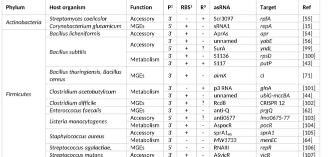
Table 2: List of studied cases of asRNAs in Gram-positive bacteria organized by phylum.
Phylum Host organism Function P1 RBS2 R3 asRNA Target Ref
Streptomyces coelicolor Accessory 3' - + Scr3097 rpfA [55]
Actinobacteria Corynebacterium glutamicum MGEs 5' + - sRNA1 repA [15] Bacillus licheniformis Accessory 3' + - AprAs apr [54]
3' + - unnamed yabE [56]
Accessory 5' + ? SurA yndL [99]
3' + - S1136 rpsD [100]
Bacillus subtilis
Metabolism 3' + + S117 putP [43]
Bacillus thuringiensis, Bacillus
cereus MGEs 3' + - aimX cI [71]
3' - + p3 RNA glnA [101]
Clostridium acetobutylicum Metabolism 3' + - unnamed ubiG-mccBA [44]
Clostridium difficile MGEs 3' + ? Rcd8 CRISPR 12 [102]
Enteroccocus faecalis MGEs 3' + - anti-Q prgQ [62] Accessory 5' + ? anti0677 lmo0675-77 [103]
Listeria monocytogenes Metabolism 3' + - AspocR pocR [104]
Accessory 3' + - sprA1AS sprA1 [105]
Staphylococcus aureus Metabolism 3' - - MW1733 menEC [64] Streptococcus agalactiae,
Staphylococcus aureus MGEs 5' - - RNAIII repR [106] Firmicutes
Streptococcus mutans Accessory 3' + - ASvicR vicR [107]
1Promoter of the asRNA located before the stop codon of the target (5’) or after (3’) the stop codon of the
target
2Coverage of the RBS by the major form of the asRNA.
3Major regulatory effect of the asRNA on the target, either activation (+), repression (-) or unknown (?).
Note: There is no consistent nomenclature for asRNAs and we have used the term most frequently found in the literature.
References
[1] S. Gottesman, Trouble is coming: Signaling pathways that regulate general stress responses in bacteria, J. Biol. Chem. 294 (2019) 11685-11700. https://doi.org/10.1074/jbc.REV119.005593
[2] I.M. Keseler, A. Mackie, A. Santos-Zavaleta, R. Billington, C. Bonavides-Martínez, R. Caspi, C. Fulcher, S. Gama-Castro, A. Kothari, M. Krummenacker, M. Latendresse, L. Muñiz-Rascado, Q. Ong, S. Paley, M. Peralta-Gil, P. Subhraveti, D.A. Velázquez-Ramírez, D. Weaver, J. Collado-Vides, I. Paulsen, P.D. Karp, The EcoCyc database: reflecting new knowledge about Escherichia coli K-12, Nucleic Acids Res. 45 (2016) 543-550. https://doi.org/10.1093/nar/gkw1003
[3] P. Mandin, M. Guillier, Expanding control in bacteria: interplay between small RNAs and transcriptional regulators to control gene expression, Curr. Opin. Microbiol. 16 (2013) 125-132.
https://doi.org/10.1016/j.mib.2012.12.005
[4] E.G.H. Wagner, P. Romby, Chapter Three - Small RNAs in Bacteria and Archaea: Who They Are, What They Do, and How They Do It, in: Adv. Genet., 90 (2015) 133-208
https://doi.org/10.1016/bs.adgen.2015.05.001
[5] M. Lejars, A. Kobayashi, E. Hajnsdorf, Physiological roles of antisense RNAs in prokaryotes, Biochimie 164 (2019) 3-16. https://doi.org/10.1016/j.biochi.2019.04.015
[6] D.L. Court, J. Gan, Y.H. Liang, G.X. Shaw, J.E. Tropea, N. Costantino, D.S. Waugh, X. Ji, RNase III: Genetics and function; structure and mechanism, Annu. Rev. Genet. 47 (2013) 405-431.
https://doi.org/10.1146/annurev-genet-110711-155618
[7] M. Levinthal, H. Nikaido, Consequences of deletion mutations joining two operons of opposite polarity, J. Mol. Biol. 42 (1969) 511-520. https://doi.org/10.1016/0022-2836(69)90239-3
[8] K. Bøvre, W. Szybalski, Patterns of convergent and overlapping transcription within the b2 region of coliphage λ, Virology 38 (1969) 614-626. https://doi.org/10.1016/0042-6822(69)90181-0
[9] F.A. Kolb, H.M. Engdahl, J.G. Slagter‐Jäger, B. Ehresmann, C. Ehresmann, E. Westhof, E.G.H. Wagner, P. Romby, Progression of a loop–loop complex to a four‐way junction is crucial for the activity of a regulatory antisense RNA, EMBO J. 19 (2000) 5905-5915. https://doi.org/10.1093/emboj/19.21.5905
[10] B.A. Berghoff, E.G.H. Wagner, RNA-based regulation in type I toxin-antitoxin systems and its
implication for bacterial persistence, Curr. Genet. 63 (2017) 1011-1016. https://doi.org/10.1007/s00294-017-0710-y
[11] K.E.L. Cox, J.F. Schildbach, Sequence of the R1 plasmid and comparison to F and R100, Plasmid 91 (2017) 53-60. https://doi.org/10.1016/j.plasmid.2017.03.007
[12] J.A. Ross, M.J. Ellis, S. Hossain, D.B. Haniford, Hfq restructures RNA-IN and RNA-OUT and facilitates antisense pairing in the Tn10/IS10 system, RNA 19 (2013) 670-684.
https://doi.org/10.1261/rna.037747.112
[13] J.N. Mark Glover, S.G. Chaulk, R.A. Edwards, D. Arthur, J. Lu, L.S. Frost, The FinO family of bacterial RNA chaperones, Plasmid 78 (2015) 79-87. https://doi.org/10.1016/j.plasmid.2014.07.003
[14] F.A. Kolb, H.M. Engdahl, J.G. Slagter-Jager, B. Ehresmann, C. Ehresmann, E. Westhof, E.G. Wagner, P. Romby, Progression of a loop-loop complex to a four-way junction is crucial for the activity of a
regulatory antisense RNA, EMBO J. 19 (2000) 5905-5915. https://doi.org/10.1093/emboj/19.21.5905
[15] S. Hashiro, M. Mitsuhashi, H. Yasueda, High copy number mutants derived from Corynebacterium
glutamicum cryptic plasmid pAM330 and copy number control, J. Biosci. Bioeng. 127 (2018) 529-538.
https://doi.org/10.1016/j.jbiosc.2018.10.012
[16] T. Thisted, N.S. Sorensen, E.G. Wagner, K. Gerdes, Mechanism of post-segregational killing: Sok antisense RNA interacts with Hok mRNA via its 5'-end single-stranded leader and competes with the 3'-end of Hok mRNA for binding to the mok translational initiation region, EMBO J. 13 (1994) 1960-1968.
[17] S. Masachis, F. Darfeuille, Type I Toxin-Antitoxin Systems: Regulating Toxin Expression via Shine-Dalgarno Sequence Sequestration and Small RNA Binding, Microbiol. Spectr. 6 (2018).
https://doi.org/10.1128/microbiolspec.RWR-0030-2018
[18] J. Georg, W.R. Hess, Widespread Antisense Transcription in Prokaryotes, Microbiol. Spectr. 6 (2018).
https://doi.org/10.1128/microbiolspec.RWR-0029-2018
[19] Á. Kun, A. Szilágyi, B. Könnyű, G. Boza, I. Zachar, E. Szathmáry, The dynamics of the RNA world: insights and challenges, Ann. N. Y. Acad. Sci. 1341 (2015) 75-95. https://doi.org/10.1111/nyas.12700
[20] M. Vandevenne, M. Delmarcelle, M. Galleni, RNA Regulatory Networks as a Control of Stochasticity in Biological Systems, Front. Genet. 10 (2019). https://doi.org/10.3389/fgene.2019.00403
[21] S. Altman, Enzymatic cleavage of RNA by RNA, Bioscience Rep. 10 (1990) 317-337.
https://doi.org/10.1007/BF01117232
[22] J.M. Peters, R.A. Mooney, J.A. Grass, E.D. Jessen, F. Tran, R. Landick, Rho and NusG suppress pervasive antisense transcription in Escherichia coli, Genes Dev. 26 (2012) 2621-2633.
https://doi.org/10.1101/gad.196741.112
[23] K. Kavita, F. de Mets, S. Gottesman, New aspects of RNA-based regulation by Hfq and its partner sRNAs, Curr. Opin. Microbiol. 42 (2018) 53-61. https://doi.org/10.1016/j.mib.2017.10.014
[24] N. Raghunathan, R.M. Kapshikar, J.K. Leela, J. Mallikarjun, P. Bouloc, J. Gowrishankar, Genome-wide relationship between R-loop formation and antisense transcription in Escherichia coli, Nucleic Acids Res. 46 (2018) 3400-3411. https://doi.org/10.1093/nar/gky118
[25] E.G. Wagner, S. Altuvia, P. Romby, Antisense RNAs in bacteria and their genetic elements, Adv. Genet. 46 (2002) 361-398. https://doi.org/10.1016/S0065-2660(02)46013-0
[26] F.Y. Tan, M.E. Wormann, E. Loh, C.M. Tang, R.M. Exley, Characterization of a novel antisense RNA in the major pilin locus of Neisseria meningitidis influencing antigenic variation, J. Bacteriol. 197 (2015) 1757-1768. https://doi.org/10.1128/JB.00082-15
[27] F. Amman, A. D'Halluin, R. Antoine, L. Huot, I. Bibova, K. Keidel, S. Slupek, P. Bouquet, L. Coutte, S. Caboche, C. Locht, B. Vecerek, D. Hot, Primary transcriptome analysis reveals importance of IS elements for the shaping of the transcriptional landscape of Bordetella pertussis, RNA Biol. 15 (2018) 967-975.
https://doi.org/10.1080/15476286.2018.1462655
[28] K. Voigt, C.M. Sharma, J. Mitschke, S. Joke Lambrecht, B. Voß, W.R. Hess, C. Steglich, Comparative transcriptomics of two environmentally relevant cyanobacteria reveals unexpected transcriptome diversity, ISME J. 8 (2014) 2056-2068. https://doi.org/10.1038/ismej.2014.57
[29] Q. Chen, J.H. Crosa, Antisense RNA, Fur, Iron, and the Regulation of Iron Transport Genes in Vibrio
anguillarum, J. Biol. Chem. 271 (1996) 18885-18891. https://doi.org/10.1074/jbc.271.31.18885
[30] L.S. Waldbeser, Q. Chen, J.H. Crosa, Antisense RNA regulation of the fatB iron transport protein gene in Vibrio anguillarum, Mol. Microbiol. 17 (1995) 747-756.
https://doi.org/10.1111/j.1365-2958.1995.mmi_17040747.x
[31] M. Stork, M. Di Lorenzo, T.J. Welch, J.H. Crosa, Transcription Termination within the Iron Transport-Biosynthesis Operon of Vibrio anguillarum Requires an Antisense RNA, J. Bacteriol. 189 (2007) 3479-3488. https://doi.org/10.1128/JB.00619-06
[32] K. Hantke, Iron and metal regulation in bacteria, Curr. Opin. Microbiol. 4 (2001) 172-177.
https://doi.org/10.1016/S1369-5274(00)00184-3
[33] J.A. Hernández, A.M. Muro-Pastor, E. Flores, M.T. Bes, M.L. Peleato, M.F. Fillat, Identification of a
furA cis antisense RNA in the cyanobacterium Anabaena sp. PCC 7120, J. Mol. Biol. 355 (2006) 325-334.
https://doi.org/10.1016/j.jmb.2005.10.079
[34] J.A. Hernández, I. Alonso, S. Pellicer, M. Luisa Peleato, R. Cases, R.J. Strasser, F. Barja, M.F. Fillat, Mutants of Anabaena sp. PCC 7120 lacking alr1690 and α-furA antisense RNA show a pleiotropic phenotype and altered photosynthetic machinery, J. Plant Physiol. 167 (2010) 430-437.
[35] E. Sevilla, B. Martín-Luna, A. González, J.A. Gonzalo-Asensio, M.L. Peleato, M.F. Fillat, Identification of three novel antisense RNAs in the fur locus from unicellular cyanobacteria, Microbiol. 157 (2011) 3398-3404. https://doi.org/10.1099/mic.0.048231-0
[36] B. Martín-Luna, E. Sevilla, A. González, M.T. Bes, M.F. Fillat, M.L. Peleato, Expression of fur and its antisense alpha-fur from Microcystis aeruginosa PCC7806 as response to light and oxidative stress, J. Plant Physiol. 168 (2011) 2244-2250. https://doi.org/10.1016/j.jplph.2011.08.006
[37] C. Lefimil, E. Jedlicki, D.S. Holmes, α-fur, an antisense RNA gene to fur in the extreme acidophile
Acidithiobacillus ferrooxidans, Microbiol. 160 (2014) 514-524. https://doi.org/10.1099/mic.0.073171-0
[38] M.K. Thomason, G. Storz, Bacterial antisense RNAs: How many are there and what are they doing?, Annu. Rev. Genet. 44 (2010) 167-188. https://doi.org/10.1146/annurev-genet-102209-163523
[39] E.J. Lee, E.A. Groisman, An antisense RNA that governs the expression kinetics of a multifunctional virulence gene, Mol. Microbiol. 76 (2010) 1020-1033. https://doi.org/10.1111/j.1365-2958.2010.07161.x
[40] K. Okamoto, S. Hara, R. Bhasin, M. Freundlich, Evidence in vivo for autogenous control of the cyclic AMP receptor protein gene (crp) in Escherichia coli by divergent RNA, J. Bacteriol. 170 (1988) 5076-5079.
https://doi.org/10.1128/jb.170.11.5076-5079.1988
[41] C. Pennetier, J. Oberto, J. Plumbridge, An antisense transcript from within the ptsG promoter region in Escherichia coli, J. Mol. Microbiol. Biotechnol. 18 (2010) 230-240. https://doi.org/10.1159/000319598
[42] M.W. Thairu, S. Cheng, A.K. Hansen, A sRNA in a reduced mutualistic symbiont genome regulates its own gene expression, Mol. Ecol. 27 (2018) 1766-1776. https://doi.org/10.1111/mec.14424
[43] J.M. DiChiara, B. Liu, S. Figaro, C. Condon, D.H. Bechhofer, Mapping of internal monophosphate 5′ ends of Bacillus subtilis messenger RNAs and ribosomal RNAs in wild-type and ribonuclease-mutant strains, Nucleic Acids Res. 44 (2016) 3373-3389. https://doi.org/10.1093/nar/gkw073
[44] G. André, S. Even, H. Putzer, P. Burguière, C. Croux, A. Danchin, I. Martin-Verstraete, O. Soutourina, S-box and T-box riboswitches and antisense RNA control a sulfur metabolic operon of Clostridium
acetobutylicum, Nucleic Acids Res. 36 (2008) 5955-5969. https://doi.org/10.1093/nar/gkn601
[45] J.A. Opdyke, E.M. Fozo, M.R. Hemm, G. Storz, RNase III Participates in GadY-Dependent Cleavage of the gadX-gadW mRNA, J. Mol. Biol. 406 (2011) 29-43. https://doi.org/10.1016/j.jmb.2010.12.009
[46] Q. Zhang, Y. Zhang, X. Zhang, L. Zhan, X. Zhao, S. Xu, X. Sheng, X. Huang, The novel cis-encoded antisense RNA AsrC positively regulates the expression of rpoE-rseABC operon and thus enhances the motility of Salmonella enterica serovar typhi, Front. Microbiol. 6 (2015).
https://doi.org/10.3389/fmicb.2015.00990
[47] C. Xiong, X. Li, J. Liu, X. Zhao, S. Xu, X. Huang, Identification and Characterization of a Cis Antisense RNA of the rpoH Gene of Salmonella enterica Serovar Typhi, Front. Microbiol. 9 (2018).
https://doi.org/10.3389/fmicb.2018.00978
[48] T. Nomura, H. Aiba, A. Ishihama, Transcriptional organization of the convergent overlapping
dnaQ-rnh genes of Escherichia coli, J. Biol. Chem. 260 (1985) 7122-7125.
https://doi.org/jbc.org/content/260/11/7122.long
[49] F. Fontaine, E. Gasiorowski, C. Gracia, M. Ballouche, J. Caillet, A. Marchais, E. Hajnsdorf, The small RNA SraG participates in PNPase homeostasis, RNA 22 (2016) 1560-1573.
https://doi.org/10.1261/rna.055236.115
[50] K.S. Kim, R. Manasherob, S.N. Cohen, YmdB: a stress-responsive ribonuclease-binding regulator of E.
coli RNase III activity, Genes Dev. 22 (2008) 3497-3508. https://doi.org/10.1101/gad.1729508
[51] P. Mandin, F. Repoila, M. Vergassola, T. Geissmann, P. Cossart, Identification of new noncoding RNAs in Listeria monocytogenes and prediction of mRNA targets, Nucleic Acids Res. 35 (2007) 962-974.
https://doi.org/10.1093/nar/gkl1096
[52] M. Cavaiuolo, R. Kuras, F.A. Wollman, Y. Choquet, O. Vallon, Small RNA profiling in Chlamydomonas: insights into chloroplast RNA metabolism, Nucleic Acids Res. 45 (2017) 10783-10799.
[53] E. Olmedo-Verd, I.L.M. Brenes, A.N. Vioque, A.M. Muro-Pastor, A Heterocyst-Specific Antisense RNA Contributes to Metabolic Reprogramming in Nostoc sp. PCC 7120, Plant Cell Physiol. 60 (2019) 1646-1655. https://doi.org/10.1093/pcp/pcz087
[54] R. Hertel, S. Meyerjürgens, B. Voigt, H. Liesegang, S. Volland, Small RNA mediated repression of subtilisin production in Bacillus licheniformis, Sci. Rep. 7 (2017). https://doi.org/10.1038/s41598-017-05628-y
[55] R.J. St-Onge, M.A. Elliot, Regulation of a muralytic enzyme-encoding gene by two non-coding RNAs, RNA Biol. 14 (2017) 1592-1605. https://doi.org/10.1080/15476286.2017.1338241
[56] W. Eiamphungporn, J.D. Helmann, Extracytoplasmic Function σ Factors Regulate Expression of the
Bacillus subtilis yabE Gene via a cis-Acting Antisense RNA, J. Bacteriol. 191 (2009) 1101-1105.
https://doi.org/10.1128/jb.01530-08
[57] M. Giangrossi, G. Prosseda, C.N. Tran, A. Brandi, B. Colonna, M. Falconi, A novel antisense RNA regulates at transcriptional level the virulence gene icsA of Shigella flexneri, Nucleic Acids Res. 38 (2010) 3362-3375. https://doi.org/10.1093/nar/gkq025
[58] J. Gonzalo-Asensio, Á.D. Ortega, G. Rico-Pérez, M.G. Pucciarelli, F. García-del Portillo, A Novel Antisense RNA from the Salmonella Virulence Plasmid pSLT Expressed by Non-Growing Bacteria inside Eukaryotic Cells, PLOS ONE 8 (2013). https://doi.org/10.1371/journal.pone.0077939
[59] A. Arini, P. Keller Marcel, W. Arber, An Antisense RNA in IS30 Regulates the Translational Expression of the Transposase, Biol. Chem. 378 (1997) 1421-1431. https://doi.org/10.1515/bchm.1997.378.12.1421
[60] J. Hahn, O.V. Tsoy, S. Thalmann, J. Cuklina, M.S. Gelfand, E. Evguenieva-Hackenberg, Small Open Reading Frames, Non-Coding RNAs and Repetitive Elements in Bradyrhizobium japonicum USDA 110, PLOS ONE 11 (2016). https://doi.org/10.1371/journal.pone.0165429
[61] R. Lloubes, D. Baty, C. Lazdunski, The promoters of the genes for colicin production, release and immunity in the ColA plasmid: effects of convergent transcription and Lex A protein, Nucleic Acids Res. 14 (1986) 2621-2636. https://doi.org/10.1093/nar/14.6.2621
[62] R.J. Breuer, H. Hirt, G.M. Dunny, Mechanistic Features of the Enterococcal pCF10 Sex Pheromone Response and the Biology of Enterococcus faecalis in Its Natural Habitat, J. Bacteriol. 200 (2018).
https://doi.org/10.1128/jb.00733-17
[63] M.J. Ellis, D.B. Haniford, Riboregulation of bacterial and archaeal transposition, WIRES RNA 7 (2016) 382-398. https://doi.org/10.1002/wrna.1341
[64] S. Sáenz-Lahoya, N. Bitarte, B. García, S. Burgui, M. Vergara-Irigaray, J. Valle, C. Solano, A. Toledo-Arana, I. Lasa, Noncontiguous operon is a genetic organization for coordinating bacterial gene
expression, Proc. Natl. Acad. Sci. U. S. A. 116 (2019) 1733-1738.
https://doi.org/10.1073/pnas.1812746116
[65] C. Ma, R.W. Simons, The IS10 antisense RNA blocks ribosome binding at the transposase translation initiation site, EMBO J. 9 (1990) 1267-1274. https://doi.org/10.1002/j.1460-2075.1990.tb08235.x
[66] S.M. Liao, T.H. Wu, C.H. Chiang, M.M. Susskind, W.R. McClure, Control of gene expression in
bacteriophage P22 by a small antisense RNA. I. Characterization in vitro of the Psar promoter and the sar RNA transcript, Genes Dev. 1 (1987) 197-203. https://doi.org/10.1101/gad.1.2.197
[67] S. Saha, E. Haggard-Ljungquist, K. Nordstrom, The Cox protein of bacteriophage P2 inhibits the formation of the repressor protein and autoregulates the early operon, EMBO J. 6 (1987) 3191-3199.
https://doi.org/10.1002/j.1460-2075.1987.tb02631.x
[68] K. Ranade, A.R. Poteete, A switch in translation mediated by an antisense RNA, Genes Dev. 7 (1993) 1498-1507. https://doi.org/10.1101/gad.7.8.1498
[69] H.M. Krause, N.P. Higgins, Positive and negative regulation of the Mu operator by Mu repressor and
Escherichia coli integration host factor, J. Biol. Chem. 261 (1986) 3744-3752.