DEVELOPMENT OF A GENETIC SYSTEM IN
RHIZOBIUM MELILOTI
by
HARRY MELVIN MEADE
..::::::B.S., Union College
(1969)
SUBMITTED IN PARTIAL FULFILLMENT
OF THE REQUIREMENTS FOR THE
DEGREE OF
DOCTOR OF PHILOSOPHY
at the
MASSACHUSETTS IrJSTITUTE OF TECHNOLOGY
2 DEVELOPMENT OF A GENETIC SYSTEM IN
RHIZOBIUM MELILOTI
by
HARRY MELVIN MEADE
Submitted to the Department of Biology on May 5, 1977 in partial fulfillment of the requirements for the Degree of
Doctor of Philosophy
ABSTRACT
This thesis is the development of a genetic system in the symbiotic nitrogen fixing bacterium Rhizobium meliloti. Drug resistant and auxo-trophic mutants of the effective strain Rm2011 were first isolated. The genetic system is based on conjugation by the drug resistance factor RP4. During mating RP4 can promote chromosome mobilization to produce recombin-&nt progeny. By carrying out crosses between multiply marked derivatives of Rm2011, a circular linkage map of R. meliloti was constructed. This is the first genetic transfer system and resulting linkage map of Rhizobium meliloti.
During this study, a spontaneous Hfr was isolated. In an attempt to generate Hfrs, a derivative of RP4 that is temperature sensitive for repli-cation was found. Recombinants between RP4 and F'ColVBtrp were selected, which have the mating properties of RP4. One of these has been used to
introduce Xh80 into R. meliloti.
3
TABLE OF CONTENTS
Title page
Abstract 2
Table of Contents 3
List of Tables and Figures
I. INTRODUCTION 6 TI. METHODS A. Chemicals 8 B. Media 9 C. Strains 10 1. Bacteria 11 2. Phage 13 D. Growth of Bacteria 14 E. Storage 15 F. Nodulation 16 G. Mutagenesis 16 H. Mutants 19 1. Drug resistant 19 2. Auxotrophic enrichment 21 3. Tester strain 22 I. Phage techniques 23
J. Isolation of rhizobia from soil 24
K. Mating 25
1. Broth-liquid 2. Plate-surface
4 L. Selection 26 1. Reversion 2. Donor counterselection M. Linkage Data 26 1. Selection 2. Scoring recombinants III. RESULTS A. ISOLATION OF MUTANTS 27
B. UNSUCCESSFUL ATTEMPTS AT GENETIC TRANSFER 28
1. Transformation 28 2. Transduction 29 3. Conjugation 31 IV. RP4 35 A. INTRODUCTION 35 B. PROPERTIES 35 C. FREQUENCY OF TRANSFER 37
D. CHROMOSOME MOBILIZATION USING RP4 38 1. Proof of gene transfer
2. Properties of the system
E. LINKAGE 39
1. Procedure (sample cross) 39
2. Linkage map 41
V. EPILOG
APPENDICES
I. Isolation of an Hfr II. Hybrid episomes
III. Introduction of Ah80 into R. meliloti IV. Isolation of RP4ts
REFERENCES
LIST OF TABLES AND FIGURES
II-G. II-H. III-A. IV-B. AI-1. Figure IV-1 Figure IV-2 Figure AI-1 Comparative Mutagenesis
Drug and Antimetabolite Sensitivity Drug Resistant Mutants
R factor Strains Hfr Linkage Data
Linkage or Transfer Frequencies Using RP4
Circular Linkage Map of R. meliloti Hfr Interrupted Mating 5 47 49 53 58 61 18 20 27 34 51 42 43 51 Table Table Table Table Table
GENERAL INTRODUCTION
6 Rhizobia fix atmospheric nitrogen in the nodules they form on the roots of legumes. This rhizobia-legume symbiotic relationship is the main means by which biological nitrogen fixation is harnessed for agricultural production. Not only are the grain legumes (pulses) a good source of protein but when they are included in crop rotation practices, there is a net increase of available fixed nitrogen in the soil.
Even though rhizobia have been studied for many years, there has been no detailed genetic analysis of the rhizobia-legume relationship. This has been mainly due to the lack of a suitable genetic system in a nodulating strain of Rhizobium.
This thesis was begun with the goal of developing a usable genetic system, and I describe here the results of this effort. I have developed a genetic system in Rhizobium meliloti, Rm20ll that is based on
conjuga-tion. During mating the drug resistance factor RP4 can mobilize the bacterial chromosome to produce recombinant progeny. Using this system I have constructed a linkage map of R. meliloti.
This thesis is the necessary first step in the genetic analysis of the rhizobia-legume symbiosis--development of the genetic system.
A number of reports have described genetic transfer in Rhizobium.
As early as 1953, Balassa (1) reported transformation among three species, and there have been occasional reports since then (2-5). It was not
until just recently that linkage was reported using transformation in R. japonicum (6).
R. meliloti. Generalized transduction has been shown by 7 Kowalski, (8); also in R. meliloti.
There have been many reports of conjugation in Rhizobium.Higashi (10) reported the transfer of nodulation specificity between two different species and Lorkiewica (11) described the transfer of auxo-trophic markers in R. trifolii. There have been no subsequent reports in either case. Heumann (12) reported mating and a circular linkage map in a strain reported to be R. lupini. However, this strain no longer nodulated (13) and unlike other rhizobia forms colored colonies on agar plates, which makes this difficult to evaluate.
The genetic system I describe here for R. meliloti is based on conjugation promoted by the drug resistance factor RP4. Using this system I have carried out a number of crosses between multiply marked derivatives of the parental strain of R. meliloti. By scoring the selected exconjugants for cotransfer of nonselected markers, I have constructed a linkage map of R. meliloti. This is the first genetic system and resulting circular linkage map of a nodulating strain of Rhizobium.
I have presented preliminary reports of this work (14, 15) and am submitting a paper describing the system. Beringer has just reported a similar system using another P-type R factor, R68-45, in R. legumino-sarum (16).
II. METHODS 8 A. CHEMICALS 1. Lysozyme 2. Carbenicillin 3. Spectinomycin 4. Rifampin 5. Tetracycline 6. 5-Fluorouracil 7. Chloramphenicol 8. Kanamycin 9. Neomycin 10. Novobiocin 11. Penicillin G 12. Polymyxin B 13. Streptomycin
Worthington Biochem. Corp.
Pfizer Upjohn Calbiochem Sigma "I "I "I I" II
II. B. MEDIA 9
LB broth: Per liter, log tryptone, 5g yeast extract, 5g NaCl, 4ml of 1N NaOH
LB agar: Same as above except only Iml of 1N NaOH and 15g agar Soft agar: Same as LB with 6.5g agar
LBCa: Lb broth plus 0.5mM CaCl
2
M9 : Per liter, 5.8g Na2HPO4, 3g KH2PO4, 0.5 NaCi, lg NH4C1
and 1mM MgSO4 added after autoclaving M9 agar: Same as above with 15g agar
GB M9: For E. coli M9 is supplemented with 0.2% Glucose and 0.5ug/ml thiamine
SbM9: For Rm2011 M9is supplemented with 0.2% Sucrose and 0.5ug/ml biotin
All minimal media are supplemented with 20ug/ml of amino acids
or bases whenever needed SAY: Per liter, 0.5g K2HPO
4, 0.2g MgSO4'7H20, 0.5mM CaCl2,
lOg yeast extract, 2g Sucrose
SAY agar: Same as above with 15g agar
SAY top: Same as SAY with 6.5g agar
AA agar: Same as SAY with 120mg cycloheximide, 60mg
pentachloro-nitrobenzene, 1.5ml of 1% Congo Red solution
Nodulation: Per liter, 1.0g Ca2HPO4, 0.2g K2HPO4, 0.2g MgSO-7H20
0.2g NaCl, 0.lg FeCl3, Adjust to pH7.0
STAB agar: Per liter, lOg Nutrient Broth, 8g NaCl, 20mg cysteine 6g agar
II. C. STRAINS
Bacterial strains are listed in Table II-C-1. E. coli strains
carrying R factors were kindly donated by G. Jacoby at Mass. Genaeral Hospital. These strains are shown in Table III-B-3. All E. coli strains
used were from the collection of Ethan Signer.
The parental Rhizobium meliloti strain,Rm20ll str3, was donated
by Jean Denarie in Versailles, France. This strain is a streptomycin resistant mutant of R. meliloti SU47 from P. J. Nutman at Rothamsted Experimental Station, England. I isolated all of the derivatives of Rm2011 described in this this.
Phage strains were from the collection of Ethan Signer except where otherwise noted. Phage are listed in Table II-C-2.
II. C. 1 RHIZOBIUM MELILOTI STRAINS Rm2011 str3 parental wild type
All are derivates of Rm2011 Rm2011 cys11 Rm2012 gly12 Rm2013 ilv13 Rm2014 ade14 Rm2018 his18 Rm2024 met24 Rm2025 his25 Rm2026 leu26 Rm2027 ilv27 Rm2028 met28 Rm2029 pan29 Rm2031 ade3l Rm2032 cho32 Rm2049 his18 rif2
Rm2079 his18 leu79 rif2 Rm2080 cyt80 pyr80 11 str 3 Rm3300 Rm3330 Rm3339 Rm3341 Rm3342 Rm334.3 Rm3344 trp33 sp l trp33 spc rifl trp33 his39 spl rifl arg4l "I cys42 gly43 " pan44
K013Y6O cys gly
Rm3347 ser,gly Rm3348 ilv48 Rm3349 pyr49 Rm3350 cys50 Rm3351 aro51 Rm3353 " leu53 Rm3354 phe54 Rm3355 "Iys55 Rm3356 met56 Rm3357 1eu53, nov57
Rm3363 his39 pan44 glyl2 rifl / RP4 Hfr
Rm3373 trp33 his39 leu53 spc rifl
Rm3374 pyr49 Rm3375 leu53 Rm 37C Rm3377 Rm3378 Rm3379 Rm3381 Rm3382 Rm3383 Rm3386 Rm3388 Rm3389 Rm3390 I, r 12 nov57 na173 nov59 ery74 nov57 gly75 rif 1 nov57 nov59 "I " pmx86 nov57 pmx88 nov59 pmx89 " nov90 Rm2lO0/RP4 Rm2lOl/RP4 Rm2l02/RP4' Rm2l03/RP4 glyl2 (ATK) pmxl (ATK) ery2 (ATK) vani (ATK)
"I "1 "I arg76
spcl
"I "I "Iilv77
"I "I " phe78 " "i "I "I aro79
pyr49 lys8l
"I "I "1 phe82 "
"I "I "I aro83 "I "I "i lys55
" "
1eu53
arg76 " "I "I pyr49 "II. C. 2 PHAGE STRAINS 13 Xc+ XcI8 57h80 080 v5 P1Cm, clrilOO P1 vir Plkc T5 T7 Richard Goldberg "I "' "I Nancy Kleckner "1 Soil isolate P22 P22Tet XcI60 II MP1-3
II. D. GROWTH OF BACTERIA
14 E. coli
E. coli strains are routinely grown at 370C except for temperature sensitive lysogens which are grown at 300C. Rich medium is LB and minimal medium is M9 supplemented with 0.2% Glucose, 0.5ug/ml B1, and 20ug/ml of amino acids or bases when needed.
R. meliloti
Rm2011 is a fast growing strain of Rhizobium that unlike most strains grows well on standard E. coli media. LB is used for rich medium and minimal medium is M9 supplemented with 0.2% Sucrose, o.5ug/ml biotin, and
20ug/ml of amino acids or bases when needed. Rm2011 has a growth range of 220 to 390C. These bacteria are routinely grown at 320C.
The doubling time of Rm2011 in rich medium is 2 to 3 hours and single colonies appear on LB agar within 3 days. In minimal medium the doubling time is 6 hours and 5 days are required to see colonies on minimal plates.
SAY medium is a standard Rhizobium growth medium (17) which is used for phage isolation and growth. With the addition of a dye and anti-biotics this medium can be used to isolate rhizobia from the soil (AA medium) as described by Grahm (18). The rhizobia are recognized by their white mucoid colonies which do not take up the dye.
II. E. STORAGE.
Rhizobium have traditionally been stored in refrigerated slants. 15 These slants must be reinoculated every month as described by Vincent
(17). There is a constant selection that occurs each time the rhizobia
are inoculated into a new slant. This may account for the loss of
in-fectivity in those strains that have been kept under laboratory condi-tions for many years.
I have found that Rm2011 can be stored in stabs used routinely for E.coli. This has eliminated the need to repeatedly transfer the
rhizobia to new slants. Rm2011 has shown no loss of nodulation ability
II. F. NODULATION
Nodulation is carried out as described by Vincent (17). Alfalfa 16 seeds are surface-sterilized by soaking in one half strength Chlorox for 15 minutes, then washed repeatedly with sterilized water. The
seed are then placed in a growth chamber repared as follows.
A piece of #29 Whatman balck filter paper is rolled to form a cylinder that can be pushed to the bottom of a 20 x 150 m test tube so that it stands on edge. Three seeds are placed between the paper and the tube wall. Eight ml of Nodulation medium are then added and a glass cap is placed over the top of the tube.
The seeds germinate within four days and one week later their roots extend to the bottom of the tube. The growth chambers are placed in a window for daylight and also supplemented with Sylvania Gro-lite at 1000 lux for a 14 hr. day. The average temperature is 260C. The roots are kept dark.
The plants are then inoculated with about 108 rhizobia that have been washed and resuspended in sterile water. One week after inoculation
nodules begin to appear, and within a month there are several nodules on each plant.
Effectiveness can be monitored within 2 months. Since the plants are growing in nitrogen-free medium, uninoculated controls become stunted and chlorotic. Plants inoculated with effective Rm2011 are lush, green and healthy.
II. G. MUTAGENESIS
Introduction
I first determined the optimal conditions for mutagensis by measuring the survival versus mutagenic rate for EMS, NTG, and UV irradiation.
the procedures in parallel with the mutagenized cells. Survival was determined by comparing the number of rif (rifampin) resistant colonies17
found on LBrif plates to the unmutagenized control. Both the controls and the experimental cells were grown overnight before being plated on
LBrif to allow the expression of the rif resistant phenotype. The methods are essentially those described by Miller (19).
EMS (ethyl methyl sulfonate)
Cells were grown in LB to saturation, washed twice and resuspended in one half the original volume of Tris-saline pH 7.6. A control aliquot was removed, and two drops per ml of EMS was added to the cells. The mixture was incubated at 300C without shaking. At various times aliquots were removed, washed with 0.02M Na2S203, titered, diluted ten fold into
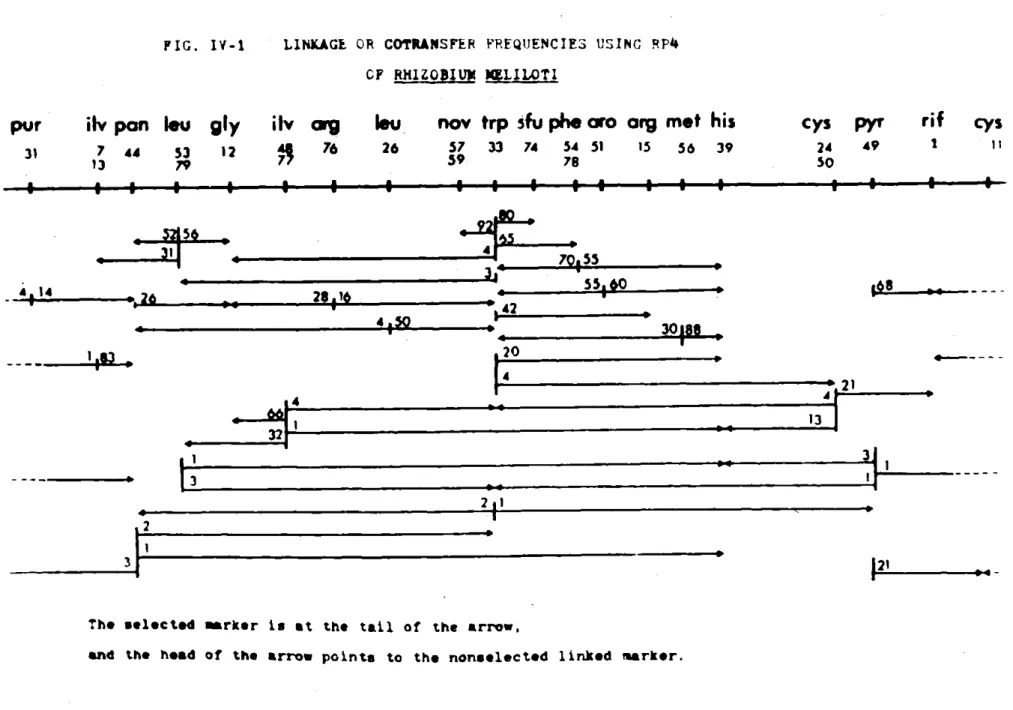
LB, and allowed to grow overnight as described above. The results are shown in Table II-G.
NTG
Cells were grown to log phase, washed twice and resuspended in the original volume of 0.01M Citrate Buffer pH 5.5. NTG from frozen stock
(2.5mg/ml) was added to a final concentration of 100ug/ml. The cells
were incubated at 300C without shaking. At various times aliquots were
removed, washed with Phosphate Buffer pH 7.0, and resuspended in LB. Survival and mutagenesis were measured as described above. The results are shown in Table II-G.
UV Irradiation
Cells were grown to log phase, washed twice and resuspended in saline.
The cells were then irradiated with a germicidal U.V. lamp at 5 ergs/min. At various times aliquots were removed, titered, and diluted into LB. The
TABLE II-G COMPARATIVE MUTAGENSIS 18 EMS Time (min) 0 30 60 90 120 Survival (5) 100 90 25 1 0.1 Rif resistant colonies 4 30 1200 1000 500 Increase (X) 1 8 300 250 125 NTG Time (min.) 0 15 30 60 90 Survival
(%)
100 23 11 1 0.5 Rif resistant colonies 7 700 2000 1100 Increase (x) 1 100 267 167 UV Time (sec.) 0 15 30 45 60 Survival(%)
100 25 3.4 0.1 0.004 Rif resistant colonies 10 200 1000 250 Increase (X) 1 20 100 25II. H. MUTANTS
1. Drug resistant 19
Mutants resistant to various antibiotics and antimetabolites have been isolated. These mutants are useful in counterselection schemes and the mutations themselves are markers that can be mapped in genetic studies. In order to isolate mutants of Rm2011 resistant to drugs and anti-metabolites it was first necessary to test this strain for sensitivity to various agents. Antibiotics were tested using either Pfizer Test Discs (small paper discs impregnated with antibiotic) or a few grains of powdered antibiotic placed on a lawn of Rm2011 growing on LB agar. Antimetabolites and analogs were tested by placing the powdered agent on Rm2011 growing on minimal agar. The results are shown in Table II-K.
Once those agents which are toxic to Rm2011 were found, they were further tested for the level at w'ich mutants could be isolated. This was done by two fold dilutions starting at 500ug/ml. I chose a level at
which a log phase culture would give individual resistant colonies and no background when plated out. The concentrations at which the mutants were isolated are listed beside each agent in Table II-H.
TABLE II-H DRUG Trimethoprin Streptolydigan Puromycin Sulfathiazole Cephalosporin Mendalamine Cloxicillin Penicillin Chloramphenicol Rifampin Specti nomycin Ampicillin Novobiocin Polymyxin B Gentamicin D-cycloserine Vancomycin Bacitracin Erythromycin Nalacixic Acid Kanamycin
DRUG AND ANTIMETABOLITE SENSITIVITY ANTIMETABOLITE R D-histidine R R R R R R S S S S S S S S R S R S S S 50ug/ml 50ug/ml 50ug/ml 1 0Oug/ml 40ug/ml 25ug/ml 2ug/ml 50ug/ml D-seri ne L-valine 5-methly tryptophane methyl anlanine thiaolyl anlanine p-fluoro-phenyl alanine methyl aspartic acid azaxanthine azaguanine azahypoxanthine azauracil 5-fluoro uracil deoxygalactose ethionine 20 R R R R R R R R R R R R S 10Oug/ml R S 100 ug/ml 50ug/ml 50ug/ml 5Oug/ml 1 0Oug/ml S--sensitive R--resistant
II. H. 2. Auxotrophic mutants
21 Various amino acid and base requiring auxotrdphic mutants have been
isolated by a modification of the Penicillin enrichment method described by Davis (20). Although Rm2011 was sensitive to Amp and Pen G (see II-H), these drugs were ineffective in enrichment procedures since they gave only 10 fold killing in minimal media. Carbenicillin (Pfizer) a synthetic derivative of penicillin was effective and could be used to enrich for auxotrophs.
Log phase mutagenized cells grown in LB were washed twice and re-suspended in supplemented SbM9 medium at 108_cells/ml. The cells were
in-cubated under growing conditions for six hours to starve the cells of
metabolites. After this time the cells were diluted to 107/ml. The mixture
was incubated under growing conditions for 5 days. The cells were then washed twice with water and survivors plated on LB. Colonies that grew up
in 3 days were replicated onto minimal medium to determine prospective auxotrophs. Requirements were determined by test streaking them on minimal plates supplemented with metabolite pools as described by Holliday (21). This technique gave 104-105 killing during the Carbenicillin treatment and
2% auxotrophs among the survivors.
Carbenicillin is effective at 2mg/ml., yet only ten fold killing takes place each day in minimal medium. This is surprising since the doubling time of Rm2011 in minimal medium is about 6 hours. One would then expect high
levels of cell death within 24 hours. Even increased levels of Carbenicillin
did not enhance killing.
Before enrichment the level of auxotrophs in the bacterial population is less than 0.1%, and afterwards the level is 2%. This shows that the
22 percentage of auxotrophs make this method more tedious than that des-cribed for E. coli (19). I have used this technique to isolate the auxotrophs Rm2012 - Rm3375 (II.C.I.).
Recently, another method has been described by Klapwijk, et al. (22) to isolate auxotrophs in Agrobactererium. In this method both Carbenicillin (500ug/ml) and lysozyme (100ug/ml) are used together to enrich for non-growing cells. I have found that PenG (lmg/ml) and lysozyme (100ug/ml) can be used in Rm2011. This method give 105 killing within 12 hours and 2% auxotrophs among the survivors. Peni-cillinase is added after incubation (2000 I.U./ml) to inactivate the
PenG. I have used this technique to isolate mutants Rm3375-90 (II.C.I) Multiply marked strains were isolated by repeated mutagenesis and enrichment.
II. H.
3. Tester strain Rm3330
A multiply marked strain of Rm2011 was isolated to be used in studies to establish genetic transfer. The strain was obtained with
str3, and successively made resistant to spectinomysin (spcl, spontaneous), tryptophan requiring (trp33, EMS), and resistant to rifampin (rifl,
spontaneous) to give strain Rm3330, genotype trp33, rifl, spc, str3. The trp33 marker reverts less than 10-10 and the drug markers can be used in counterselection schemes.
II. I. PHAGE
1. Rhizobiophage 23
Phage that lyse Rm2011 were isolated as described by Parker and Allen (26). Soil isolates of about 10 grams were added to 50ml. of
water and slowly shaken at 300C overnight. The suspension was centrifuged
to remove particulate material, chloroform was added to the supernatant,
which was then plated on Rm2011 using the overlay method in SAY medium. Plaques that formed were purified by streaking on an indicator lawn of Rm2011 growing on SAY plates.
Transduction experiments using rhizobiophage were done by absorbing
phage at moi of one to a log phase culture of Rm3330 grown in SAY medium.
After incubation at 300C for 2 hours the cells were diluted and plated on SbM9 minimal medium.
2. Enteric phage
Rm2011 was tested for sensitivity to enteric phage by spotting on a lawn of Rm2011 about 108 pfu of the phage listed in Table II-C-2. These phage included P22, Pivir, P1kc, T4, T5, T7,
X',
XcI6O6O80v5There appeared to be clearing only by P1, yet even this phage produced no single plaques.
To isolate mutants of Rm2011 sensitive to enteric phage P1 and P22, I used the methods described by Goldberg, et al. (27). P1Cm and P22Tet were added at a moi of one to log phase clutures of Rm2011. Each culture
was then plated on either chloramphenicol or tetracycline (depending on
the phage) and incubated at 300C. The Cm resistant colonies were
II. 3. ISOLATION OF R. MELILOTI FROM SOIL 24 Other rhizobia were isolated in an attempt to find naturally occurring fertile strains of R. meliloti. Bacteria were isolated from nodules of alfalfa plants that were growing on various farms. Excised nodules were first surface sterilized by dipping in ethanol for two minutes, followed by washing with water. They were then crushed and the contents streaked or diluted onto AA medium. Rhizobia can be recognized on this medium as white or clear mucoid colonies.
Several strains were isolated in this way from nodules, and several others from commercial sources. These naturally occurring rhizobia were used in liquid mating experiments with Rm3330 in an attempt to demonstrate genetic exchange. None of the strains isolated gave evidence for fertility.
These rhizobia were also screened for their ability to produce phage that would plaque on Rm2011. No phage were found.
II. K. MATING
A. LIQUID OR BROTH MATING 25
This type of mating procedure is routinely used to move RP4 into a new strain. It was also used in attempts to mate other plasmids into Rm2011.
Donors are grown to log phase in LB broth. Recipients are grown
to saturation. Equal volumes of each parent are mixed and allowed to
sit for 3 hours at 340C. The mating mixture is then washed and re-suspended in the same volume of saline. Dilutions are then plated on appropriate media.
B. PLATE OR SURFACE MATING
This mating procedure is routinely used in RP4 promoted chromosome
mobilization and linkage studies. This technique is the basis of the genetic mapping described in this thesis.
The donor which carries RP4 is grown to log phase in LB broth. The recipients are grown to saturation also in broth. One half ml. of both donor and recipient are mixed together and spread on a pre-warmed LB plate. After incubation overnight at 340C, 10 ml. of saline
is added to the resulting lawn and the resuspended bacteria are scraped into a centrifuge tube. The cells are washed twice and resuspended in 5 ml. saline, before being plated on selective media. Each parent alone
is carried through the same procedure as a control. This technique allows
mating to take place on a surface under nonselective conditions before recombinants are selected.
IT. L. SELECTION
26 After mating, the mating mixture and controls are plated on selective medium. RP4 transfer was selected on LB agar with added tetracycline
(10ug/ml). All auxotrophic markers selected for in mating experiments have a reversion frequency less than 0.1% of the recombinant frequency, except for pur-31 which gave revertants at about 1% of the recombinant frequency.
To counterselect donor bacteria, two different antibiotics are used when possible. In chromosome mobilization studies with RP4, counter-selection is done using a resistance marker as far away as possible from the auxotrophic marker being selected, (i.e. rifl and trp33, pyr49 and nov59).
M. LINKAGE DATA
All linkage data are derived from RP4 plate matings. A single marker is first selected on plates lacking the required nutrient. Then
200 colonies are picked and streaked on appropriately supplemented plates,
to score for other (i.e. unselected) markers. The cotransfer or linkage frequency for a given marker pair is defined as the fraction of selected colonies that have also inherited the unselected marker from the donor.
ii I. RESULTS
A. ISOLATION OF MUTANTS 27
In order to develop a genetic system in R. meliloti I first
iso-lated a collection of auxotrophic and drug resistant mutants of Rm20ll, as described in II. G.H. These isolates are listed in II.C.1. R.
MELILOTI STRAINS. EMS was used to isolate all auxotrophic mutants except Rm2025-32, which were isolated following NTG mutagenesis.
Spontaneous mutants to most of the drugs to which Rm2011 is sensi-tive have been isolated. The drug, concentration in the selection, and strains carrying the resistance marker are listed in Table III-A.
TABLE III-A
DRUG ABBREV. CONC. (ug/ml) STRAIN
Spectinomycin spc 100 Rm3300-90 Rifampin rif 50 Rm3330-90 Novobiocin nov 50 Rm3357, Rm3359 Polymyxin B pmx 2 Rm3388, Rm211 Gentamicin gen 50 Rm3391 Vancomycirn van 50 Rm2103 Erthromycin ery 50 Rm3374, Rm2102
Naladixic Acid nal 50 Rm3373
Chloramphenicol crm 50 Rml009
5-Fluorouracil 5fu 100 Rm2074
Ethionine eth 100 Rm3301
Even after heavy mutagenesis, I was unsuccessful in isolating mutants
resistant to NaN3, Tet, or HgCl 2.
III. B. UNSUCCESSFUL ATTEMPTS AT GENETIC TRANSFER
28 I tried a number of other methods before succeeding with RP4. To do this, I used the tester strain Rm3330 trp33, rifl, spcl, str3, (see II.H.3).
1. Transformation
There have been many reports of transformation in Rhizobium. Balassa (1) first described transfer between three species using drug
resistance markers. Lorkiewicz (5) and Dunican (23) have reported transformation in fast growing strains while Raina and Modii (4) have established techniques for the slow growing species R_. japonicum.
I used DNA from the parental strain Rm2011, isolated as described by Marmur (24). I used the techniques described by Balassa, Dunican, and Raina and Modii in my attempts to transform Rm3330 to prototrophy. None of these experiments were successful.
Dunican has also described the transformation of the R-factor RP4 into R. trifolii. RP4 DNA was isolated using the Cleared Lysate pro-cedure, and transformation by the technique previously described by Dunican (25), was used in an attempt to transform Rm2011 to TetR a
drug resistance carried by RP4. RP4 can express TetR in Rm2011 following conjugation, and Rm2011 is very sensitive to Tet (2ug/ml). Nevertheless
no transformants were detected among 109 cells.
I should point out that, although I followed published transformation
procedures, I used Rm2011 rather than those particular strains used in those reports. However, other laboratories have also had difficulty
re-producing published results, (J. Beringer, F. Cannon, pers. comm.).
There has been described a transformation technique in E. coli
plasmids, some of which carry Tet resistance into E. coli and Klebsiella pnemoniae. Chakrabarty (29) has used this method to transform RP4 into
Pseudomonas. 29
J. Denarie's laboratory (pers. comm.) has carried out extensive studies with Rm2011 in order to demonstrate transformation. However, neither
the classical rhizobia transformation procedures nor the cold shock, Cacd2 technique have been successful in this strain.
I did not continue to work on transformation once the RP4 conjugation system began working.
2. Transduction
Generalized transduction has been described by Kowalski (8) who used the temperate phage L5, which he isolated after testing 30 different
strains for lysogeny. L5 can transduce many markers at a frequency of
10-6/pfu, but cotransduction has been demonstrated in only one instance (9). Sik and Orosz (7) have used the specialized transducing phage 16-3 to
move a cys marker in R. meliloti 41. Rm2011 is not sensitive to either of. these phage and since there were already laboratories using them I decided to look elsewhere for a transducing phage.
Phage that lyse Rm201 were isolated as described in II and I (Phage). I have recently isolated three phage (HP1-3) from soil around alfalfa
roots. None of these transduce Rm3330 to prototrophy. Other strains of
rhizobia were screened for their ability to produce phage, by spotting chloroformed cultures on a lawn of Rm2011. No plaques were found. To test for transducing phage that were not lytic on Rm2011 derivatives, I
grew these new R. meliloti strains with the tester strain Rm3330 for three days in mixed culture, and then tested for trp on SbM9 rif, str plates where only Rm3330 could grow. I found no evidence for genetic exchange.
30 I then tried to isolate mutants of Rm2011 sensitive to the generalized transducing phages P1 and P22 of enteric bacteria. Goldberg et al, (27) have described how to select mutants sensitive to P1 among a population of bacteria not normally sensitive to this phage. PlKm clr1OO carries, a gene for kanamycin resistance. By adding this phage to a culture of bacteria and selecting for kanamycin resistance, one can isolate the rare Pl-sensitive mutants that can be lysogenized by Pkm phage. Since this phage is thermoinducible the lysogen can be induced by raising the
tempe-rature to 370.
Since Rm2011 is resistant to Km, I used another thermoinducible P1 derivative, namely PlCm clrlOO, which carries resistance to Chloramphenicol instead. Infection of Rm2011 gave CmY colonies at 20 fold above background. However, at 370, where the phage is normally induced in E. coli, these
colonies were still viable and Cm resistant. No phage release was detect-able on lawns of either E. coli or Rm20ll. Similar results were found by Goldberg et al. with Erwinia and Plkm, and by Kaiser and Dworkin (30) with
Myxococcus and PlCm.
I tried to test if the Cm determinant had separated from the Pl
phage genome and become integrated into the Rm20ll chromosome, thus giving rise to CmR Rm2011 with no phage produced. 200 of the colonies selected at 300 on LBCm after the addition of PlCm were tested for additional auxotrophic markers, which might have arisen due to PlCM insertion, by
0 0
streaking on SbM9 Cm Plates at 30 and 37 . All of the colonies, grew on these plates thus giving no evidence for "hopping" by the Cm resistance. P22Tet is a derivative of the generalized phage P22 which carries a
gene conferring resistance to Tetracycline. Infection of a mutagenized
9
culture of Rm2011 with 10 phage gave no Tet resistant colonies on LBTet plates.
3. Conjugation
31
Conjugation has the advantage in that large sections of the chromo-some may be transferred. This allows the mapping of genes that are
some-what distant from one another. I will describe here my attempts to find sex factors in other strains of R. meliloti, and to introduce known fertility factors from other bacteria into Rm2011.
a. Natural sex factors
I isolated ten strains of R. meliloti from soil samples and alfalfa nodules. I also used strains sold commercially as inoculant, and strains from other laboratories. Each prospective donor strain was grown in SAY medium to log pahse, mixed with an equal volume of Rm3330 recipients grown in the same manner, and incubated for three days under growing conditions. At the end of this time the saturated cultures were washed and plated on SbM9 rif str to select for trp Rm3330 derivatives. None were found.
b. Sex factors from other bacteria
The fertility factor F from E. coli is also fertile in Salmonella
and Pasteurella, therefore I tried to mate it into Rm2011. Since there
is no selection for F+ cells, I used the episome F'ColVB trp isolated by Fredericq (31). I mated E. coli WD5017/F'ColVB trp with Rm3300 in liquid and plate matings as described in II.L. and selected for trp on SbM9 str plates.
I found no trp+ exconjugates of Rm3300 in 1010 cells. However in a different type of experiment described in Appendix II, I did isolate a
recombinant of RP4 and F'ColVB trp that will mate into Rm3300. This shows
that the selection was appropriate, since E. coli trp genes are expressed in R. meliloti.
32 I tried a similar experiment with Ftshis from E. coli. Using the his auxotroph Rm2049, I selected his recombinants after liquid mating with E. coli RW11/ Ftshis. This selection was limited by the reversion of
-8 +
Rm2049 to prototrophy at 10 I found no his recombinants above the
reversion frequency.
I also tried to mate into R. meliloti the fertility factor FP found in Pseudomonas. In Pseusdomonas FP increases resistance to HgCl2 from
0.01mM to 0.1mM. Since this sex factor carries resistance to Hg , I could select those mutants of Rm2011 which would accept FP by selecting for increased resistance to HgCl2. The minimal toxic level of HgCl2 is
o.1mM. Both liquid and plate matings were done with Pseudomonas PAO/FP and Rm3330, and selection was done on LB rif, str, o.1mM HgCl2 plates.
No colonies were found in 1010 cells. Therefore, either there is no mating, or FP cannot confer resistance to Hg upon Rm3330.
c. R-factors
Many R-factors promote chromosome mobiliation in various bacteria. F-type (R1) and I-type (r64) have been used in E. coli and Salmonella. The I-type (R144-drd3) was used to mobilize Klebsiella genes from K.
pnemoniae to E. coli by Dixon and Postgate (32). P-type R-factors mobilize chromosomal genes in Pseudomonas (33). I will describe here my attempts to introduce R-factors of many different compatibility groups into Rm2011 from E. coli. Of all the R-factors tested, only RP1 and RP4 could mate into Rm2011.
E. coli carrying the R-factors are listed in III.B.3. Each was used
as a donor in liquid mating with Rm2011 for 3 hours at 370. Selection was
done on LB supplemented with str to counterselect the donor and with a drug to which Rm2011 is sensitive that is carried by the R-factor being tested.
33 Controls were done of each parent alone. The R-factors, their drug re-sistances, compatibility group, and the concentration of antibiotic used in the selection in LB str plates are listed in Table III-B-3. In order to be detected an R-factor would have to mate into Rm2011 at a frequency above 10-8/ donor since 108 donors were plated in each mating mixture.
R-factor TABLE III-B-3 Compatibili ty Resistances J5-3 pro Met J5-3/RI FI /Rl-16 FI /R1-19 FI /r386 FI /R124 FI /R64 I /R64-11 I /N3 N "/S-a W /R40a C /R55 C /R135 M /R6K X /R7la Y /R402 T "/RA1 A /R387 K /RP4 P E.coli C/R6886 P AB1932-1/R1822 or RP1 P Symbols: A-ampicillin, Sp-spectinomy( I I oc I i V drd ACSSu "' T T TS TS TSSu CSKSu ATK ACKGSu TSGSu AS ATCSSu ASSp Ts u SC ATK ATK
Drug selected Conc. in LB strep ug/ml plates C C C T T T T T C T C T Carb. T Carb. T C T T 50 50 50 10 10 10 10 10 50 10 50 10 50 10 50 10 50 10 10 34 Freq. (107 (10-8 "I "I "I "I ", "I "I "I "' "I "I
1i-
6 4. 0-ATK T 10 io-6C-chloramphenicol, T-tetracycline, S-streptomycin,
IV. RP4 35 A. INTRODUCTION
RP4 is a P-type drug resistance factor that confers resistance to Carbenicillin or Ampicillin, Tetracycline, and Kanamycin/Neomycin.
It was found in a clinical isolate of Pseudomonas that had become re-sistant to Carbenicillin (34). Datta et al. (35) showed that RP4 will
mate into most Gram-negative bacteria including Rhizobium. Stanisich
and Holloway (33) used P-type R-factors to mobilize the Pseudomonas chromosome. RP1 and RP4 both were the only R-factors that would mate into Rm2011, and since no difference has been found between them, I chose RP4 for further study. I will describe here some of the properties of RP4 and its effect on Rm2011.
B. PROPERTIES
Rm2011 is very sensitive to Tet so this drug was used to select for the transfer of RP4 into Rm2011. Mating mixtures were plated on 10ug/ml Tet, colonies appearing were tested on LB plates supplemented with various drug concentrations by streaking for single colonies. Resistance is de-fined as the highest concentration of a drug at which single colonies can grow. The results are shown below.
Amp Tet Kan Neo
Rm2011 10 less 1 100
Rm2011/RP4 20 10 150 50
Amp resistance is not expressed very well in Rm2011. Beringer
re-ported similar results with RP4 in R. leguminosarum (36). Kanamycin is
also not very useful for selection of RP4 since the level of Rm2011 is so high, and the increment of resistance added by RP4 is so small. For these
36 reasons transfer of RP4 was always selected using Tet 10ug/ml.
Olsen (37,38) has isolated two phage, PRD-1 (DNA) and PRR-1 (RNA),
that plate only on bacteria carrying P-type R-factors. Neither phage
lyses Rm201I/RP4, although in my hands both lysed the control E. coli J5-3/RP4.
Nodulation tests were carried out as described in If.F. RP4 does not alter the nodulation properties of Rm2011. This is essential for the
future use of the RP4 mating system in studies of the genetics of nodulation.
Stability of RP4
RP4 is very stable in Rm2011. Even after repeated subculture in drug
-4 free LB no cured cells were found (fewer than 10 ).
I tried to cure RP4 from Rm2011 using Acridine Orange, Ethidium. Bromide, SDS, and Sarkosyl. Each agent was added to tubes of LB in
two-3
fold dilutions from 200ug/ml to 1.5ug/ml. 10 cells of Rm201/RP4 were added, and each tube was then incubated at 320 for 4 days. Cells from the tube with the highest concentration that allowed any visual growth were plated onto LB. The colonies that appeared were replicated onto LBTet plates to screen for curing. No Tet sensitive cells were found among 10
tested for each agent. Growth at 390, the high temperature limit for this strain, also gave no evidence for curing.
Datta, et al. (35) found that RP4 could not be cured in E. coli,
C. FREQUENCY OF TRANSFER
37 Liquid matings were done as described in 1I.L.1 and plated on LB
agar supplemented as indicated. The results are shown below.
Donor Recipient Selection Counter Frequency/donor selection
E. coli
J5-3/RP4 Rm2011 Tet 10g/ml Str 106
Rm2011/RP4 E. coli EG47 Kan 25ug/ml 420 10-5 -4
Rm2011/RP4 Rm3330 Tet 10ug/ml rif 10
Controls with no RP4 in the donor gave no evidence of drug transfer.
Frequency/ donor is defined as the number of selected markers per number of donor cells at the end of the mating.
Thus, RP4 can mate both into and out of Rm2011.
I found no evidence for chromosome mobilization in liquid matings as described in II.k.l. Rm2011/RP4 was mated overnight with Rm3330 trp33, rifl, spcf, with selection for Tet (RP4) transfer or trp (chromosome mobilization). Tet transfer was 10-/ donor, consistent with earlier findings, but no trp+ transfer was found (less than 10-9 trp+ colonies/ donor).
In E. coli chromosomal markers are mobilized during matings at about
10-5 of the frequency of episome transfer. In R. meliloti liquid matings, -4
RP4 is transferred at 10~/ donor. Therefore chromosomal mobilization at 10-5 of that frequency would give 10-9 recombinants per donor cell. This
would put the level of gene transfer at the borderline of detectability.
Attempts to improve RP4 transfer led me to develop the plate mating technique which increases transfer to 10-1/ donor. This method and the resulting
D. CHROMOSOMAL MOBILIZATION USING RP4 38 RP4 promoted chromosomal mobilization is the basis of the genetic system I have developed in R. meliloti. The plate mating technique is described in II.K.2. I describe here proof of mobilization and some properties of the system.
1. Proof of gene transfer
First, I mated Rm2011/RP4 with the tester strain Rm3330.
IrT
recombinants were selected on SbM9 rif, spc plates and RP4 transfer was monitored by plating on LB rif Tet. Unmated parents were carried through
as controls. In these condition, R-factor transfer was increased to
10- / donor. In addition, trp+ colonies were now found at 10-6/ donor
cell. Since Rm3330 reverts to jrp+ at less than 10-10, and the donor cell
Rm2011/RP4 mutates to simultaneous double resistance to rif and spc at
less than 10-11, the trp_+ colonies appeared to be due to genetic exchange. As a control, a mock mating of Rm2011 (carrying no RP4) with Rm3330 gave no trp colonies. Therefore, the trp colonies appearing in the cross
Rm2011/RP4 X Rm3330 appear to represent mobilization of trp by RP4.
Next, I was able to demonstrate the transfer of a nonselected marker. The donor Rm2074/RP4 ade14, 5fu74 was crossed with Rm333O in a plate mating, with selection for trp only. The trp colonies were then tested for
resistance to 5-fluorouracil. One hundred sixty of the 200 trp+ colonies tested were also 5fu resistant. This transfer of a nonselected marker is additional proof that chromosomal mobilization has taken place.
2. Properties
These colonies appear to represent true haploid chromosomal
re-combinants by several criteria: a) when purified, colonies are stable and
39 the selected colonies at characteristic and reproducible frequencies; c) the unselected markers may be auxotrophic and therefore presumably
recessive. (see sample cross in next section).
The reason for the success of the plate mating technique has not been studied. Broth mating experiments using enough cells to detect 10-10 chromosome transfer were without success. It is possible that in plate matings the surface stabilizes pair formation since R-factor transfer
itself is increased 1000 fold (104 to 101 per donor).
I find approximately the same frequency of recombination for all markers selected, (10-6 / donor). Since RP4 transfer is 10-1 / donor
the recombination frequency is 10- per RP4 transferred. This is compar-able to the frequency of mobilization in E. coli by other R-factors and F.
Fifty per cent of the recipients become R+ after the overnight mating. However, when the recombinants are scored for the presence of RP4, 98% of them carry the R-factor. This has been found for every marker selected. The recombinants which are R+ have the same donor ability as Rm2011/RP4. Recombinants which remain RC have not yet been tested.
E. LINKAGE
Using the plate mating technique (II.K.2) I have crossed pairs of differently marked strains, selecting for one marker and then testing the recombinants for linkage of unselected markers (II.L.,M.). From the linkage frequencies I observed, I have constructed a linkage map of R.
meliloti.
1. Procedure
RP4 was mated into each of the auxotrophic strains Rm2012 to Rm2032 from E. coli J5-3/RP4. These episomal donor strains were then crossed in
40
various pairs with the multiply marked recipient strains, Rm3339 to Rm3391.
In each mating only one of the prototrophic markers from the donor was selected for. Two hundred recombinants were then scored for the presence
of each unselected donor marker. The unselected donor markers were found among the selected colonies at characteristic and reproducible linkage frequencies.
A sample cross is performed as follows:
Rm2012/RP4 gly12 is plate mated with Rm3344 trp33, his39, pan44 rifl,
spcl.
1) select for t p+ on SbM9 gly, his pan, rif plates
2) select for pan on SbM9 gly, his, trp, rif plates Linkage is determined as follows:
1) tr+ recombinants are scored for gly, his, pan
+ + + +)
out of 200 trp --8y, 45 his , 5 pan (all pan_ are gly
2) pan recombinants are scored for gy, his, trp_ out of 200 pan -- 34jfy, 6 trp , 2 his
Since 200 recombinants of each selected marker were scored, linkage below 0.5% would not be detected. No linkage (less than 0.5%) was found between the following pairs of markers: pyr49--glyl2, pur31--trp33,his39; ilvl3--his39; leu53--rifl.
In the earliest experiements I had of course not even a rough idea of
where markers were located on the map. Therefore, there was inevitably some distortion in cases where a counterselected marker was actually located
near a selected marker, a fact which would emerge from several experiments
done with different combinations. Ultimately I was able to arrange crosses
choosing a counterselective marker that was located far from the selected
41
recombinants were selected, fewpyr colonies were found. It was only by
using nov to counterselect the donor when selecting pyr and scoring for rif that the high linkage between pyr49 and rnfl was found. Similarly,
nov maps near trp33 and cannot be used as a counterselection when selecting
for trp recombinants.
All map positions shown in Fig IV-1 and IV-2 were determined in crosses where the counterselective marker mapped at least 1200 on the circle from
the selected marker.
2. Linkage Map
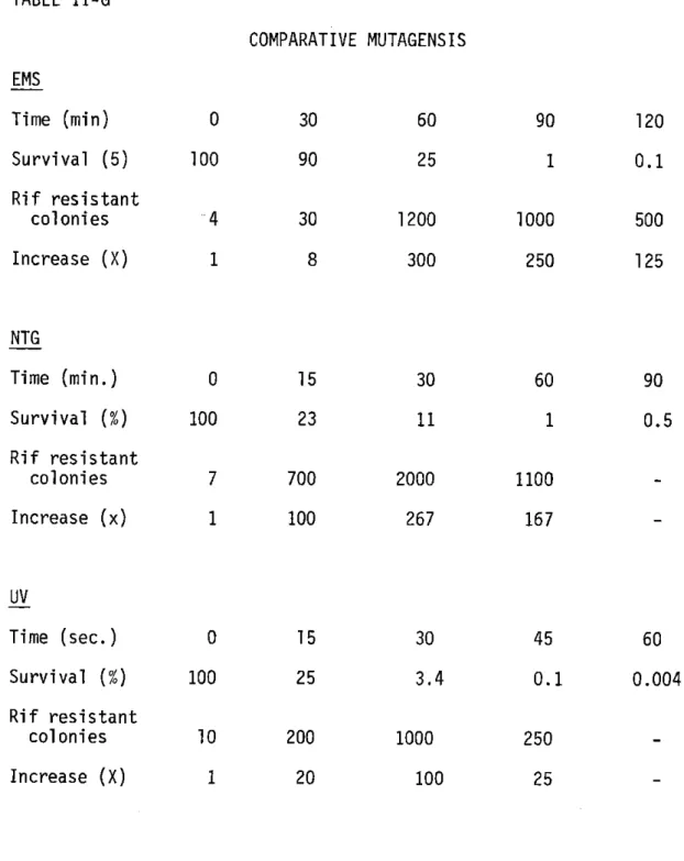
All the linkage results can be represented in a circular linkage map (Fig. IV-2). In Fig. IV-1 the circle has been broken arbitrarily between pur3l and cys11 so that the linkage frequencies from 1-5 crosses for each marker can be shown. The selected marker is at the tail of the arrow, and the head of the arrow points to the nonselected linked marker. The numbers on each tail are the cotransfer or linkage frequency in percent.
F. DISCUSSION
These results demonstrate transfer of genetic markers between R. meliloti strains.
The exconjugants with recombinant phenotype appear to be haploids re-sulting from true crossing-over, since they do not segregate, and since recessive markers may be introduced from the donor.
As expected, the empirical linkage map is one-dim6nsional. Further, selection of a pair of donor markers implies in most cases inheritance of
a marker located between them on the map: for example in the cross .gjj15 X trp33, his39, simultaneous selection for trp and his gives recombinants
LINKAGE OR COTRANSFER FREQUENCIES USING RP4
CF RHIZO 3 SL1lTI
ilv
pan
leu
7 44 53
13 79
pA p
gly
iIV
rg
leu
12 76 26
p
tz 291*6
nov trp sfu phe aro
arg
met his
57 33 74 54 51 15 56 39 59 78 me4 20
ZILw0-IF4
I-A~t 4 '1 21 2 31The selected marker is at the tail of the arrow,
and the head of the arrow points to the nonselected linked marker.
FIG. IV-1
pur
31 4 1l4 -w cys 24 50 pyr 49rif
I cys AIL-55 301S U -3 21 13 rz I 42 qp I I43 Fig. IV-2
Linkage Map of Rhizobium meliloti
(lys)
leu
pan
-l
lv(met)
ilv
(h
pur
arg
leu
cys
nov
trp
rif
5fu
phe
py
r
aro
(aro)
arg
cys
his
met
44 These results deal entirely with genetic linkage and hence prove
nothing about the physical structure of the R. meliloti chromosome.
Nevertheless, since the R. meliloti map is circular like those of enteric
bacteria such as E. coli, it would not be surprising to find that the R. meliloti chromosome is circular too. Comparison of Fig. IV-E-1 with the map of E. coli (39) does have some rough similarities. For example the relative positions of 1eu53, trp33, his39, phe54 and aro51 seem to be close to that of E. colt. However, there are at least three markers that
seem to be grouped at single loci in E. coli but at more than one in R.
meilioti, (his, leu, and ilv).
Recently linkage of auxotrophic markers has been shown in R.
japonicum using transformation techniques (6). Two linkage groups are found, one containing arg and ura, the other with ileu val, leu. No linkage was found between the two groups. This may be similar to the map I have constructed for R. meliloti, (i.e. ilvi3, 1eu53 and arl5,'pyr80 which maps close to his39 on Fig. IV-E-2).
Beringer and Hopwood (16) have reported preliminary linkage data in
R. leguminosarum, using the R factor, R68-45, to promote transfer. Three
of the four markers which they have studied map in an order consistent with markers found on the R. meliloti map.
I have as yet little information about the mechanism of transfer. Since I found a wide continuum of linkage frequencies ranging from 0.5 to 92%,
there is no reason to suppose that more than one mechanism is involved. Recombination frequencies for all markers are roughly the same and give no
indication of a polarity of transfer. This is similar to F+ X F~ matings
in E. coli, where the origin and polarity of transfer that are
45
the large number of such events with independent origins that take place
simultaneously. This is likely to be the case here too.
This is unlike the RP4 promoted conjugation described in Acineto-bacter (40) in which a gradient of recombination frequencies was found for different markers on the linkage map. However, I have isolated one R. meliloti derivative, that gives higher (100-loX) frequency of transfer of some markers, apparently with both an origin and polarity like an E. coli Hfr male, (see Appendix I). Thus at this early stage of under-standing, the E. coli F system seems to be a useful model for RP4-promoted transfer in .R. meliloti, although the situation in Acinetobacter might be different.
One disadvantage of this system is that almost all recombinants
be-come R+. Since I have been unable to cure Rm2011 of RP4, any strains that
have had new markers crossed into them, can only be used as donors. This limits strain construction.
F-type Hfrs can be isolated selectively through the use of an Ft5 derivative that is thermosensitive for episomal replication (41). I
have isolated an analogous RP4ts and have attempted this kind of selection, (see Appendix IV). Other experiments which may elucidate the mechanisms of chromosome transfer, such as donor/ recipient ratio, time course of RP4
and chromosomal gene transfer, and the relationship of chromosomal
mobilization to RP4 trnasfer itself have yet to be done.
This genetic system allows me to locate chromosomal markers anywhere on the linkage map of Rm2011. All of the 25 sites constitute a single
linkage structure, and there is no evidence for any additional linkage groups. Therefore I expect to be able to link any new markers to markers
46 already mapped, and the linkage frequency should be no less than the
fre-quency for the pair that is currently the most loosely linked, namely 3%
(for cysjj and pan44).
I believe that this genetic system will greatly aid our study of Rhizobium genetics and physiology. But more importantly it now allows us to begin a genetic analysis of the Rhizobium-legume symbiosis. The most interesting properties of Rhizobium, namely nodulation and nitrogen fixation, cannot be easily selected in free-living bacteria. The mating system will allow the relevent genes to be manipulated by their linkage to more easily handled markers. This will allow us to construct strains with interesting combinations of mutations in the symbiotic pro-cess, a procedure which is vital to the genetic analysis of such a complex system.
V. EPILOG 47 When I began this research in 1972, there was no workable genetic system in a nodulating strain of Rhizobium. I chose R. meliloti strain
Rm2011 for a number of reasons. First, this species nodulates alfalfa which is a small seeded legume and allows nodulation assays to be
carried out in a laboratory of limited space. Second, Scherrer and
Denarie had already begun working with this strain and were studying the effect of auxotrophic mutations on the nodulatior process (42).
Finally, and most important, R. meliloti grows rapidly and, unlike other rhizobia, on standard E. coli media.
The early parts of this thesis, whichwere unsuccessful attempts to show genetic exchange, were carried out when I was just learning
microbiological techniques and genetics. For this reason I feel that
some of these experiemnts warrent repeating. A transformation procedure has recently been described for Azotobacter (43). This technique might
be effective with Rm2011. In my early attempts at phage isolation none
were found, yet recently I succeeded in isolating three phage from soil samples. This project should be continued since the development of a
transduction system would complement the RP4 mating genetics.
I spent a lot of time trying to find conditions where RP4 would
pro-mote chromosome mobilization. In fact, the hybrid episomes (see Appendix II) were isolated with the hope that more bacterial DNA on the episome would increase homology and therefore lead to chromosome mobilization. I found
this to be true in E. colt, but could not demonstrate increased mobilization
48 The plate mating technique is very simple and obvious in retrospect, since Holloway had used this method earlier in Pseudomonas (33). Both John Beringer in England and I hit upon this idea late in 1974. The reason for the success of the technique has as yet not been studied.
There is some concern over the use of a drug resistance factor in genetic studies. This is particularly true of RP4 because of its wide host range. Therefore I have autoclaved all cultures of bacteria carrying R-factors before discarding them, and petri plates with R bacteria are incinerated. R. meliloti does have an important ecological niche. I have isolated many drug resistant mutants of Rm2011. To prevent the intro-duction of new genetic material carrying drug resistance information into the genetic pool of bacteria in the environment, I also sterilize cultures of Rm2011 before discarding.
The approach that I have taken to develop this genetic system may be of general importance. RP4 which was isolated originally from Pseudomonas is not native to R. meliloti. Therefore there should be nothing unique about R. meliloti which makes it amenable to genetic studies using RP4 promoted chromosome mobilization. For this reason it would seem that RP4 could be used in other bacteria. Because of its wide host range, RP4 may be used to develop genetic systems in a wide range of bacteria, many of which have interesting properties, yet no known genetic system.
Appendix I ISOLATION OF AN fifr
Early mating experiments in E. coli were not easily interpretable until the isolation of an Hfr. Because of their higher frequency of recombination and directed transfer Hfrs make mapping much easier.
In an attempt to construct a strain carrying both pan and gly I accidently isolated an exconjugant that seems to have the properties of
an Hfr. This strain gave 50-100 fold higher recombination frequencies for some markers, yet normal low levels for other markers.
STRAINS:
Rm2012/RP4 glyl2, (ATK)
Rm3344 trp33, his39, pan44, spcl, rifl
Rm3357 " I leu53, " " nov57
Rm3359 " " pyr49, " " nov59
Rm3363 his39, pan44, glyl2, rifl (ATK)
CONSTRUCTION OF Hf r
My original goal was to construct a strain carrying both pan and
gly . To do this I crossed Rm20l2/RP4 gly with Rm3344 trp, his, pan~, rifl, selected for trp and scored for other markers. Three out of 200
were gly and pan. One of these, Rm3363 his~, pan-, gly-, rifl, (ATK) was chosen for further study.
Initially I crossed this new strain (Rm3363) with Rm3359 trp, his, pyr , nov59, rifl and selected for either trp or pyr recombinants.
Sur--4 +
prisingly, 100 fold higher (10 /donor) frequency of pyr recombinants was
found, yet a normal (10-6/donor) level of trp colonies appeared. This was the first evidence that Rm3363 may be an Hfr.
50
For this reason I did an interrupted plate mating with both Rm20l2/RP4 and Rm3363 crossed with Rm3359. I selected for trp and pyr recombinants in order to determine the kinetics of chromosome mobilization. The time
course of interrupted mating was done as follows:
Donor and recipient cultures were mixed together and one ml. of the mating mixture was spread onto each of six identical LB plates, (one for each time point). The plates were incubated and at each time point one was removed and the mating interrupted. This was done by adding 10 ml of Saline, scraping the lawn into a centrifuge tube, spinning, resuspending in lml of
Saline, vortexing for 30 sec. and finally plating on selective media using F top agar. Controls of each parent alone and t=0 time point were done and showed no background trp or pyr colonies. This procedure was done at
each time point. The results are shown in Fig. AI-i.
From this it appears that the pyr marker from Rm3363 enters within 2 hours of the beginning of the mating process. However, the trp
recom-binants appear much later, at the same time and frequency as trp and pyr
from Rm20l2/RP4.
A three hour plate mating was then done between Rm3363 and recipients
Rm3357 and Rm3359, in order to determine if leu+ also mates in early, and
to study the linkage of leu53 to pan44 and glyl2. Under these conditions, leu recombinants were found at 10 5/donor while the trp recombinants were
-6 +
below 10-. The leu were scored for gly and pan. The results are shown in Table AI-l. Thus Rm3363 can mate both pyr and leu early in the mating process, and at a higher frequency than trp+
TABLE AI-1 Hfr Linkage Data 51 Donor Recipient Selected Marker Rm2012/RP4 Rm3344/RP4 Rm3363 Hfr
Recombination Frequencies for Three
Donor Recipient
Rm3363 Rm3357
Rm3359
5
5
Hour Plate Mating
trp leu x lo x 1o 5 X 10⁻⁵ 10-4 Figure AI-1 Hfr Rm3363 _pyr = $ Time course X Rm3359 trp =
of interrupted plate matings Rm20l2/RP4 X Rm3359 pyr =# # trp 70 1 $ $ f-s ~ 'i JL.. 4f* ~1#* 1 2 3 4
Time of plate mating (hours) leu53 " " pan 48% 86% gly 56% 26% + n-YE 0 0 4-1 0 C-) Q) 60 50 40 30 20 10 0 5