HAL Id: inserm-02421294
https://www.hal.inserm.fr/inserm-02421294
Submitted on 20 Dec 2019
HAL is a multi-disciplinary open access archive for the deposit and dissemination of sci-entific research documents, whether they are pub-lished or not. The documents may come from teaching and research institutions in France or abroad, or from public or private research centers.
L’archive ouverte pluridisciplinaire HAL, est destinée au dépôt et à la diffusion de documents scientifiques de niveau recherche, publiés ou non, émanant des établissements d’enseignement et de recherche français ou étrangers, des laboratoires publics ou privés.
patients with septic shock
Fanny Vardon-Bounes, Marie-Pierre Gratacap, Samuel Groyer, Stéphanie
Ruiz, Bernard Georges, Thierry Seguin, Cédric Garcia, Bernard Payrastre,
Jean-Marie Conil, Vincent Minville
To cite this version:
Fanny Vardon-Bounes, Marie-Pierre Gratacap, Samuel Groyer, Stéphanie Ruiz, Bernard Georges, et al.. Kinetics of mean platelet volume predicts mortality in patients with septic shock. PLoS ONE, Public Library of Science, 2019, 14 (10), pp.e0223553. �10.1371/journal.pone.0223553�. �inserm-02421294�
Kinetics of mean platelet volume predicts
mortality in patients with septic shock
Fanny Vardon-BounesID1,2*, Marie-Pierre GratacapID2, Samuel Groyer1,Ste´phanie Ruiz1,2, Bernard Georges1, Thierry Seguin1, Ce´dric Garcia3, Bernard Payrastre2,3, Jean-Marie Conil1, Vincent Minville1,2
1 Anesthesiology and Critical Care Unit, Toulouse University Hospital, Toulouse, France, 2 INSERM UMR
1048, Institut des Maladies Me´taboliques et Cardiovasculaires, Equipe 11, Toulouse, France, 3 Hematology laboratory, Toulouse University Hospital, Toulouse, France
*bounes.f@chu-toulouse.fr
Abstract
Introduction
Thrombocytopenia is well recognized as a poor prognosis sign associated with increased mortality and prolonged Intensive Care Unit (ICU) stay, particularly in septic patients. Mean platelet volume (MPV) could represent a relevant predictive marker of mortality. Here we investigated whether MPV kinetics during the first 15 days after hospital admission has a potential prognostic value for clinical outcome in septic shock.
Methods
We performed a retrospectively analysis of a cohort of 301 septic patients admitted in ICU. Three-month mortality was the primary endpoint. The prognostic value of the covariates of interest was ascertained by multidimensional analysis. We proposed a classification and regression trees analysis to predict survival probability.
Results
MPV kinetics was significantly different between 90-day survivors and non-survivors when
followed during 15 days (except on day 3). 10-day MPV>11.6fL was an independent
predic-tive factor of 90-day mortality (Hazard Ratio (HR) 3.796, 95% Confidence Interval (CI)
[1.96–7.35], p = 0.0001) in multivariate analysis. Base excess on day 4<1.9mmol/L was
also a predictive factor of mortality (HR 2.972, 95%CI [1.38–6.40], p = 0.0054.
Conclusion
MPV increase during the first 15 days after ICU admission in non-survivors was observed
during septic shock and 10-day MPV>11.6fL was an independent predictive factor of
90-day mortality. This could be explained by the emergent response to acute platelet loss dur-ing septic shock, leaddur-ing to megakaryocyte rupture to produce new but potentially immature platelets in the circulation. Therefore, continuous monitoring of MPV may be a useful param-eter to stratify mortality risk in septic shock.
a1111111111 a1111111111 a1111111111 a1111111111 a1111111111 OPEN ACCESS
Citation: Vardon-Bounes F, Gratacap M-P, Groyer
S, Ruiz S, Georges B, Seguin T, et al. (2019) Kinetics of mean platelet volume predicts mortality in patients with septic shock. PLoS ONE 14(10): e0223553.https://doi.org/10.1371/journal. pone.0223553
Editor: Martina Crivellari, Vita Salute University of
Milan, ITALY
Received: May 15, 2019 Accepted: September 5, 2019 Published: October 17, 2019
Copyright:© 2019 Vardon-Bounes et al. This is an open access article distributed under the terms of theCreative Commons Attribution License, which permits unrestricted use, distribution, and reproduction in any medium, provided the original author and source are credited.
Data Availability Statement: All relevant data are
within the manuscript and its Supporting Information files.
Funding: The author(s) received no specific
funding for this work.
Competing interests: The authors have declared
Introduction
Sepsis has recently been defined as a “life-threatening organ dysfunction caused by a dysregulated host response to infection” [1]. Septic shock constitutes a “subset of sepsis in which both circula-tory and cellular metabolism abnormalities occur”. Sepsis-related mortality is linked to multiple organ failure (MOF) development. MOF is partly due to microvascular thrombosis and endothe-lial dysfunction, involving thrombocytes. Thrombocytopenia is the most common hematologic disorder in the Intensive Care Unit (ICU) with a prevalence of around 50% [2]. Thrombocytope-nia is well recognized as a poor prognosis sign and is associated with increased mortality and with a prolonged ICU stay [3–6]. Mean platelet volume (MPV) describes the average size of platelets in a blood sample. Many physicians have recently shown interest in MPV in several human studies in term of prognosis on short periods (one or two days) [7–10]. A mechanism that could explain the changes in MPV values is an adapted response to acute platelet loss during an inflammatory condition. Indeed, in physiological conditions, platelet count and thrombopoiesis from bone mar-row megakaryocytes (MK) are tightly inter-regulated processes. In the presence of thrombopoie-tin (TPO), MK exhibit microtubule-dependent extensions of elongated pseudopodal structures called proplatelets allowing the release of newly generated platelets in the blood stream [9,11,12]. It is well known that younger platelets have a higher MPV. However, using intravital microscopy, Nishimura etal. have suggested that this process may not be sufficient to support a rapid platelet
turnover, especially when the platelet need is acute [13]. Their team highlighted a mechanism for the rapid production of platelets and their release into the bloodstream thanks to a MK rupture process which leads to the rapid fragmentation of cytoplasmic prolongations. This leads to the release of a large number of thrombocytes into circulation. These platelets exhibit an increase in MPV and their morphology is somewhat different. This mode of release of young platelets from bone marrow megakaryocytes would restore a pool of circulating platelets in acute consumption situations such as sepsis. However, these platelets may have important functional differences due to a lesser organization of their microtubules and could therefore contribute to a poor clinical impact in septic patients. Little is known about the potential influence of MPV changes on mortal-ity in a homogenous group of septic patients. Therefore, we focused our study on the MPV kinet-ics during the first 15 days after hospital admission to check if this parameter has a prognostic value for clinical outcome in septic shock.
Materials and methods
Patients and study design
This retrospective cohort study included patients with microbiologically proven septic shock who were admitted to the Intensive Care Unit (ICU) of a tertiary-care teaching hospital between January 2012 and January 2016. Patients received early gold-directed therapy (EGDT) as recommended.
Patients were excluded if they met these criteria: age <16, hemorrhagic shock, hemorrhagic surgery, patient under extracorporeal life support (ECLS), cardiac arrest, platelet transfusion and pre-existing thrombocytopenia or thrombocytopenia induced by chemotherapy.
This study was reviewed and approved by the institutional review board of Toulouse Uni-versity Teaching Hospital, France (n˚09–0916). All data were fully anonymized before we accessed them. No consent was necessary for this retrospective study.
The primary end-point was 90-day mortality.
Data collection
We collected baseline characteristics including demographic information, neurologic and hemodynamic factors, laboratory data, duration of mechanical ventilation, renal replacement
therapy, stay in ICU and clinical outcome 28 days after ICU admission, at 3 months (e.g. 90 days) and intrahospital mortality. For disease severity assessment, both new Simplified Acute Physiology Score (SAPS II) and Sequential Organ Failure Assessment (SOFA) were deter-mined according to the worst values within the initial 24 hours of ICU admission.
Platelet count, serum mean platelet volume, hemoglobin (Hb), white blood cell (WBC) count, lactic acid, pH and base excess were measured during the 15 days after admission. Venous blood samples for laboratory counts were collected from all patients in tubes contain-ing ethylenediamine tetra-acetic acid (EDTA) and analyzed with an SYSMEX XN 1000 hema-tology analyzer (Canada) within 30 minutes of sample collection. The normal reference range for MPV in our laboratory hospital is 8.5 to 12.2fL. Thrombocytopenia was defined by platelet count <150 G/L.). Day -1 corresponded to the day before the onset of sepsis for the patients already in hospital for another reason such as elective surgery.
Statistical analysis
Continuous variables are expressed as median, 95% confidence interval (95% CI) and extreme values and categorical variables as numbers with percentages. Patients who died within 90 days after ICU admission were defined as “non-survivors”. Baseline characteristics are pre-sented according to the occurrence of the primary outcome (survivorsvs. non-survivors) and
were compared between the 2 groups. We compared continuous variables using Mann-Whit-ney test and categorical variables usingχ2tests or Fisher exact tests. Correlations between the quantitative variables were realized by the Spearman Rank method.
Considering time-to-event, we constructed time-dependent receiver operating characteris-tics (ROC) curves to assess threshold and predictive values of the covariates of interest. Kaplan-Meier survival curves were produced using 90-day mortality based on MPV threshold value.
Covariate selection for the multivariate analysis was based onP value <0.2 in univariate
analysis. The prognostic value of the covariates of interest was ascertained by Cox proportional hazards model, using covariates whose AUC contained 0.8 in the 95% confidence interval and the results are presented as hazard ratios (HR) with 95% CI. To highlight patients with the best survival chances, a partitioning of the population was represented using a Classification and Regression Trees (CART) analysis. The advantage of this approach is to describe the means of distribution of the population in homogeneous groups according to 90-day survival and the covariates selected from the multidimensional analysis[14].
Statistical analyses were conducted using SPSS1 for Window version 23.0 (IBM Corpora-tion, Chicago, IL). AP value <0.05 was considered statistically significant.
Results
Baseline characteristics
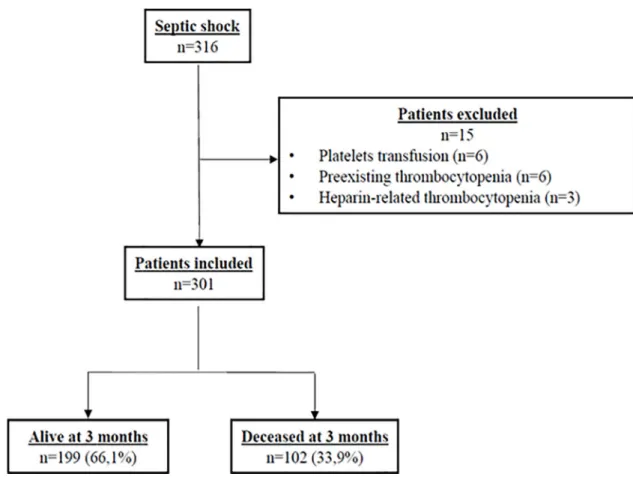
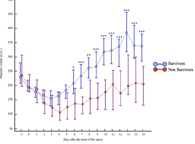
As shown inFig 1illustrating the flow chart of the enrollment of patients used in our study, of the 316 consecutive patients admitted to the ICU, 15 were excluded. Among the 301 patients included, 102 (33.9%) were deceased at 3 months. The baseline demographic, clinical, and bio-logical data of each group stratified by 90-day all-cause mortality are presented inTable 1. The main infection sites were lung (45.5%), and intra-abdominal cavity (29.2%), followed by uri-nary tract (20.9%). As shown inTable 1, 90-day survivors were younger than 90-day non-sur-vivors. As expected, SOFA and SAPS II scores were higher in non-surnon-sur-vivors. At admission, platelet count was not different between the 2 groups. However, MPV value, serum creatinine, total serum bilirubin and lactic acid were higher in non-survivors. The proportions of patients
who received renal replacement therapy (RRT) and who required mechanical ventilation were significantly higher in non-survivors.
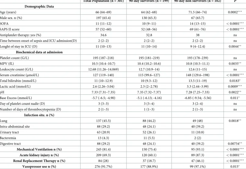
Kinetics of MPV during the first 15 days in ICU
Monitoring the MPV during the 15 days following admission in ICU indicated a significant difference between 90-day survivors and non-survivors since their admission for sepsis (10.4fL 95%CI [10.2–10.6]vs 10.8fL 95% CI [10.5–11.1] (p = 0.035) (Fig 2). Strikingly, the
non-survi-vors exhibited a higher MPV all along the kinetics. Except for day 3, this difference was signifi-cant for all time points and was particularly clear after day 7. In the survivors group, MPV values were stabilizing as early as day 2 then declining after 7 days in ICU. MPV value was inversely significantly correlated with the value of the platelet count at each time (S1 Table). MPV was also significantly correlated with SOFA score at each time (S2 Table).
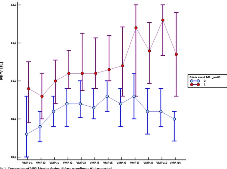
Kinetics of platelet count during the first 15 days in ICU
Platelet count decreased significantly during the first days of ICU admission to reach a nadir on day 3 in the survivors group and on day 4 in the non-survivors group (Fig 3). The differ-ence between the two groups was significant from day 6 and was particularly exacerbated on day 10 with 326 [16–993] G/L, 95% CI [276–361] in the 90-day survivors groupvs 198 [11–
681] G/L, 95% CI [131–224] in the 90-day non-survivors group (p = 0.0001). In the survivors group, the platelet count returned to the admission value at the end of the first week and con-tinued to rise to become significantly greater than that at admission. In the non-survivors group, the platelet count did not return to the admission values, even after 15 days.
Fig 1. Flowchart of the enrolment of the patients in the study. https://doi.org/10.1371/journal.pone.0223553.g001
Kinetics of blood pH and lactic acid
The study of metabolic parameters kinetics revealed that blood pH and serum lactic acid were different between survivors and non-survivors, from the day of admission in ICU to day 4 for lactic acid and to day 6 for blood pH (S1andS2Figs). The main differences were observed at day 1 and day 2.
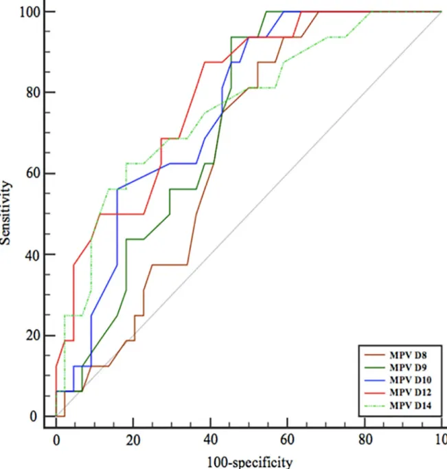
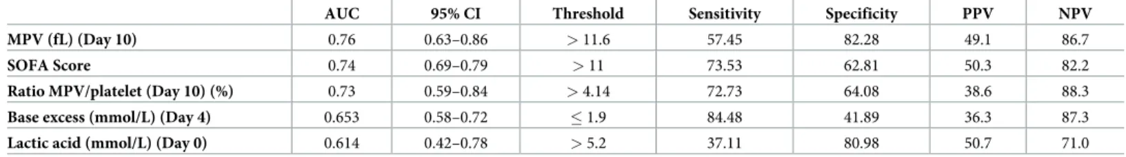
ROC curves analysis
Considering time-to-event, we constructed time-dependent receiver operating characteristics (ROC) curves to assess on which day after the onset of the sepsis MPV provided the better prognostic value for 90-day mortality (Fig 4). Areas under the curves (AUC) of the main con-tinuous clinical and biological variates discriminating survivors and non-survivors are shown
Table 1. Characteristics of the patients.
Total Population (n = 301) 90-day survivors (n = 199) 90-day non survivors (n = 102) P
Demographic Data
Age (years) 66 (64–69) 64 (62–68) 71.5 (66–74) 0.0002���
Male sex. n (%) 197 (65.4) 130 (65.3) 67 (65.7)
SOFA 11 (11–12) 10 (9–11) 14 (13–15) < 0.0001���
SAPS II score 57 (52–60) 52 (48–56) 69 (61–76) < 0.0001���
Antiplatelet therapy: yes (%) 34.6 32.8 38 ns
Time between onset of sepsis and ICU admission(D) 2 (2–2) 2 (2–2) 2 (2–2) ns
Lenght of stay in ICU (D) 11 (10–13) 11 (10–14) 9 (4–12.4) 0.0044��
Biochemical data at admission
Platelet count (G/L) 195 (187–218) 195 (181–219) 193 (178–239) ns
MPV (fL) 10.5 (10.4–10.7) 10.4 (10.2–10.6) 10.8 (10.5–11.1) 0.0035��
Leukocyte count (G/L) 12.68 (11.26–14.069) 12.7 (10.9–14) 12.6 (11–15) ns
Serum creatinine (μmol/L) 127 (119–140) 115 (99.6–127) 148 (129.6–198) < 0.0001���
Total bilirubin (mmol/L) 11 (10–12.9) 10 (9.3–12) 13.5 (11–19) 0.0183�
Lactic acid (mmol/L) 2.6 (2.26–3.04) 2.3 (2–2.78) 3.3 (2.44–3.99) 0.0009���
pH 7.33 (7.31–7.35) 7.35 (7.32–7.37) 7.28 (7.25–7.33) 0.0022��
Base Excess (mmol/L) -5.7 (-6.5; -4.90) -5.1 (-6.13; -4.16) -6.85 (-9.54; -5.56) 0.011�
Day of platelet count nadir (D) 3 (3–3) 3 (3–4) 3 (2–4) ns
Number of days of thrombocytopenia (D) 2 (1–3) 1 (1–3) 2 (1–3) ns
Infection site. n (%) Lung 137 (45.5) 88 (44.2) 49 (48) 0.0018�� Intra-abdominal site 88 (29.2) 48 (24.1) 40 (39.2) Urinary tract 63 (20.9) 52 (26.1) 11 (10.8) Bacteremia 13 (4.3) 11 (5.5) 2 (2) Digestive tract 88 (29.2) 48 (24.1) 40 (39.2) 0.00754�� Mechanical Ventilation n (%) 245 (81.4) 150 (75.4) 95 (93.1) < 0.0001���
Acute kidney injury n (%) 209 (69.3) 120 (60.1) 89 (87.3) < 0.0001���
Renal Replacement Therapy n (%) 84 (28) 37 (18.7) 47 (46.1) < 0.0001���
Vasopressor use n (%) 276 (91.7%) 177 (88.9%) 99 (97.1%) 0.015� Data are median (CI 95%) or n (%). SOFA: Sequential Organ Failure Assessment; SAPS II: Simplified Acute Physiology Score Index 2; MPV: Mean Platelet Volume �p<0.05
��p<0.01 ���p<0.001 ns: non-significant.
inTable 2. Best MPV value was the 10-day value with a Youden index >11.6fL. Other parame-ters did not provide acceptable prognostic value in view of AUC or sensitivity/specificity.
Kaplan-Meier curve
Kaplan-Meier survival curves were produced using 90-day mortality based on 10-day MPV value >11.6 or � 11.6fL. As shown inFig 5, survival probability was better in patients with 10-day MPV�11.6fL (p<0.0001) with a survival probability of 86.3%.
Multivariate analysis
Among the 301 patients included in the study, 102 (33.9%) died within 90 days following admission in ICU. We used multivariate Cox proportional analysis to assess the effect of sev-eral covariates on 90-day mortality by adjusting for other significant variables. Covariates included in the multivariate analysis were SOFA score >11, serum lactic acid on admission
>5.2 mmol/L, day 4 base excess �1.9 mmol/L, 10-day MPV >11.6fL and 10-day/platelet >4.14 (Table 2). Cox Model exhibited that 10-day MPV >11.6fL was an independent predic-tive factor of 90-day mortality (Hazard Ratio (HR) 3.796, 95% CI [1.96–7.35], p = 0.0001) and
Fig 2. Comparison of MPV kinetics during 15 days according to 90-day survival. https://doi.org/10.1371/journal.pone.0223553.g002
base excess on day 4 <1.9mmol/L was also a predictive factor of mortality (HR 2.972, 95%CI [1.38–6.40], p = 0.0054 (Table 3).
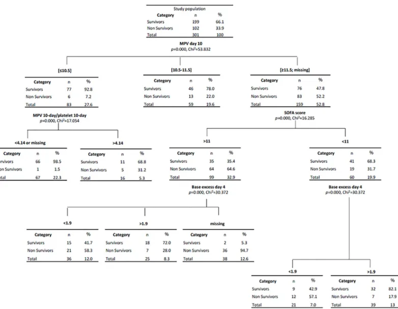
Results of Classification and Regression Trees (CART) are shown inFig 6. CART have been used extensively as an alternative to the classical linear and additive prediction models. Results are presented in tree form of a decision rule with a hierarchical sequential structure that can be easily understood and applied in clinical practice. The percentage of estimation of this CART analysis was greater than 80%. For example, patients with 10-day MPV �10.5fL and MPV/platelet ratio <4.14 had a 90-day predicted survival of 98.5%. This corresponded to 66 patients from the study (21.9%). In contrast, patients with 10-day MPV >11.6fL and with SOFA score >11 at the day of admission presented a 90-day risk of mortality of 64.6%.
Discussion
Septic shock is a major cause of mortality in ICU; therefore, it is crucial to detect patients at high risk of death in order to improve their management. In this context, the main findings of
Fig 3. Comparison of platelet counts during 15 days according to 90-day survival. https://doi.org/10.1371/journal.pone.0223553.g003
this study bring new insight that may be useful for the monitoring of septic patients. First, we found that 10-day MPV >11.6 fL was an independent predictive factor of 90-day mortality. Second, we verified the significant increase of MPV during the first 15 days after ICU admis-sion in non-survivors compared to survivors. Third, we confirmed that platelet count in the survivors group, after an early drop, returned to the admission value during the first week whereas it did not in the non-survivors group. The AUC values and multidimensional analysis (COX and CART) showed that MPV was an important factor linked to mortality not suffi-ciently discriminating if used alone. Other covariates (usually used for mortality prediction in sepsis shock) should be associated, including base excess and lactic acid, as shown by the thresholds of these covariates for the partitioning of patients using CART analysis. This is also
Fig 4. Comparison of areas under ROC curves according to Mean Platelet Volume. https://doi.org/10.1371/journal.pone.0223553.g004
Table 2. Comparison of area under the curves of continuous clinical and biological variates discriminating survivors and nonsurvivors.
AUC 95% CI Threshold Sensitivity Specificity PPV NPV MPV (fL) (Day 10) 0.76 0.63–0.86 > 11.6 57.45 82.28 49.1 86.7
SOFA Score 0.74 0.69–0.79 > 11 73.53 62.81 50.3 82.2
Ratio MPV/platelet (Day 10) (%) 0.73 0.59–0.84 > 4.14 72.73 64.08 38.6 88.3
Base excess (mmol/L) (Day 4) 0.653 0.58–0.72 � 1.9 84.48 41.89 36.3 87.3
Lactic acid (mmol/L) (Day 0) 0.614 0.42–0.78 > 5.2 37.11 80.98 50.7 71.0 MPV: Mean Platelet Volume; SOFA: Sequential Organ Failure Assessment; PLT: Platelet; AUC: Area under the curve; CI: Confidence Interval; PPV: positive predictive value, NPV: Negative predictive value
https://doi.org/10.1371/journal.pone.0223553.t002
Fig 5. Survival probability Kaplan Meier curve depending on 10-Day MPV. https://doi.org/10.1371/journal.pone.0223553.g005
the first time that a multidimensional analysis has been completed by a population partitioning in a study on MPV, thus refining the predictability of survival in septic shock with relevant covariates.
Table 3. Multivariate analysis (Cox Model).
Significant Covariates Hazard-Ratio 95% CI P value
10-day MPV >11.6 fL 3.796 1.96–7.35 0.0001
4-day Base Excess <1.9 mmol/L 2.972 1.38–6.40 0.0054
Variables not included
Renal Replacement Therapy 1.596 0.77–3.29 ns
SOFA > 11 0.957 0.47–1.96 ns
MPV: Mean Platelet Volume; SOFA: Sequential Organ Failure Assessment; CI: Confidence Interval; ns: not significant. Overall model significance with P—0.0001—AUC 0.76 [0.69 to 0.82]
https://doi.org/10.1371/journal.pone.0223553.t003
Fig 6. Classification and regression tree (CART) according to 10-day MPV, 10-day MPV/platelet ratio, SOFA score and 4-day base excess. https://doi.org/10.1371/journal.pone.0223553.g006
Concerning the MPV values, we observed a significant difference from the first day of admission in ICU for septic shock between survivors and non-survivors. Moreover, in the deceased patients group, there was a gradual increase in the MPV values during the first 15 days following the onset of septic shock, whereas, in the survivors group, MPV increased dur-ing two days, remained stable until day 7 and then decreased. An increase in MPV value could be associated with uncontrolled infection, as well as linked to illness severity and patient out-come [15,16]. Some studies have highlighted an increase in MPV in infectious endocarditis associated with embolic complications and death while the MPV value decreased in completely healed patients [15,16]. The dosage of MPV and its monitoring during sepsis is relatively sim-ple and would track the evolution of the disease. Several studies have previously demonstrated that the increase in MPV was statistically significant in the first 3 days of gram-positive sepsis [7], could predict 28-day mortality in septic shock [8], and was a risk factor for poor clinical outcome.
Daily monitoring of MPV value would stratify the risk of death in these patients. Special attention should be paid to the evolution of MPV during the first week after the onset of sepsis because the lack of return to its starting value is correlated to an unfavorable outcome. Indeed, septic patients with increased MPV value without a return to the base value are most likely to die and should be tracked in order to improve their management. Some authors propose to use MPV/platelet ratio on admission and at 24 hours to predict mortality at 28 days [10] and that is why this ratio was included in our CART analysis.
The increase in MPV is an important platelet production index that has been shown to cor-relate with increased platelet reactivity. Beside the physiological platelet production process, previous study has shown that in response to a sharp fall in the platelet count, a rapid produc-tion of platelets was possible after megakaryocyte rupture with cytoplasmic fragmentaproduc-tion [13]. Thanks to this alternative mechanism, a large number of platelets is released rapidly into the blood stream with a high proportion of thrombocytes with a large VMP and hence a risk of procoagulant phenotype[16]. As a result, these large platelets are more activated than smaller platelets. An explanation can be that larger platelets, indicating an increased MPV, have more intracellular thromboxane A2 and increased levels of procoagulant surface proteins, such as P-selectin and glycoprotein IIIa, thus presenting a greater prothrombotic potential [17].
MPV is a strong predictive factor of mortality, particularly after day 10. However, rather than the absolute value, kinetics of MPV values appeared to be of major significance, with an absence of decrease in the non-survivors group. Our findings are consistent with a study show-ing that an increase in MPV after admission to an ICU was independently associated with higher hospital mortality [18]. These findings suggest that progressive MPV increase during sepsis without returning to the initial value is linked to a more severe illness. The trends in changes in MPV and platelets counts are more reliable markers of poor prognosis than the cor-responding absolute values.
Platelet count was not different during the first 4 days of the disease between the two groups, and became significantly different at day 6 until the end of the observation (day 15). Platelet count kinetics generally corresponds to a biphasic course that has previously been reported in patients after surgery [19,20] and acute myocardial infarction [21] and could be a physiologic response to stress. In critically ill patients, a similar biphasic pattern has been reported in a study of 18 surgical patients with severe sepsis [22]. Indeed, non-survivors had persistent thrombocytopenia, whereas survival was related to the degree of thrombocytosis within 2 weeks. In critically ill patients, the evolutionary profile of the platelet count is different depending on whether the patient will survive or not. It has even been shown that late throm-bocytopenia was more predictive of death than its early onset [22]. In our cohort, the platelet count decreased significantly in the first days after admission to reach a nadir on day 3 in
survivors or day 4 in non-survivors. In the survivor group, the platelet count returned to the admission value by the end of the first week and continued to rise to become significantly greater than the admission value by day 8 whereas, in the non-survivors group, platelet count never returned to the admission value during follow-up.
Aydemir et al. have evaluated kinetics of platelet counts and MPV in adult with sepsis to determine whether the responses were specific to the type of infection [7]. They found a day of nadir different between Gram-positive septic patients, gram-negative septic patients and fun-gal septic patients. They concluded that funfun-gal sepsis had a stronger association with thrombo-cytopenia and increased MPV. We did not find a difference in platelet counts according to the pathogen agent. This may be explained by the fact that the authors excluded all patients deceased before day 10 or with negative blood cultures contrary to our study. Previous studies have already reported such conflicting results [23,24]; therefore, it is difficult to predict the type of pathogen agent depending on the kinetics of MPV or platelet count.
Mean platelet volume therefore represents a prognostic marker of interest in septic shock and its value is higher in patients who will die. Similar results have previously been shown in other diseases than sepsis. MPV appeared to be as a useful marker for early mortality and neu-rologic outcomes in patients who achieved return of spontaneous circulation after out-of-hos-pital cardiac arrest [25]. An elevated MPV was independently associated with increased 30-day mortality, with the highest discriminative value being obtained upon admission after cardiac arrest. An elevated MPV on admission was also associated with poor neurologic outcomes [26]. In another study, MPV was an independent predictor of the risk of stroke among individ-uals with a history of stroke or transient ischemic attack. Concerning inflammatory diseases like rheumatoid arthritis, MPV has been correlated with inflammatory markers and with the measures of disease activity [26,27].
Other indices of platelets have been described, including platelet volume distribution width (PDW), plateletcrit (PCT), and platelet large cell ratio (PLCR). All these indices can be mea-sured by an inexpensive and readily available routine blood count; however their use and application in septic shock remains unknown [7] and these parameters are not routinely done in our lab, and have not been reported for this study.
The novelty of the study is the use of classification and regression tree (CART) methodol-ogy that is a recursive partitioning method for predicting continuous dependent variables (regression) and categorical predictor variables (classification). It is a simple, accurate predic-tion model for outcome in patients with septic shock easily usable for clinicians [14]. Models are easy to read and interpreted using a flow chart diagram.
Our study presents limitations. The retrospective and monocentric nature of the study can limit the external validity of the results. The variation of standard MPV value between different laboratories is a weak point for the realization of a larger scale study. However, our population was similar to that reported in the literature in terms of age, severity and site of infection [10,28]. 90-day mortality in our study was 33.9% which was comparable with most studies [8,10] and in a recent publication reporting the “third International Consensus definitions for sepsis and septic shock”[1]. We did not have plateletcrit values, at the time of the study that could have been markers of interest.
Conclusion
Mortality related to sepsis and septic shock remains high. Our work confirms the existence of early biological anomalies such as thrombocytopenia and biological markers of tissue hypoper-fusion (pH, base excess, lactic acid). Our results suggest that an increase in MPV value is corre-lated with mortality. 10-day MPV and 4-day base excess values were the main covariates
predicting 90-day survival. Based on these data, we propose a segmentation analysis with 80% predictability of 90-day mortality. This study shows that particular attention must be paid to platelet counts and MPV value variations in septic shock.
Supporting information
S1 Table. Spearman rank correlation between platelet count and MPV.
(DOCX)
S2 Table. Correlation coefficients between SOFA score at admission and MPV.
(DOCX)
S1 Fig. Comparison of serum lactic acid kinetics according to 90-day survival.
(TIF)
S2 Fig. Comparison of blood pH kinetics according to 90-day survival.
(TIF)
Acknowledgments
We thank Ce´dric Garcia and Jennifer Se´ries for their kind help in the lab.
Author Contributions
Conceptualization: Fanny Vardon-Bounes, Samuel Groyer, Vincent Minville. Data curation: Fanny Vardon-Bounes, Samuel Groyer, Jean-Marie Conil. Formal analysis: Fanny Vardon-Bounes, Samuel Groyer, Jean-Marie Conil. Investigation: Fanny Vardon-Bounes, Thierry Seguin, Jean-Marie Conil.
Methodology: Fanny Vardon-Bounes, Bernard Georges, Thierry Seguin, Jean-Marie Conil. Supervision: Bernard Georges, Vincent Minville.
Validation: Ste´phanie Ruiz, Jean-Marie Conil, Vincent Minville. Visualization: Vincent Minville.
Writing – original draft: Fanny Vardon-Bounes, Samuel Groyer, Ste´phanie Ruiz. Writing – review & editing: Marie-Pierre Gratacap, Ce´dric Garcia, Bernard Payrastre.
References
1. Singer M, Deutschman CS, Seymour CW, Shankar-Hari M, Annane D, Bauer M, et al. The Third Inter-national Consensus Definitions for Sepsis and Septic Shock (Sepsis-3). JAMA. 2016; 315: 801–10. https://doi.org/10.1001/jama.2016.0287PMID:26903338
2. Strauss R, Wehler M, Mehler K, Kreutzer D, Koebnick C, Hahn EG. Thrombocytopenia in patients in the medical intensive care unit: Bleeding prevalence, transfusion requirements, and outcome*. Critical Care Medicine. Critical Care Medicine; 2002; 30: 1765–1771. https://doi.org/10.1097/00003246-200208000-00015PMID:12163790
3. Vanderschueren S, De Weerdt A, Malbrain M, Vankersschaever D, Frans E, Wilmer A, et al. Thrombo-cytopenia and prognosis in intensive care. Critical Care Medicine. 2000; 28: 1871–1876.https://doi.org/ 10.1097/00003246-200006000-00031PMID:10890635
4. Baughman RP, Lower EE, Flessa HC, Tollerud DJ. Thrombocytopenia in the intensive care unit. Chest. 1993; 104: 1243–1247.https://doi.org/10.1378/chest.104.4.1243PMID:8404200
5. Ste´phan F, Hollande J, Richard O, Cheffi A, Maier-Redelsperger M, Flahault A. Thrombocytopenia in a surgical ICU. Chest. 1999; 115: 1363–1370.https://doi.org/10.1378/chest.115.5.1363PMID:10334154
6. Moreau D, Timsit J-F, Vesin A, Garrouste-Orgeas M, de Lassence A, Zahar J-R, et al. Platelet count decline: an early prognostic marker in critically ill patients with prolonged ICU stays. Chest. 2007; 131: 1735–1741.https://doi.org/10.1378/chest.06-2233PMID:17475637
7. Aydemir H, Piskin N, Akduman D, Kokturk F, Aktas E. Platelet and mean platelet volume kinetics in adult patients with sepsis. Platelets. 2015; 26: 331–335.https://doi.org/10.3109/09537104.2012. 701027PMID:22731700
8. Kim CH, Kim SJ, Lee MJ, Kwon YE, Kim YL, Park KS, et al. An Increase in Mean Platelet Volume from Baseline Is Associated with Mortality in Patients with Severe Sepsis or Septic Shock. Lazzeri C, editor. PLoS ONE. Public Library of Science; 2015; 10: e0119437.https://doi.org/10.1371/journal.pone. 0119437PMID:25742300
9. Patel SR, Hartwig JH, Italiano JE. The biogenesis of platelets from megakaryocyte proplatelets. J Clin Invest. 2005; 115: 3348–3354.https://doi.org/10.1172/JCI26891PMID:16322779
10. Oh GH, Chung SP, Park YS, Hong JH, Lee HS, Chung HS, et al. Mean Platelet Volume to Platelet Count Ratio as a Promising Predictor of Early Mortality in Severe Sepsis. Shock. 2017; 47: 323–330. https://doi.org/10.1097/SHK.0000000000000718PMID:27504801
11. Thon JN, Montalvo A, Patel-Hett S, Devine MT, Richardson JL, Ehrlicher A, et al. Cytoskeletal mechan-ics of proplatelet maturation and platelet release. J Cell Biol. 2010; 191: 861–874.https://doi.org/10. 1083/jcb.201006102PMID:21079248
12. Machlus KR, Thon JN, Italiano JE. Interpreting the developmental dance of the megakaryocyte: a review of the cellular and molecular processes mediating platelet formation. Br J Haematol. 2014; 165: 227–236.https://doi.org/10.1111/bjh.12758PMID:24499183
13. Nishimura S, Nagasaki M, Kunishima S, Sawaguchi A, Sakata A, Sakaguchi H, et al. IL-1αinduces thrombopoiesis through megakaryocyte rupture in response to acute platelet needs. J Cell Biol. 2015; 209: 453–466.https://doi.org/10.1083/jcb.201410052PMID:25963822
14. Speiser JL, Karvellas CJ, Shumilak G, Sligl WI, Mirzanejad Y, Gurka D, et al. Predicting in-hospital mor-tality in pneumonia-associated septic shock patients using a classification and regression tree: a nested cohort study. Journal of Intensive Care. 2018; 6: 66.https://doi.org/10.1186/s40560-018-0335-3PMID: 30349726
15. Gunebakmaz O, Kaya MG, Kaya EG, Ardic I, Yarlioglues M, Dogdu O, et al. Mean platelet volume pre-dicts embolic complications and prognosis in infective endocarditis. Int J Infect Dis. 2010; 14: e982–5. https://doi.org/10.1016/j.ijid.2010.05.019PMID:20851017
16. Cho SY, You E, Lee HJ, Lee W-I, Park TS. Mean platelet volume shows a significant positive correlation with hs-CRP in Korean patients with various disease conditions. Clin Lab. 2014; 60: 1775–1777. PMID: 25651726
17. Colkesen Y, Muderrisoglu H. The role of mean platelet volume in predicting thrombotic events. Clin Chem Lab Med. 2012; 50: 631–634.https://doi.org/10.1515/CCLM.2011.806PMID:22112054
18. Zampieri FG, Ranzani OT, Sabatoski V, de Souza HP, Barbeiro H, da Neto LMC, et al. An increase in mean platelet volume after admission is associated with higher mortality in critically ill patients. Ann Intensive Care. 2014; 4: 20.https://doi.org/10.1186/s13613-014-0020-1PMID:25520853
19. Breslow A, Kaufman RM, Lawsky AR. The effect of surgery on the concentration of circulating megakar-yocytes and platelets. Blood. 1968; 32: 393–401. PMID:5675977
20. Leibovitch I, Ben Chaim J, Raviv G, Mor Y, Avigad I, Goldwasser B. Quantitative changes in platelet counts following major urological pelvic surgery. Eur Urol. 1993; 24: 350–354.https://doi.org/10.1159/ 000474327PMID:8262101
21. Fagher B, Sjo¨gren A, Sjo¨gren U. Platelet counts in myocardial infarction, angina pectoris and peripheral artery disease. Acta Med Scand. 1985; 217: 21–26.https://doi.org/10.1111/j.0954-6820.1985.tb01629. xPMID:3976430
22. Smith-Erichsen N. Serial determinations of platelets, leucocytes and coagulation parameters in surgical septicemia. Scand J Clin Lab Invest Suppl. 1985; 178: 7–14. PMID:3867123
23. Guida JD, Kunig AM, Leef KH, McKenzie SE, Paul DA. Platelet count and sepsis in very low birth weight neonates: is there an organism-specific response? Pediatrics. 2003; 111: 1411–1415.https://doi.org/ 10.1542/peds.111.6.1411PMID:12777561
24. Manzoni P, Mostert M, Galletto P, Gastaldo L, Gallo E, Agriesti G, et al. Is thrombocytopenia suggestive of organism-specific response in neonatal sepsis? Pediatr Int. 2009; 51: 206–210.https://doi.org/10. 1111/j.1442-200X.2008.02689.xPMID:19405917
25. Chung SP, Yune HY, Park YS, You JS, Hong JH, Kong T, et al. Usefulness of mean platelet volume as a marker for clinical outcomes after out-of-hospital cardiac arrest: a retrospective cohort study. J Thromb Haemost. 2016; 14: 2036–2044.https://doi.org/10.1111/jth.13421PMID:27437641
26. Yazici S, Yazici M, Erer B, Calik Y, Ozhan H, Ataoglu S. The platelet indices in patients with rheumatoid arthritis: mean platelet volume reflects disease activity. Platelets. 2010; 21: 122–125.https://doi.org/10. 3109/09537100903474373PMID:20050760
27. Yazici S, Yazici M, Erer B, Erer B, Calik Y, Bulur S, et al. The platelet functions in patients with ankylos-ing spondylitis: anti-TNF-alpha therapy decreases the mean platelet volume and platelet mass. Plate-lets. 2010; 21: 126–131.https://doi.org/10.3109/09537100903470306PMID:20050759
28. Thiery-Antier N, Binquet C, Vinault S, Meziani F, Boisrame´ -Helms J, Quenot J-P, et al. Is Thrombocyto-penia an Early Prognostic Marker in Septic Shock? Critical Care Medicine. 2016; 44: 764–772.https:// doi.org/10.1097/CCM.0000000000001520PMID:26670473