HAL Id: hal-02513155
https://hal.archives-ouvertes.fr/hal-02513155
Submitted on 20 Mar 2020HAL is a multi-disciplinary open access archive for the deposit and dissemination of sci-entific research documents, whether they are pub-lished or not. The documents may come from
L’archive ouverte pluridisciplinaire HAL, est destinée au dépôt et à la diffusion de documents scientifiques de niveau recherche, publiés ou non, émanant des établissements d’enseignement et de
Synthesis, Characterization, Cytotoxic Activity, and
Metabolic Studies of Ruthenium(II) Polypyridyl
Complexes Containing Flavonoid Ligands
Alexandra-Cristina Munteanu, Anna Notaro, Marta Jakubaszek, Joseph
Cowell, Mickaël Tharaud, Bruno Goud, Valentina Uivarosi, Gilles Gasser
To cite this version:
Alexandra-Cristina Munteanu, Anna Notaro, Marta Jakubaszek, Joseph Cowell, Mickaël Tharaud, et al.. Synthesis, Characterization, Cytotoxic Activity, and Metabolic Studies of Ruthenium(II) Polypyridyl Complexes Containing Flavonoid Ligands. Inorganic Chemistry, American Chemical Society, 2020, �10.1021/acs.inorgchem.9b03562�. �hal-02513155�
Synthesis, Characterisation, Cytotoxic Activity and Metabolic
Studies of Ruthenium(II) Polypyridyl Complexes containing
Flavonoid Ligands
Alexandra-Cristina Munteanu,a,# Anna Notaro,b,# Marta Jakubaszek,b,c Joseph Cowell,b Mickaël
Tharaud,d Bruno Goud,c Valentina Uivarosi,a and Gilles Gasserb,*
a Department of General and Inorganic Chemistry, Faculty of Pharmacy, “Carol Davila”
University of Medicine and Pharmacy, 020956 Bucharest, Romania.
b Chimie ParisTech, PSL University, CNRS, Institute of Chemistry for Life and Health Sciences, Laboratory for Inorganic Chemical Biology, F-75005 Paris, France.
c Institut Curie, PSL University, CNRS UMR 144, Paris, France.
d Université de Paris, Institut de Physique du Globe de Paris, CNRS, F-75005 Paris, France.
# these authors have contributed equally to the work
* Corresponding author: E-mail: [email protected]; WWW:
www.gassergroup.com; Phone: +33 1 44 27 56 02 ORCID Number Alexandra-Cristina Munteanu: 0000-0003-4704-5401 Anna Notaro: 0000-0003-0148-1160 Marta Jakubaszek: 0000-0001-7590-2330 Joseph Cowell: 0000-0001-8750-7223
Bruno Goud: 0000-0003-1227-4159 Valentina Uivarosi: 0000-0002-7165-5069 Gilles Gasser: 0000-0002-4244-5097
Keywords: Bioinorganic Chemistry, Cancer, Flavonoid, Medicinal Inorganic Chemistry,
Abstract
Four novel monocationic Ru(II) polypyridyl complexes have been synthesized with the general
formula [Ru(DIP)2flv]X, where DIP is 4,7-diphenyl-1,10-phenanthroline, flv stands for the
flavonoid ligand (5-hydroxyflavone in [Ru(DIP)2(5-OHF)](PF6), genistein in
[Ru(DIP)2(gen)](PF6), chrysin in [Ru(DIP)2(chr)](OTf), and morin in [Ru(DIP)2(mor)](OTf))
and X is the counterion, PF6̄, and OTf ̄ (triflate, CF3SO3̄ ), respectively. Following the chemical
characterisation of the complexes by 1H and 13C-NMR, mass spectrometry and elemental analysis,
their cytotoxicity was tested against several cancer cell lines. The most promising complex,
[Ru(DIP)2(gen)](PF6), was further investigated for its biological activity. Metabolic studies
revealed that this complex severely impaired mitochondrial respiration and glycolysis processes,
contrary to its precursor, Ru(DIP)2Cl2, which showed a prominent effect only on the
mitochondrial respiration. In addition, its preferential accumulation in MDA-MB-435S cells (a
human melanoma cell line previously described as mammary gland/breast; derived from metastatic
site: pleural effusion), that are used for the study of metastasis, explained the better activity in this
Introduction
Cancer, listed as a chronic degenerative non-communicable disease by the World Health
Organization (WHO), is a leading cause of death worldwide.1 Despite the clinical success of
several platinum-based drugs (e.g., cisplatin, carboplatin and oxaliplatin).2 their efficacy is
impeded by intrinsic and acquired resistance, and dose-limiting toxicity.3 Therefore, the search for
more effective therapeutic strategies has led to the development of other metal complexes with
anticancer properties.4 Ruthenium (Ru)-based compounds have emerged as potential anticancer
drug candidates due to their unique physico-chemical and biological properties, 5–8 generally lower
systemic toxicity (in animal models) and higher cellular uptake compared to platinum complexes.5
NAMI-A, 9,10 KP1019 11,12 and its water-soluble sodium salt IT-139 (formerly KP1339)13 are Ru
complexes that have been evaluated in clinical trials as chemotherapeutic agents for the treatment
of cancer. NAMI-A is an antimetastatic drug candidate with diverse mechanisms of action.14–17
Unfortunately, during a phase I/II study, its clinical activity was found to be disappointing, which
led to the discontinuation of the trials. These poor results were mainly attributed to dose-limiting
adverse events associated with the treatments.10
Therefore, current trends in the development of novel Ru-based anticancer drug candidates aim to
meet the need for more efficient treatments and improved toxicological profiles for the emergent
drugs. For instance, Ru(II) polypyridyl complexes have shown great potential,18,19 finding
applications in tumour diagnosis,20 as antineoplastic agents19,21 or photosensitizers for PDT.22,23
The most successful compound bearing a Ru(II) polypyridyl scaffold, TLD-1433,24 has recently
entered phase II clinical studies as a photosensitizer for intravesical photodynamic therapy (PDT)
Moreover, very interesting results have been found for heteroleptic complexes of Ru(II), bearing
an O,O-chelating ligand. For instance, RAPTA complexes with curcuminoid ligands (IC50 values
≤ 1 μM) displayed novel binding modes with biomolecular targets and high, cancer cell selective
activity.27 In addition, RuII(η6-p-cymene) complexes with flavonol-derived ligands were found to
have potent cytotoxic activity against several human cancer cell lines, with IC50 values in the low
micromolar range.28
These recent discoveries have prompted us towards the study of the therapeutic potential of Ru(II)
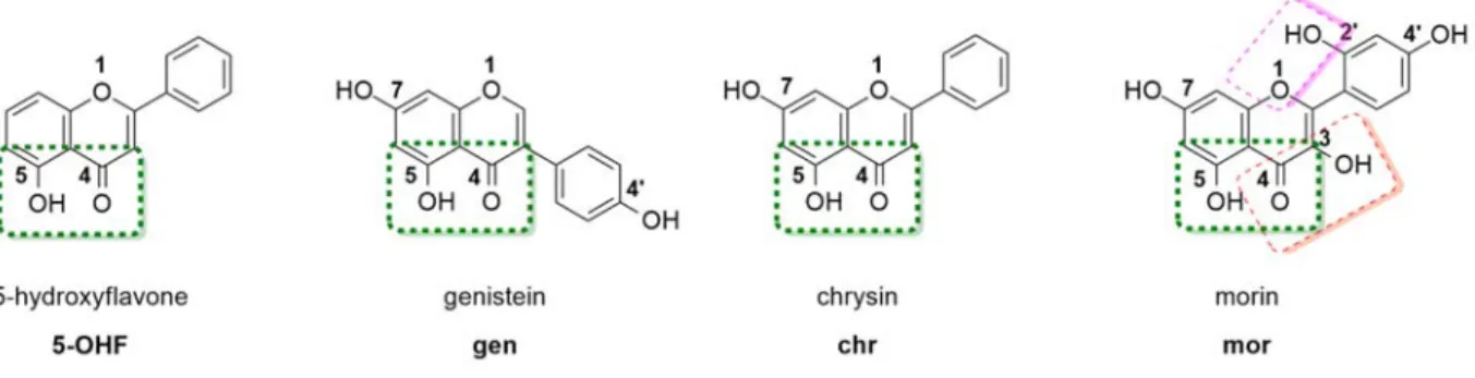
polypyridyl complexes with the flavonoids shown in Figure 1, as O,O-chelating ligands.
Flavonoids are a naturally occurring subclass of polyphenols, with high structural versatility.29
They have been extensively studied in the design of novel anticancer drug candidates. As a result,
two derivatives of the flavonoid chrysin (Figure 1), namely flavopiridol and P276-00, have entered
clinical trials.30,31 Although not yet fully understood, the cytotoxic activity of flavonoids is
believed to rely upon the modulation of cellular processes that include proliferation,
differentiation, apoptosis, metastasis and oxidative stress.29,32,33 Moreover, naturally occurring
flavonoid aglycons display exceptionally low, if any, systemic toxicity. It should be noted,
however, that the absence of acute toxic effects is related to their low water solubility and
bioavailability. 34,35
The present work focuses on the synthesis of four novel monocationic Ru(II)-polypyridyl
complexes with the general formula [Ru(DIP)2flv]X, where DIP is
4,7-diphenyl-1,10-phenanthroline, flv stands for the flavonoid ligand (5-hydroxyflavone in [Ru(DIP)2
(5-OHF)](PF6), genistein in [Ru(DIP)2(gen)](PF6), chrysin in [Ru(DIP)2(chr)](OTf), and morin in [Ru(DIP)2(mor)](OTf)) and X is the counterion (PF6̄ or OTf ̄ (triflate)). Following the successful
different cell lines. For the most potent compound of the series, metabolic studies were performed
Results and Discussion
Synthesis and characterization of the Ru(II) complexes
The synthesis of the Ru(II) complexes was achieved in a 2-step process for [Ru(DIP)2
(5-OHF)](PF6), a 3-step process for [Ru(DIP)2(gen)](PF6) and [Ru(DIP)2(chr)](OTf) and a 4-step
process for [Ru(DIP)2(mor)](OTf), respectively (Scheme 1). Briefly, RuCl2(dmso)436, DIP and
LiCl were refluxed in DMF to afford Ru(DIP)2Cl2 in a 72% yield after precipitation with
acetone.37 Ru(DIP)
2Cl2 was then refluxed in a nitrogen atmosphere for 1.5-2 hours with the
appropriate flavonoid in the presence of sodium ethoxide in dry ethanol. Complexes [Ru(DIP)2
(5-OHF)](PF6) and [Ru(DIP)2(gen)](PF6) (25% and 13%, respectively) were obtained after
precipitation with a large excess of NH4PF6 and further purification. Complexes
[Ru(DIP)2(chr)](OTf) and [Ru(DIP)2(mor)](OTf) (16% and 35%, respectively) were obtained
via a ruthenium triflate intermediate. Briefly, Ru(DIP)2Cl2 and silver triflate were stirred to afford
[Ru(DIP)2(OTf)2], and the appropriate flavonoid was added after filtration of AgCl in the presence
of sodium ethoxide.
Figure 1. Chemical structures of flavonoids 5-hydroxyflavone, chrysin, genistein and morin.
Worthy of note, morin bears three possible coordination sites (Figure 1) and literature data suggests
allow for comparison to the Ru(II) complexes of 5-OHF, genistein and chrysin, where the
flavonoids coordinate via the 4,5-O,O site, the selective protection of the oxygen atoms at the 3,
7, 2’ and 4’ positions was necessary.
Scheme 1. Synthesis of complexes of the type [Ru(DIP)2(flv)]X, where flv = flavonoid and X =
counterion. I) DIP, LiCl, DMF, reflux, 24 h, 78%. II) (i) NaOH, 5-hydroxyflavone, ethanol, reflux,
2 h; (ii) NH4PF6, ethanol/H2O (1:10), 25%. III) (i) silver triflate, ethanol, RT, 1h. IV) sodium
ethoxide, genistein, ethanol, reflux, 2 h; (ii) NH4PF6, ethanol/H2O (1:10), 13%. V) sodium
ethoxide, chrysin, ethanol, reflux, 2 h, 16%. VI) NEt3, TMSBr, THF, RT, 1h. VII) sodium
Therefore, the synthesis of [Ru(DIP)2(mor)](OTf) involved an additional protection step shown
in Scheme 1. Following a similar procedure to Qi et al.,42 the selective protection at the 2’, 4’, 3
and 7 positions with trimethylsilyl (TMS) protecting group was achieved. The protection step was
performed in the presence of triethylamine and TMS-Br in THF and, following an aqueous
work-up, the protected morin was used in the complexation step without any further purification. The
complexation reaction was performed as described above. Interestingly, during the course of the
complexation reaction, the TMS protecting groups were hydrolysed, negating the need for a
deprotection step. Following the successful synthesis of [Ru(DIP)2(mor)](OTf), coordination at
the 4, 5-O,O site was confirmed by 1D and 2D NMR studies. It was noticed during the course of
the NMR experiments that [Ru(DIP)2(mor)](OTf) exists as a mixture of two isomers in solution.
The second isomer is presumed to be the result of the morin binding via the 3,4-O,O site. The rate
of isomerisation between the two isomers, however, is slow, with approximately 25% of the
3,4-O,O complex being visible by 1H NMR after 5 days in solution (Figure S5). It should be noted that
[Ru(DIP)2(mor)](OTf) is stable if stored as a powder at -20 oC for over 6 months.
The identity of the compounds was confirmed by ESI-MS and NMR spectroscopy (Figures
S1-S9) and their purity confirmed by microanalysis. All complexes are chiral and were isolated as a
racemic mixture of ∆ and Λ enantiomers. No attempt to obtain enantiopure complexes was made
in this work. All four complexes are stable in the solid state and soluble in methanol, DCM,
DMSO, DMF and moderately soluble in acetone, acetonitrile. Since the stability and aggregation
of metal-based drug candidates is an important parameter, stability studies were undertaken.43–45
Preliminary studies (Figures S10-S13) showed that [Ru(DIP)2(5-OHF)](PF6),
[Ru(DIP)2(gen)](PF6), and [Ru(DIP)2(chr)](OTf) are stable in DMSO over 5 days. The stability
rate when compared to DMSO. Taking this into account, NMR analysis in DMF over 5 days shows
no degradation of the product (Figure S13).
Cytotoxicity, cellular uptake and metabolic studies
The biological activity of the complexes was tested on MDA-MB-435S (human, melanoma),
FaDU (human, pharynx carcinoma), MCF-7 (human, ductal carcinoma), U87 (human,
glioblastoma), RPE-1 (human, normal retinal pigmented epithelium) and HEK 293 (human
embryonic kidney) cell lines using a fluorometric cell viability assay.46 Cisplatin and doxorubicin
were tested in the same conditions as positive controls.47,48 Ru(DIP)
2Cl2 as well as the flavonoids
5-hydroxyflavone, genistein, chrysin and morin were used as additional controls. The IC50 (half
maximal inhibitory concentration) values obtained in this study are reported in Table 1 (all
cytotoxicity graphs are available in Figure S14).
Table 1. IC50 values for flavonoid ligands, cisplatin, doxorubicin, [Ru(DIP)2(5-OHF)](PF6),
[Ru(DIP)2(gen)](PF6), [Ru(DIP)2(chr)](OTf), [Ru(DIP)2(mor)](OTf), and Ru(DIP)2Cl2 in
different cell lines (48 h treatment).
Compounds IC50 (μM)
MCF-7 FaDU
MDA-MB-435S
U87 RPE-1 HEK293
5-Hydroxyflavone >100 >100 >100 >100 >100 >100 Genistein >100 >100 >100 >100 >100 75.85 ± 0.84 Chrysin 62.59 ± 3.23 95.06 ± 11.55 79.37 ± 8.13 91.14 ± 13.76 >100 26.80 ± 2.79 Morin >100 >100 >100 >100 >100 >100
Cisplatin 19.69 ± 1.63 5.17 ± 0.21 17.62 ± 0.54 6.94 ± 0.46 39.9 ± 9.14 2.27 ± 0.67 Doxorubicin 9.39 ± 1.37 1.55 ± 0.18 5.55 ± 1.37 0.59 ± 0.03 14.9 ± 1.31 0.21 ± 0.03 Ru(DIP)2Cl2 >50 >50 27.73 ± 5.33 25.59 ± 0.29 3.13 ± 0.28 12.11 ± 1.30 [Ru(DIP)2 (5-OHF)](PF6) >50 38.21 ± 5.22 24.48 ± 1.92 30.72 ± 1.48 19.72 ± 8.23 26.46 ± 3.20 [Ru(DIP)2(gen)](PF6) 16.67 ± 3.93 5.21 ± 0.73 2.64 ± 0.43 5.21 ± 1.74 2.36 ± 0.77 0.72 ± 0.10 [Ru(DIP)2(chr)](OTf) >50 >50 27.73 ± 5.33 25.59 ± 0.29 23.21 ± 8.08 33.02 ± 3.25 [Ru(DIP)2(mor)](OTf) >50 >50 >50 >50 >50 >50
The literature cites good to excellent cytotoxic activity for other 5-hydroxyflavone, chrysin and
morin metal complexes,41,49–52 results that prompted us to the design of these compounds. Worthy
of note, complexes of morin (bound via the 3,4-O,O site) and chrysin bearing a Ru(II) polypyridyl
scaffold have been previously reported. Their cytotoxic activity was studied on HeLa (cervical
carcinoma), SW620 (colorectal adenocarcinoma, metastatic), HepG2 (hepatocellular carcinoma)
and MCF-7 cell lines with IC50 values ranging from 7.64 to >100 μM.41 [Ru(DIP)2(mor)](OTf),
however, was found to be essentially non-toxic, with IC50 values above 50 μM in all cell lines
tested, while [Ru(DIP)2(5-OHF)](PF6) and [Ru(DIP)2(chr)](OTf) exerted moderate toxicity
towards some of the cell lines tested. Interestingly, the most promising complex identified in this
study is the complex bearing the flavonoid genistein, ([Ru(DIP)2(gen)](PF6)), with IC50 values
comparable to those of both cisplatin and doxorubicin. Genistein is considered a suitable lead for
anticancer drug development and derivatives have been synthesised in order to enhance its
cytotoxic activity. 53–57 It should be stated that among all chemical derivatives of genistein, scarce
was reported to enhance the cytotoxic activity of the ligand against four cancer cell lines, including
518A2 melanoma and MCF-7/Topo breast carcinoma cell lines.52 Unfortunately,
[Ru(DIP)2(gen)](PF6) exerted no selectivity between cancerous and non-cancerous cell lines with
comparable IC50 values. However, this drawback is commonly faced in medicinal chemistry and
could be improved by the introduction of a targeting moiety.
[Ru(DIP)2(gen)](PF6) showed good activity towards the MDA-MB-435S cell line, with an IC50
of 2.64 μM. Currently, this cell line is identified as a melanoma cell line, which derives from the
pleural effusion of a 31-year-old female with metastatic, ductal adenocarcinoma of the breast and
considered still valuable for the study of metastasis.58,59 The lower activity expressed by the
complex towards the MCF-7 cell line (IC50=16.67 μM) led us to study the cellular uptake and
mechanism of uptake of this complex in two different cell lines derived from breast tissue. In these
experiments, cells were treated with 5 μM of [Ru(DIP)2(gen)](PF6) for 2 h and the metal content
was analysed via inductively coupled plasma mass spectrometry (ICP-MS). Cisplatin and
Ru(DIP)2Cl2 were tested in the same conditions as controls. The viability of the cells after the 2 h
treatment was additionally tested, confirming that the acquired results were obtained from living
cells (Figure S14). Figure 2a shows that the cellular uptake is much lower for the MCF-7 cell line
when compared to MDA-MB-435S for all the tested compounds. Interestingly, Ru(DIP)2Cl2
accumulates more in MDA-MB-435S compared to [Ru(DIP)2(gen)](PF6), in the same cell line,
but shows lower cytotoxicity than the flavonoid complex. This observation can be rationalised by
the explanation provided by Policar et al. in 2014 where they state that IC50 is a resultant value of
cellular uptake, interaction with cellular target and its intrinsic toxicity.60 Therefore, one could
argue that the higher activity expressed by [Ru(DIP)2(gen)](PF6) towards MDA-MB-435S when
kinetics of the tested compounds in the chosen cell lines, we have performed time-dependent
accumulation experiments. Ruthenium and platinum contents in treated cells were measured by
ICP-MS after 2 h, 12 h, 24 h and 48 h. In this analysis, the concentration of the tested compounds
was decreased to 1 µM to reduce cell loss during the experiment. Figures 2b and 2c show the
changes in cellular accumulation in the two cell lines tested. The obtained results confirm previous
conclusions that all tested compounds accumulate more in the MDA-MB-435S cell line than in
MCF-7 cells. After 24 h incubation time, a similar uptake of Ru(DIP)2Cl2 and
[Ru(DIP)2(gen)](PF6) was found in MDA-MB-435S (~ 30 ng of metal in 106 cells) in comparison
with cisplatin (~ 4 ng of metal in 106 cells). On the other hand, [Ru(DIP)
2(gen)](PF6) accumulates
much more in MCF-7 cells than the two other compounds after 24 h (~ 2 ng of metal in 106 cells
as compared to ~ 1 ng) and 48 h (~ 5 ng of metal in 106 cells compared to ~ 1 ng). Notably, there
is a discrepancy between the amount of metal detected in the total accumulation and the time
dependent accumulation experiments in both cell lines at the 2 h time point (shown in Figures 2a,
2b and 2c). This can be explained by the different mechanisms of uptake of the Ru complexes (see
below) and the availability of the complexes in cellular media (5 times lower concentration of the
compounds in the time dependent experiments).
To understand the nature of the mechanism of uptake (passive or active) of the tested complexes,
cells were pre-treated with various inhibitors or kept at different temperatures. A temperature of 4
ºC was used to slow down passive diffusion, as well as active transportation. To block cellular
metabolism, pre-treatments with ATP production inhibitors 2-deoxy-D-glucose and oligomycin
were performed. Chloroquine or ammonium chloride (NH4Cl) impede endocytic pathways and
incubated with [Ru(DIP)2(gen)](PF6) or Ru(DIP)2Cl2 (2 h, 5 μM) and subsequently analysed via
ICP-MS (Figures 2d and 2e).
Inhibition of active uptake mechanisms did not significantly perturb accumulation of
[Ru(DIP)2(gen)](PF6) in both cell lines tested, demonstrating that the mechanism responsible for
its accumulation is energy independent (passive). On the other hand, Ru(DIP)2Cl2 is taken up via
a passive mechanism by the MCF-7 cell line and an active mechanism by the MDA-MB-435S cell
line. As shown for other similar ruthenium complexes, this observation indicates that slight
changes in lipophilic properties and structure play a decisive role in the cellular uptake of Ru(II)
Figure 2. ICP-MS data of cellular uptake of tested compounds in MDA-MB-435S and MCF-7 cell
lines. (a) Total cellular accumulation (2 h treatment, 5 µM) (b) Time-dependent cellular
accumulation in MDA-MB-435S cell line (c) Time-dependent cellular accumulation in MCF-7
cell line (d) Mechanism of cellular uptake of Ru(DIP)2Cl2 in tested cell lines (2 h treatment, 5
5 µM). Data of (a), (d) and (e) is presented as the mean ± SD of at least 3 technical replicates. Data
of (b) and (c) is presented as the mean ± SD of at least 3 biological replicates
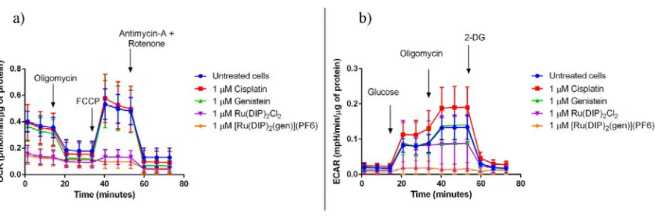
To better understand the effect of the flavonoid complex of interest on the cellular metabolism of
MDA-MB-435S cells, a Seahorse XF Analyser was used. This device allows for the real time
measurement of the oxygen consumption rate (OCR) and extracellular acidification rate (ECAR)
in cells. Firstly, the influence on the oxidative phosphorylation was measured. As shown in Figures
3a and S15, 24 h treatment with flavonoid complex [Ru(DIP)2(gen)](PF6) and its precursor
Ru(DIP)2Cl2 strongly inhibit mitochondrial respiration. Cells do not respond to the oligomycin
injection, which inhibits ATP synthase,64 nor to the FCCP which will interfere with the
mitochondrial membrane proton gradient.65 ATP production, as well as spare respiratory capacity
(calculated as the difference between maximal and basal respiration), are extremely low, further
confirming non-functioning mitochondria in treated MDA-MB-435S cells.
Next, the effect on the glycolysis process was investigated. Figures 2b and S16 show interesting
differences between the modes of action of [Ru(DIP)2(gen)](PF6) and Ru(DIP)2Cl2. During the
glycolysis stress test the first injection is made with a saturated solution of glucose. This treatment
should trigger the glycolysis process in cells and consequently lead to higher ECAR. Surprisingly,
MDA-MB-435S cells treated with [Ru(DIP)2(gen)](PF6) showed no increase in ECAR values
following injection of the saturated glucose solution. This observation is a clear indication of the
impaired glycolytic process. On the other hand, cells treated with Ru(DIP)2Cl2 showed similar
glycolysis levels when compared to those of the untreated cells. This suggests that the cytosolic
process of ATP production is impaired in [Ru(DIP)2(gen)](PF6) treated cells, but not in those
treated with Ru(DIP)2Cl2.Furthermore, the lack of response to the oligomycin injection in cells
suggests non-functioning mitochondria after both treatments. Interestingly, the complexes
[Ru(DIP)2(sq)](PF6), [Ru(DIP)2(mal)](PF6) and [Ru(DIP)2(3-methoxysq)](PF6), recently
reported by our group, also showed impaired mitochondrial function but did not show any effect
on the glycolysis process.66–68 This illustrates how subtle structural changes in the complexes
bearing the same Ru(DIP)2 core but different dioxo ligands, can result in significantly different
behaviour of the complexes in living cells.
Figure 3. a) Mito Stress Test profile in MDA-MB-435S cells after 24 h treatment. Oxygen
consumption rate changes after treatment with specific electron transport chain inhibitors.
Oligomycin (inhibitor of ATP synthase (complex V)), FCCP (uncoupling agent), antimycin-A
(complex III inhibitor) and rotenone (complex I inhibitor). b) Glycolysis Stress Test profile in
MDA-MB-435S cells after 24 h treatment. Extracellular acidification rate that corresponds to the
glycolysis process changes after treatment with glucose (basal level of glycolysis in cells),
oligomycin (inhibitor of ATP synthase (complex V) - mitochondria inhibition), 2-deoxyglucose
Conclusions
Briefly, four monocationic Ru(II) polypyridyl complexes with the general formula [Ru(DIP)2flv]X
have been synthesised. The cytotoxicity of these complexes was tested against different cancerous
and healthy cell lines and the most promising compound identified is [Ru(DIP)2(gen)](PF6) with
cytotoxicity comparable to that of cisplatin and doxorubicin. The complex displayed good activity
towards the MDA-MB-435S cell line (IC50 = 2.64 μM), a melanoma cell line derived from the
pleural effusion of a female with metastatic breast adenocarcinoma, used for the study of
metastasis. Interestingly, genistein was not cytotoxic (IC50 > 100 μM) and the precursor,
Ru(DIP)2Cl2, was only moderately active (IC50 = 27.73 μM).[Ru(DIP)2(gen)](PF6) was found to
be taken up more efficiently by MDA-MB-435S cell lines than MCF-7, a commonly used breast
cancer cell line, in both cases via a passive transportation mechanism. Further metabolic studies
in the MDA-MB-435S cell line revealed that [Ru(DIP)2(gen)](PF6) not only inhibits
mitochondrial respiration, but also interferes with the cytosolic glycolysis process in comparison
to Ru(DIP)2Cl2. This result suggests that addition of the flavonoid moiety changes the behaviour
of the complex in living cells and allows for a more complex mode of action, leading to cell death.
Therefore, we consider [Ru(DIP)2(gen)](PF6) to be a suitable candidate for further studies, which
will aim to identify the cellular targets of the complex and possible interactions with protein
transporters. Since the current treatment of advanced melanoma provides modest results, this work
may open new opportunities in the search for chemopreventive and/or chemotherapeutic agents
Experimental Section
Materials
All chemicals were either of reagent or analytical grade and used as purchased from commercial
sources without additional purification. Ruthenium trichloride hydrate was provided by I2CNS,
4,7-diphenyl-1,10-phenanthroline, lithium chloride (anhydrous, 99%), the flavonoids and
tetrabutylammonium hexafluorophosphate were provided by Sigma-Aldrich. All solvents were
purchased of analytical, or HPLC grade. When necessary, solvents were degassed by purging with
dry, oxygen-free nitrogen for at least 30 minutes before use. Preparative thin layer chromatography
(TLC) glass plates (Analtech, Sigma-Aldrich, Steinheim, Germany, 20 cm × 20 cm; 1500 𝜇m thickness).
Instrumentation and methods
Amber glass or clear glassware wrapped in tin foil were used when protection from the light was
necessary. Schlenk glassware and a vacuum line were employed when reactions sensitive to
moisture/ oxygen had to be performed under a nitrogen atmosphere. Thin layer chromatography
(TLC) was performed using silica gel 60 F-254 (Merck) plates with detection of spots being
achieved by exposure to UV light. Eluent mixtures are expressed as volume to volume (v/v) ratios.
1H and 13C NMR spectra were measured on Bruker Avance III HD 400 MHz or Bruker Avance
Neo 500 MHz spectrometers using the signal of the deuterated solvent as an internal standard.69
The chemical shifts δ are reported in ppm (parts per million) relative to tetramethylsilane (TMS)
or signals from the residual protons of deuterated solvents. The following abbreviations were used
to designate multiplicities: s = singlet, d = doublet, app t = apparent triplet, m = multiplet, dd =
out using a 6470 Triple Quad (Agilent Technologies). Elemental analysis was performed at Science
Centre, London Metropolitan University using Thermo Fisher (Carlo Erba) Flash 2000 Elemental
Analyser, configured for %CHN. IR spectra were recorded with a SpectrumTwo FTIR
Spectrometer (Perkin–Elmer) equipped with a Specac Golden GateTM ATR (attenuated total
reflection) accessory; applied as neat samples; 1/λ in cm–1.
Synthesis and characterization RuCl2(dmso)4
RuCl2(dmso)4 was synthesised following an adapted literature procedure.36 Spectroscopic data
were in agreement with the literature.36
Ru(DIP)2Cl2
Ru(DIP)2Cl2 was synthesised following an adapted literature procedure.36 Spectroscopic data were
in agreement with the literature.37,66
[Ru(DIP)2(5-OHF)](PF6)
Ru(DIP)2Cl2 (0.20 g, 0.24 mmol) and aq. NaOH (0.38 mL, 1 M) were dissolved in ethanol
(20 mL). The solution was degassed for 20 min and 5-hydroxyflavone (0.09 g, 0.38 mmol) was
added. The resulting mixture was heated to reflux for 1.5 h under a N2 atmosphere and protected
from light. The mixture was cooled to RT, while still protected from light, and the solvent was
removed under vacuum. The residual solid was redissolved in ethanol (10 mL), and H2O (100 mL)
and NH4PF6 (1.00 g, 6.13 mmol) were added. The precipitate formed was filtered, washed with
H2O (3 × 50 mL) and Et2O (3 × 50 mL) and collected. The solid with Et2O (10 mL) and then
heptane (10 mL), was sonicated for 10 min and then centrifuged. This procedure was repeated
[Ru(DIP)2(5-OHF)](PF6) (0.07 g, 0.061 mmol, 25 % yield) as a purple solid.1H NMR (400 MHz, CD2Cl2): /ppm = 9.54 (d, J = 5.5 Hz, 1H), 9.38 (d, J = 5.5 Hz, 1H), 8.27 (d, J = 8.7 Hz, 2H), 8.21 – 8.16 (m, 3H), 8.11 (d, J = 5.5 Hz, 1H), 7.96 (dd, J = 9.4, 5.5 Hz, 2H), 7.92 – 7.89 (m, 2H), 7.78 – 7.50 (m, 23H), 7.42 (dd, J = 10.5, 5.5 Hz, 2H), 7.35 (app t, J = 8.3 Hz, 1H), 6.74 (s, 1H), 6.65 (dd, J = 11.6, 8.3 Hz, 2H); 13C NMR (125 MHz, CD2Cl2): /ppm = 179.9, 168.1, 160.0, 158.1, 153.5, 153.1, 151.6, 151.1, 151.0, 150.2, 149.8, 149.6, 148.0, 147.7, 146.3, 146.2, 136.2, 136.2, 136.0, 136.0, 134.3, 131.8, 131.0, 129.9, 129.9, 129.7, 129.7, 129.6, 129.5, 129.5, 129.4, 129.2, 129.1, 128.6, 128.6, 128.4, 126.0, 125.9, 125.8, 125.7, 125.6, 125.4, 124.7, 124.5, 118.3, 113.0,
105.9, 100.3. MS (ESI+): m/z 1003.22 [M]+. Elemental Analysis: calcd. for C
63H41F6N4O3PRu =
C, 65.91; H, 3.60; N, 4.88. Found = C, 65.70; H, 3.58; N, 4.55.
[Ru(DIP)2(gen)](PF6)
Ru(DIP)2Cl2 (0.20 g, 0.24 mmol) was dissolved in ethanol (20 mL). The solution was degassed
for 20 min and silver triflate (0.13 g, 0.52 mmol) was added. The mixture was stirred at RT for 1 h
protected from light, under a N2 atmosphere. The crude reaction mixture was filtered and the
filtrate was degassed for 20 min. To the degassed solution, genistein (0.10 g, 0.38 mmol) and an
ethanolic solution of sodium ethoxide (21%, 285 μL) were added. The mixture was heated to reflux
for 2 h under N2 atmosphere whilst protected from light. The mixture was cooled to RT and the
solvent was removed under vacuum. The residual solid was dissolved in ethanol (10 mL), and H2O
(100 mL) and NH4PF6 (1.00 g, 6.13 mmol) were added. The precipitate which formed was filtered
and washed with H2O (3 x 50 mL), heptane (3 x 50 mL) and Et2O (2 x 50 mL). The solid was
collected with DCM and dried under vacuum to deliver the crude product. Purification was
from the prep TLC with methanol and the solvent was subsequently removed under reduced
pressure. The solid with Et2O (10 mL) and then heptane (10 mL), was sonicated for 10 min and
then centrifuged. This procedure was repeated three times for each solvent. The solid was collected
with DCM and dried under vacuum to deliver [Ru(DIP)2(gen)](PF6) (0.04 g, 0.033 mmol, 14%)
as a deep purple solid. 1H NMR (400 MHz, CD
3OD): /ppm = 9.59 (d, J = 5.5 Hz, 1H), 9.21 (d, J = 5.5 Hz, 1H), 8.42 (d, J = 5.5 Hz, 1H), 8.28 (dd, J = 9.4, 1.4 Hz, 2H), 8.20 (dd, J = 9.4, 3.7 Hz, 2H), 8.10 (dd, J = 5.5, 2.3 Hz, 2H), 8.00 (d, J = 5.5 Hz, 1H), 7.82 – 7.73 (m, 5H), 7.72 – 7.53 (m, 18H), 7.50 (d, J = 5.5 Hz, 1H), 7.38 (d, J = 5.5 Hz, 1H), 6.50 (d, J = 8.7 Hz, 2H), 6.26 (d, J = 8.7 Hz, 2H), 6.10 (s, 1H); 13C NMR (125 MHz, CD3OD): /ppm = 178.2, 169.5, 165.5, 160.9, 158.1, 155.2, 155.1, 153.0, 152.7, 152.6, 152.1, 151.2, 150.9, 150.9, 149.6, 149.1, 147.8, 147.5, 137.7, 137.6, 137.6, 137.5 131.1, 131.1, 131.0, 130.8, 130.5, 130.4, 130.3, 130.2, 130.1, 130.1, 130.1, 129.7, 129.7, 129.6, 129.5, 126.9, 126.8, 126.7, 126.7, 126.6 125.9, 125.8, 124.2, 123.6, 115.3,
109.3, 92.4, 58.3. MS (ESI+): m/z 1035.5 [M]+. Elemental Analysis: calcd. for C
63H41F6N4O5PRu
= C, 64.12; H, 3.50; N, 4.75. Found = C, 64.51; H, 3.45; N, 4.48.
[Ru(DIP)2(chr)](OTf)ꞏ4H2O
Ru(DIP)2Cl2 (0.50 g, 0.60 mmol) was dissolved in ethanol (30 mL). The solution was degassed
for 20 min and silver triflate (0.34 g, 1.32 mmol) was added. The mixture was stirred at RT for 1 h
protected from light, under a N2 atmosphere. The crude reaction mixture was filtered and the
filtrate was degassed for 20 min before chrysin (0.24 g, 0.96 mmol) and an ethanolic solution of
sodium ethoxide (21%, 717 μL) were added. The mixture was heated to reflux for 2 h under N2
atmosphere and protected from light. The mixture was cooled to RT, while still protected from
mL) and filtered through celite. The solvent was removed under vacuum to deliver the crude
product. Purification was achieved via preparative TLC (DCM/ethylacetate/methanol 79/20/1).
The product was collected from the prep TLC with methanol and the solvent was subsequently
removed under reduced pressure. The solid with Et2O (10 mL) and then heptane (10 mL), was
sonicated for 10 min and then centrifuged. This procedure was repeated three times for each
solvent. The solid was collected with DCM and dried under vacuum to afford
[Ru(DIP)2(chr)](OTf) (0.12 g, 0.09 mmol, 16% yield) as a deep purple solid. 1H NMR (400 MHz,
CD2Cl2-d2): /ppm = 9.56 (d, J = 5.5 Hz, 1H), 9.32 (d, J = 5.5 Hz, 1H), 8.20 – 8.09 (m, 4H), 8.09 – 7.99 (m, 2H), 7.84 – 7.80 (m, 2H), 7.76 (d, J = 7.3 Hz, 2H), 7.69 – 7.36 (m, 24H), 7.34 (d, J = 5.5 Hz, 1H), 7.28 (d, J = 5.5 Hz, 1H), 6.48 (s, 1H), 6.17 (br d, J = 2.2 Hz, 1H), 6.04 (br d, J = 2.2 Hz, 1H). 13C NMR (125 MHz, CD 2Cl2): /ppm = 178.2, 169.1, 160.0, 159.4, 153.7, 153.4, 152.3, 152.0, 151.6, 150.7, 150.2, 150.2, 147.9, 147.7, 146.3, 146.2, 136.9, 136.8, 136.7, 136.6, 131.8, 131.7, 130.4, 130.4, 130.2, 130.1, 129.9, 129.8, 129.7, 129.6, 129.6, 129.5, 129.0, 129.0, 128.8, 126.3, 126.2, 126.1, 125.8, 125.1, 107.7, 105.5, 104.6, 92.3. MS (ESI+): m/z 1019.6 [M]+, (ESI-):
m/z 149.2 [OTf]̄. Elemental Analysis: calcd. for C64H49F3N4O11RuS= C, 61.97; H, 3.99; N, 4.51.
Found = C, 62.09; H, 3.93; N, 4.28.
[Ru(DIP)2(mor)](OTf)
A. Morin (0.56 g, 1.85 mmol) was suspended in dry tetrahydrofuran (50 mL) and triethylamine
(1.55 mL, 11.1 mmol) was added. The mixture was stirred at RT under a N2 atmosphere for 15
minutes before TMS-Br (1.47 mL, 11.1 mmol) was added. The mixture was stirred at RT under a
the product was extracted in DCM and dried on Na2SO4. The solvent was removed under vacuum
to yield the crude product A.
B. Ru(DIP)2Cl2 (0.83 g, 1.00 mmol) was dissolved in ethanol (50 mL). The solution was degassed
for 20 min and silver triflate (0.56 g, 2.20 mmol) was added. The mixture was stirred at RT for 1 h
protected from light, under a N2 atmosphere. The crude reaction mixture was filtered and the
filtrate was degassed for 20 min before product A and an ethanolic solution of sodium ethoxide
(21%, 750 μL) were added. The mixture was heated to reflux for 2 h under N2 atmosphere and
protected from light. The mixture was cooled to RT, while still protected from light, and the solvent
was removed under vacuum. The residual solid was collected in DCM (20 mL) and filtered through
celite. The solvent was removed under vacuum to deliver the crude product. Purification was
achieved via preparative TLC (DCM/ethylacetate/methanol 79/20/1). The product was collected
from the prep TLC with methanol and the solvent was subsequently removed under reduced
pressure. The solid with Et2O (10 mL) and then heptane (10 mL), was sonicated for 10 min and
then centrifuged. This procedure was repeated three times for each solvent. The solid was collected
with DCM and dried under vacuum to afford [Ru(DIP)2(mor)](OTf) (0.42 g, 0.35 mmol, 35%
yield) as a deep purple solid. 1H NMR (400 MHz, DMF-d7): /ppm = 11.85 (s, 1H), 9.73 (dd, J =
10.1, 5.5 Hz, 2H), 8.53 (d, J = 5.5 Hz, 1H), 8.45 (d, J = 5.5 Hz, 1H), 8.42 – 8.20 (m, 7H), 7.93 – 7.49 (m, 25H), 6.45 (dd, J = 8.7, 2.4 Hz, 1H), 6.06 (d, J = 2.4 Hz, 1H), 5.99 (s, 1H), 5.76 (s, 1H). 13C NMR (125 MHz, DMF-d 7): /ppm = 158.9, 158.0, 155.0, 154.7, 151.9, 151.8, 151.8, 151.5, 149.7, 149.6, 147.3, 147.0, 145.7, 145.5, 143.3, 136.4, 136.1, 136.0, 130.3, 130.2, 130.0, 129.4, 129.3, 129.2, 129.1, 128.8, 128.2, 128.0, 126.4, 126.3, 125.9, 125.9, 125.8, 125.7, 125.1, 125.0,
Analysis: calcd. for C64H41F3N4O10RuS= C, 63.20; H, 3.40; N, 4.60. Found = C, 62.77; H, 3.33;
N, 4.45.
Stability studies
The stability in DMSO-d6 or DMF-d7 at room temperature was assessed by 1H NMR over 96 h.
Cytotoxicity assay using a 2D cellular model
Cytotoxicity of [Ru(DIP)2(5-OHF)](PF6), [Ru(DIP)2(gen)](PF6), [Ru(DIP)2(chr)](OTf),
[Ru(DIP)2(mor)](OTf), Ru(DIP)2Cl2, cisplatin and doxorubicin was assessed by a fluorometric
cell viability assay using Resazurin (ACROS Organics). Briefly, cells were seeded in triplicate in
96-well plates at a density of 4×10³ cells/well in 100 μL. After 24 h, cells were treated with
increasing concentrations of the ruthenium complexes. Dilutions were prepared as follows: 0.250
mM stock in DMSO ([Ru(DIP)2(5-OHF)](PF6), [Ru(DIP)2(gen)](PF6), [Ru(DIP)2(chr)](OTf))
or DMF ([Ru(DIP)2(mor)](OTf), Ru(DIP)2Cl2, which were further diluted to 100 μM in cell
media. After 48 h incubation, the medium was removed and 100 μL of complete medium
containing resazurin (0.2 mg/mL final concentration) was added. After 4 h of incubation at 37 °C,
the fluorescence signal of resorufin product was read (ex: 540 nm em: 590 nm) in a SpectraMax
M5 microplate Reader. IC50 values were then calculated using GraphPad Prism software.
GraphPad Prism calculations of IC50 values
XY analysis with three replicate values in side by side sub-columns were chosen. Inserted raw data
obtained from SpectraMax M5 microplate reader was treated as follows: X values were
transformed into logarithm; data was normalised to the lowest Y value. Data was then analysed
Cytotoxicity assay using a 2D cellular model f (2 h incubation)
Cytotoxicity of [Ru(DIP)2(gen)](PF6) and cisplatin was assessed by a fluorometric cell viability
assay using Resazurin (ACROS Organics). Briefly, cells were seeded in triplicate in 96-well plates
at a density of 4×10³ cells/well in 100 μL. After 24 h, cells were treated with increasing
concentrations of the complexes. Dilutions were prepared as described in the section “Cytotoxicity
assay using a 2D cellular model”. After 2 h incubation, the medium was removed and 100 μL of
complete medium containing resazurin (0.2 mg/mL final concentration) was added. After 4 h of
incubation at 37 °C, the fluorescence signal of resorufin product was read (ex: 540 nm em: 590
nm) in a SpectraMax M5 microplate Reader. IC50 values were then calculated using GraphPad
Prism software as stated before.
Sample Preparation for cellular uptake
MDA-MB-435S and MCF-7 cells were seeded at a density of 2×10⁶ in 10 cm plates. Next day,
cells were treated with 5 μM concentration of [Ru(DIP)2(gen)](PF6), Ru(DIP)2Cl2 or cisplatin.
Dilutions were prepared as described in the section “Cytotoxicity assay using a 2D cellular model”.
After 2 h, cells were washed, collected, counted and snap frozen in liquid nitrogen and stored at
-20 ºC. ICP-MS samples were prepared as follows: samples were digested using 70% nitric acid (1
mL, 60 ºC, overnight). Samples were then further diluted 1:100 (1% HCl solution in MQ water)
Sample preparation for studies on the mechanism of cellular uptake
Samples were prepared as previously reported.66 Briefly, MDA-MB-435S and MCF-7 cells were
seeded at a density of 2×10⁶ in 10 cm dishes and were pre-treated the following day with the
corresponding inhibitors or kept at a specific temperature for 1 h. Next, cells were washed with
PBS and were incubated with 5 μM of [Ru(DIP)2(gen)](PF6) or Ru(DIP)2Cl2 for 2 h (low
temperature samples were still kept at 4 ºC). Dilutions were prepared as described in the section
“Cytotoxicity assay using a 2D cellular model”. Subsequently, cells were washed with PBS,
collected, counted and snap frozen in liquid nitrogen. Pellets were stored at -20 ºC. ICP-MS
samples were prepared as follows: samples were digested using 70% nitric acid (1 mL, 60 ºC,
overnight), further diluted 1:100 (1% HCl solution in MQ water) and analysed using ICP-MS.
Sample Preparation for time-dependent cellular accumulation
MDA-MB-435S and MCF-7 cells were seeded at a density of 3×10⁶ in 10 cm plates. The next day,
cells were treated with 1 μM concentration of [Ru(DIP)2(gen)](PF6), Ru(DIP)2Cl2 or cisplatin.
Dilutions were prepared as described in the section “Cytotoxicity assay using a 2D cellular model”.
After 2 h, 12 h, 24 h and 48 h, respectively, the cells were washed, collected, counted and snap
frozen in liquid nitrogen and stored until further use at -20 ºC. ICP-MS samples were prepared as
follows: samples were digested using 70% nitric acid (0.5 ml for the 2 h and 12 h samples; 1 mL
for the 24 h and 48 h samples, 65 ºC, overnight). The samples were further diluted 1:50 (2 h
samples) or 1:100 (12 h, 24 h, 48 h samples) in 1% HCl solution in MQ water and analysed using
ICP-MS studies
All ICP-MS measurements were performed on a high resolution ICP-MS (Element II,
ThermoScientific) located at the Institut de physique du globe de Paris (France). The monitored
isotopes are 101Ru and 195Pt. Daily, prior to the analytical sequence, the instrument was first tuned
to produce maximum sensitivity and stability while also maintaining low uranium oxide formation
(UO/U ≤ 5%). The data were treated as follows: intensities were converted into concentrations
using uFREASI (user-FRiendly Elemental dAta proceSsIng ).70 This software, developed for
HR-ICP-MS users community, is free and available on http://www.ipgp.fr/~tharaud/uFREASI.
ICP-MS data analysis
Cellular uptake studies: The amount of metal detected in the cell samples was transformed from
ppb into μg of metal. Data were subsequently normalised to the number of cells and expressed as
ng of metal/ amount of cells.
Mechanism of uptake: The amount of ruthenium detected in cell samples was transformed from
ppb into μg of ruthenium and values obtained were normalised to the number of cells used for
specific treatment. The value for the ruthenium found in the 37 ºC sample was used as a 100%.
Metabolic Studies
HeLa cells were seeded in Seahorse XFe96 well plates at a density of 10×103 cells / well in 80 μL.
After 24 h, the medium was replaced with fresh medium and cisplatin (1 μM), genistein (1 μM),
Ru(DIP)2Cl2 (1 μM) or [Ru(DIP)2(gen)](PF6) (1 μM) were added. Dilutions were prepared as
the regular medium was removed, cells were washed thrice using Seahorse Base Media and
incubated in a non-CO2 incubator at 37 °C for 1 h.
Mito Stress Test: Mitostress assay was run using oligomycin, 1 μM, FCCP 1 μM and mixture of
antimycin-A/ rotenone 1 μM each in ports A, B and C respectively using Seahorse XFe96
Extracellular Flux Analyzer.
Glycolysis Stress Test: Glycolytic stress test was run using glucose (10 mM), oligomycin (1 μM)
and 2-Deoxyglucose (50 mM) in ports A, B and C respectively using Seahorse XFe96 Extracellular
Flux Analyzer.
Supporting Information
The Supporting Information is at DOI: XXXXX.
1H-NMR spectrum of [Ru(DIP)
2(5-OHF)](PF6) (Figure S1), 1H-NMR spectrum of [Ru(DIP)2(gen)](PF6) (Figure S2), 1H-NMR spectrum of [Ru(DIP)2(chr)](OTf) (Figure S3), 1
H-NMR spectrum of [Ru(DIP)2(mor)](OTf) (Figure S4), 1H-NMR spectrum of
[Ru(DIP)2(mor)](OTf) after 5 days in solution (Figure S5), 13C-NMR spectrum of [Ru(DIP)2
(5-OHF)](PF6) (Figure S6), 13C-NMR spectrum of [Ru(DIP)2(gen)](PF6) (Figure S7), 13C-NMR
spectrum of [Ru(DIP)2(chr)](OTf) (Figure S8), 13C-NMR spectrum of [Ru(DIP)2(mor)](OTf)
(Figure S9), Overlap of 1H-NMR spectra of [Ru(DIP)
2(5-OHF)](PF6) in DMSO (Figure S10),
Overlap of 1H-NMR spectra of [Ru(DIP)
2(gen)](PF6) in DMSO (Figure S11), Overlap of 1
H-NMR spectra of [Ru(DIP)2(chr)](OTf) in DMSO (Figure S12) Overlap of 1H-NMR spectra of
[Ru(DIP)2(mor)](OTf) in DMF (Figure S13), Fluorometric cell viability assay (Figure S14),
Oxygen consumption rates and different respiration parameters in MDA-MB-435S cells alone or
different parameters during glycolysis in MDA-MB-435S cells alone or after treatment with
various test compounds (Figure S16).
Acknowledgements
This work was financially supported by an ERC Consolidator Grant PhotoMedMet to G.G.
(GA 681679) and has received support under the program Investissements d’Avenir
launched by the French Government and implemented by the ANR with the reference
ANR-10-IDEX-0001-02 PSL (G.G.). Ile de France Region is gratefully acknowledged for
financial support of 500 MHz NMR spectrometer of Chimie ParisTech in the framework
of the SESAME equipment project. We acknowledge the loan of Agilent's equipment to
Chimie ParisTech. Part of this work was supported by IPGP multidisciplinary program
PARI and by Region Île-de-France SESAME Grant no. 12015908. This project was also
financially supported by “Carol Davila” University of Medicine and Pharmacy through
Contract no. 23PFE/17.10.2018 funded by the Ministry of Research and Innovation within
PNCDI III, Program 1 – Development of the National RD system, Subprogram 1.2 –
References
(1) Organization, W. H. Noncommunicable diseases
https://www.who.int/news-room/fact-sheets/detail/noncommunicable-diseases (accessed Sep 2, 2019).
(2) Hartinger, C. G.; Zorbas-Seifried, S.; Jakupec, M. A.; Kynast, B.; Zorbas, H.; Keppler, B.
K. From Bench to Bedside – Preclinical and Early Clinical Development of the Anticancer
Agent Indazolium Trans-[Tetrachlorobis(1H-Indazole)Ruthenate(III)] (KP1019 or
FFC14A). J. Inorg. Biochem. 2006, 100 (5), 891–904.
https://doi.org/https://doi.org/10.1016/j.jinorgbio.2006.02.013.
(3) Oun, R.; Moussa, Y. E.; Wheate, N. J. The Side Effects of Platinum-Based Chemotherapy
Drugs: A Review for Chemists. Dalton Trans. 2018, 47 (19), 6645–6653.
https://doi.org/10.1039/c8dt00838h.
(4) Boros, E.; Dyson, P. J.; Gasser, G. Classification of Metal-Based Drugs According to Their
Mechanisms of Action. Chem 2020, 6 (1), 41–60.
https://doi.org/https://doi.org/10.1016/j.chempr.2019.10.013.
(5) Mari, C.; Pierroz, V.; Ferrari, S.; Gasser, G. Combination of Ru(II) Complexes and Light:
New Frontiers in Cancer Therapy. Chem. Sci. 2015, 6 (5), 2660–2686.
https://doi.org/10.1039/C4SC03759F.
(6) Smith, G. S.; Therrien, B. Targeted and Multifunctional Arene Ruthenium
Chemotherapeutics. Dalt. Trans. 2011, 40 (41), 10793–10800.
https://doi.org/10.1039/C1DT11007A.
(7) Nazarov, A. A.; Hartinger, C. G.; Dyson, P. J. Opening the Lid on Piano-Stool Complexes:
An Account of Ruthenium(II)–Arene Complexes with Medicinal Applications. J.
https://doi.org/https://doi.org/10.1016/j.jorganchem.2013.09.016.
(8) Zeng, L.; Gupta, P.; Chen, Y.; Wang, E.; Ji, L.; Chao, H.; Chen, Z.-S. The Development of
Anticancer Ruthenium(II) Complexes: From Single Molecule Compounds to
Nanomaterials. Chem. Soc. Rev. 2017, 46 (19), 5771–5804.
https://doi.org/10.1039/C7CS00195A.
(9) Rademaker-Lakhai, J. M.; van den Bongard, D.; Pluim, D.; Beijnen, J. H.; Schellens, J. H.
M. A Phase I and Pharmacological Study with
Imidazolium-Trans-DMSO-Imidazole-Tetrachlororuthenate, a Novel Ruthenium Anticancer Agent. Clin. Cancer Res. 2004, 10
(11), 3717–3727. https://doi.org/10.1158/1078-0432.CCR-03-0746.
(10) Leijen, S.; Burgers, S. A.; Baas, P.; Pluim, D.; Tibben, M.; Van Werkhoven, E.; Alessio,
E.; Sava, G.; Beijnen, J. H.; Schellens, J. H. M. Phase I/II Study with Ruthenium Compound
NAMI-A and Gemcitabine in Patients with Non-Small Cell Lung Cancer after First Line
Therapy. Invest. New Drugs 2015, 33 (1), 201–214.
https://doi.org/10.1007/s10637-014-0179-1.
(11) Hartinger, C. G.; Jakupec, M. A.; Zorbas-Seifried, S.; Groessl, M.; Egger, A.; Berger, W.;
Zorbas, H.; Dyson, P. J.; Keppler, B. K. KP1019, A New Redox-Active Anticancer Agent
– Preclinical Development and Results of a Clinical Phase I Study in Tumor Patients. Chem.
Biodivers. 2008, 5 (10), 2140–2155. https://doi.org/10.1002/cbdv.200890195.
(12) Lentz, F.; Drescher, A.; Lindauer, A.; Henke, M.; Hilger, R. A.; Hartinger, C. G.; Scheulen,
M. E.; Dittrich, C.; Keppler, B. K.; Jaehde, U. Pharmacokinetics of a Novel Anticancer
Ruthenium Complex (KP1019, FFC14A) in a Phase I Dose-Escalation Study. Anticancer.
Drugs 2009, 20 (2), 97–103. https://doi.org/10.1097/CAD.0b013e328322fbc5.
NKP-1339, the First Ruthenium-Based Anticancer Drug on the Edge to Clinical Application.
Chem. Sci. 2014, 5 (8), 2925–2932. https://doi.org/10.1039/C3SC53243G.
(14) Levina, A.; Mitra, A.; Lay, P. A. Recent Developments in Ruthenium Anticancer Drugs.
Metallomics 2009, 1 (6), 458–470. https://doi.org/10.1039/b904071d.
(15) Sava, G.; Bergamo, A.; Dyson, P. J. Metal-Based Antitumour Drugs in the Post-Genomic
Era: What Comes Next? Dalt. Trans. 2011, 40 (36), 9069–9075.
https://doi.org/10.1039/C1DT10522A.
(16) Bergamo, A.; Gaiddon, C.; Schellens, J. H. M.; Beijnen, J. H.; Sava, G. Approaching
Tumour Therapy beyond Platinum Drugs: Status of the Art and Perspectives of Ruthenium
Drug Candidates. J. Inorg. Biochem. 2012, 106 (1), 90–99.
https://doi.org/10.1016/j.jinorgbio.2011.09.030.
(17) Alessio, E. Thirty Years of the Drug Candidate NAMI-A and the Myths in the Field of
Ruthenium Anticancer Compounds: A Personal Perspective. Eur. J. Inorg. Chem. 2017,
2017 (12), 1549–1560. https://doi.org/10.1002/ejic.201600986.
(18) Mital, M.; Ziora, Z. Biological Applications of Ru(II) Polypyridyl Complexes. Coord.
Chem. Rev. 2018, 375, 434–458. https://doi.org/https://doi.org/10.1016/j.ccr.2018.02.013.
(19) Notaro, A.; Gasser, G. Monomeric and Dimeric Coordinatively Saturated and
Substitutionally Inert Ru(II) Polypyridyl Complexes as Anticancer Drug Candidates. Chem.
Soc. Rev. 2017, 46 (23), 7317–7337. https://doi.org/10.1039/C7CS00356K.
(20) Lin, K.; Zhao, Z.-Z.; Bo, H.-B.; Hao, X.-J.; Wang, J.-Q. Applications of Ruthenium
Complex in Tumor Diagnosis and Therapy. Front. Pharmacol. 2018, 9, 1323.
https://doi.org/10.3389/fphar.2018.01323.
2011, 2011 (32), 4931–4947. https://doi.org/10.1002/ejic.201100376.
(22) Heinemann, F.; Karges, J.; Gasser, G. Critical Overview of the Use of Ru(II) Polypyridyl
Complexes as Photosensitizers in One-Photon and Two-Photon Photodynamic Therapy.
Acc. Chem. Res. 2017, 50 (11), 2727–2736. https://doi.org/10.1021/acs.accounts.7b00180.
(23) Jakubaszek, M.; Goud, B.; Ferrari, S.; Gasser, G. Mechanisms of Action of Ru(II)
Polypyridyl Complexes in Living Cells upon Light Irradiation. Chem. Commun. 2018, 54
(93), 13040–13059. https://doi.org/10.1039/C8CC05928D.
(24) Monro, S.; Colón, K. L.; Yin, H.; Roque, J.; Konda, P.; Gujar, S.; Thummel, R. P.; Lilge,
L.; Cameron, C. G.; McFarland, S. A. Transition Metal Complexes and Photodynamic
Therapy from a Tumor-Centered Approach: Challenges, Opportunities, and Highlights
from the Development of TLD1433. Chem. Rev. 2019, 119 (2), 797–828.
https://doi.org/10.1021/acs.chemrev.8b00211.
(25) Intravesical Photodynamic Therapy (PDT) in BCG Refractory/Intolerant Non-Muscle
Invasive Bladder Cancer (NMIBC) Patients
https://clinicaltrials.gov/ct2/show/NCT03945162 (accessed Sep 5, 2019).
(26) McFarland, S. A.; Mandel, A.; Dumoulin-White, R.; Gasser, G. Metal-Based
Photosensitizers for Photodynamic Therapy: The Future of Multimodal Oncology? Curr.
Opin. Chem. Biol. 2020, 56, 23–27. https://doi.org/10.1016/j.cbpa.2019.10.004.
(27) Pettinari, R.; Marchetti, F.; Condello, F.; Pettinari, C.; Lupidi, G.; Scopelliti, R.;
Mukhopadhyay, S.; Riedel, T.; Dyson, P. J. Ruthenium(II)–Arene RAPTA Type
Complexes Containing Curcumin and Bisdemethoxycurcumin Display Potent and Selective
Anticancer Activity. Organometallics 2014, 33 (14), 3709–3715.
(28) Kurzwernhart, A.; Kandioller, W.; Bachler, S.; Bartel, C.; Martic, S.; Buczkowska, M.;
Muhlgassner, G.; Jakupec, M. A.; Kraatz, H.-B.; Bednarski, P. J.; Arion, V. B.; Marko, D.;
Keppler, B. K.; Hartinger, C. G. Structure-Activity Relationships of Targeted RuII(η6
-p-Cymene) Anticancer Complexes with Flavonol-Derived Ligands. J. Med. Chem. 2012, 55
(23), 10512–10522. https://doi.org/10.1021/jm301376a.
(29) Uivarosi, V.; Munteanu, A.-C.; Nițulescu, G. M. Chapter 2 - An Overview of Synthetic and
Semisynthetic Flavonoid Derivatives and Analogues: Perspectives in Drug Discovery;
Atta-ur-Rahman, B. T.-S. in N. P. C., Ed.; Elsevier, 2019; Vol. 60, pp 29–84.
https://doi.org/https://doi.org/10.1016/B978-0-444-64181-6.00002-4.
(30) Senderowicz, A. M. Flavopiridol: The First Cyclin-Dependent Kinase Inhibitor in Human
Clinical Trials. Invest. New Drugs 1999, 17 (3), 313–320.
(31) Jain, S. K.; Bharate, S. B.; Vishwakarma, R. A. Cyclin-Dependent Kinase Inhibition by
Flavoalkaloids. Mini Rev. Med. Chem. 2012, 12 (7), 632–649.
(32) Chahar, M. K.; Sharma, N.; Dobhal, M. P.; Joshi, Y. C. Flavonoids: A Versatile Source of
Anticancer Drugs. Pharmacognosy Reviews. India 2011, pp 1–12.
https://doi.org/10.4103/0973-7847.79093.
(33) Singh, M.; Kaur, M.; Silakari, O. Flavones: An Important Scaffold for Medicinal Chemistry.
Eur. J. Med. Chem. 2014, 84, 206–239.
https://doi.org/https://doi.org/10.1016/j.ejmech.2014.07.013.
(34) Kumar, S.; Pandey, A. K. Chemistry and Biological Activities of Flavonoids: An Overview.
Sci. J. 2013, 2013, 1–20. https://doi.org/10.1070/RC2004v073n07ABEH000856.
(35) Hostetler, G. L.; Ralston, R. A.; Schwartz, S. J. Flavones: Food Sources, Bioavailability,
https://doi.org/10.3945/an.116.012948.
(36) Brastos, I.; Alessio, E.; Ringenberg, M. E.; Rauchfuss, T. B. Ruthenium Complexes. Inorg.
Synth. 2010, 35 (Ii), 148–163. https://doi.org/10.1002/9780470651568.ch8.
(37) Caspar, R.; Cordier, C.; Waern, J. B.; Guyard-Duhayon, C.; Gruselle, M.; Le Floch, P.;
Amouri, H. A New Family of Mono- and Dicarboxylic Ruthenium Complexes [Ru(DIP) 2
(L 2 )] 2+ (DIP = 4,7-Diphenyl-1,10-Phenanthroline): Synthesis, Solution Behavior, and
X-Ray Molecular Structure of Trans -[Ru(DIP) 2 (MeOH) 2 ][OTf] 2. Inorg. Chem. 2006, 45
(10), 4071–4078. https://doi.org/10.1021/ic0601236.
(38) Porter, L. J.; Markham, K. R. The Aluminium(III) Complexes of Hydroxyflavones in
Absolute Methanol. Part II. Ligands Containing More than One Chelating Site. J. Chem.
Soc. C Org. 1970, 1970, 1309–1313. https://doi.org/10.1039/J39700001309.
(39) Panhwar, Q. K.; Memon, S. Synthesis and Properties of Zirconium (IV) and Molybdate (II)
Morin Complexes. J. Coord. Chem. 2012, No. 65, 37–41.
(40) Naso, L. G.; Lezama, L.; Rojo, T.; Etcheverry, S. B.; Valcarcel, M.; Roura, M.; Salado, C.;
Ferrer, E. G.; Williams, P. A. M. Biological Evaluation of Morin and Its New
Oxovanadium(IV) Complex as Antio-Xidant and Specific Anti-Cancer Agents. Chem. Biol.
Interact. 2013, 206 (2), 289–301. https://doi.org/10.1016/j.cbi.2013.10.006.
(41) Zahirović, A.; Kahrović, E.; Cindrić, M.; Kraljević Pavelić, S.; Hukić, M.; Harej, A.;
Turkušić, E. Heteroleptic Ruthenium Bioflavonoid Complexes: From Synthesis to in Vitro
Biological Activity. J. Coord. Chem. 2017, 70 (24), 4030–4053.
https://doi.org/10.1080/00958972.2017.1409893.
(42) Qi, C.; Xiong, Y.; Eschenbrenner-Lux, V.; Cong, H.; Porco, J. A. Asymmetric Syntheses
2016, 138 (3), 798–801. https://doi.org/10.1021/jacs.5b12778.
(43) Huang, H.; Humbert, N.; Bizet, V.; Patra, M.; Chao, H.; Mazet, C.; Gasser, G. Influence of
the Dissolution Solvent on the Cytotoxicity of Octahedral Cationic Ir(III) Hydride
Complexes. J. Organomet. Chem. 2017, 839, 15–18.
https://doi.org/https://doi.org/10.1016/j.jorganchem.2016.12.010.
(44) Patra, M.; Joshi, T.; Pierroz, V.; Ingram, K.; Kaiser, M.; Ferrari, S.; Spingler, B.; Keiser, J.;
Gasser, G. DMSO-Mediated Ligand Dissociation: Renaissance for Biological Activity of
N-Heterocyclic-[Ru(Η6-Arene)Cl2] Drug Candidates. Chem. – A Eur. J. 2013, 19 (44),
14768–14772. https://doi.org/10.1002/chem.201303341.
(45) Keller, S.; Ong, Y. C.; Lin, Y.; Cariou, K.; Gasser, G. A Tutorial for the Assessment of the
Stability of Organometallic Complexes in Biological Media. J. Organomet. Chem. 2019,
121059. https://doi.org/10.1016/j.jorganchem.2019.121059.
(46) Frei, A.; Rubbiani, R.; Tubafard, S.; Blacque, O.; Anstaett, P.; Felgenträger, A.; Maisch, T.;
Spiccia, L.; Gasser, G. Synthesis, Characterization, and Biological Evaluation of New
RU(Ii) Polypyridyl Photosensitizers for Photodynamic Therapy. J. Med. Chem. 2014, 57
(17), 7280–7292. https://doi.org/10.1021/jm500566f.
(47) Cepeda, V.; Fuertes, M.; Castilla, J.; Alonso, C.; Quevedo, C.; Perez, J. Biochemical
Mechanisms of Cisplatin Cytotoxicity. Anticancer. Agents Med. Chem. 2007, 7 (1), 3–18.
https://doi.org/10.2174/187152007779314044.
(48) Keizer, H. G.; Pinedo, H. M.; Schuurhuis, G. J.; Joenje, H. DOXORUBICIN
(ADRIAMYCIN): A CRITICAL REVIEW OF FREE RADICAL-DEPENDENT
MECHANISMS OF CYTOTOXICITY. Pharmac. Ther 1990, 47, 219–231.
(49) Munteanu, A.-C.; Badea, M.; Olar, R.; Silvestro, L.; Mihaila, M.; Brasoveanu, L. I.; Musat,
M. G.; Andries, A.; Uivarosi, V. Cytotoxicity Studies, DNA Interaction and Protein Binding
of New Al (III), Ga (III) and In (III) Complexes with 5-Hydroxyflavone. Appl. Organomet.
Chem. 2018, 32 (12), e4579. https://doi.org/10.1002/aoc.4579.
(50) Deka, B.; Bhattacharyya, A.; Mukherjee, S.; Sarkar, T.; Soni, K.; Banerjee, S.; Saikia, K.
K.; Deka, S.; Hussain, A. Ferrocene Conjugated Copper(II) Complexes of Terpyridine and
Traditional Chinese Medicine (TCM) Anticancer Ligands Showing Selective Toxicity
towards Cancer Cells. Appl. Organomet. Chem. 2018, 32 (4), e4287.
https://doi.org/10.1002/aoc.4287.
(51) Roy, S.; Sil, A.; Chakraborty, T. Potentiating Apoptosis and Modulation of P53, Bcl2, and
Bax by a Novel Chrysin Ruthenium Complex for Effective Chemotherapeutic Efficacy
against Breast Cancer. J. Cell. Physiol. 2019, 234 (4), 4888–4909.
https://doi.org/10.1002/jcp.27287.
(52) Spoerlein, C.; Mahal, K.; Schmidt, H.; Schobert, R. Effects of Chrysin, Apigenin, Genistein
and Their Homoleptic Copper (II) Complexes on the Growth and Metastatic Potential of
Cancer Cells. J. Inorg. Biochem. 2013, 127, 107–115.
https://doi.org/10.1016/j.jinorgbio.2013.07.038.
(53) Fotsis, T.; Pepper, M.; Adlercreutz, H.; Hase, T.; Montesano, R.; Schweigerer, L. Genistein,
a Dietary Ingested Isoflavonoid, Inhibits Cell Proliferation and in Vitro Angiogenesis. J.
Nutr. 1995, 125 (3 Suppl), 790S-797S. https://doi.org/10.1093/jn/125.suppl_3.790S.
(54) Peterson, G.; Barnes, S. Genistein Stimulated Inhibits Both Proliferation Estrogen and
Growth of Human Breast Cancer. Cell Growth Differ. 1996, 7 (October), 1345–1351.
Isoflavone Genistein Induces Cell Death in Breast Cancer Cells through Mobilization of
Endogenous Copper Ions and Generation of Reactive Oxygen Species. Mol. Nutr. Food
Res. 2011, 55 (4), 553–559. https://doi.org/10.1002/mnfr.201000329.
(56) Marverti, G.; Andrews, P. A. Stimulation of Cis-Diamminedichloroplatinum(II)
Accumulation by Modulation of Passive Permeability with Genistein: An Altered Response
in Accumulation-Defective Resistant Cells. Clin. Cancer Res. 1996, 2 (6), 991–999.
(57) Spagnuolo, C.; Russo, G. L.; Orhan, I. E.; Habtemariam, S.; Daglia, M.; Sureda, A.; Nabavi,
S. F.; Devi, K. P.; Loizzo, M. R.; Tundis, R.; Nabavi, S. M. Genistein and Cancer: Current
Status, Challenges, and Future Directions. Adv. Nutr. 2015, 6 (4), 408–419.
https://doi.org/10.3945/an.114.008052.
(58) Chambers, A. F. MDA-MB-435 and M14 Cell Lines : Identical but Not M14 Melanoma ?
Perspect. Cancer Res. 2009, 63 (13), 5292–5294.
https://doi.org/10.1158/0008-5472.CAN-09-1528.
(59) Rae, J. M.; Creighton, C. J.; Meck, J. M.; Haddad, B. R.; Johnson, M. D. MDA-MB-435
Cells Are Derived from M14 Melanoma Cells –– a Loss for Breast Cancer , but a Boon for
Melanoma Research. Breast Cancer Res Treat 2007, 60, 13–19.
https://doi.org/10.1007/s10549-006-9392-8.
(60) Clède, S.; Lambert, F.; Saint-fort, R.; Plamont, M.; Bertrand, H.; Vessières, A.; Policar, C.
Influence of the Side-Chain Length on the Cellular Uptake and the Cytotoxicity of Rhenium
Triscarbonyl Derivatives : A Bimodal Infrared and Luminescence Quantitative Study.
Chem. - A Eur. J. 2014, 3, 8714–8722. https://doi.org/10.1002/chem.201402471.
(61) Puckett, C. A.; Barton, J. K. Methods to Explore Cellular Uptake of Ruthenium Complexes.
(62) Puckett, C. A.; Barton, J. K. Mechanism of Cellular Uptake of a Ruthenium Polypyridyl
Complex. Biochemistry 2008, 47 (45), 11711–11716. https://doi.org/10.1021/bi800856t.
(63) Gill, M. R.; Cecchin, D.; Walker, M. G.; Mulla, R. S.; Battaglia, G.; Smythe, C.; Thomas,
J. A. Targeting the Endoplasmic Reticulum with a Membrane-Interactive Luminescent
Ruthenium(II) Polypyridyl Complex. Chem. Sci. 2013, 4 (12), 4512–4519.
https://doi.org/10.1039/c3sc51725j.
(64) Shchepina, L. A.; Pletjushkina, O. Y.; Avetisyan, A. V; Bakeeva, L. E.; Fetisova, E. K.;
Izyumov, D. S.; Saprunova, V. B.; Vyssokikh, M. Y.; Chernyak, B. V; Skulachev, V. P.
Oligomycin , Inhibitor of the F 0 Part of H + -ATP-Synthase , Suppresses the TNF-Induced
Apoptosis. Oncogene 2002, 21, 8149–8157. https://doi.org/10.1038/sj.onc.1206053.
(65) Inho, K. P.; Youngmi, J.; Pak, K.; Hyewhon, S. B.; Suh, R. S.; Jin, S.; Mei, P.; Zhu, H.; So,
I.; Kim, K. W. FCCP Depolarizes Plasma Membrane Potential by Activating Proton and Na
+ Currents in Bovine Aortic Endothelial Cells. 2002, 344–352.
https://doi.org/10.1007/s004240100703.
(66) Notaro, A.; Frei, A.; Rubbiani, R.; Jakubaszek, M.; Basu, U.; Koch, S.; Mari, C.; Dotou,
M.; Blacque, O.; Gouyon, J.; Bedioui, F.; Rotthowe, N.; Winter, R. F.; Goud, B.; Ferrari,
S.; Tharaud, M.; Řezáčová, M.; Humajová, J.; Tomšík, P.; Gasser, G. A Ruthenium(II)
Complex Containing a Redox-Active Semiquinonate Ligand as Potential Chemotherapeutic
Agent: From Synthesis to In Vivo Studies. ChemRxiv 2019.
https://doi.org/10.26434/chemrxiv.9582527.v1.
(67) Notaro, A.; Jakubaszek, M.; Rotthowe, N.; Maschietto, F.; Felder, P. S.; Goud, B.; Tharaud,
M.; Ciofini, I.; Bedioui, F.; Winter, R. F.; Gasser, G. Increasing the Cytotoxicity of Ru(II)
Prepr. 2019. https://doi.org/10.26434/chemrxiv.10280507.
(68) Notaro, A.; Jakubaszek, M.; Koch, S.; Rubbiani, R.; Domotor, O.; Enyedy, É. A.; Dotou,
M.; Bedioui, F.; Tharaud, M.; Goud, B.; Ferrari, S.; Alessio, E.; Gasser, G. A
Maltol-Containing Ruthenium Polypyridyl Complex as a Potential Anticancer Agent. ChemRxiv.
Prepr. 2019. https://doi.org/10.26434/chemrxiv.10008917.v1.
(69) Fulmer, G. R.; Miller, A. J. M.; Sherden, N. H.; Gottlieb, H. E.; Nudelman, A.; Stoltz, B.
M.; Bercaw, J. E.; Goldberg, K. I. NMR Chemical Shifts of Trace Impurities: Common
Laboratory Solvents, Organics, and Gases in Deuterated Solvents Relevant to the
Organometallic Chemist. Organometallics 2010, 29 (9), 2176–2179.
https://doi.org/10.1021/om100106e.
(70) Tharaud, M.; Gardoll, S.; Khelifi, O.; Benedetti, M. F.; Sivry, Y. UFREASI: User-FRiendly
Elemental DAta ProcesSIng. A Free and Easy-to-Use Tool for Elemental Data Treatment.
Microchem. J. 2015, 121, 32–40. https://doi.org/10.1016/j.microc.2015.01.011.
TOC
Synopsis
We report the synthesis, characterisation and biological activity of four heteroleptic Ru(II)
polypyridyl complexes containing flavonoid ligands. The most promising compound identified in
An interesting parallel between this compound and its dichloro precursor highlights the impact of
, [Ru(DIP) 2 (gen)](PF 6 ), [Ru(DIP) 2 (chr)](OTf), [Ru(DIP) 2 (mor)](OTf), and Ru(DIP) 2 Cl 2 in different cell lines (48 h treatment)](https://thumb-eu.123doks.com/thumbv2/123doknet/14778986.595318/11.892.107.789.780.1068/table-values-flavonoid-ligands-cisplatin-doxorubicin-different-treatment.webp)