HAL Id: tel-01057047
https://tel.archives-ouvertes.fr/tel-01057047
Submitted on 21 Aug 2014HAL is a multi-disciplinary open access archive for the deposit and dissemination of sci-entific research documents, whether they are pub-lished or not. The documents may come from teaching and research institutions in France or abroad, or from public or private research centers.
L’archive ouverte pluridisciplinaire HAL, est destinée au dépôt et à la diffusion de documents scientifiques de niveau recherche, publiés ou non, émanant des établissements d’enseignement et de recherche français ou étrangers, des laboratoires publics ou privés.
Role of stroma and Wound Healing in carcinoma
response to ionizing radiation
Adnan Arshad
To cite this version:
Adnan Arshad. Role of stroma and Wound Healing in carcinoma response to ionizing radiation. Human health and pathology. Université Paris Sud - Paris XI, 2014. English. �NNT : 2014PA11T032�. �tel-01057047�
École doctorale de Cancérologie
Noattributé par la Bibliothéque
PhD Thesis
To obtain the degree of
DOCTEUR DE L’UNIVERSITE PARIS XI
Spécialité: Cancerologie et Radiobiologie
(Radiation Oncology)
Submitted by
Adnan ARSHAD
Date 3rdJuly 2014
Role of Stroma and Wound Healing in Carcinoma
Response to Ionizing Radiation
Thesis Supervisor
Dr Marie-Catherine VOZENIN
Jury
Pr Dr EricDEUTSCH President Dr DominiqueTHIERRY Reporter Dr YannickSAINTIGNY Reporter
Roussy Grant. Inserm U1030 is a member of the Laboratory of Excellence LERMIT
supported by a grant from ANR (ANR-10-LABX-33)
Acknowledgements
Foremost, I would like to thank ALMIGHTY ALLAH for making me what I was, am and what I will be. Completing a PhD is truly a marathon event, and I would not have been able to complete this journey without the aid and support of countless people over the past four years. First of all i would like to express special appreciation and gratitude to my advisor Dr. Marie Catherine Vozenin. Your continuous support of my PhD study and research, your patience, motivation, enthusiasm, and immense knowledge for allowing me to grow as a research scientist is really beyond words. Your advice on both research as well as on my career have been priceless. I could not have imagined having a better advisor and mentor for my PhD study.
I would like to convey my heartiest thanks to Prof. Dr Eric Deutsch, Dr Dominique Thierry, Dr Yannick Saintigny and Dr Eric Morel for serving as my committee members even at hardship. I also want to thank you for letting my defence be an enjoyable moment, and for your brilliant comments and suggestions, thanks to you.
I would especially like to thank all my colleagues and labmates in Inserm U1030. All of you have been there to support me and made my stay a memorable one.
I am thankful to all of my friends (too many to list here but you know who you are!) for providing support and friendship that I needed.
Last but not the least, a special thanks to my family, my sister, my brother and my in-laws. They were always supporting me and encouraging me with their best wishes. Words cannot express how grateful i am to my parents for all of the sacrifices that you’ve made on my behalf. Your prayers for me were what sustained me thus far. At the end I would like express appreciation to my beloved wife Sara who spent sleepless nights with and was always there cheering me up and for my support in the moments when there was no one to answer my queries. I am so in love with my little angel (BABA ki Jaan) Hamna who has been continuously enlightening my life with her cute smile and little magical word BABA.
.
Adnan ARSHAD May 2014
TABLE OF CONTENTS ---7 LIST OF ABBREVATIONS ---11 LIST OF FIGURES ---13 LIST OF TABLES ---15 FOREWORD ---17 I-INTRODUCTION ---21 Lung Cancer ---22
Incidence and prevalence---22
Causes---22
Classification of lung cancer---23
Management of lung cancer---25
Surgery---25 Chemotherapy---25 Radiation---25 Radiotherapy ---26 Mode of action---27 Methods of delivery---27 Side effects---29 Wound Healing ---31
Stages of wound healing---31
Phase 1: Inflammation---32
Phase II: Proliferative phase---33
Phase III: Maturation and remodelling---33
Radiotherapy and Wound Healing ---34
Radiotherapeutic injury and wound healing---34
Effect of radiotherapy on early wound matrix---34
Delayed wound healing---35
Microenvironment ---37
Tumor microenvironment---37
Overview of microenvironment components---38
A) Tumor cells---39
B) Stromal tissue---40
Fibroblasts---40
EMT---41
C) Extracellular matrix---42
D) Cytokines, chemokines and growth factors---42
Tumor Microenvironment (cellular players) ---45
Fibroblasts---45
Myofibroblasts---47
CAF---47
Tumor Microenvironment (molecular players) ---49
TGF ---49
TGF signaling pathways---49
TGF paradox ---50
TGF role in tumor progression---50
TGF and EMT---51
TGF-β induced EMT in driving stem cell phenotype---53
TGF , RhoGTPases and fibroblasts---53
Matrix Metalloproteinases (MMPs) --- 54
Role in ECM modulation---54
Expression of MMPs in cancer---56
Regulation of MMP activity---56
Role of AP-1 in regulation of MMP---57
Role of MMPs in EMT---58
Extracellular functions of MMPs on tumor cells---59
a) Activation of growth factors and receptors---59
b) TGF proteolytic activation by MMPs---60
c) MMPs and integrins ---60
d) Effects of differential MMP expression---61
MMPs and metastatic niche formation---61
RhoB ---62
RhoB characteristics---62
Mechanism of activation---62
Downstream mediators of Rho---64
Complexcity in the role of RhoB---65
RhoB as tumor suppressor---65
RhoB as tumor promoter---66
RhoB as a metastatic effector---66
TGF and RhoB axis ---68
Non Smad and Smad Pathways in RhoB activation by TGF ---68
RhoB/TGF
axis
---68Mechanism of short and long term cytoskeleton reorganization---69
Therapeutic Targeting of Tumor Microenvironment ---71
1. The ECM antagonists---72
MMP inhibitors---72
2. Anti-CAF therapy---73
3. Interfering with growth factor/cytokine-mediated crosstalk between CAFs and cancer cells---75
a. Disabling TGF-β signalling---76
b. Inhibiting the HGF/Met axis---77
c. Stifling angiogenesis---77
III-MATERIALS AND METHODS ---83 In vitroExperiments ---84 1- Cells---84 2- Chemicals---84 3- Irradiation---84 4- Clonogenic assay ---85
5- Wound healing assay---85
6- Electrophoresis and western-blotting---85
7- Cytokine array analysis---85
8- Zymography---86 9- Statistical analysis---86 In vivoExperiments ---87 10-Cells---87 11-Animals---87 12-Tumorigenicity ---88
13-Transpleural orthotopic injection ---88
14-Ionizing radiation ---88
15-Bioluminescent imaging ---88
16-CTC analysis in collaboration with C Decreane ---89
Blood sampling---89
PCR---89
CellSearch epithelial cell kit (Veridex LLC, USA)---89
17-Histopathological analysis---89
IV-RESULTS ---91
Paracrine signals secreted by fibroblasts do not modulate TC-1 radiosensitivity---92
Wound healing signals secreted by fibroblasts enhance TC-1 migration---93
TC-1 migration is mediated by MMP---94
Wt but not RhoB -/- fibroblasts enhance TC-1 invasiveness by secretion of TGF β1---97
Irradiation and TC-1 stimulated the myofibroblastic differentiation in Wt fibroblasts --100
TGF production induces EMT markers in TC-1---101
Tumor growth kinetics and tumor bed effect (TBE)---102
Gross and microscopic appearance of tumor---103
Histology of tumor and lung parenchyma in 3 Groups---104
CTC enumerationin after orthotopic implantation and irradiation ---105
Difference in CTCs detection by blood collection sites---107
Metastatic potential of CTCs---108
V-DISCUSSION---109
Perspectives ---115
VI-BIBLIOGRAPHY---121
VII- ANNEX (Articles) ---149
Article I---152
Article II--- 175
ABSTRACT ---205
ADAM A Disintegrin and Metalloproteinase bFGF basic Fibroblast Growth Factor
BM Basement Membrane
BMDCs Bone Marrow-Derived Cells CAF Cancer-Associated Fibroblast
COX-2 Cyclooxygenase-2
COXIBs COX-2 Inhibitors
CTGF Connective Tissue Growth Factor
CXCL CXC Chemokine Receptor
CXCR CXC Chemokine Receptor
DNA Deoxyribo Nucleic Acid
ECM Extracellular Matrix
EGF Epidermal Growth Factor
EGFR Epidermal Growth Factor Receptor EMT Epithelial To Mesenchymal Transition
EMMPRIN Extracellular Matrix Metalloproteinase Inducer ERKs Extracellular-Signal-Regulated Kinases
FAP Fibroblast Activation Protein FDA Food and Drug Administration FGFR Fibroblast Growth Factor Receptor
FMT Fibroblast to Myofibroblast Transdifferentiation FSP-1 Fibroblast Specific Protein 1
G-CSF Granulocyte Colony-Stimulating Factor
GAP GTPase-Activating Proteins
GBM Glioblastoma Multiforme
GDI Guanine Nucleotide Dissociation Inhibitors
GEF Guanine Exchange Factors
GTP Guanosine-5'-Triphosphate Gy Gray
HCC Hepatocellular Carcinoma HGF Hepatocyte Growth Factor
HNSCC Head and Neck Squamous Cell Carcinomas IGFs Insulin-Like Growth Factors
IL Interleukin
IL-1 Interleukin 1 Beta
IFN Interferon Gamma
IR Ionizing Radiation
JNK c-Jun N-Terminal Kinases kDa KiloDalton
LOH Loss of Heterozygosity
LOX Lysyl Oxidase
mAb Monoclonal Antibody
mRNA Messenger RNA (Ribonucleic acid)
mDia Mammalian Diaphanous
MCP Monocyte Chemoattractant Protein MGMT Methylguanine-DNA Methyltransferase
MLC Myosin Light Chain
MMP Matrix Metalloproteinases
MSC Mesenchymal Stem Cell
MVB Multivesicular Body
NF-κB Nuclear Factor Kappa B
NO Nitric Oxide
NSAIDs Nonsteroidal Anti-Inflammatory Drugs NSCLC Non-Small-Cell Lung Cancer
p21waf Cyclin-Dependent Kinase Inhibitor 1 or CDK-Interacting Protein 1 PAI Plasminogen Activator Inhibitor
PCR Polymerase Chain Reaction
PDGF Platelet-Derived Growth Factor
PDGFR Platelet-Derived Growth Factor Receptor PGE2 Prostaglandin E2
PI3K Phosphoinositide 3-Kinase PMNs Poly Morpho Nuclear Cells PTEN Phosphatase and Tensin Homolog
RCC Renal Cell Carcinoma
ROCK Rho-Associated Protein Kinase RhoB-/- RhoGTPase B Deficient RILI Radiation Induced Lung Injury ROCK Rho Associated Protein Kinase
ROS Reactive Oxygen Species
R-Smad Receptor-Regulated SMADs
RT Radiation Therapy
SDF-1 Stromal-Derived Factor-1 siRNA Small Interfering RNA
SOD Superoxide Dismutase
STAT3 Signal Transducer and Activator Of Transcription 3 α-SMA Alpha-Smooth Muscle Actin
TGF-β Transforming Growth Factor-β
TGFβR Transforming Growth Factor-β Receptor TIMP Tissue Inhibitor of Matrix Metalloproteinase
TK Tyrosine Kinase
TKI Tyrosine Kinase Inhibitor
TME Tumor Microenvironment
TNF-α Tumor Necrosis Factor-α
TNFR Tumor Necrosis Factor Receptor
TSP Thrombospondin
uPA Urokinase Plasminogen Activator
uPAR Urokinase Plasminogen Activator Receptor VEGF Vascular Endothelial Growth Factor
VEGFR Vascular Endothelial Growth Factor Receptor
WT Wild Type
Figure 1: Treatment algorithm for NSCLC (T, tumor; Ν, lymphnodes; Μ, metastasis). Adapted from[61]---26 Figure 2: DNA damage due to radiotherapy. Radiation induces single and double stranded DNA
breaks and DNA strand cross-linking. Adapted from[65]---27 Figure 3: Stages of wound Healing. Adapted from[84] ---31 Figure 4: Normal wound healing requires multiple events, many of which occur simultaneously
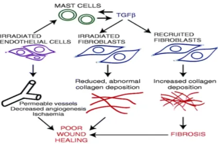
or overlap in time. Adapted from[87]---32 Figure 5: Myofibroblast differentiation in healing wounds. Adapted from[101]---33 Figure 6: Cellular cmponents involved in radiation induced poor wound healing.
Adapted from[65]---35 Figure 7: Comparison of the microenvironments of a healing wound and an invading tumor
margin. Adapted from[111]---38 Figure 8:Changes to the normal microenvironment promote tumour invasion.
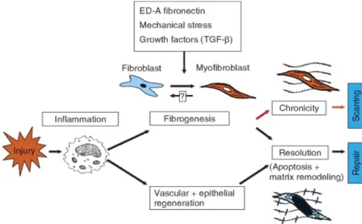
Adapted from[114]---39 Figure 9: Hypoxic microenvironment regulation of tumor survival. Adapted from[120a]---40 Figure 10: Generalized model to show the role of fibroblast in wound healing and scarring versus pathological fibrosis. Adapted from[151]---45 Figure 11: Possible origins of cancer-associated fibroblasts (CAFs) in TME.
Adapted from[165]---47 Figure 12:Transition of TGF- as a growth inhibitor to an inducer of epithelial to mesenchymal
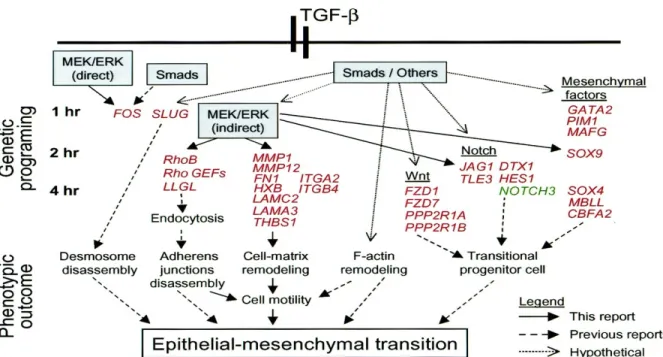
transition in tumorigenesis. Adapted from[212]---51 Figure 13: Signaling modules and genetic programs underlying EMTs induced by TGF-β.
Adapted from[234]---52 Figure 14: Expression pattern of proteinases and their physiological inhibitors in non-malignant
stromal cells.Adapted from[256]---55 Figure 15: Proteolytic cascades regulate MMP function from inflammatory cells.
Adapted from[256]---55 Figure 16: Established and recently uncovered modes of MMP, ADAM and TIMP regulation.
Adapted from[273]---57 Figure 17: Regulatory elements of MMPs promoter. Adapted from[270]---58
Figure 18: Modulation of the Tumor Microenvironment by MMPs. Adapted from [256]---60
Figure 19: Multiple functions of MMPs in the tumor microenvironment. Adapted from[256]---61
Figure 20: Basic RhoGTPase activation cycle. Adapted from[330]---62
Figure 21: (a) Stress fibers and the contractile ring, (b) Presumed Rho regulated assembly of actin myosin bundles. Adapted from[333]---63
Figure 22: Regulators and effectors of the Rho GTPases. Adapted from[366]---65
Figure 23: Mechanisms of short-term and long-term actin cytoskeleton reorganization induced by TGFβ in fibroblasts. Adopted from[408]---69
Figure 24: Several currently therapeutic strategies aim at the tumor microenvironment. Adapted from[417]---71
Figure 25:TGF signaling pathway inhibitors under development for potential cancer therapy. Adapted from[436]---76
Figure 26: Experimental scheme for coculture experiments ---84
Figure 27: Experimental scheme for invivo experiments ---88
Figure 28: TC-1 clonogenic survival curve ---92
Figure 29: Wound healing signals secreted by fibroblasts enhance TC-1 migration ---93
Figure 30A: Cytokine array analysis ---94
Figure 30B: MMP secretion from TC 1 cells ---95
Figure 30C: MMP secretion from RhoB-/- fibroblasts ---95
Figure 31: Modulation of TC-1 migration by O-Phenanthroline ---96
Figure 32: Effect of irradiation and coculture on TGF 1 expression in Wt fibroblasts ---97
Figure 33: Effect of irradiation and coculture on TGF 1 expression in RhoB-/-fibroblasts ----98
Figure 34: Modulation of TC-1 migration by SB 41542 with Wt fibroblasts ---99
Figure 35: Modulation of TC-1 migration by SB 41542 with RhoB -/- fibroblasts ---99
Figure 36: Effect of Irradiation and coculture on SMA expression in Wt and RhoB-/-fibroblasts ---100
Figure 37: TC-1 enhanced migration is associated with induction of EMT markers ---101
Figure 38: Bioluminescence imaging (BLI) for 3 groups at different time points ---102
Figure 39: Tumor bed effect ---103 Figure 40: Gross and microscopic appearance (HES) of orthotopic lung implantation of A549
Figure 43: Immunohistochemical analysis of mice CTCs clusters strong positive cytokeratin
staining ---106
Figure 44: Comparison of CTCs count before A) and 24Hrs after irradiation B) between different blood collection sites ---107
Figure 45: Cytokeratine staining ---108
Figure 46: Summary of findings ---115
LIST OF TABLES
Table 1:Specific terminology and criteria for adenocarcinoma, squamous cell carcinoma, and
NSCLC-NOS in small biopsies and cytology. Adapted From[55, 56]---23
Table 2: IASLC/ATS/ERS classification for small biopsies/cytology comparing 2004 WHO. Adapted from[55, 56]---24
Table 3: IASLC/ATS/ERS classification of lung adenocarcinoma in resection specimens. Adapted from[55, 56]---24
Table 4: Complications of radiotherapy. Adapted from[65]---29
Table 5: Tissue components affected by radiotherapy. Adapted from[65]---30
Table 6: Comparison between the hallmarks of cancer and wound healing. Adapted from[111]---36
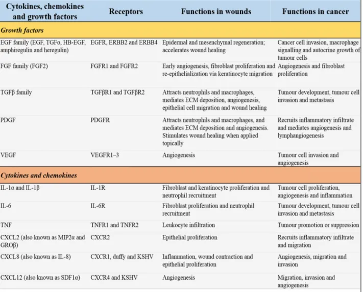
Table 7: Cytokines, chemokines and growth factors that influence wound healing and tumor progression. Adapted from[111]---43
Table 8: Regulation of epithelial growth, differentiation and apoptosis. Adapted from[157]---46
Table 9: Transcriptomic selection of relevant candidate genes. Adapted from[405]---67
Table 10: MMP inhibitors in clinical development. Adapted from[425]---73
Table 11: Stroma targeted anti-cancer strategies. Adapted from[430]---74
Foreword
Wound healing and carcinogenesis are defined as complex, adaptive processes which are controlled by intricate communications between the host and the tissue microenvironment. During a normal wound healing process, regeneration and repair of a wound, depends on a variety of signals which coordinate the response to injury. These processes entail cell proliferation, survival, and migration which are controlled by growth factors, cytokines as well as inflammatory and angiogenic signals. These signals are derived from multiple intra and extracellular components embedded in the microenvironment of wounds and are also involved in cancer. Therefore, a number of phenotypic similarities are shared by wounds and cancers in cellular signaling and gene expression. These similarities between wound healing and carcinogenesis were first recognized by Haddow, and the
notion that ‘cancer are wounds that do not heal’ was defined by Dvorak [1, 2].Most recently genomic
approaches revealed that wound healing and stromal signatures were able to predict for metastatic
progression [3] and resistance to neo adjuvant chemotherapy in breast cancers [4]. This underscores
the importance of microenvironment in tumor progression [5] and response to antitumor treatments
and suggests that the radiation induced wound healing response could be responsible for tumor radio resistance.
Radiotherapy is the second most effective modality of cancer treatment after surgery and can be used either alone or in combination with chemotherapy. The main anti-tumor effect of radiation therapy is the induction of tumor cell death but recent findings suggest that radiotherapy also rapidly and persistently modifies the tissue microenvironment. These modifications affect cell phenotype, tissue metabolism, bidirectional exchanges and signalling events between cells [4]. While there is evidence indicating that these changes might contribute to the antitumor effects of radiotherapy, some clinical and experimental observations indicates that irradiated stroma might exert tumor-promoting effects[4].
It has been shown previously that in glioblastoma, radioresistance signals are transmitted by
integrins 3, 5 ILK and then relayed by HIF-1 and RhoB[6, 7]. R Kolesnick and F Paris groups
showed the importance of the vascular microenvironment in tumor response to radiotherapy [8, 9],
which is further strengthened by the fact that under the influence of PI3K a portion of tumor cells within the perivascular niche presents an extended shutdown of the cell cycle in G2/M and becomes resistant to IR; while majority of the tumor cells die after radiation [10]. These data suggest the existence of a link between tumor microenvironment, radioresistance and cell cycle control after irradiation.
Foreword
Indeed in a mouse model mimicking tumor recurrence after irradiation where the tumor bed is irradiated tumor recurrence seems to depend upon the development of immature blood vessels initiated by immobilization of circulating cells from bone marrow and MMP-9 induced matrix
remodeling. [11]. These later data is in line with our previous work showing the importance of cell
cycle regulation and activation of the PI3K/AKT pathway after irradiation[12-14].
MH. Barcellos-Hoff`s Group has indeed shown a major contribution of TGF-1 produced by
irradiated stroma to carcinogenesis [15-17]as high dose of radiotherapy is known to stimulate
TGF-1 production [18]. TGF-1 is the prototype of pro-wounding molecules shown to be the main
inducer of reactive stroma, by not only affecting chemotaxis of fibroblasts, but also their
trans-differentiation into reactive fibroblasts, termed myofibroblasts [19]. TGF-1 also regulates epithelial
phenotype and has been especially described as a potent stimulatory molecule during the late phase of carcinogenesis and metastasis dissemination.
Beside TGF-signal, the contribution of the Rho pathway to radiation response has been
proposed by our group and others [20]. Rho GTPases are a family of signalling mediators implicated
in regulating cytoskeletal dynamics, motility, cell division, and transcriptional regulation. Amonst various Rho GTPases, RhoB has been described as a determinant of intrinsic tumor radiosensitivity
[21, 22]; which acts downstream of integrins alpha 3, 5 [23] and HIF-1 [24] in tumor cells. More specifically, RhoB expression is increased by a variety of extra-cellular stimuli which include
irradiation, epidermal growth factor (EGF) and transforming growth factor (TGF-)[18, 25]. Most
Rho proteins are modified by the covalent attachment of a geranylgeranyl group, but RhoB can exist in either a geranylgeranylated (RhoB-GG) or a farnesylated (RhoB-F) form. RhoB-F localizes to the cell membrane, modulates actin cytoskeleton, activates nuclear factor kappa B and promotes cell
growth[26-28]. In contrast, RhoB-GG localizes to endosomes and induces cell apoptosis [26].A role
for RhoB in TGF-induced cell responses such as epithelial-mesenchymal transition (EMT) and apoptosis was suggested by a series of DNA microarray studies, which showed that RhoB expression was upregulated by TGF- in a variety of cell types such as keratinocytes, mouse mammary gland
epithelial cells, hepatoma cells, and dermal fibroblasts [29]. TGF-β also stimulates actin stress fiber
formation in Ras-transformed cells in a way which is associated with upregulation of RhoB[30, 31].
Moreover, our previous genomic studies allowed us to identify the members of the Rho/ROCK cascade involved and showed a preferential activation of RhoB and ROCK 1 in tissues
and cells from fibrotic tissues [32, 33]. Infact pharmacological inhibition of Rho by statins prevents
the development of pulmonary fibrosis and radiation induced intestinal fibrosis [34, 35]and reverses
Studies on tumor radiation sensitivity have focused for about 2 decades on intrinsic tumor radiation sensitivity which highlighted the roles of the EGF/Ras/PI3k/AKT pathways in tumor radiation sensitivity. In parallel, several groups have also highlighted the role of tumor microenvironment of tumor radiosensitivity. Mostly these groups are focusing on anti angiogenic approaches which may potentially increase the normal tissue toxicities [37-40]. More recently our group have highlighted the importance of mitotic catastrophe in tumor response to radiotherapy, we demonstrated that agents targeting the G2/M check points are good candidates for radiosensitization
[41, 42]. We have also showed that normal tissue response to radiotherapy can activate a wound healing response. The current project aims at merging our expertise on both normal tissue and tumor; studying the role of the tumor microenvironment on tumor cure after radiation. This work will enable us to provide information about the involvement of the wound healing response to radiation on tumor radiation sensitivity.
Furthermore, in recent decade a lot of work has been focused on circulating tumor cells
(CTCs), which acts as a liquid biospsy and are detectable in many cancers [43]( including non-small
cell lung cancer (NSCLC) ). Detection of CTCs had been linked to prognostic significance[44, 45]in
different cancers. Previously, not much work has been done on the effect of radiation therapy on CTC level related to tumor microenvironmental changes because of the lack of a suitable detection method, as there is no useful data available on CTCs in patients undergoing RT.
Currently, the only US FDA approved technique is CellSearch platform which uses epithelial
cellular adhesion molecule EpCAM[46]for CTC detection. However, it lacks the detection of tumor
cell clusters or “circulating tumor microemboli” (CTMs) [47] and can therefore, significantly
underestimate total CTC numbers. Furthermore, CTCs in NSCLC often lose epithelial markers
[48-50]allowing them to evade detection. Moreover, there were major inconsistencies between different
methods. In samples where CellSearch detected few or no cells, manual counts typically detected abundant CTCs. These findings are consistent with another report indicating limited efficiency of
CellSearch in NSCLC CTC detection [51]. Therefore we also evaluated and compared a newer PCR
Introduction Lung Cancer
Lung Cancer
Incidence and prevalence
Lung cancer has been the most common cancer in the world for several decades. According to
the most recent statistics published (GLOBOCAN 2008) [52]. Worldwide 1,092,000 men and 516,
000 women were diagnosed with lung cancer which represents 12.7% of all new cancer cases. About
1.4 million people died from the disease, which represented 18% of all cancer deaths that year[53]. In
2010, the number of deaths from lung cancer worldwide increased to 1.5 million, representing 19% of all cancer death that year [54]. There are now fewer lung cancer cases in more developed regions (Northern America, Japan, Europe, and Australia/New Zealand) than in rest of the world. The statistics showed that there are 16.2% or 479,000 of all cancers in males in the developed countries as compared to 16.8% or 612,000 in developing countries. In both men and women, the incidence of lung cancer is low in persons under age 40 and increases up to age 70 or 75.
Causes
Lung cancer is often treacherous, as it may not produce any symptoms until the disease is well advanced. Approximately 7-10% of patients with lung cancer are asymptomatic, and their cancers are diagnosed incidentally after a chest radiograph performed for other reasons.
Causes of lung cancer include the following:
Smoking (78% in men, 90% in women)
Asbestos exposure
Radon exposure
Halogen ether exposure
Chronic interstitial pneumonitis
Inorganic arsenic exposure
Radioisotope exposure, ionizing radiation
Atmospheric pollution
Chromium, nickel exposure
Classification of lung cancer
Recently a significant amendment in pathologic classification of lung cancer arose after the publication of a new lung adenocarcinoma classification in 2011, under the sponsorship of the American Thoracic Society (ATS), International Association for the Study of Lung Cancer (IASLC) and the European Respiratory Society (ERS). This new classification has paved the path for the role of personalized medicine for patients with lung cancer. This classification takes into account the importance of histologic classification and molecular testing in stratifying patients for specific therapies. This has been the central theme of this new classification.
This classification is divided into two modules based on the primary lung cancer diagnosis:
1) Small biopsy and cytology specimens for patients with advanced-stage lung cancer
Tables 1 and 2
Table 1: Specific terminology and criteria for adenocarcinoma, squamous cell carcinoma, and
NSCLC-NOS in small biopsies and cytology. IASLC/ATS/ERS, International Association for the Study of Lung Cancer/American Thoracic Society/European Respiratory Society; NOS, not otherwise specified; NSCLC, non–small-cell carcinoma; TTF-1, thyroid transcription factor 1.Adapted from[55, 56]
Introduction Lung Cancer
Table 2: IASLC/ATS/ERS classification for small biopsies/cytology comparing 2004 WHO. Adapted from[55, 56]
2) Resection specimens for early-stage patients who are eligible for surgical resectionTable 3
An important point in this classification is the concept that personalized medicine in advanced lung cancer is determined by genetics and histology and that strategic tissue handling of small biopsies is critical for diagnosis and for molecular studies.
The majority of late stage lung cancer patients die within 18-months of diagnosis[52]. These
subtype differerences can be attributed to the site of origin and patient characteristics, SCC being associated with smoking and originates from bronchial epithelial cells, whilst adenocarcinoma is mainly derived from alveolar/bronchial cells [57]. Principal sites for NSCLC metastasis are bone, brain, adrenal gland and the liver. Evidence states that these sites of preferential metastasis are determined by interactions between cancer-cell-surface proteins and capillary lining endothelial-cell receptors at distant sites. Although a uniformly accepted staging of lung cancer is crucial for management of the disease, a considerable proportion of patients with lung cancer show tumour spread at the time of diagnosis and 40% of patients with non-small-cell lung cancer have distant metastases at presentation[58].
Management of lung cancer
Surgery, chemotherapy, and radiation are the main stay treatment options for NSCLC. As most lung cancers cannot be cured with currently available therapeutic modalities, the appropriate application of skilled palliative care is an important part of the treatment of patients with NSCLC.
Surgery
Surgery is the treatment of choice for stage I and stage II NSCLC. Several different types of surgery can be offered such as:
Lobectomy – removing a section of the lung
Pneumonectomy – removing the entire lung
Wedge resection – removing part of a lobe
Chemotherapy
At some point during the course of their illness, approximately 80% of all patients with lung cancer are considered for chemotherapy.
Radiation
In the treatment of stage I and stage II NSCLC, radiation therapy alone is considered only
when surgical resection is not possible [59]. Among those who are not candidates for surgery,
Introduction Radiotherapy
Radiotherapy
The radiotherapeutic approach for the eradication of malignant cells was first introduced during the late 19th century. Today radiotherapy is used as the primary therapy or in combination with surgery, chemotherapy, hormone therapy, and therapy with antibodies or a mixture of all strategies. Most cancer types can be treated with radiotherapy. Radiotherapy can be given either as a curative modality, adjuvant, or as a palliative treatment in lung cancer.
Non-small cell lung cancer (NSCLC) constitutes approximately 85% of all lung cancers. Roughly 1/3 of these patients have early stage disease (stages I and II) at the time of presentation and surgery is the preferred mode of treatment, whereas radiotherapy is reserved for medically inoperable patients. Radiotherapy is reserved as a palliative treatment for another 1/3 of patients presenting with disease at advance stage. The rest of the patients with locally advanced disease (stage III) and unresectable tumor, radical intent radiotherapy is the mainstay of treatment. A brief overview of
treatment schematic for NSCLC is shown below (Figure 1), as clinical staging and histopathological
grading greatly influence the choice of treatment modalities and outcome.
Mode of action
Radiation damage is a complex multi-faceted biological effect. Radiation damage occurs either through DNA damage caused directly by ionizing the atoms which make up the DNA chain or it can be a result of indirect ionization by ionization of water, forming free radicals, which then
damages the DNA[62]. Several treatment specific factors (e.g., overall treatment time, total radiation
dose, dose per fraction,) and patient-specific characteristics (e.g., performance status, disease stage,
histology) are known to influence the probability of local tumor control by radiation therapy[63, 64].
It is well known that the biological response to radiotherapy influences the outcome of radiation therapy; e.g. clonogenic cell growth can occur following fractionation, and if the cancer is growing exceedingly fast then it may grow between each daily fraction of treatment.
Figure 2: DNA damage due to radiotherapy. Radiation induces single and double stranded DNA
breaks and DNA strand cross-linking.Adapted from[65]
Methods of delivery
Following methods are used in clinical practice to deliver radiotherapy, depending upon tumor size, location, patient status and stage of cancer
a) Conventional external beam radiation therapy (2DXRT) is delivered via
Introduction Radiotherapy
b) Stereotactic radiation is a specialized type of external beam radiation therapy. It uses
focused radiation beams targeting a well-defined tumor using extremely detailed imaging scans. Radiation oncologists perform stereotactic treatments, often with the help of a neurosurgeon for tumors in the brain or spine. There are two types of
stereotactic radiation.Stereotactic radiosurgery (SRS) is when radiation oncologists
use a single or several stereotactic radiation treatments of the brain or
spine.Stereotactic body radiation therapy (SBRT) refers to one or several
stereotactic radiation treatments with the body, such as the lungs[66].
c) Virtual simulation, 3-dimensional conformal radiation therapy, and intensity-modulated radiation therapy
Radiation therapy has been revolutionized due to the ability to delineate tumors and adjacent normal structures in three dimensions using specialized CT and/or MRI scanners and planning software.
Virtual simulation, the most basic form of planning, allows more accurate placement of radiation beams than is possible using conventional X-rays. 3-dimensional conformal radiation therapy (3DCRT), is an enhancement of
virtual simulation, in which the profile of each radiation beam is shaped to fit the profile of the target. Thus the relative toxicity of radiation to the surrounding normal tissues is reduced, allowing a higher dose of radiation to
be delivered to the tumor than conventional techniques would allow[67]
Intensity-modulated radiation therapy (IMRT) is an advanced type of
high-precision radiation that is the next generation of 3DCRT. IMRT also improves the ability to adapt to the treatment volume to concave tumor shapes, for example when the tumor is wrapped around a vulnerable structure such as the
spinal cord or a major organ or blood vessel[68].
d) Particle therapy (proton therapy being one example), energetic ionizing particles
(protons or carbon ions) are directed at the target tumor. Proton beams differ from photon beams mainly in the way they deposit energy in living tissue. Whereas photons deposit energy in small packets all along their path through tissue, protons deposit much of their energy at the end of their path (called the Bragg peak) and deposit less energy along the way (into the healthy tissue surrounding the target tissue). In theory, use of protons should reduce the exposure of normal tissue to radiation, possibly
e) Brachytherapy uses high-dose intracavitatory radiation or radioactive implants and is
used to deliver radiotherapy directly to prostate tumours, soft tissue sarcoma, breast
tumours and cervical tumours[69, 70].
f) Radioisotope therapy (RIT) is a form of targeted therapy. Targeting can be achieved
by attaching the radioisotope to another molecule or antibody to guide it to the target tissue. The radioisotopes are delivered through infusion or ingestion. Examples are the infusion of metaiodobenzylguanidine (MIBG) to treat neuroblastoma, of oral iodine
131 to treat thyroid cancer or thyrotoxicosis, lutetium-177 and yttrium-90 to
treat neuroendocrine tumors (peptide receptor radionuclide therapy)[71].
Side effects
Radiation therapy is associated with both acute toxicity and long-term sequel and depend on the radiosensitivity of the body sites being treated, the volume of normal tissue irradiated, the total dose and the rate of dose accumulation [74]. Side effects are most evident in rapidly proliferating
tissues, such as the skin, mucosa and bone marrow, but may arise in almost any organ system Table 4.
Early effects are related to the loss of stem cells in quickly renewing tissues (such as epithelial layers)
or from inflammatory cytokine release (such as oedema, erythema, fatigue) [72]. Renewal of stem cells occurs either through division of surviving cells within the treatment area or through migration of stem cells from outside the treated area[75].
Introduction Radiotherapy
Late effects occur due to processes that take months to years to develop, either because the
tissue renews slowly due to chronic inflammatory processes, their relation to connective tissue cells or
genomic damage. These include but not limited to fibrosis [73] and tissue necrosis. Patients
undergoing radiotherapy display a large patient-to-patient variability in their risk of developing tissue reactions as normal body tissue varies in its response to radiation.
Table 5: Tissue components affected by radiotherapy. Adapted from[65]
Radiotherapy protocols should balance the expected outcome of the radiotherapy with the expected side effects. More severe side effects may be tolerated following curative radiotherapy but are not acceptable for palliative radiotherapy.
Wound Healing
Injury to the tissues initiates an erudite repair process that restores the damage. The different processes involved in repair are tightly regulated and synchronized to restore the integrity of the affected tissue. Wound healing is a dynamic interactive process that involves a series of molecular and cellular events. All of which revolves around the reorganization of cells and extracellular matrix, involving biological signs to repair the tissue. Defects in wound repair frequently affects not only aged individuals, patients with diabetes or immunosuppression, but patients who receive
chemotherapy or radiotherapy [76] are also affected; owing to the formation of
hypertrophic scars and keloids. Malignant transformation is a particularly severe complication of
non-healing wounds and a frequent event is the development of cancer in fibrotic tissue[77].
This suggests that common cellular and molecular mechanisms are active in wounds and in cancer tissue. Alexander Haddow suggested that "tumor production is a possible overhealing" [78]. A decade later Dvorak postulated that "tumors are wounds that do not heal" [1]. He further on clarified that the composition of the tumour stroma strongly resembles the granulation tissue of healing skin wounds. However, in contrast to healing wounds, the process is not self-limiting in cancer tissue, resulting in uncontrolled cell proliferation, invasion and metastasis. Recently, microarray technology revealed not only remarkable similarities between early wounds and cancer, but important differences
are also highlighted [80-83]. So understanding the mechanisms of normal wound healing will be an
asset in understanding tumor biology.
Stages of wound healing
Introduction Wound Healing
Wound healing of the tissue follows a specific time sequence (Figure 4), and this very
complex repair processe is sub-divided into three phases that overlap in time and space. The normal
wound healing response is depicted in(Figure 3 and 4). It represents a range of events that overlap in
time and activity with one stage merging into the next [85, 86].
Figure 4: Normal wound healing requires multiple events, many of which occur simultaneously
or overlap in time.Adapted from[87]
Phase I: Inflammation
The inflammatory phase is a period of active cellular migration and it takes place within the
first three days post injury[88]. It accomplishes both hemostasis and formation of a temporary matrix
on which fibroblasts and monocytes migrate[89]. At the time of wounding, hemostasis is achieved by
the formation of a platelet plug [90]. Blood products released into the wound initiate the coagulation
cascade. Platelets not only assist in clotting, but also release cytokines such as PDGF and TGF-[86, 91], which act as chemotactic agents in the recruitment of inflammatory cells, epithelial cells and
fibroblasts [92]. Blood clots consist predominantly of crosslinked fibrin and plasma fibronectin
secreted primarily by fibroblasts but also include other components, such as the ECM proteins vitronectin and thrombospondins. The PMNs (macrophages, monocytes, neutrophils, and
lymphocytes) are activated by the proinflammatory cytokines IL-1, TNF- and IFN-γ [93]. About
two days after injury, macrophages remain the predominant cells in the wound. They are stimulated by the hypoxia in the wound and release IL-1 (an angiogenesis promotor) and bFGF (a chemotattractant for fibroblasts and endothelial cells). Worth noting here is that deposition of the fibrin and fibronectin matrix is an acute and transient event in normal wound repair, but a chronic
Phase II: Proliferative phase (Tissue formation)
The phase of proliferation (formation of granulation tissue), occurs in the fourth to seventh day after the injury. It is characterized by angiogenesis, the proliferation of fibroblasts and the formation of collagen resulting in complete wound closure. It continues for at least 3 weeks, and the major event is the accelerated production of collagen, which is enhanced in the presence of PDGF and
TGF-[95]. Capillary formation continues, fibroblasts proliferate whereas mast cells and
macrophages remain activated[96]. Angiogenesis is further stimulated by local hypoxic conditions
which may be present[86]. Myofibroblasts also appear within the wound and stimulate contraction of
the wound and reduce the distance that must be bridged by forming collagen [97]. In addition, the
presence of growth hormone may stimulate enhanced collagen deposition by fibroblasts [98].
Phase III: Maturation and remodelling
The maturation stage begins around week three and lasts for 2 years. A subset of wound
fibroblasts differentiates into myofibroblasts(Figure 5), which are responsible for wound contraction
and for the deposition of additional matrix proteins. Collagen continues to mature with additional crosslink formation[86].
The acellular matrix is actively remodelled from a mainly type III collagen backbone to one
which predominantly composed of type I collagen [99]. This process is carried out by matrix
metalloproteinases (MMPs) which are secreted by fibroblasts, macrophages and endothelial cells
[100]. To sum up, there is a reduction in the number of fibroblasts and macrophages, an increase in collagen content, and the wound regains most but not all of the strength of normal tissue.
Introduction Radiotherapy and Wound Healing
Radiotherapy and Wound Healing
Despite the fact that radiation therapy is used to kill cancerous cells, it also damages healthy cells as well. This leads to many acute and chronic side effects. One such serious complication is difficulties in wound healing. The long-term effects of radiotherapy include but not limited to skin atrophy, microvascular damage and soft tissue fibrosis.
Radiotherapeutic injury is a multifaceted process that occurs in organised tissues consisting of a large number of interrelating cellular lineages, as well as a multitude of biologically active extracellular molecules. The radiotherapeutic response of normal tissues comprise of two partially
interacting components. The first is a process that resembles the healing of traumatic wounds after
perturbation by the radiation treatment. The second is a set of specific injuries that affect almost all
cellular and extracellular components within the irradiated volume.
Radiotherapeutic injury and wound healing
The response of normal tissues radiation injury diverge in many instances from a traumatic wound healing response, yet many processes are similar and/or occur in a similar sequence. A major differential effect between radiation injury and traumatic wounds is the accumulating and repetitive nature. That’s why the tissue irradiated at the beginning of the radiotherapy is very different from the tissue that is irradiated towards the end. During radiation therapy, the inflammatory phenomena does
not dissipate within 24 hrs, thus leading to an accumulating response known as ‘fractionated
inflammatory insult’. This Inflammation further aggravates the radiation response by augmenting
endothelial dysfunction and by increasing the levels of cytokines and growth factors, such as TGF-, thus delaying the process of re-epithelialisation.
Effect of radiotherapy on early wound matrix
High dose radiotherapy does not induce the rapid granulation tissue response that occurs in the milieu of acute traumatic wounds. Radiation inhibits fibrogenesis and angiogenesis in a dose and fractionation-dependent manner. Wound contraction can hasten the process of re-epithelialisation of
traumatic wounds and suppression of the evolving wound matrix by radiation [102, 103] results in
disruption of the actin microfilaments within wound fibroblasts. This could lead to impaired wound contraction(Figure 6).
Figure 6: Cellular components involved in radiation induced poor wound healing. Adapted from[65]
Delayed wound healing
Wound healing is orchestrated in an ordered sequence of cellular interactions. A key difference between wound healing and cancer is that wound healing is a self-limiting process;
whereas, tumours continue to expand, evolve and spread Table 6. This difference is correlated with
the alterations in components of the microenvironment. In wound healing, inflammation resolves once re-epithelialization is complete, but this is not the case during tumorigenesis. Radiation injury interrupts this highly controlled sequence of events, resulting in persistent inflammatory responses and
delayed wound healing [104]. The early effects of radiation may be due to disruption of the
inflammatory and proliferative phases. Late effects such as reactive fibrosis occur in face of radiation induced wound milieu formation and/or chronic inflammation, because high radiation doses can affect fibroblasts and other cells involved in tissue repair permanently.
It has also been suggested, that overexpression of TGF-1 in irradiated tissues, induces fibroblast proliferation through an expansion of the progenitor fibroblast pool and premature
differentiation of progenitor fibroblasts [105]. Pathological radiation fibrosis in humans is associated
with increased collagen synthesis, altered remodelling and sequential activation of key fibrogenic growth factors and cytokines, including TGF 1 and CTGF. It is assumed that processes deregulated a few weeks post-radiotherapy are responsible for complications presenting decades later. It is likely that molecular pathways exist that are common to many fibrotic responses, and that these involve the
Introduction Radiotherapy and Wound Healing
Table 6: Comparison between the hallmarks of cancer and wound healing.Adapted from[111]
* The absence of invasion and metastasis in wound healing is linked to the absence of epithelial–mesenchymal transition.
‡ In addition to angiogenesis, lymphangiogenesis is stimulated in both cancer and wound
Microenvironment
Like the developmental processes and tumor growth, wound healing involves intricate and
balanced interactions between cells and their microenvironment[107-109]. Through these interactions
the cells are being directed to differentiate, proliferate or remain quiescent, and assume the
architecture and function of that organ [107-110]. Wound healing and tumorigenesis are dynamic
events that require interactions with a wide variety of different cell types, including epithelial cells, fibroblasts, endothelial cells and immune cells. Chemokines and cytokines that are released from epithelial cells during injury are very similar to the ones found in invading tumours. All this integral relationship occurs in the milieu of microenvironment of host and depicts the outcome of therapeutic modalities in cancer treatment.
Recently it has been proposed that tumor microenvironment displays a striking resemblance to
the disrupted wound healing in normal tissues [111]. The tumor microenvironment was lately
recognized as the product of a developing crosstalk between different cells types. So, understanding the biology of microenvironment is an important asset for future cancer treatment modalities such as radiotherapy.
Tumor microenvironment
The concept of the tumor microenvironment originated from Paget’s “seed and soil” theory. It implies that tumor microenvironment is as important as tumor cell itself for metastasis initiation and progression. The tumor microenvironment is an emergent concept that defines the behavior of tumor
importantly by the surrounding milieu along with the genetics of the tumor [112]. The tumor
microenvironment is a vibrant network composed of the tumor cells, stromal tissue (immune cells, fibroblasts, cytokines, and vascular tissue), as well as the surrounding extracellular matrix. It is also characterized by unique properties namely hypoxia, low extracellular pH, low glucose concentration and necrosis.
The microenvironment of each organ and tissue develops to specifically support the function of those cells, while tumor microenvironments are subverted to promote tumor growth and expansion at the expense of normal tissue. Radiation therapy changes both, frequently in a manner that is reminiscent of processes associated with wound healing or inflammation. Defining the nature of the irradiated microenvironment provides a means of further targeting therapies directed towards
Introduction Microenvironment
Figure 7: Comparison of the microenvironments of a healing wound and an invading tumour margin. Adapted from[111]
Overview of microenvironment components
Basically, the tumor microenvironment is composed, in addition to the tumor cells themselves, of resident cells such as fibroblasts and endothelial cells, infiltrating cells such as macrophages and lymphocytes and of released products of all these cells. Among these products are extra cellular matrix (ECM) components, growth factors, cytokines, chemokines antibodies, proteases, other types of enzymes and various metabolites. These molecules are released from the tumor cells themselves as well as from the non-tumor cells mentioned above. The cross-talk between tumor cells and microenvironmental factors may result in diametrically opposed effects which could either enhance or block tumor formation or progression.
Recent evidence indicate that premalignant tumor cells may in fact prime their own
microenvironments, that is, form the metastatic niche in situ[113]. Tumor cells collaborate with local
stromal cells to recruit myeloid cells and initiate the formation of a metastatic niche. Following
extravasation and invasion at the secondary site, tumor cell survival and proliferation may be influenced by cell–cell and cell–matrix interactions in the metastatic niche. The disseminated tumor cell can successfully seed at metastatic lesion by evading the cell death signals, survive in the
Figure 8: Changes to the normal microenvironment promote tumour invasion. Adapted from[114]
A) Tumor cells
Tumor cells are characterized by a variety of biological functions including deregulated proliferation, suppression of apoptosis, evasion of host immune response, neovascularization and metastatic spread to distant sites[115].
Tumor Cells normally survive in hypoxic environment[116, 117]through adaptive phenotype by
virtue of their genetic, epigenetic instability and mutations[118]. Previous studies showed that cells
grown in a tumor undergo a significantly higher rate of mutations than the same grown in cell culture
[119]. This shows that the microenvironment of the tumor is responsible for inducing changes in the surrounding and intervening cells.
The micro-environment induce genetic and epigenetic instability in the cancer cells in several ways. One way, is the oxygen tension of the microenvironment which is generally characterized by series of hypoxia and reoxygenation. This leads to the formation of reactive oxygen species that induce damage by single and double strand DNA breaks, aberrant DNA synthesis, single and point mutations and amplifications, and so on. In addition, hypoxic conditions cause a deficiency in DNA damage repair in the form of decreased mismatch and decreased nucleotide excision repair[120]. These alterations produce tumor cells that have escaped regulatory control leading to an aggressive
Introduction Microenvironment
Figure 9: Hypoxic microenvironment regulation on tumor survival. Adapted from[120a]
Secondly, by regulating cancer genetics in relation to two gene classes. Class I genes such as the tumor suppressor genes and oncogenes that are mutated or deleted and controlled at the DNA level. Class II genes that have altered epigenetics, or a change in phenotype and are not changed at the DNA
[121]. These cause the cells to be more aggressive and malignant. The microenvironment is thought to modify these Class II genes, particularly late in the course of cancer progression and metastasis.
B) Stromal tissue
Stroma is the supportive and connective tissue of the host tissue. It is composed of different cell types including fibroblasts, myofibroblasts, vascular and lymphovascular endothelial cells, and cells of the immune system such as macrophages. There is a bidirectional, dynamic, very intricate interaction between the cells of the stromal tissue and the cancer cells [122].
Fibroblasts
The wound-healing response of fibroblasts has many similarities to the activation of fibroblasts in the tumour stroma. Fibroblasts secrete soluble factors that act in a paracrine or autocrine fashion on the tumor cells resulting in a phenotype change that is more aggressive. These activated fibroblasts that interact with the tumor cells are called cancer associated fibroblasts CAFs [123, 124].
One very well studied soluble factor is TGF-β (transforming growth factor-β). The effects of
tumor cells and the fibroblasts have receptors for TGF-β. The receptors for TGF-β on the epithelial cells can function in tumor suppression causing apoptosis and cytostasis by causing arrest of the cell cycle at G1, thus resulting in an inhibition of epithelial cell cycle progression. On the other hand, two molecular signaling pathways (SMAD and MAPK) have been identified in which TGF-β leads to an
epithelial–mesenchymal transition (EMT) which results in tumor progression and metastasis [126,
127]. Other paracrine factors secreted by fibroblasts include EGF (epidermal growth factor), IGF (
insulin like growth factor), FGF (fibroblast growth factor), and HGF (hepatocyte growth factor). Other cells of the stromal tissue are also involved in promoting tumor proliferation and motility. For example, myoepithelial cells secrete CXCL14 and myofibroblasts secrete CXCL12 which both bind to receptors on epithelial cells which in turn cause enhanced proliferation and invasion. The receptor on the epithelial cell for CXCL12 has been defined as CXCR4 and in vivo and in vitro studies link this interaction to breast cancer metastasis[124].
EMT
Changes in epithelial cells and fibroblasts that are transient during wound healing can be sustained in tumors. The increase in epithelial cell migration and proliferation that is required for wound healing returns to normal on wound closure as the basement membrane is rebuilt. However, in tumors, those processes can continue unchecked; epithelial cells sustain oncogenic mutations that can result in immortalization, and they may undergo an epithelial–mesenchymal transition (EMT), which is associated with gaining the properties of cancer stem cells[128].
The transition from epithelial to mesenchymal cell produces a tumor cell with increased motility, the ability to penetrate the basement membrane, and the ability to form foci of cancer at
distant locations in the body [127]. The epithelial-mesenchymal transition (EMT) is a process by
which epithelial cells lose their cell polarity and cell-cell adhesion, and gain migratory and invasive properties to become mesenchymal stem cells; these are multipotent stromal cells that can differentiate into a variety of cell types. EMT has also been shown to occur in embrogenesis, wound healing, organ fibrosis and in the initiation of metastasis for cancer progression. Furthermore, EMT is
divided into three types.Type 1 EMT occurs during the life of an embryo and organogenesis, Type 2
EMT is involved in wound healing and tissue regeneration, and Type 3 EMT is that associated with
cancer progression and metastasis[126].The properties of the microenvironment have been shown to
Introduction Microenvironment
C) Extracellular matrix
The extracellular matrix is composed of collagen, elastin, proteoglycans, and other specialized structural proteins that provide support to and division of the cells of the host tissue. Many of these components are actually synthesized by fibroblasts. However, in cancer, fibroblasts are transformed into myofibroblasts and secrete proteins that degrade this ECM. One of the first steps involved in formation of the tumor microenvironment is remodeling of the extracellular matrix. This is considered a seditious event in the formation of this dynamic network that supports tumor initiation and proliferation. The structure of the ECM is remodeled by enzymes called MMPs (matrix metalloproteinases) that are capable of hydrolyzing protein macromolecules. Many studies have
shown that MMPs are overexpressed in human cancers[113]. These molecules are known to be
pro-angiogenic and metastatic because the digestion of the ECM by proteases such as MMPs allow the entry of cancer cells into the host tissue and passage of tumor cells through the barrier of the host tissue as well as migration of endothelial cells into the matrix that results in neovascularization. In the process of degradation the MMPs also cause a release of growth factors that amplify the process of tumor growth and invasion.
The ECM contains other proteins such as integrins, which are cell surface receptors made up of various compositions of alpha and beta subunits responsible for binding components of the ECM and thus altering the structure of the ECM and transferring information between cells and the ECM.
In effect, integrins are the receptors for the tumor microenvironment of the cancer cell and then effect cell migration, proliferation, and metastasis via tyrosine kinases. In studies on the tumor microenvironment, integrins have been shown to promote angiogenesis, tumor proliferation, and
metastasis[130]. As a part of the epithelial–mesenchymal transition described above, the subtypes of
integrins found on the epithelial tumor cells change or undergo “integrin switching” which further
potentiate the effects of TGF-β in regards to tumor proliferation and metastasis.
D) Cytokines chemokines and growth factors in microenvironment
There are striking similarities between the growth factors, cytokines and chemokines that are
present in healing wounds and those present in tumours [131], but the kinetics of expression differ
Table 7. In solid tumours, the same signalling pathways that are transiently upregulated to repair wounds are hijacked and activated constitutively.
CSF1, colony stimulating factor 1; CSF1R, CSF1 receptor; HB-EGF, heparin-binding ; MCP1, monocyte chemoattractant protein 1; MIP2α, macrophage inflammatory protein 2α; PDGF, platelet-derived growth factor; PDGFR, PDGF receptor; *The cytokines, chemokines and growth factors were included on the basis that they have been shown to influence both wound healing and tumour invasion or progression in vivo
Table 7: Cytokines, chemokines and growth factors that influence wound healing and tumour
progression.Adapted from[111]
Communication between cells and their microenvironment occurs through a complex network of signals generated by cell-ECM and cell-cell adhesion and junctional molecules, as well as by collaboration between the epithelial, stromal and other organ-specific cell types. These ECM-molecules, together with the enzymes that remodel them, organize and sculpt tissues but also directly signal to the cells. The cells respond to both soluble and insoluble factors and in turn change their microenvironment in a fugue-like reciprocity, the end result of which is a magnificent and still somewhat mysterious integrated system that guides and allows maintenance of the differentiated state. If the microenvironment were not dominant, each cell would have its own way and the result would be either a uniform lump of similar fate or absolute chaos.
Introduction Microenvironment
IL-6 and bFGF
Interleukin (IL)-6 is one of the best-characterized pro-tumorigenic cytokines. It affects cell proliferation, survival, differentiation, migration, invasion, metastasis, angiogenesis, inflammation and
metabolism[132]and is elevated in many cancers [133]. Major signal transducer downstream of IL-6
is STAT3 which acts as a key player linking inflammation and cancer[134-136]. IL-6 has a direct
growth stimulatory effect on many cancer cells through several signaling pathways[137]. IL-6 family
stimulates tumor invasion and migration (metastatic spread) through induction of EMT phenotype
[138-140]. bFGF is a multipotential glycoprotein which exerts its mitogenic and angiogenic characteristics, through tissue remodelling, wound healing and neovascularization. The bFGF efficiently inhibits terminal differentiation of fibroblasts to myofibroblast, by a significant decrease of
alpha–smooth muscle actin (α-SMA) positive cells [141]. The majority of human NSCLC cell lines
produce elevated levels of bFGF which in turn stimulate the growth of these tumor cells by intracrine
mechanisms[142-145]. bFGF is also regarded as one of the most specific and crucial regulators of
Tumor Microenvironment (cellular players)
The microenvironment of a tumor is a vital part of its structure, physiology and function. An essential unbalanced relationship between tumor and stromal cells is required for tumor cell growth, progression, and metastasis. Improved understanding of this interaction may provide valuable clinical targets for cancer management. Secreted proteins from tumor, stromal cells and non-malignant cells are dynamic participants in cancer progression. Apart from others cells such as macrophages, endothelial cells present in tumor microenvironment fibroblasts and its transformed forms still remains the most important determining factor for the fate of tumor and its response to radiotherapeutic interventions.
Fibroblasts
Fibroblasts are elongated, spindle shaped and metabolically active cells in many tissues in the body and are mainly responsible for the production and turnover of extracellular matrix (ECM) by expressing collagens, fibronectins, laminins, elastins, proteoglycans, integrins, matrix metalloproteinases (MMPs), tissue inhibitors of metalloproteinases (TIMPs) and a host of other ECM
proteins that are expressed in a tissue-specific manner[148, 149].
Fibroblasts activity and their consequent differentiation into myofibroblasts is dependent upon different growth factors, extracellular matrix components, and mechanical stress. Worth noticing is the expression of receptors for a number of cytokines including PDGF, TGFβ1, and TNFα by fibroblasts. The heterogeneous nature of the fibroblast phenotype derived from different anatomical
locations had been described by Alvarez et al[150].
Figure 10: Generalized model to show the role of fibroblast in wound healing and scarring versus
Introduction Tumor Microenvironment (cellular players)
Fibroblast activity is crucial during processes of wound healing and inflammation. Under such conditions, fibroblasts are generally considered to be “activated”. In particular, as the healing process progresses, fibroblasts turn on expression of afilamentous actin, alpa-smooth muscle actin (-SMA), which enables them to exert contractile forces to close the wound. Local tissue contractility is mediated by focal adhesions between the activated fibroblasts called myofibroblasts and the ECM.
After wound closure, the balance of MMPs and TIMPs secreted by fibroblasts is changed to favor ECM degradation (as opposed to synthesis) which leads to massive apoptosis of the myofibroblast population. Instead of deactivation, the myofibroblast population persists during fibrosis or tumorigenesis for reasons that are not clear. This aspect of fibroblasts can be regarded as bimodal. Because, in early tumorigenesis, they negatively regulate malignant progression, but in advance stages they are subverted to promote tumor growth-being referred to as cancer-associated fibroblasts (CAFs). It is important to underline that a cross talk and reciprocal relations between tumor and its stroma are necessary for tumor formation and its progression. Recent advances showed that fibroblasts in stroma actively drive tumorigenesis and cancer progression[151-156], negating the previous notion that fibroblast behavior is dictated by the epithelium. Fibroblasts play important roles during tumor development, from the pre-neoplastic state until metastasis. In vitro co-culture and in vivo xenograft systems demonstrate that factors derived from tumour fibroblasts contribute to the transformation of
immortalized epithelia [157]. Table 8 shows soluble factors from fibroblasts effecting tumor


![Table 6: Comparison between the hallmarks of cancer and wound healing.Adapted from [111]](https://thumb-eu.123doks.com/thumbv2/123doknet/14455027.519436/37.918.103.773.107.433/table-comparison-hallmarks-cancer-wound-healing-adapted.webp)

![Figure 8: Changes to the normal microenvironment promote tumour invasion. Adapted from [114]](https://thumb-eu.123doks.com/thumbv2/123doknet/14455027.519436/40.918.169.717.108.439/figure-changes-normal-microenvironment-promote-tumour-invasion-adapted.webp)