A Comparative Look at Structure-Function Roles in Light-Harvesting
Dynamics of Purple Bacteria
By Ashley Tong B.S. Biochemistry
Indiana University South Bend, 2014
SUBMITTED TO THE DEPARTMENT OF CHEMISTRY IN PARTIAL FULFILLMENT OF THE REQUIREMENTS FOR THE DEGREE OF
DOCTOR OF PHILOSOPHY IN CHEMISTRY AT THE
MASSACHUSETTS INSTITUTE OF TECHNOLOGY September 2019
@2019
Massachusett Instityte of Technology. All rights reserved.Signature redacted
Signature of Author: L ",I
I
)
Ashley Tong July03,2019Signature redacted
Certified by:I
C
Gabriela Schlau-CohenAssistant Professor of Chemistry
Accepted by: MASSACHUSETT INSTITUTE OF TECHNOLOGY_ 0CT
0 3
2019_
LIBRARIES
Thesis Supervisor Robert Field Hslam and Dewey Professor of Chemistry Chair, Department Committee on Graduate StudentsC:r
A
This doctoral thesis has been examined by a committee of the Department of Chemistry as follows
Signature
redacted
Professor Alex Shalek:
Thesis Commitee Chair Associate Professor of Chemistry
Signature redacted
Professor Gabriela Schlau-Cohen:___
(
(7
Thesis Supervisor Assistant Professor of Chemistry
Signature redacted.
Professor Cathy Drennan:
Thesis Committee Member Professor of Biology and Chemistry \,,
A Comparative Look at Structure-Function Roles in Light-Harvesting
Dynamics of Purple Bacteria
By Ashley Tong
Submitted to the Department of Chemistry on July 03, 2019 in Partial Fulfillment of the Requirements for the Degree of Doctor of Philosophy in Chemistry
Abstract
Using a unique approach to solar energy conversion, photosynthetic organisms have developed a light-harvesting process with near unity quantum efficiency. Light-harvesting proteins transfer energy from the sun to reach a central location, the reaction center, where charge separation occurs and energy is converted to chemical energy. Moreover, these proteins are able to carry out this efficient transfer in cellular membranes despite the complex environment found in these membranes. Particularly, light-harvesting in photosynthetic purple bacteria uses a diverse set of tools from species to species to efficiently transfer energy through this protein network. Induced by their habitats, external environmental pressures on the fitness of purple bacteria have caused species to evolve different mechanisms in order to deal with thesel pressures. Although these complexes have been studied for some time, there is still very little known about particular species. Additionally, most previous work has been on non-native samples, such as detergent solubilized proteins, or on complex membranes such as vesicles, chromatophores, or whole
membranes that contain multiple proteins with multiple processes occurring simultaneously. This work investigates how photosynthetic light-harvesting complexes are able to achieve their
impressive efficiency using ensemble ultrafast spectroscopy to measure energy transfer dynamics and near-native discoidal model membrane-discs. These model membrane-discs provide a controlled environment to effectively study how energy is transferred in a single protein and between particular sets of proteins, allowing individual steps in the light-harvesting process to be probed without other processes interfering. They also provide a near-native system to explore how lipid-protein and protein-protein interactions affect the energy transfer kinetics in these proteins. Additionally, this work explores the differences in energy transfer kinetics of
light-harvesting proteins between species of purple bacteria. Overall, this provides new insights into the role the membrane plays in light-harvesting and how the composition of proteins within the native membrane of different species of purple bacteria can add variation to energy transfer kinetics.
Thesis Supervisor: Gabriela Schlau-Cohen Title: Assistant Professor of Chemistry
Contents
1 Chapter 1: Model Membrane-Discs 12
1.1 Membrane Protein Studies ... . 12
1.2 N anodiscs . . . . 12
1.3 Other Discoidal Model-Membrane Platforms ... 13
1.4 Conclusion . . . .. . . . .. . 16
2 Chapter 2: Impact of the Lipid Bilayer on Energy Transfer Kinetics in the Photosynthetic Protein LH2 17 2.1 Sum m ary . . . . 17
2.2 Results and Discussion ... . 18
2.2.1 LH2 in Membrane Discs ... 18
2.2.2 Time-Correlated Single Photon Counting Measurements ... 18
2.2.3 Transient Absorption Spectroscopy and Transient Absorption Anisotropy . . 18
2.3 Conclusion . . . . 25
2.4 M aterials and M ethods . . . . 25
2.4.1 Purification of MSP1E3D1 . . . . 25
2.4.2 Lipid Preparation . . . . 26
2.4.3 LH2 Preparation . . . . 26
2.4.4 LH2 Disc Assembly . . . . 27
2.4.5 Time-Correlated Single Photon Counting Measurements . . . . 27
2.4.6 Sample Preparation for Ultrafast Spectroscopy . . . . 28
2.4.7 Transient Absorption and Transient Anisotropy Measurements . . . . 28
3 Chapter 3: Comparison of Energy Transfer Kinetics for Light-Harvesting Pro-teins of Rhodobacter sphaeroides and Phaeospirillum molischianum 29 3.1 Sum m ary . . . . 29
3.2 Results and Discussion . . . . 29
3.2.1 Linear Absorption Analysis . . . . 29
3.2.2 Transient Absorption Spectroscopy . . . . 30
3.3 Conclusion . . . . 33
3.4 M aterials and M ethods . . . . 34
3.4.1 Purple Bacteria Growth . . . . 34
3.4.2 Lysis and Purification of LH2 and LH3 . . . . 34
3.4.3 Linear Absorption and Fluorescence Measurements . . . . 34
3.4.4 Sample Preparation for Ultrafast Spectroscopy . . . . 35
3.4.5 Transient Absorption Measurements . . . . 35
4 Chapter 4: Inter-Protein Energy Transfer Between Antenna Complexes of Pur-ple Bacteria 36 4.1 Sum m ary . . . . 36
4.2 Results and Discussion . . . . 37
4.3 Conclusion . . . . 43
4.4 M aterials and M ethods . . . . 43
4.4.1 LH2 and LH3 Doubly Loaded Membrane-Discs . . . . 43
4.4.2 Gold labeling of Doubly Loaded Membrane-Discs . . . . 44
4.4.4 C ryo-EM . . . .. 45
5 Conclusions and Future Outlooks 46 6 Appendix 1: Purification of Light-Harvesting Complexes of Purple Bacteria 47 6.1 Growth of R. sphaeroides ... . 47
6.2 Growth of Ph. moliscianum . . . . 47
6.3 Lysis of Light-Harvesting Complexes . . . 47
6.4 Protein Purification . . . . 48
6.5 Linear Absorption Spectral Decomposition . . . 49
7 Appendix 2: Spectroscopy Experimental Set-Up 51 7.1 Spectroscopy System Layout . . . . 51
7.2 Transient Absorption Apparatus . . . . 51
7.3 850 nm Pulse Generation . . . . 51
7.4 Power Dependence Measurements . . . . 52
7.5 Transient Absorption and Transient Anisotropy Measurements . . . . 52
7.6 Data Analysis M ethods . . . . 53
7.7 Calculation of Relative Energy Transfer Rates . . . . 54
8 Appendix 3: Assembly of Model-Membrane Discs 56 8.1 Production of Membrane-Disc Belting Protein MSP1E3D1 . . . . 56
8.2 Production of Membrane-Disc Belting Protein ApoE422K . . . . 57
8.3 Lipid Preparation . . . . 57
8.4 Loaded Membrane-Disc Reactions . . . . 58
8.5 Purification and Characterization of LH2 Embedded Membrane-Discs Using MSP1E3D1 59 8.6 Purification and Characterization of LH2 and LH3 Embedded Membrane-Discs Using A poE 422K . . . . 60
8.7 Optimization of Membrane-Disc Ratios by FPLC . . . . 61
8.8 M embrane-Disc Stability . . . . 63
8.9 Linear Absorption and Fluorescence Measurements . . . . 63
8.10 Transmission Electron Microscopy . . . . 63
8.11 Cryo-EM to Discern Relative Orientation of LH2 and LH3 in Membrane-Discs . . . 63
Introduction
Photosynthesis powers most life on Earth and is responsible for the generation of over 100 gigatons of biomass on an annual basis [1, 2]. Photosynthetic purple bacteria convert light energy into chemical energy via a cyclic electron transport mechanism with >90% quantum efficiency [3,4. The photosynthetic machinery in purple bacteria contain light-harvesting antenna proteins LH2 and LH1 (Figure 1) [5-7]. LH2 is peripheral to the reaction center, while LH1 encircles the reaction center and directly interacts. These antennae absorb energy from light and funnel it to the reaction center where it is used to power the electron transport chain and synthesize ATP. It is in the first few steps of energy transfer within the photosynthetic antennae that >90% quantum efficiency is achieved. One of the fascinating features of the light-harvesting complexes of purple bacteria is that this efficiency is always maintained with major differences between complexes of different species and under various light conditions, even with light restriction. Although vital to photosynthetic biomass production, the in vivo dynamics of the initial energy transfer steps within this network have not been determined due to the intrinsic difficulties associated with production, purification, and sample preparation of membrane proteins. Specifically, producing spectroscopically amenable samples that mimic the in vivo membrane architecture and environment has proven challenging. While much research has been done to characterize the energy transfer dynamics within light-harvesting complexes, there has been little direct comparison of these complexes under the exact same experimental conditions. Also, given the wide diversity of differences between proteins across species, most research thus far has been done on only Rhodopseudomonas (Rps.) acidophila and
Rhodobacter (R.) sphaeroides. >90% Quantum Efficiency
LH2
RC
21
Figure 1: Light-Harvesting energy flow through the photosynthetic protein network in purple bac-teria.
LH2"(Figure 2A) is a nonomer or an octomer of heterodimers (Figure 2B) consisting of two peptides (~100 amino acids each) known as the a and
#
peptides [8]. It contains two rings of bacteriochlorophyll a (BChla) that mediate the energy flow within the protein, three BChla per heterodimer. Each BChla has a central Mg2+ atom that serves a structural role [9]. One ringabsorbs at 800 nm and is known as the B800 band. The other ring absorbs at 850 nm and is known as the B850 band. Two of the BChla in each heterodimer (one per peptide) make up the B850 band, while the third forms the B800 band. The BChla that makes up the B800 band is singly ligated by a carboxylated N-terminal methionine of the a peptide and is hydrogen bonded
to
#-Arg20,
0-His12 and a-Gln3. The BChla of the B850 band are ligated by a single histidine,one from the a peptide and one from the
#
peptide, and hydrogen bonded by residues in both peptides as well. The B800 band BChlas lie parallel to the surface of the membrane, while those in the B850 band lie perpendicular. The BChla in the B850 band are held closer together and are more strongly coupled than those in the B800 band. One carotenoid per dimer is also present and aids in broadening the spectral window for light energy collection and also serves a photoprotectiveA
BK
Figure 2: Structure of LH2. (A) Complete structure of LH2 from Rsp. acidopillus showing the organization of pigments. Dark gray shows the a peptide and light gray shows the peptide. BChla in blue are part of the B800 ring and those in red are part of the B850 ring. (B) Hetero-dimer of LH2. Colors are the same as (A).
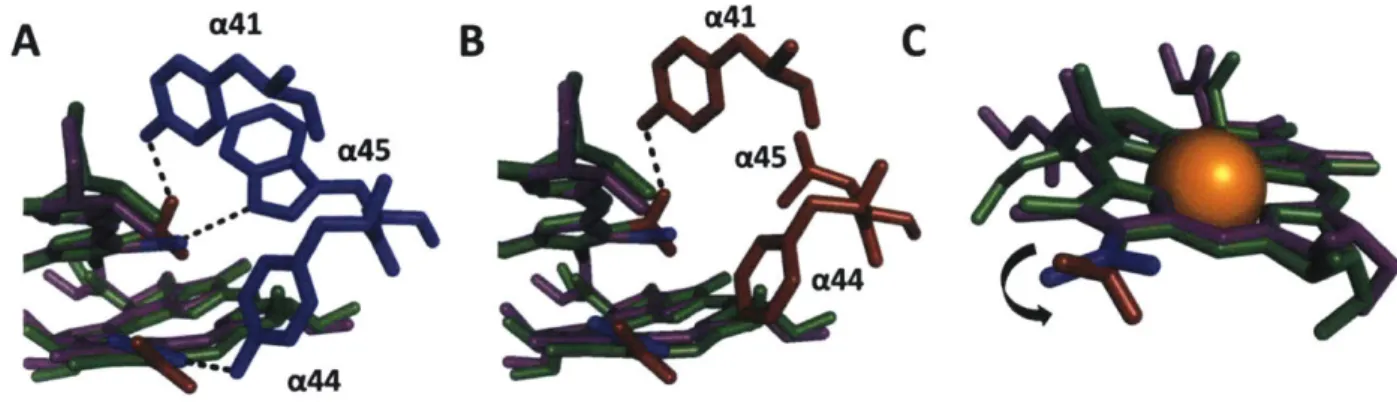
role [8,10]. Some species of purple bacteria contain spectral variations of the LH2 antennae known as LH3 [11-13]. These spectral variants have a blue-shifted B850 band, absorbing at 820 nm (B820 band) as shown in Figure 3. LH3 is produced under stressed conditions, such as low light and low temperature. Its major roles in vivo are to broaden the spectral range over which light can be absorbed and to increase the energy gap from the peripheral antennae to LH1, prohibiting back transfer to LH3 and increasing the overall efficiency of energy transfer [14,15]. LH3 can be present in varying ratios that depend on the stressed condition. The membrane can contain anywhere from
100% LH2 to almost 100% LH3 [16].
A high resolution structure of LH2 from R. sphaeroides is not available but projection struc-tures from negative stain electron microscopy with resolution at 8
Adetermined
it to be a nonomer of heterodimers in resemblance to LH2 from Rps. acidophilus [17,18]. Additionally, these LH2 variants have a high sequence percent identity of 45% and 66% between the a and#
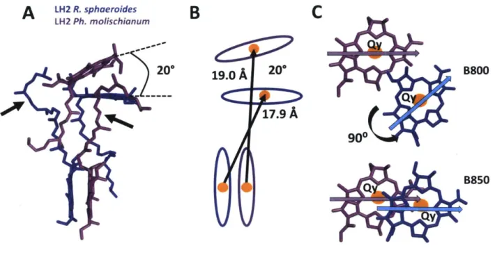
subunits, respectively [19]. Thus, these two LH2 variants have similar energy transfer dynamics. In this work, LH2 from Rps. acidophilla will be used as the R. sphaeroides model. The structure of LH2 from Ph. molischianum reveals that is is an octomer of heterodimers, producing B800 pigments that are rotated 90 and have a 20° tilt with respect to R. sphaeroides B800 pigments (Figure 4A) [20]. The inter-ring distances of the B800 BChla and the closest B850 BChla are shorter for R.sphaeroides with Mg-Mg distances between B800 BChla and B850 BChla within a heterodimer
be-ing 18.4 and 20.2
A
for R. sphaeroides and Ph. moliscianum, respectively (Figure 4B). The Mg-Mg distances between B800 BChla and B850 BChla between adjacent heterodimers are 19.0 and 17.1A
for R. sphaeroides and Ph. moliscianum, respectively. The 90° rotation of the B800 BChlas inPh. molischianum align the transition dipoles to be parallel to the B850 BChlas' dipole moment,
whereas the transition dipoles lie more perpendicular to each other in R. sphaeroides (Figure 4C). The intra-ring Mg-Mg distances are 21.2 and 22.0
Afor
the B800 pigments and 9.2 and 8.7A
for the B850 pigments for Ph. molischianum and R. sphaeroides, respectively [18,20].'4 LH2 R. sphaeroides
0
0.5
LH2 Ph. molischianum
C
LH3
Ph. molischianum
0 °750
800
850
Wavelength(nm)
Figure 3: Linear absorption of all proteins showing difference in their spectral properties. In LH3 the 850 nm absorption is shifted to 820 nm.
from the same species [11,14]. Interestingly, the overall pigment-protein interactions between the BChlas in the B800 and B850 are very similar in the two complexes. CD spectra report on the electronic structure of the B850 and B820 bands and are almost identical between LH2 and LH3 besides the wavelength dependent shift of the peak location in LH3 from 850 to 820 nm [21]. In fact, the peptide sequences are identical except for two amino acids in the a peptide of the heterodimer [22,23]. The residue a-Tyr44 in LH2 is switched to a-Phe44 in LH3 and a-Trp45 of LH2 is changed to a-Leu45 in LH3. These two changes in amino acids restructures the hydrogen bonding network of the- BChlas in the B850 band, blue-shifting the absorption to 820 nm (Figure 5A,B). Through site-directed mutagenesis and Raman spectroscopy, this has been attributed to the rotation of the C3-acetyl chain of a BChla pigment out of the plane of the macrocycle (Figure 5C) [11,14,24,25]. This rotated bond reduces the number of conjugated bonds in the macrocycle thus increasing the BChla's site energy [7,26,27].
To date, there have been a significant number of studies using spectroscopic methods to determine energy transfer rates within LH2 and other components of the photosynthetic apparatus of purple non-sulfur bacteria [15,28-45]. Energy transfer rates from LH2 in R. sphaeroides were previously determined with transient absorption spectroscopy and anisotropy [46]. Energy transfer within the B800 and B850 bands was found to be -450 fs and -60 fs, respectively. Inter-band energy transfer from B800 to B850 was determined to be ~850 fs in detergent solubilized complexes. This rate was enhanced by 30% in a native-like membrane model system as will be discussed more in Chapter 2 [46]. These rates were in agreement with many past works [30,33,37,47-51]. Energy transfer rates of LH2 from Ph. moliscianum has not been as well established as that from R. sphaeroides but has been shown to have similar rates as observed in R. sphaeroides with the B800-B850 energy transfer slightly longer (1-1.2 ps) [52-55]. Energy transfer taking place in the B800 band of LH2 has been described by de Caro et al. as a two-pool model [56]. In this model the B800 pigments are divided into two groups, one on the blue side and one on the red side of the B800 band. Pigments of the red pool can transfer exclusively to B850 and those in the blue pool can transfer energy via two pathways that compete with one another. The blue pool of BChlas can transfer to the red pool of BChlas in the B800 band and can also transfer energy directly to the
B
A
LH2 R. sphaeroides LH 2 Ph. molischianum20°0
C
B800
90°
B850
Y
Figure 4: Differences in BChla orientations in Rsp. acidophilla (our R. sphaeroides model) and Ph.
moliscianum. (A) Closest B800 and B850 BChlas showing 20° tilt between two species. Arrows
point to a macrocycle substituent that is a long alkyl chain known as the phytol tail. (B) BChla schematic showing differences in Mg-Mg distances for two species. BChlas are depicted as ovals and Mg 2atoms are depicted as orange circles. (C) Closest BChlas of the B800 and B850 bands showing changes in Qy dipole moments due to 90 rotation. The Qy dipole of B800 BChla in Ph.
moliscianum better aligns with the Qy dipole of the B850.
B850 band [54,56].
Energy transfer dynamics for LH3 Ph. molischianum have not been characterized in any pre-vious work. Some information is available for LH3 in Rps. acidophilus which ultimately concludes that the energy transfer dynamics of LH3 is very similar to LH2 and that the large blue-shifted B820 does not have a substantial effect on the interband energy transfer [42,57]. However, single-molecule fluorescence studies have shown distinctive heterogeneous spectral features in the B820 band of LH3 that are significantly different than LH2 [58]. This heterogeneity was ascribed to the sensitivity of the B820 BChlas to light-induced local conformational changes. The change in hydrogen bonding and rotation of the C3-acetyl bond was suggested to be the cause of observed
heterogeneity in the B820 band of single-molecule experiments because of the increase in confor-mational flexibility [58]. Compared to LH2, there is a red-shift of the upper excitonic energy level from 755 to 790 nm which is attributed to -2-fold decrease in B820 intra-pigment coupling [42].
The energy transfer in LH2 and LH3 can be viewed as two types. The first is a non-coherent hopping mechanism between weakly coupled pigments as described by F6rster theory, which can be described as energy transfer from a donor to an acceptor molecule whose rate is determined by the distance between the two [59]. The second is a coherent energy transfer mechanism between pigments or chromophores that are coupled to one another that is better described using Redfield
19.0A
20°
17.9
A
A
*B
4lC
a45 a45
a"4
Figure 5: Restructuring of hydrogen bonding network in LH3 of Rsp. acidophilla. (A) B850 (purple) and B820 (green) BChlas showing hydrogen bonding framework of LH2 (blue). (B) B850 (purple) and B820 (green) BChlas showing hydrogen bonding framework of LH3 (red). (C) Rotation induced at C3-acetyl bond due to loss of two hydrogen bonds.
theory [60]. Excitation energy transfer (EET) occurs within the B800 band, within the B850 band, and from the B800 to B850 band within LH2. The B850 band has a higher lying set of excited states known as B850* that absorb near 800 nm (770-815 nm), overlapping with the B800 band [61). EET is driven by electrostatic interactions between pigments. The BChla of the B800 band of LH2 in R. sphaeorides are weakly coupled (J = -1-20 cm-1), whereas the coupling of the BChla in the B850 band are quite strongly coupled (J = -300-') [16,29]. Additionally, the bands are weakly coupled to each other (J= ~ 20-50 cm- 1) [29]. Using F6rster theory, energy transfer is inversely proportional to the sixth power of the distance between donor and acceptor. For coherent energy transfer, the distance between donor and acceptor equally affects the energy transfer but the intrapigment coupling must also be considered. Therefore these subtle changes in pigment distances, pigment tilt, and pigment coupling between species would predict differences in energy transfer rates. In practice, because the B800 pigments are somewhat coupled as well as the interband pigments between B800 and B850, modified F6rster and Redfield theory best describes energy transfer within LH2 and LH3 [44,62-64].
Ensemble transient absorption (TA) spectroscopy is used in this work to study energy trans-fer kinetics. TA monitors the dynamics of an excited state, which can then be fit with a sum of exponentials to extract EET rates. Each exponential component has a timescale (r) and a rela-tive percent amplitude (A). For each component a physical process must be assigned, such as a specific energy transfer step. This can be determined theoretically, experimentally using control experiments, and through molecular intuition. In some cases assignments are limited to the likely physical process, as it is difficult to prove definitively that the assignment is correct. The percent amplitudes report on whether a process is non-negligible. If the amplitude is very small it may be an insignificant process or even an artifact of the fitting altogether. If it is very large that may indicate that process dominates the kinetics or the technique was not sensitive enough to discern some processes. Additionally, the sign (-/+) of the amplitude reports on which processes are domi-nating at that specific energy transition. TA kinetic curves are composed of ground state bleach (-),
stimulated emission (-), and excited state absorption (+). The net sign of the curve (-/+) provides some information about which of these processes dominate and when energy transfer is occurring. TA is ideally suited because EET occurs on a femtosecond to picosecond timescale. In pump-probe TA, the pump excites the sample while the probe measures the signal. LH2 is excited and the decay
of the difference spectrum (probe with pump - probe without pump) reports on the timescale for this EET process. In a one color experiment, the pump and probe are the same wavelength. In one variation of this experiment, the polarization of the pump and probe was set orthogonal and, from monitoring anisotropy decay of the transition dipole moments, EET rates within degenerate states around the ring are extracted. In a two-color experiment, the wavelength of the pump and probe vary. Pump and probe wavelengths and bandwidths can be selectively chosen. Because of the competitive pathways of energy transfer in B800, experimentally determined B800 to B850 rates vary according to the pump wavelength, getting faster as the pump wavelength shifts from 790 to 830 nm causing conflicting information in literature [53]. To mitigate ambiguity, in this work, a comparative analysis between species was done under the same experimental conditions with a narrow (10 nm) pump bandwidth. In contrast, due to the spectral overlap of the B800 and B850 band, experiment has found that LH3 is insensitive to choice in pump wavelength [42].
One color and two color pump-probe TA was used to investigate energy transfer dynamics within several light-harvesting proteins. Due to experimental flexibility, this technique is ideally suited to answer fundamental questions about changes in EET dynamics in light-harvesting systems.
A
BCD
E
100-500
nm
9-12
nm
20-50
nm
6-60
nm
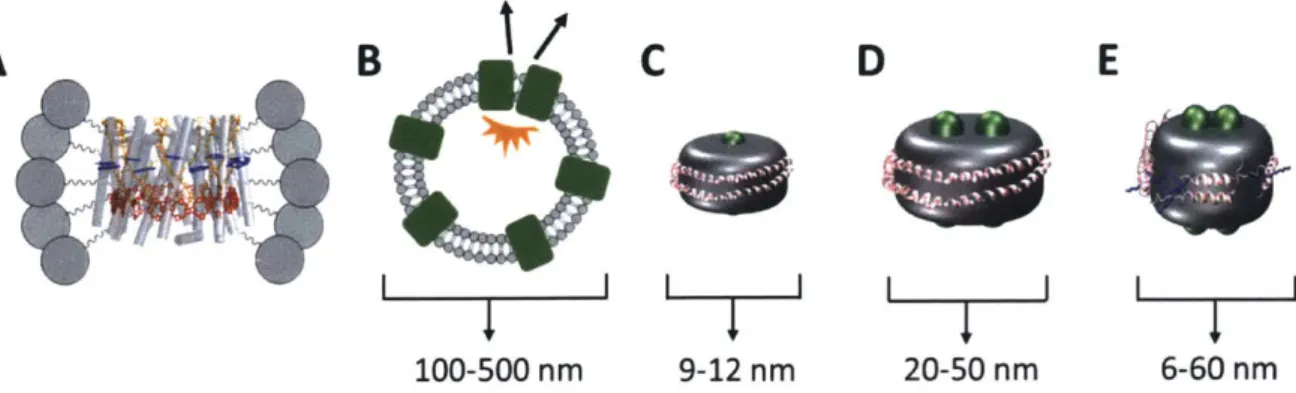
Figure 6: Membrane-model systems. Membrane proteins (green) are solubilized with various mem-brane mimicking tools. Memmem-brane discs are surrounded by a belting protein (pink). Lipids are shown in gray. (A) LH2 in a detergent micelle. (B) Membrane proteins in a liposome or chro-matophore. (C) Single membrane protein in a Nanodisc. (D) Two membrane proteins in ApoE422K or covalently circularized disc. (E) Telodendrimer Nano-Lipoparticles. Telodendrimers are shown in blue.
1
Chapter 1: Model Membrane-Discs
1.1
Membrane Protein Studies
Previous works have employed three predominant methodologies to solve the problems of photo-synthetic membrane protein production and purification as well as appropriate sample preparation. The first relies on detergent micelle formation surrounding the hydrophobic portion of the protein to produce a soluble protein-detergent complex (Figure 6A) [65,66]. Although effective in solubiliz-ing membrane proteins, the harsh conditions of detergents can produce drastic changes to protein structure, including loss or change of function as well as denaturation [67-69]. The second method reconstitutes membrane proteins into lipid vesicles in vitro (Figure 6B) [47,70-72]. Although this provides a hospitable membrane environment, these systems are heterogeneous in size, have var-ied and uncontrolled local membrane curvature, and are highly scattering, which can overwhelm spectroscopic signals. Furthermore, membrane vesicles often incorporate multiple proteins, which have been shown to introduce effects from protein-protein interactions that alter the dynamics in photosynthetic light-harvesting complexes [73]. The third method uses LH2-only chromatophores, membrane sections, or live cells [34, 68, 72, 74-78]. Similar to vesicles, this provides a hospitable membrane environment but also includes effects from protein-protein interactions. Furthermore, the membrane morphology and composition can be heterogeneous or even unknown [79]. Thus, the intrinsic energy transfer dynamics of light-harvesting complexes in the membrane environment, without the effects of protein-protein interactions, have not been determined before this work. These effects illustrate that benchmarking the dynamics of individual light-harvesting complexes in the membrane environment requires a near-native system with isolated proteins.
1.2
Nanodiscs
One emerging platform developed by the Sligar lab at the University of Illinois Urbana-Champaign, membrane-discs commonly known as Nanodiscs, overcomes the limitations of the sample prepara-tion methods commonly employed by the spectroscopic community (Figure 6C). Nanodiscs provide a simplified near-native environment to solubilize individual membrane proteins [80-84]. They are
similar in structure to nascent discoidal high-density lipoprotein particles in humans that transport lipids, cholesterol, and fats through the circulatory system [85]. Nanodiscs are biochemically pro-duced from subcomponents that are comprised of an amphiphilic belting protein, termed membrane scaffolding protein (MSP), a mixture of lipids used to form a bilayer, and the target membrane protein of interest [82,86,87]. The MSPs are derivatives of the human protein ApoAl which as-sembles the high-density lipoprotein particles in situ. The resultant Nanodiscs exhibit remarkably homogeneous diameters that can be straightforwardly characterized [86]. These Nanodiscs range in outer diameter from 9.8 to 17 nm (Table 1). The size of the Nanodisc can be tuned by the length of the MSP [81]. The type of lipids used to form the Nanodiscs can be selected for length and head group composition to best mimic the native environment. When mixed in precise stoichiometric ratios, these components self-assemble such that the MSP surrounds a lipid bilayer with the target membrane protein embedded (Figure 7) [87]. Key features in the self-assembly process have been identified previously [88]. The two main factors affecting size and shape of Nanodiscs are the choice of solubilizing detergent and the addition of detergent-absorbing resin beads, which removes the detergent from solution to allow the Nanodiscs to form without resolubilization. Because the size of Nanodiscs are still small relative to the wavelengths of UV through near-IR light, they produce very little scattering, making the membrane proteins embedded within this platform amenable to spectroscopic studies [89]. The number of proteins embedded can be controlled by the ratio of target protein to belting protein and the choice of MSP, which determines Nanodisc size. Finally, the lipid bilayer produced within the Nanodiscs forms a natively flat landscape versus an irregularly curved environment as in the membrane vesicles [84].
Although little is known about how well the Nanodisc mimics the native membrane, the flat landscape and bilayer nature suggests they mimic more sufficiently than vesicles or detergent. There are some disadvantages in using Nanodiscs. The first is that it is unknown if the belting proteins interact with the target protein inserted within the membrane-disc. While structural studies have not suggested this occurs in systems studied thus far, it is possible to occur in other systems. The second difficulty is incorporating multiple proteins in the correct orientation. This requires some additional chemical biology to ensure correct protein orientation and stoichiometry within the membrane-discs. Additionally, because the Nanodiscs are formed by a self-assembly process, it can prove difficult to assemble a homogeneous sample with the same stoiciometry of proteins embedded. For example, some discs may contain one protein while others contain two or even none. Lastly, if one would wish to study the reactivity of both hydrophillic ends of the protein which require different concentrations of reagents or different reagents all together, the Nanodiscs do not provide the ability for this type of investigation because both the hydrophillic portions are exposed to the exact same solution. These types of experiments can be done in vesicles, entrapping a different solution on the inside of the vesicle versus outside the vesicle.
Using a small Nanodisc size and substoichiometric ratios of target protein to belting protein allows a single protein to be incorporated into the membrane-discs. The control over membrane-disc composition allows the energy transfer dynamics of individual proteins to be explored without the complexity of protein-protein interactions [82,90,91]. The work in the Sligar lab was the beginning of discoidal model membrane-discs. Since their establishment, a wide variety of different types of membrane-discs have emerged.
1.3 Other Discoidal Model-Membrane Platforms ApoE422K
-4 hrs
purification
W
Figure 7: Assembly of model membrane-discs. Specific ratios of target protein (green), lipids (gray), and belting proteins (pink) are mixed together. Upon detergent removal, membrane-discs are spontaneously formed.
A similar protein to ApoAl named ApoE422K (N-terminal 22 kDa fragment of ApoE4) forms discs of varying size determined by the ratios of belting protein and lipid mixed together in a similar fashion as Nanodiscs (Figure 6D, Table 1) [92,93]. Although ApoE422K is shorter than the largest MSP belting protein MSP1E3D1 for Nanodics, the purified ApoE422K protein was shown to elute in a single peak in size-exclusion chromatography at an elution volume indicative of a hexameric form (data not shown). MSP1E3D1 elutes as monomers, dimers, and trimers in three peaks (data not shown). This suggests that the initial oligomerization of the belting protein, in addition to its length, contributes to the size of membrane-disc formed. Because ApoE422K can form larger membrane-discs and is assembled by the self assembly process the size distribution of final particles can be quite large. Additionally, other larger aggregates form more readily during the assembly process. This makes ApoE422K membrane-discs much more sensitive to assembly stoichiometries of belting protein, lipids, and target protein.
Model Membrane-Discs Size (nm) Nanodiscs -10-17 SMALPS ~5-15 Covalently Circularized ~9-50 ApoE422K ~15-35 TD-NLPs ~6-60
Table 1: Model membrane-discs toolkit and their various sizes. Telodendrimer Nanolipoparticles
In addition to using only belting proteins to form membrane-discs, other types of model membrane systems have been developed. The first type of model-membrane discs are known as telodendrimer nanolipoparticles (TD-NLPs) established by the Coleman lab at University of Cali-fornia Davis School of Medicine (Figure 6E). These membrane-discs utilize a telodendrimer (TD) polymer in addition to the belting protein that improves stability of the discs by allowing the
polymer to interweave with the belting protein, increasing the size of the membrane-discs [94]. The TD polymer consists of polyethylene glycol (PEG) with cholic acid (CA) moieties. Changing the composition of the telodendrimer allows for adjustment of disc size (6-60 nm). A49ApoA1 (1-49 fragment of Apo Al) and ApoE422K belting proteins produce 16-25 nm discs by including a telodendrimer polymer [94]. Due to the larger sizes, these membrane-discs are ideally suited for experiments measuring energy transfer between multiple proteins. However, the larger sizes have a greater size distribution of final particles. The main concern with such a large size distribution is the possibility of a heterogeneous sample with variable numbers of target protein embedded. The increased sample stability is very beneficial in experiments that take longer periods of time. The TD polymer does add the ability to ligate various ligands onto the polymer allowing for many types of additional experiments. Dyes can be added for FRET experiments. Specific tags can be added to attach the membrane-discs to a variety of different surfaces. Soluble proteins or additional ligands can be attached via a linker to investigate how two proteins function together or how a protein interacts with its ligand. However, caution must be used to determine whether the TD polymer and any additional substituents will interact with your target protein during the assembly process. It is possible that either of these could interfere with the assembly process or cause damage to your target protein.
Styrene Maleic Acid Lipid Particles
Another type of model-membrane disc uses a styrene maleic acid co-polymer (SMA) as a belting tool to form discoidal membrane particles [95-97]. The advantage to these particles is that the polymer can be used to directly remove membrane proteins from the native membrane without the use of detergent. This prohibits any interaction of the protein with detergent which can cause alterations to the protein structure and loss of function. Additionally, it provides the membrane-disc with the most native lipid composition. However, because the SMA essentially extracts a section of the native membrane, the composition of the membrane-discs can be completely unknown. While incorporating protein-tags onto the in vivo protein of interest can allow one to select the membrane-discs with their target protein embedded, it is still unknown how many of the target proteins are present and if any additional proteins are present. Because protein-protein interactions can affect biological processes this may make any results difficult to determine definitively. Finally, these are limited to only smaller sized membrane-discs (Table 1).
Covalently Circularized Membrane-Discs
Finally, the Wagner group at Harvard has created a covalently circularized membrane-disc [98]. Using a sortase reaction, newly made variants of ApoAl were covalently linked at the C and N termini. Varying lengths of ApoAl variants led to membrane-discs of sizes varying from 9-50 nm (Table 1). These were shown to have increased stability as well as a more homogeneous size distribution. However, the larger membrane-discs have much less stability overall in comparison to Nanodiscs. In practice, forming the larger particles is very sensitive to reagents, stoichiometries, and the incubation with the detergent-absorptive beads. Additionally, the covalently linked belting proteins do not produce only the size of membrane-disc expected. They produce smaller sized membrane-discs in the same reaction as well. It is unclear how the covalently linked belts made for a precise size are generating membrane-discs of a variety of sizes. Perhaps the covalently linked protein wraps around the disc twice, but this would yield membrane-discs much smaller than what is actually observed. More likely, the covalently circularized belt may assemble in a fashion that it does not actually encircle the target protein but forms the membrane-disc by interacting in a similar way to Nanodiscs, in a side on side fashion.
<I 25 0 10 20 30 40 50
B
Ur
Diameter (nm) 10 15' 10 0 10 20 30 40 50 Diameter (nm) 010 20 30 40 50 Diameter (nm)Figure 8: Optimal ratios of belting protein and lipids were determined to produce a platform of empty model-membrane discs. (A) 12 nm membrane-discs produced with MSP1E3D1. (B) 25 nm membrane-discs produced with NW50. (C) 30-45 nm Discs produced with ApoE422K.
1.4 Conclusion
Belting proteins MSP1E3D1, ApoE422K, and covalently circularized belting protein NW50 were used with DMPC and POPC to establish a platform of membrane-discs for various studies (Figure 8). MSP1E3D1 produces 12 nm membrane-discs as expected (Figure 8A). NW50 produced 25 nm membrane-discs (Figure 8B). ApoE422K was shown to produce 30-50 nm membrane-discs by adjusting the ratio of protein to lipids (Figure 8C). Figure 8 illustrates the variety of different sizes available for future studies. In this work, using ApoE422k and a Nanodisc belting protein MSP1E3D1, membrane-discs are utilized to achieve both a spectroscopically viable sample and a simplified near-native membrane. With the combination of model-membrane discs and spectroscopy the effect that specific lipid-protein and protein-protein interactions have on the energy transfer dynamics within photosynthetic light-harvesting proteins was investigated.
2
Chapter 2: Impact of the Lipid Bilayer on Energy Transfer
Kinetics in the Photosynthetic Protein LH2
2.1
Summary
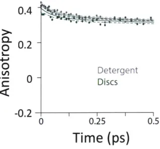
The energy transfer dynamics of LH2 are highly sensitive to intermolecular distances and relative organizations. As a result, minor structural perturbations can cause significant changes in these dynamics. Previous experiments have primarily been performed in two ways. One uses non-native samples where LH2 is solubilized in detergent, which can alter protein structure. The other uses complex membranes that contain multiple proteins within a large lipid area, which make it difficult to identify and distinguish perturbations caused by protein-protein interactions and lipid-protein interactions. Here, we introduce the use of the biochemical platform of model membrane-discs to study the energy transfer dynamics of photosynthetic light-harvesting complexes in a near-native environment. We incorporate a single LH2 (nonamer of heterodimers) from R. sphaeroides into membrane discs that provide a spectroscopically amenable sample in an environment more physiological than detergent but less complex than traditional membranes. This provides a sim-plified system to understand an individual protein and how the lipid-protein interaction affects energy transfer dynamics. We compare the energy transfer rates of detergent-solubilized LH2 with those of LH2 in membrane-discs using transient absorption spectroscopy and transient absorption anisotropy (Figure 9A). For one key energy transfer step in LH2, we observe a 30% enhancement of the rate for LH2 in membrane-discs compared to that in detergent. Based on experimental results and theoretical modeling, we attribute this difference to tilting of the peripheral bacteriochlorophyll in the B800 band. These results highlight the importance of well-defined systems with near-native membrane conditions for physiologically-relevant measurements. This work has been published in Chemical Science [46].
A
450fs
B
I
A-1~Discs
Detergent 80.6875/670
fs
M
e
0.4 ;0.2 40 fs 0s750 800 850 900 Wavelength (nm)Figure 9: Summary of results. (A) Time constants for energy transfer as measured by transient absorption spectroscopy on both the detergent solubilized LH2 and the membrane disc embedded LH2. Energy transfer within the B800 and B850 bands is similar for both samples (blue and red, respectively) but energy transfer between bands indicates structural differences induced by the membrane condition (detergent - gray; membrane discs - green). (B) UV-VIS linear absorption spectra for LH2 in LDAO detergent (gray) and solubilized in DMPC membrane discs (green) in the B800/B850 region. The data are normalized to the B800 peak on the wavelength scale and shows a peak shift of the B850 band from 849 nm to 852 nm for LH2 in detergent and discs, respectively. Insert: the full spectrum of LH2 in detergent and discs from 250 to 900 nm showing the nearly identical structure of the two LH2 samples, independent of solubilization condition.
2.2 Results and Discussion 2.2.1 LH2 in Membrane Discs
A single LH2 from R. sphaeroides (nonomer of heterodimers) was embedded into a membrane-disc as described in Chapter 1. The linear absorption spectra (Figure 9B) show that the LH2 complexes maintain integrity within the membrane-disc. The same peaks are observed in detergent-solubilized and disc-embedded LH2, demonstrating that the structure of the protein is robust to the disc assembly process. However, the linear absorption spectra of LH2 in detergent and in discs shows a consistent shift of the B850 absorption peak from 849 nm in detergent to 852 nm in membrane-discs, as previously reported [72, 99]. Pressure-dependent spectral shifts have also been previously reported [100]. The electronic structure of the pigments is highly sensitive to the protein environment so structural perturbations can alter the absorption spectra. The 3 nm shift highlights that the local environment of the B850 pigments changes due to solubilization environment. The complex and varied interactions of membrane proteins with detergents are both protein and detergent specific. As a result, it is difficult to identify the molecular origin of spectral shifts.
2.2.2 Time-Correlated Single Photon Counting Measurements
Time-correlated single-photon counting (TCSPC) was used to determine the fluorescence kinet-ics of detergent-solubilized and disc-embedded LH2 (Figure 10). All four samples exhibit mono-exponential decays with a time constant of ~1 ns, consistent with previous work (Table 2) [72].
Fluorescence decays of detergent-solubilized and membrane-embedded LH2 have previously been shown to have a mono-exponential decay and exponential decay, respectively, where the bi-exponential decay exhibits shorter time constants [72,79]. The shorter time constants are attributed to protein-protein energy transfer enabling exciton-exciton annihilation, which does not occur in the membrane-discs. The mono-exponential decays observed here, therefore, are consistent with previous work and illustrate the utility of membrane-discs as model systems for energy transfer kinetics of single LH2s in a near-physiological environment.
TCSPC IAlI
r
#-OG
1 1.018 ns LDAO 1 1.113 ns DMPC 1 1.119 ns POPC 1 0.974 nsTable 2: Parameters extracted from fits of the TCSPC data. Each fluorescence decay curve was fit mono-exponentially. The amplitudes and time constants are shown.
2.2.3 Transient Absorption Spectroscopy and Transient Absorption Anisotropy To probe the effect of solubilization environment on LH2, the energy transfer kinetics were measured in two detergents, LDAO and
#-OG,
and two lipid compositions of the membrane, DMPC and POPC. Detergents that have been shown to solubilize LH2 while maintaining the integrity of the complex are LDAO, DDM, and O-OG. LDAO and DDM are the most commonly used detergents, while -OG is more rarely used. However, the detergent tails of LDAO and DDM are the same length, while the tail of -OG is significantly shorter [74,102,103]. Specifically, the hydrocarbon chain of /-OG is six carbons shorter than that of LDAO.#-OG
also has a much bulkier headA
B
DMPC 100 POPC LDAO 1 c0.5
10
P-OG U CU 01020
5
10
0
1
2
Ui-Time (ns)
Figure 10: Time-correlated single photon counting (TCSPC) experiments and curve fitting were performed as described previously [101]. For the experiments presented here, the direct output of the Ti:sapphire oscillator (Vitara-S, Coherent, Inc.) was used as the excitation source (A=800 nm, 80 MHz). The excitation wavelength was selected with a 800 nm center-wavelength bandpass (FF01-7851/62-25, Semrock Inc.). The emission wavelength was selected with a 875 center-wavelength bandpass filter (FF01-834/LR 25-L2, Semrock Inc.). Excitation density for these experiments was 6.22 mW/cm2. Fluorescence lifetime data for all four samples in (A) full linear scale and (B) logarithmic scale to 2 ns. Data are shown in color and fits are shown as red lines. All samples were fit to a mono-exponential decay.
group than LDAO (Figure 11). Therefore, LDAO and 3-OG were selected to provide two distinct detergent tail lengths. The hydrocarbon chain of POPC is two carbons longer than that of DMPC, although both are similar lengths to the majority of native lipids in R. sphaeroides.
0 0 o o oDMPC OH 0 X °0 N- POPC LDAO HO HO, " OH O OH
1-OG
Figure 11: Lipid and detergent structures for the samples used in these experiments.
Figure 12A presents 800 nm pump - 850 nm probe transient absorption spectra recorded with the pulse polarization set to the magic angle (MA = 54.70) for all four samples. This pulse combination directly probes the time evolution of the population of states at 850 nm (in the B850 ring) after initial excitation of states at 800 nm (in the B800 ring). The spectra are fit to a sum of three exponentials, the results of which are shown in Table 3. The spectrum of LH2 in 3-OG collapses to a sum of two exponentials, indicating different energy transfer pathways than the other three samples. For the three component spectra, the fast decay component is assigned to B850* to B850 (as described in the introduction) and decays over the course of the first 0.5 ps, in line with previous experimental and theoretical work [62]. The second decay is assigned to the transfer
A
A
DMPC 0 POPC LDAO 0-OOOP
E-0.8
PO
-1.8
B
0
2 40.4
0.2
-0-0.2
0
0.5
1.0
1.5
Time (ps)
Figure 12: Transient absorption data of LH2 in different solubilization environments. (A) Magic angle 800 nm pump - 850 nm probe transient absorption spectra for LH2 in DMPC membrane discs (green), POPC membrane discs (teal), LDAO detergent (gray), and 3-OG detergent (black). (B) 800 nm pump - 800 nm probe anisotropy for the four LH2 solubilization conditions. Data is shown as points and three exponential fits are overlaid as lines with 95% confidence intervals indicated by the shaded region around each line.
of population between the B800 and B850
Q,
excited states (lowest lying energy states), directly reporting on inter-band energy transfer dynamics. According to previous work, transfer from B800 to B850 occurs directly and via B850* with approximately the same timescale [62]. A 30% increase in the timescale of this decay component was observed in membrane-discs, revealing a difference in B800 to B850 energy transfer between detergent-solubilized and membrane-reconstituted LH2 due to one or both of the energy transfer routes. Longer timescale processes, i.e. vibrational relaxation (Stokes' shift) and the decay back to the ground state, are collectively fit by the third time component of picoseconds.While the rate of energy transfer from B800 to B850 changes with local environment, the dynamics of energy transfer within both the B800 and B850 rings do not. Transient absorption anisotropy measurements decay due to the orientational change of the excitation as it transfers within the band. Thus, the experiment identifies and quantifies energy migration around the ring. The 800 nm anisotropy (Figure 12B) is initially fit to a sum of two exponential decays. The long time
r Weight r Weight 400 fs 24% 425 fs 49% 5 ps -76% >5 ns 51% 280 fs 19% 420 fs 58% LDAO 875 fs 36% >5 ns 42% >10 ps -45% DMPC 325 fs 8% 419 fs 58% Disc 670 fs 32% >5 ns 42% >10 ps -60% P 0 325 fs 16% 413 fs 43% .ic 670 fs 15% >5 ns 57% Discs 9 ps -69%
Table 3: Time constants and relative weights for magic angle 800 nm pump - 850 nm probe transient absorption (left column, data shown in Figure 12A) and 800 nm pump - 800 nm probe anisotropy (right column, data shown in Figure 12B) for the four solubilization conditions. Each spectrum was initially fit to a sum of three exponential decays. When two components collapsed to a single decay value, a two exponential fit was performed.
component (>5 ns) is then fixed as an offset to improve the fit for the short time decay to produce the timescales shown in Table 3. For all four LH2 solubilization conditions, these components are similar in time and relative weight. The faster decay component of -420 fs corresponds to energy transfer within the B800 ring. The 850 nm anisotropy is fit in the same manner as the 800 nm anisotropy with an additional decay component. The results are shown in Figure 13 and Table 4. The data show a rapid decay component corresponding to energy transfer within the B850 ring of ~55 or -65 fs for LH2 solubilized in LDAO and in DMPC discs, respectively. The energy transfer dynamics within the B850 ring are much faster because the pigments are much strongly coupled [29]. In addition to the <1 ps energy transfer dynamics discussed here, in all data sets there is a long (nanoseconds) component that arises from excited state decay and in the B850 anisotropy there is a picoseconds component that arises from vibrational relaxation.
850 Pump - 850 Probe T Weight 65 fs 24% Detergent 1700 fs 53% >5 ns 41% 55 fs 22% Discs 1340 fs 31% > 5 ns 47%
Table 4: Anisotropy decay rates and relative weights. Each anisotropy curve was fit to a sum of three exponential decays and the time constants and relative weights of each component are shown in the table.
The energy transfer rates are similar not only in DMPC and POPC membrane-discs, as observed here, but are also consistent with previously published experiments for LH2 in LH1-knockout R. sphaeroides [30]. Collectively, these results suggest that protein-protein interactions
A
0.4
0
0.2
Detergent
'E
0Discs
-0.02
0
0.25
0.5
Time (ps)
Figure 13: Anisotropy for LH2 in LDAO detergent (grey) and in DMPC discs (green) calculated from simultaneously measured parallel and perpendicular components (V-V and V-H, respectively) for 850 nm pump, 850 nm probe.
are not impacting the photophysics, in contrast to green plants [73]. Furthermore, the similarity in energy transfer rates illustrates that a membrane maintains a similar local environment for LH2 regardless of lipid composition. This similar local environment is likely due to the structured assembly of a membrane where the acyl chains predominantly interact with the protein in a side-by-side orientation, thus decreasing lipid-pigment interactions. The additional two carbon chain length of the POPC versus DMPC (Figure 11) does not have a significant effect on energy transfer between the B800 and B850 bands. This result excludes the possibility that the membrane height is the driving factor behind the increase in the rate of energy transfer in the membrane-disc samples. Interestingly, although DMPC discs are in a structured gel phase while POPC discs are in a liquid crystalline phase at the temperature of our experiments (4°C), there is no change to the energy transfer rates. This lack of change suggests that membrane phase has little effect on the energy transfer rates, and thus on the protein structure. Furthermore, these results highlight that energy transfer dynamics are robust to the dynamic lipid composition of the purple bacterial membrane.
To determine the molecular origin of the effect of solubilization environment, the energy trans-fer rates within the B800 band and from B800 to the B850 band were calculated using generalized F6rster theory as a function of tilt of the BChla in the B800 band (See Appendix 2," Calculation of Relative Energy Transfer Rates") [105]. Because the BChla in the B800 band protrude from the protein scaffold (Figure 2A) and interact with the lipid or detergent used for solubilization, they are much more susceptible to perturbation due to solubilization environment than the BChla in the B850 band. Figure 14 shows the calculated relative energy transfer rates within the B800 band and between the B800 and B850 band as a function of the tilt of the BChlas in the B800 band. These calculations determine relative rates, and so take into account all the experimental energy transfer steps, including those via the B850* states. The rate of energy transfer from B800 to B850 is much more sensitive (purple) than the rate within the B800 ring (blue). Flattening the B800 Bchlas by 2° causes an enhancement of the B800-B850 rate by more than 30%, while the B800 rate remains almost constant with less than 1% enhancement. The sharp dependence of the B800-B850 relative rate on the tilt angle arises from sensitivity to the distance between the coupled dipoles,
T1.4 -B800- B850 C q) 1.2 1.0 --- - - -B800-B800 0.8 0.6-I . . . I . . . . I -10 0 10 20 Tilt angle AG (0)
Figure 14: Theoretical energy transfer rates within LH2. The energy transfer rates within B800 (blue) and between B800 and B850 (purple) as a function of a tilt in the orientation of the B800 transition dipole moments relative to the original structure [104]. A positive tilt results in a steady increase of the B800 rate from the original geometry, up to a maximum obtained for a tilt of AO =
9° corresponding to a flat B800 ring. The shaded domains illustrate the range for which the B800 rate increases by 1% (blue) and the corresponding, drastic change in the inter-ring transfer rate (purple). Flattening the B800 BChls by 2° enhances the B800-B850 rate by more than 30%. The presented B800-B850 rate displays the average rate from one B800 BChl to the six nearest B850 BChls.
and hence on how the tilt is simulated. It can become smoother by including disorder [106]. These theoretical predictions are consistent with our experimental results, which find a change in B800 to B850 energy transfer but similar energy transfer rates within the B800 band in the anisotropy mea-surements at 800 nm as a function of solubilization (Table 3). This model would also predict similar energy transfer rates within the B850 band as a function of solubilization environment as observed in the short time component of the anisotropy measurements at 850 nm (Table 4). Essentially, the B800 BChla tilt towards the orientation of the B850 BChla, which increases the inter-band coupling, speeding up the overall B800 to B850 energy transfer step including contributions from both B800 to B850 and B800 to B850*. Because the B800 BChla tilt symmetrically together, their orientation relative to each other is largely unchanged, which leaves the intra-band coupling the same.
While the microscopic origin of the perturbation cannot be definitively determined, three pos-sibilities are hydrophobic mismatch, lateral membrane pressure, or direct interaction of solubilizing environment with pigments. We consider these three physical processes. Hydrophobic mismatch occurs when the height of the membrane or detergent used does not match the hydrophobic region of the protein. In vivo, the hydrophilic headgroups of the bilayer interact with the hydrophilic protein regions, which are the N- and C- terminal regions of the
#
subunit in the case of LH2. The hydrophobic acyl chain region of the bilayer associates with the hydrophobic protein regions, which are the center of transmembrane alpha helices. The in vivo membrane height surrounding LH2 in R. sphaeroides is measured to be between 40 and 45A[107].
To properly emulate the native environment, the solubilizing membrane or detergent should span a similar distance andhave the hydrophilic and hydrophobic regions properly matched to the corresponding regions of the membrane protein [74,108]. Hydrated DMPC and POPC (Figure 11) bilayers measure -44A in thickness, have a phosphatidylcholine headgroup, and a two acyl chain tail, which produce an environment that matches the native conditions [109]. LDAO is an intermediate detergent with a weakly polar headgroup and a tail with 12 carbons [110]. Although advantageous for solubilization, purification, and crystallization, LDAO produces micelles with a single acyl chain length of ~15-16
A
[111, 112]. This reduced lipid height means that LDAO is shorter than the hydrophobic region of native LH2, producing a hydrophobic mismatch of ~7A
as measured from the crystal struc-ture [104, 110]. Hydrophobic mismatch has been shown to alter membrane protein function, and thus is a likely candidate to change the tilt of the Bchla in the B800 band [113,114]. Hydrophobic mismatch would also explain the greater perturbation of#-OG
due to its even shorter hydrocarbon tail and bulkier head group.The lateral membrane pressure profile is a second possible cause of the changing tilt of the B800 band BChlas. A blue shift of the B850 band has been previously observed with increasing pressure [100]. Here, we observe the same shift in moving from membrane solubilized to detergent solubilized LH2, as shown in Figure 9B. In addition, only a very small shift is observed in the B800 band in either the membrane solubilized LH2 or the pressure-dependent absorption spectra. These similarities suggest that pressure may be the physical origin of the differences observed here. Fur-thermore, LH2 can induce curvature to the membrane that changes the lateral membrane pressure profile, as seen in previous work [115]. Although difficult to measure in situ, the lateral pressure profile for membrane bilayers has been extensively studied using computational methods, which have included the effects on integral membrane proteins [116-119]. In a lipid bilayer, such as the native environment or the membrane-discs used in this work, the lateral stress profile has a positive (inward toward the membrane protein) pressure at the furthest extent due to the electrostatic in-teractions of the headgroups, a negative pressure due to the interfacial tension at the polar-apolar interface, and a positive pressure in the acyl chain region due to the repulsion between the hydro-carbon chains [120. In a protein-detergent system, it has been suggested that single chain, small headgroup detergents increase the pressure in the headgroup region while decreasing the pressure in the acyl chain relative to the bilayer system [121]. The LDAO headgroup (Figure 11) is an amine oxide zwiterionic group which is small (2.8 nm2) compared to the phosphatidylcholine headgroup of the DMPC (5.7 nm2) and POPC (6.9 nm 2) [87]. The O-OG headgroup is a glucoside that is nonionic, polar, and similar in size (~5.1 nm2) compared to DMPC or POPC but is much bulkier due to its ring structure. In combination, LDAO's small headgroup, 3-OG's bulkier headgroup, and the single acyl chain of detergents produce a very different intra-membrane region and thus a very different lateral membrane pressure profile than the DMPC or POPC discs [122]. Finally, direct intermolecular interactions of single chain detergent molecules with pigments rather than the protein scaffold is a third possible cause of the changing tilt of the B800 band BChlas. Similar effects have previously been reported in other photosynthetic systems (LHCII from Pisum sativum, peas) and perturbations in protein-pigment interactions due to detergent have been shown to alter the excited state lifetimes of LH2 [123,124].
Our data reveals a change in energy transfer kinetics which suggests that varying the solubi-lization environment alters the structure of LH2 in such a way as to increase the B800-B850 energy transfer rate. The energy transfer rates within individual proteins also determine the pathways of energy transfer between proteins to reach the reaction center. Energy transfer from one LH2 to another via the B850 rings is thought to occur on a 1-5 ps timescale [16]. Currently, inter-protein energy transfer kinetics have been done in model systems that have multiple proteins incorporated and have multiple other processes occurring that may influence those kinetics. Because the B800
![Figure 10: Time-correlated single photon counting (TCSPC) experiments and curve fitting were performed as described previously [101]](https://thumb-eu.123doks.com/thumbv2/123doknet/14403630.510419/19.917.208.699.115.349/figure-correlated-counting-experiments-fitting-performed-described-previously.webp)