ORIGINAL ARTICLE
A novel animal model for external anal sphincter insufficiency
Lukas Brügger&Roman Inglin&Daniel Candinas&Tullio Sulser&Daniel Eberli
Accepted: 22 August 2014 / Published online: 4 September 2014 # Springer-Verlag Berlin Heidelberg 2014
Abstract
Purpose Reliable animal models are essential to evaluate future therapeutic options like cell-based therapies for external anal sphincter insufficiency. The goal of our study was to describe the most reliable model for external sphincter muscle insufficiency by comparing three different methods to create sphincter muscle damage.
Methods In an experimental animal study, female Lewis rats (200–250 g) were randomly assigned to three treatment groups (n=5, each group). The external sphincter muscle was weakened in the left dorsal quadrant by microsurgical excision, cryosurgery, or electrocoagulation by diathermy. Functional evaluation included in vivo measurements of rest-ing pressure, spontaneous muscle contraction, and contraction in response to electrical stimulation of the afferent nerve at baseline and at 2, 4, and 6 weeks after sphincter injury. Masson’s trichrome staining and immunofluorescence for skeletal muscle markers was performed for morphological analysis.
Results Peak contraction after electrical stimulation was sig-nificantly decreased after sphincter injury in all groups. Con-traction forces recovered partially after cryosurgery and electrocoagulation but not after microsurgical excision. Mor-phological analysis revealed an incomplete destruction of the external sphincter muscle in the cryosurgery and
electrocoagulation groups compared to the microsurgery group.
Conclusions For the first time, three different models of ex-ternal sphincter muscle insufficiency were directly compared. The animal model using microsurgical sphincter destruction offers the highest level of consistency regarding tissue damage and sphincter insufficiency, and therefore represents the most reliable model to evaluate future therapeutic options. In addi-tion, this study represents a novel model to specifically test the external sphincter muscle function.
Keywords Anal incontinence . Animal model . External sphincter . Microsurgery
Introduction
Fecal incontinence limits daily activities and strongly influ-ences the patient’s psychological, emotional, and social well-being [1,2]. The high prevalence [2,3,4] and the significant socioeconomic impact of the disease on one hand [5] and the limitations of current therapeutic options especially for large defects of the external sphincter on the other hand highlight the importance of ongoing research in the field [6–8].
Research concentrates on the external sphincter for several reasons: This part of the sphincter muscle is much more voluminous in humans in comparison to the internal sphincter muscle and is frequently affected after obstetric traumas [9, 10]. The external sphincter is relevant in preventing urge incontinence [11,12] and contributes to the maintenance of passive continence [13].
However, in previous studies, a variety of methods includ-ing sphincterotomy [14, 15], resection of the internal and external sphincter [16], electrocoagulation [17], or cryoinjury [18] to generate models for sphincter muscle insufficiency
This work was presented at the 99th Annual Congress of the Swiss Society of Surgery, Davos, Switzerland, 20–22 June 2012.
L. Brügger
:
R. Inglin:
T. Sulser:
D. EberliLaboratory for Urologic Tissue Engineering and Stem Cell Therapy, Department of Urology, University Hospital Zürich,
Frauenklinikstrasse 10, 8091 Zürich, Switzerland L. Brügger (*)
:
R. Inglin:
D. CandinasDepartment of Visceral Surgery and Medicine, Bern University Hospital, University of Bern, Freiburgstrasse 4, 3010 Bern, Switzerland
have been used. Therefore, data comparison across the litera-ture is difficult.
Besides creation of a distinct defect of the external sphinc-ter, this muscle has to be accessible to functional evaluation in order to test new therapeutic procedures. In vivo measurement of the resting pressure has been described previously [15,19]. However, since the resting pressure is generated by the inter-nal and exterinter-nal sphincter [13], this examination allows no individual functional evaluation of both muscles. A rat animal model that permits assessment of squeeze pressure to specif-ically evaluate the external sphincter muscle does not exist so far.
This study was designed to describe a rodent animal model with a consistent isolated defect of the external sphincter, which offers functional in vivo evaluation of this muscle. We therefore compared three different techniques of isolated and irreversible external sphincter injury including microsur-gical excision which has not been described before, cryosur-gery, and electrocoagulation.
Materials and methods Study design and protocol
The study was performed with the approval of the Animal Care Committee of the Canton of Zurich, Switzerland. Pre-liminary experiments were conducted to determine the opti-mal settings for the in vivo assessment of sphincter function. These experiments included evaluation of the most precise localization for electrical nerve stimulation to induce external sphincter muscle contraction.
For the final evaluation, a total of 15 adult female Lewis rats (200–250 g) were randomly placed into three groups (n= 5, each group) for functional analysis. Another six animals served for morphological investigations. Baseline manometric assessment included resting pressure, spontaneous contrac-tions, and contractions in response to electrical stimulation. As shown in Fig.1, the left dorsal quadrant of the external sphincter muscle was microsurgically removed (MS group) or destroyed by cryosurgery (CS group) or electrocoagulation using diathermy (EC group). Manometric evaluation was repeated immediately after surgery and at 2, 4, and 6 weeks after sphincter damage. Additionally, after 2 and 4 weeks, one animal and, after 6 weeks, four animals per group were sacrificed for tissue retrieval and histological analysis. Sam-ples from the untreated right dorsal quadrant served as internal controls.
Animal anesthesia and surgery
All animals received carprofen (Rimadyl®, Pfizer AG, Zürich, Switzerland; 5 mg/kg bodyweight) and buprenorphine
(Temgesic®, Reckitt Benckiser AG, Wallisellen, Switzerland; 0.01–0.05 mg/kg bodyweight) by subcutaneous injection 30 min prior to surgery. Anesthesia was induced by inhalation of 5 % isoflurane and maintained using 1–2 % isoflurane. The surgical area was shaved and disinfected with povidone-iodine solution. After a left circum-anal incision, the fat was separated from the lower rectum and anal canal under an operating microscope and the left dorsolateral quadrant of the sphincter muscle was exposed.
For partial sphincter damage, approximately 25 % of the circumferential external sphincter muscle fibers were removed or destroyed. Before microsurgical excision, the external sphincter muscle was separated from the underlying internal sphincter muscle. Cryosurgery of the anal sphincter was in-duced in anesthetized rats using an aluminum rod measuring 2×5 mm, which was chilled in liquid nitrogen as was previ-ously described [18]. The chilled probe was held toward the anal sphincter for 15 s. Sphincter injury by electrocoagulation was induced with the use of an electrical surgical bipolar forceps, similar to a technique used for urinary sphincter injury [17]. Electrocoagulation of the left dorsal external sphincter muscle was performed on an area of 5×2 mm for 1 s. Resorbable suture material was used for skin closure.
After surgery, observations were made every 15 min until the animals were fully awake and active. Thereafter, animals
Fig. 1 Model generation. A cross section of the anal canal in Masson’s trichrome staining. The left dorsal quadrant of the external sphincter muscle resected by microsurgery (between arrowheads). The internal sphincter muscle (arrow) was left intact
were returned to their normal housing. Carprofen (5 mg/kg bodyweight once daily) and buprenorphine (0.01–0.05 mg/kg bodyweight twice daily) were administered subcutaneously for 48 h. Animals were carefully monitored for signs of infection.
Anal manometric evaluation
All examinations were done under general anesthesia without any muscle relaxation in the supine position. Based on previ-ous studies [15, 19], we used a saline-filled latex balloon connected through PE100 tubing to a pressure transducer (SP844, Memscap, Crolles Cedex, France) and a digital data acquisition system (PowerLab 4/35 with LabChart Pro, ADInstruments, Spechbach, Germany) for anal manometric measurements. After the balloon was inflated to 10 cm H2O,
pressure, calibration of the system was accomplished. After adaption of 15 to 30 min, spontaneous contraction waves were recorded for 5 min. The peak contraction, area under the curve (AUC), and frequency of the contraction waves were recorded (Fig. 2a). To determine the resting pressure, the balloon probe was drawn out in a contraction interval and the resulting difference to the baseline pressure (mean of five intervals) was calculated. For the assessment of sphincter contraction after electrical stimulation of the inferior rectal
nerve, a bipolar electrode was inserted through a 2-mm incision at the ventral verge of the anus. We used a Grass S48 stimulator (Grass Technologies, Astro-Med, Rodgau, Germany) at 30 Hz (pulse duration 1 ms), 30 V for 2 s for the electrical stimulation. The best stimulation location was defined as the place with minimal leg twitching and maximal sphincter response. The peak contraction force and the max-imal slope of the contraction waves were registered (Fig.2b). An average of five contractions was used for statistical analysis.
Histology and immunohistochemistry
Animals were sacrificed under anesthesia by thoracotomy and incision of the heart. After a circum-anal incision, the anus and lower rectum were removed en bloc. The total specimen was fixed in formalin for 24 h and rinsed in phosphate-buffered saline. After dehydrating overnight in a histokinette (TP1020 tissue processor, Leica Microsystems, Wetzlar, Germany), the samples were embedded in paraffin.
The blocks were sectioned into 3-μm slices using a micro-tome (Model RM 2155, Leica Microsystems, Wetzlar, Ger-many). The sections were fixed and labeled with Masson’s trichrome stain. Sphincter muscle cells were identified via immunocytochemistry using an anti-α-actinin antibody
Fig. 2 a Representative example of spontaneous contractions of the sphincter muscles registered by balloon manometry in a normal animal. b Representative example of contractions of the sphincter muscles after electrical
stimulation of the branches of the inferior rectal nerve registered by balloon manometry
(Sigma-Aldrich, Buchs, Switzerland) at a dilution of 1:100. Native sphincter tissue sections served as positive controls. Tissue sections without primary antibody were used as nega-tive controls.α-Actinin-labeled cells were identified by fluo-rescence microscopy (Leica DM 6000 B/DFC 350 FX, Leica Microsystems, Wetzlar, Germany) using an excitation wave-length of 550 nm. Nuclei were stained with 4′,6-diamidino-2-phenylindole (DAPI; Sigma-Aldrich, Buchs, Switzerland). Image analysis to determine signal intensity per high power field was performed (ImageJ 1.45s, Wayne Rasband, National Institutes of Health, Bethesda, MD, USA).
Statistical analysis
All experimental groups contained five animals for functional analysis at each time point. The presented data are expressed as averages ± the corresponding standard error of the mean. The repeated measures analysis of variance (ANOVA) was performed to assess the group effect for quantitative variables. The Bonferroni pairwise multiple comparison test was used to compare measurements after the intervention with the baseline values within the groups (time effect). Measurements at the end of the experiment were compared with unpaired Student’s t tests. All tests were performed bilaterally with a 5 % signif-icance threshold. The NCSS/PASS software package (NCSS, Kaysville, UT, USA) was used for all calculations.
Results
No intraoperative or postoperative complications occurred in either group. In particular, no rectal perforation, anal stricture, or wound infection occurred.
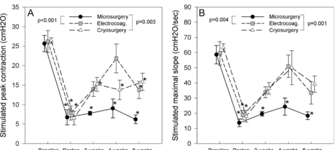
Manometric evaluation during electrical nerve stimulation Peak contractions were significantly reduced in all groups after model generation when compared to normal pressures (MS, 6.7±1.9 vs 25.7±2.1 cm H2O; EC, 7.9±1.8 vs 26.2±
2.8 cm H2O; CS, 6.6±1.8 vs 26.5±0.7 cm H2O). Peak
con-traction pressures were only reduced after cryosurgery and microsurgery over all time points investigated as is demon-strated in Fig. 3a. However, the values after microsurgery were significantly lower. The slope of the contraction waves recovered over time after sphincter damage by cryosurgery and electrocoagulation (Fig.3b). The values in the microsur-gery group were significantly lower for peak pressures (MS, 6.2 ± 1.1 cm H2O; EC, 13.8 ± 2.2 cm H2O; CS, 15.8 ±
2.2 cm H2O) and slope of the contraction waves (MS, 18.4±
2.5 cm H2O/s; EC, 33.5 ± 7.4 cm H2O/s; CS, 39.2 ±
5.4 cm H2O/s) compared to the other groups at the end of
the experiment (6 weeks after model generation).
Spontaneous manometric evaluation
Spontaneous, rhythmic contraction waves of the sphincter muscles demonstrated great variability between time points and animals (Fig.4). Peak contraction, area under the curve (impulse), frequency, and resting pressures did not differ from baseline values or between the groups. Resting pressures demonstrated a trend toward temporary recovery after 2 and 4 weeks, respectively (Fig.4d).
Histology and immunohistochemistry
The external sphincter muscle was consistently removed at all time points after microsurgery (Fig.5a–d). After cryosurgery and diathermy, skeletal musculature from the external sphinc-ter muscle was partially recognizable at all time points (Fig. 5f–h and j–l). No significant inflammatory reaction was registered at later time points.
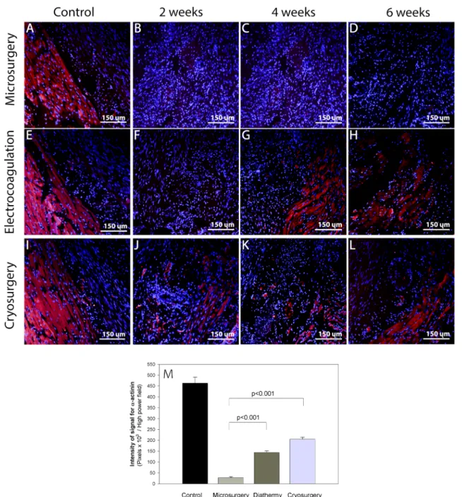
A sustained loss of muscle tissue in the MS group (28.0± 4.9 pixels×103/high power field 6 weeks after model genera-tion) compared to the control group (462.9±28.4 pixels×103/ high power field) was demonstrated in immunofluorescence analysis usingα-actinin as a specific musculoskeletal marker (Fig. 6d, m). Reduction of α-actinin expression was less pronounced in the EC and CS groups, showing values of 143.4±8.3 and 204.8±9.2 pixels×103/high power field, re-spectively (Fig.6h, l, m).
Discussion
In this study, three rat animal models for the evaluation of the external anal sphincter function have been explored. The results from this study objectively reveal functional and mor-p h o l o g i c a l r e g e n e r a t i o n a f t e r c r y o s u r g e r y a n d electrocoagulation after 6 weeks, which was not found after microsurgical excision.
The advantage of microsurgery seems to be that skeletal muscle tissue can be removed under direct visual control until no muscle fibers are left. On the other hand, after cryosurgery and electrocoagulation, necrotic tissue remains, and the depth of the injury cannot be precisely controlled. Thus, potentially vital skeletal muscle fibers remain underneath or in the ne-crotic area and repair mechanisms based on satellite cells might help to quickly regenerate muscular tissue [20]. In order to prevent such a mechanism, the interval and intensity of cryosurgery or electrocoagulation must be increased. Howev-er, individual anatomical differences and lacking possibilities to control the depth of penetration increase the risk of going beyond the target by destroying the adjacent internal sphincter muscle.
We did not observe any changes at the different time points or between models after either method of external sphincter destruction for frequency, peak force, and impulse (area under curve) of spontaneous contraction waves in the anal canal. These results support the current understanding that these spontaneous contraction waves are provoked by contractions of the internal sphincter [13], which was not intended to be damaged in our model. Involvement of the internal sphincter using cryosurgery and electrocoagulation is possible since the depth of destruction with these methods is more difficult to
control. Therefore, differences between models during spon-taneous contractions may be detected with a larger sample size. However, since our model was developed for external sphincter assessment, our experiments were not designed to find differences in spontaneous contractions, which are related to internal sphincter function.
After destruction of the external sphincter, a significant impairment of the resting pressure would be expected [13]. Conversely, only a non-significant reduction of sphincter pressure could be demonstrated in all groups. This might be
Fig. 4 Comparing spontaneous contractions measured by balloon manometry. Peak forces (a), area under the curve (b), frequency of spontaneous contractions (c), and resting pressures (d) were subject to great variability and did not differ from baseline or between treatment groups. A trend toward a temporary recovery of the resting pressure was shown in all groups (d)
Fig. 3 Differences between models (repeated measures ANOVA) for contraction (a) and maximal slope of the contraction wave (b) demonstrating the most reliable and long-term pressure reduction after microsurgery. Asterisks indicate a significant difference from baseline value. Values are given as means± standard error of the mean
due to a distinct anatomical difference between humans and rats, among which the external sphincter is less voluminous. With regard to the resting pressure, it seems that in rats, the internal sphincter can compensate the damaged external sphincter. Moreover, a transient trend toward recovery of the resting pressure with a peak after 2 and 4 weeks might be explained by postoperative edema.
Indeed, the ratio of thickness between the internal and external sphincter muscles in rats is not exactly comparable to humans. This fact does not signify a limitation of the present study since this model was specifically developed to study damage of the external sphincter muscle. This muscle can be selectively assessed for functional and morphological analysis, which underlines the relevance of this model. Selec-tive assessment of the external sphincter has been described in larger animals [21] such as rabbits or dogs. Compared to these experiments, our rat model seems to provide similar possibil-ities of external sphincter evaluation in the lowest possible species. One of the main restrictions of our model is the translation of the resting pressure measurements to humans. However, resting pressure is always a product of internal and, to a minor part, of external sphincter muscle function [13] and is therefore not suitable for external sphincter assessment.
It might further be argued that when it comes to evaluation of fecal incontinence, results from a horizontally walking animal could hardly be transferred to upright walking humans. Gravitation requires more sophisticated mechanisms in humans to maintain continence. However, since we do not quantify incontinence by itself, but concentrate on isolated measurement of the external sphincter muscle, this limitation can be avoided. We intended to describe a model for external sphincter muscle function and not for incontinence. This point should be respected when this model will be used for future studies.
A limitation of this study might further be represented by the very critical exact position of the probe for electrical stimulation of the afferent nerve for muscle contraction. In order to encounter this potential bias, the best stimulation location was defined as the place with minimal leg twitching and maximal sphincter response. An average of five contrac-tions was then used for statistical analysis.
Our study has a number of important strengths. First, a reliable animal model facilitates both evaluation of novel therapy options and physiological studies of underlying mech-anisms. By comparing different methods of sphincter destruc-tion, this study represents an important landmark to encounter
Fig. 5 Histomorphological assessment at three time points (a–l). Masson’s trichrome staining showing smooth muscle cells in purple-red, skeletal muscle cells in purple-red, and extracellular matrix in blue. The
predominance of skeletal muscle to extracellular matrix at all postopera-tive time points is higher after model generation by electrocoagulation (f– h) or cryosurgery (j–l)
efforts for standardization and rationalization of animal models and evaluation methods for studying the anal sphincter.
Second, with regard to testing of future therapy and options including cell-based therapies [14,18,22] or tissue engineer-ing [23] for external sphincter muscle regeneration, we chose to induce sustained long-term external sphincter muscle dam-age. Previous studies evaluating such therapies were based on animal models with destruction of both sphincter muscles [14, 18, 22]. Still, in such a model, the function of one muscle might be influenced by the destruction of the other muscle, which complicates interpretation of outcome measures. Our
animal model presented in this study consists of an isolated defect of the external sphincter and will therefore allow unbi-ased evaluation of this muscle.
Third, with this study, we describe a method to selectively assess external sphincter function of rats in vivo by electrical stimulation of the inferior rectal nerve and balloon manome-try. Functional evaluation of the external sphincter muscle was performed so far ex vivo using organ bath studies [14,18]. However, conclusions on in vivo function from a denervated piece of muscle, which is isolated from its surroundings, have consequently to be drawn with caution.
Fig. 6 a–l Cross sections of the anal canal at the level of the sphincter injury in the different groups are shown at different time points. Immu-nostaining (red) with the skeletal muscle marker (α-actinin) demonstrates persistence and/or recovery of skeletal muscle in the damaged area after
cryosurgery or electrocoagulation. Nuclear counterstaining was done with DAPI. (m) Intensity (n=4) of the signal for immunofluorescence ofα-actinin 6 weeks after model generation (pixels per high power field). Error bars indicate standard error of the mean
This new model facilitates evaluation of various current and future therapies meant for regeneration and reinforcement of the external sphincter muscle. Possible applications would be com-parison of different techniques for surgical repair (e.g., overlap vs direct adaption, early vs late surgery), evaluation of bulging agents [24], sacral nerve stimulation, injection of stem or pre-cursor cells [14,18,22], or implantation of an ex vivo-created piece of muscle using tissue engineering methods [23].
In conclusion, this study presents a rodent animal model offering a morphologically well-defined external sphincter muscle defect, which is mirrored in a corresponding loss of contraction force. For that purpose, a novel method to specif-ically assess external sphincter function in rats was described. The objective analysis of three distinct methods for creating a model of external sphincter muscle insufficiency provides evidence that sphincter destruction by microsurgery repre-sents the most reliable model in terms of persisting morpho-logical and functional deficiency compared to cryosurgery and electrocoagulation. Therefore, this animal model should be considered when evaluating novel therapeutic options for external sphincter muscle defects.
Acknowledgments The authors thank Meline Stoelting, Souzan Salemi, and Fatma Kivrak for the introduction in analyzing techniques and for technical assistance. This research project was financially sup-ported by grants from the EMDO foundation and the Insula Foundation, Switzerland.
References
1. Miner PB Jr (2004) Economic and personal impact of fecal and urinary incontinence. Gastroenterology 126(1 Suppl 1):S8–13 2. Perry S, Shaw C, McGrother C, Matthews RJ, Assassa RP, Dallosso
H, Williams K, Brittain KR, Azam U, Clarke M, Jagger C, Mayne C, Castleden CM (2002) Prevalence of faecal incontinence in adults aged 40 years or more living in the community. Gut 50(4):480–484 3. Nelson R, Norton N, Cautley E, Furner S (1995) Community-based
prevalence of anal incontinence. JAMA 274(7):559–561
4. Whitehead WE, Borrud L, Goode PS, Meikle S, Mueller ER, Tuteja A, Weidner A, Weinstein M, Ye W (2009) Fecal incontinence in US adults: epidemiology and risk factors. Gastroenterology 137(2):512– 517. doi:10.1053/j.gastro.2009.04.054
5. Hetzer FH, Bieler A, Hahnloser D, Lohlein F, Clavien PA, Demartines N (2006) Outcome and cost analysis of sacral nerve stimulation for faecal incontinence. Br J Surg 93(11):1411–1417. doi:10.1002/bjs.5491
6. Belyaev O, Muller C, Uhl W (2006) Neosphincter surgery for fecal incontinence: a critical and unbiased review of the relevant literature. Surg Today 36(4):295–303. doi:10.1007/s00595-005-3159-4
7. Chapman AE, Geerdes B, Hewett P, Young J, Eyers T, Kiroff G, Maddern GJ (2002) Systematic review of dynamic graciloplasty in the treatment of faecal incontinence. BrJ Surg 89(2):138–153
8. Mundy L, Merlin TL, Maddern GJ, Hiller JE (2004) Systematic review of safety and effectiveness of an artificial bowel sphincter for faecal incontinence. BrJ Surg 91(6):665–672
9. Dudding TC, Vaizey CJ, Kamm MA (2008) Obstetric anal sphincter injury: incidence, risk factors, and management. Ann Surg 247(2): 224–237. doi:10.1097/SLA.0b013e318142cdf4
10. Wheeler TL 2nd, Richter HE (2007) Delivery method, anal sphincter tears and fecal incontinence: new information on a persistent prob-lem. Curr Opin Obstet Gynecol 19(5):474–479. doi:10.1097/GCO. 0b013e3282ef4142
11. Lewicky-Gaupp C, Hamilton Q, Ashton-Miller J, Huebner M, DeLancey JO, Fenner DE (2009) Anal sphincter structure and func-tion relafunc-tionships in aging and fecal incontinence. Am J Obstet Gynecol 200(5):559–e551. doi:10.1016/j.ajog.2008.11.009
12. Rao SS (2004) Pathophysiology of adult fecal incontinence. Gastroenterology 126(1 Suppl 1):S14–22
13. Rasmussen OO (2003) Fecal incontinence. Studies on physiology, pathophysiology and surgical treatment. DanMed Bull 50(3):262– 282
14. Lorenzi B, Pessina F, Lorenzoni P, Urbani S, Vernillo R, Sgaragli G, Gerli R, Mazzanti B, Bosi A, Saccardi R, Lorenzi M (2008) Treatment of experimental injury of anal sphincters with primary surgical repair and injection of bone marrow-derived mesenchymal stem cells. Dis Colon Rectum 51(4):411–420
15. Zutshi M, Salcedo LB, Zaszczurynski PJ, Hull TL, Butler RS, Damaser MS (2009) Effects of sphincterotomy and pudendal nerve transection on the anal sphincter in a rat model. Dis Colon Rectum 52(7):1321–1329. doi:10.1007/DCR.0b013e31819f746d
16. Yamaguchi I, Fujita F, Yamanouchi K, Mishima T, Kawahara D, Sakai Y, Ito S, Kanetaka K, Takatsuki M, Kuroki T, Eguchi S (2013) A novel animal model of long-term sustainable anal sphincter dys-function. J Surg Res. doi:10.1016/j.jss.2013.04.010
17. Yiou R, Yoo JJ, Atala A (2003) Restoration of functional motor units in a rat model of sphincter injury by muscle precursor cell autografts. Transplantation 76(7):1053–1060
18. Kang SB, Lee HN, Lee JY, Park JS, Lee HS (2008) Sphincter contractility after muscle-derived stem cells autograft into the cryoinjured anal sphincters of rats. Dis Colon Rectum 51(9):1367– 1373. doi:10.1007/s10350-008-9360-y
19. Vinograd I, Hanani M, Hadary A, Merguerian P, Nissan S (1985) Animal model for the study of internal anal sphincter activity. Eur Surg Res 17(4):259–263
20. Yablonka-Reuveni Z (2011) The skeletal muscle satellite cell: still young and fascinating at 50. J Histochem Cytochem 59(12):1041– 1059. doi:10.1369/0022155411426780
21. Shafik A (1995) Inferior rectal nerve stimulation for anal sphincteric control: experimental study. J Surg Res 59(4):441–445. doi:10.1006/ jsre.1995.1188
22. Kajbafzadeh AM, Elmi A, Talab SS, Esfahani SA, Tourchi A (2010) Functional external anal sphincter reconstruction for treatment of anal incontinence using muscle progenitor cell auto grafting. Dis Colon Rectum 53(10):1415–1421. doi:10.1007/DCR.0b013e3181e53088
23. Raghavan S, Gilmont RR, Miyasaka EA, Somara S, Srinivasan S, Teitelbaum DH, Bitar KN (2011) Successful implantation of bioengineered, intrinsically innervated, human internal anal sphinc-ter. Gastroenterology 141(1):310–319. doi:10.1053/j.gastro.2011.03. 056
24. Maeda Y, Laurberg S, Norton C (2010) Perianal injectable bulking agents as treatment for faecal incontinence in adults. Cochrane Database Syst Rev 5, CD007959. doi:10.1002/14651858. CD007959.pub2