9 Springer-Verlag 1999 Printed in Austria
Transmembrane segment proteases
B. Martoglio*Institut f~ir Biochemie II, Eidgen6ssische Technische Hochschule Ziirich, Zurich Received March 1, 1999
Accepted March 23, 1999
Summary. Transmembrane segment proteases comprise a novel class of proteases that cleave substrates within hydrophobic membrane-spanning segments. They cleave in parts of proteins that upon first glance should be protected by the hydrophobic environ- ment of the lipid bilayer. At present, no such protease has been iso- lated and biochemically characterized. They are defined according to the appearance of the respective cleavage products. All trans- membrane segment proteases seem to participate in a regulated two-step proteolytic process that plays a central role in cellular reg- ulation or is part of a protein degradation pathway.
Keywords: Endoplasmic reticulum; Signal peptide peptidase; 7-Secretase; SREBR
Abbreviations: 13-APP 13-amyloid precursor protein; S1P site-1 protease; S2P site-2 protease; SPase signal peptidase; SPPase signal peptide peptidase; SREBP sterol regulatory element-binding protein; SCAP SREBP cleavage-activating protein.
degradation in addition to the proteasome (Jensen et al. 1995, Moriyama et al. 1998). Particular interest in transmembrane segment proteases arose from findings showing that some of these proteases generate prod- ucts that regulate cell functions.
This review will emphasize the common principles of the new type of proteases and focus on the three transmembrane segment proteases that seem to be localized in the ER of mammalian cells. These pro- teases are (i) site-2 protease that activates sterol reg- ulatory element-binding proteins, (ii) 7-secretase that is involved in the generation of amyloidogenic peptide AI3, and (iii) signal peptide peptidase that releases signal peptide fragments.
Introduction
Proteases are found in almost every organelle of eukaryotic cells. They play constructive roles by proteolytic processing of precursor proteins or act in degradative processes. Wherever proteases hydrolyse peptide bonds, cleavage occurs in an aqueous com- partment because the hydrophobic environment of the lipid bilayer precludes hydrolysis. Nevertheless, it has become clear that various organelles contain a new class of proteases that cleave substrate proteins within membrane-spanning segments. Such transmembrane segment proteases can be involved in the degradation of membrane proteins like the m A A A protease in the mitochondrial inner membrane (Leonhard et al. 1996) and may contribute to endoplasmic-reticulum (ER)
* Correspondence and reprints: Institut fiir Biochemie II, ETH- Zentrum, Universit~itstrasse 16, CH-8092 Ztirich Switzerland.
Common features
All transmembrane segment proteases known at present act in a characteristic two-step proteolytic process. In a first step, the substrate protein is cleaved within its hydrophilic ectodomain. The residual membrane-bound fragment is next cleaved within the transmembrane segment and the resulting products are released into the cytosol or the exoplasmic space (Lyko et al. 1995, Bunnell et al. 1998, Duncan et al. 1998). The two-step process usually involves auxiliary factors that may activate the proteolytic cascade, reg- ulate the proteases, or deliver the substrate.
According to the cleavage sites, transmembrane segment proteases can be devided in two groups (Table 1). Group 1 transmembrane segment proteases cleave close to the cytoplasmic end of membrane- spanning segments, in the region of the hydrophilic phospholipid head groups. Group 2 proteases cleave
Table 1. Classes and substrates of transmembrane segment proteases a
Protease Sequence of membrane-spanning segment and cleavage sites Substrate protein Intracellular localization Group 1 (cleavage close to the cytosolic surface of the membrane)
S2P . . . DRSRILLCVLTFLCLSFNPLTSLLQWGGAH... nn b . . . Q L H L M Y V A A A A F V L L F F V G C G V L L S R K R . . . Group 2 (cleavage in the centre of the transmembrane region)
y-Secretase c . . . S N K G A I I G L M V G G W l A T V I V I T L V M L K K K . . . SPPase a . . . K G S R L L L L L V V S N L L L C Q G . . . m A A A protease e . . . HDNIMYYLVVILFVGWILLSIIR_N_Y... SREBP-2 mNotch-1 13-APP signal sequence (bovine preprolactin) COXII-DHFR endoplasmic reticulum (?) plasma membrane (?) endoplasmic reticulum (?), endosomes (?) endoplasmic reticulum
mitochondrial inner membrane " In boldface, cleavage sites in the hydrophobic transmembrane segment; solid underline, exoplasmic side; broken underline, cytosolic side b Notch proteins are ligand-activated transmembrane receptors in the plasma membrane. Cleavage by an unidentified protease in the trans- membrane segment enables the released fragment to enter the nucleus (Schroeter et al. 1998)
c 7-Secretase(s) can cleave the transmembrane segment of J3-APP at alternative sites, predominantly at the indicated positions, generating A[340 (at I) or A~42 (at T)
d SPPase cleaves the preprolactin signal peptide within the region of the small residues in the centre of the transmembrane segment. The exact cleavage site is not known (Lyko et al. 1995)
e m A A A protease is part of a proteolytic system for the ATP-dependent degradation of inner membrane proteins in mitochondria Leonhard et al. 1996). COX II-DHFR, hybrid protein of cytochrome oxidase subunit II (1-74) and dihydrofolate reductase
an the centre of transmembrane segments. Actually, it is not known so far whether cleavage of a transmem- brane segment occurs within the lipid bilayer. It is con- ceivable that the active site of group 1 proteases is placed within a groove that faces the cytosolic side of the membrane and is accessible to water for hydroly- sis. Cleavage by group 2 proteases may require a more complex mechanism. Transfer of the substrate protein to either side of the membrane prior to cleavage seems unlikely since fragments can be released to both sides of the membrane. These proteases may be part of a multisubunit membrane protein complex similar to mitochondrial m A A A protease and provide water through the inside of the complex to the active site.
Site-2 protease: activation o f a transcription factor The best characterized transmembrane segment pro- tease at present is site-2 protease (S2P) that cleaves sterol regulatory element-binding proteins (SREBPs) within one of their two membrane-spanning segments (Rawson et al. 1997). SREBPs are membrane-bound transcription factors that regulate the expression of proteins involved in synthesis and uptake of choles- terol and fatty acids in mammalian cells (Brown and Goldstein 1997). They share a common structure comprising three domains: (i) a cytosolic N-terminal domain containing a basic helix-loop-helix leucin
zipper element and an acidic transcriptional activation sequence, (ii) a central segment with two membrane- spanning helices separated by a hydrophilic loop that is exposed toward the E R lumen, and (iii) a C- terminal regulatory domain that is exposed into the cytosol. SREBPs are anchored in the E R membrane and nuclear envelope and are released from the mem- brane in a two-step proteolytic process.
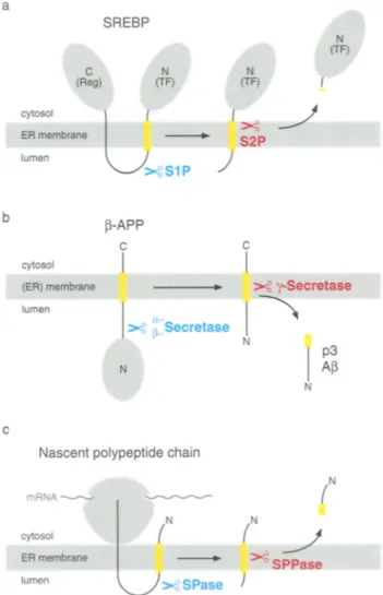
Processing of SREBPs is initiated by a protease, termed S1R when sterols are depleted in a cell (Sakai et al. 1996). S1P cleaves SREBPs in the lumenal loop and generates two fragments, each anchored with a single membrane-spanning segment (Fig. 1 a) (Sakai et al. 1998). Next, a second protease, S2R cleaves the N-terminal intermediate at a leucine-cysteine bond that is located within the membrane-spanning segment (Duncan et al. 1998). This liberates the transcription factor domain that is released into the cytosol, enters the nucleus, and activates gene transcription.
Cleavage by S1P involves the regulatory protein SCAP (for SREBP cleavage-activating protein). This putative sterol sensor is a polytopic membrane protein that is localized in the E R membrane (Nohturfft et al. 1998). It consists of two domains: (i) an N-terminal domain with several membrane-spanning segments containing a putative sterol-sensing subdomain and (ii) a C-terminal cytosolic domain containing several WD repeats that bind to the regulatory domain of
SIP and S2P have both been identified recently by complementation cloning. According to the deduced amino acid sequence, S1P is a C-terminally anchored type I membrane protein. Its large exoplasmic domain contains a catalytic triade that is typical for the subtil- isin family of serine proteases (Sakai et al. 1996). Sen- sitivity toward endoglycosidase H suggests that SIP is N-glycosylated and localized in the ER. The analysis of the amino acid sequence of S2P revealed a H E X X H consensus sequence that is found in many families of metalloproteases (Rawson et al. 1997). Unlike any other known metalloprotease, S2P shows extensive hydrophobicity. It has up to five putative transmem- brane segments, one of which containing the metallo- protease consensus sequence. The transmembrane segments may assemble to a substrate-binding groove that places the catalytic site into the membrane.
Fig. la-c. Transmembrane segment cleavage is part of a two-step process, a Processing of SREBR S1P cleaves SREBP in the lumenal loop between the regulatory domain (Reg) and the transcription factor domain (TF). S2P next cleaves the N-terminal membrane- anchored intermediate within the membrane-spanning segment and liberates the transcription factor domain, b Proteolytic processing of 13-APE a- or [~-secretase cleaves [~-APP in its exoplasmic domain. The membrane-anchored fragment is next cleaved by 7-secretase within the transmembrane segment and peptide p3 or A[3 is released from the membrane, c Cleavage and processing of signal sequences. After membrane insertion, the signal sequence of a nascent polypeptide chain is cleaved off by SPase and further processed by SPPase. The N-terminal signal peptide fragment is released into the cytosol. Hydrophobic membrane-spanning segments are indicated in yellow
SREBPs. In sterol-depleted cells, SCAP and SREBP form a tight complex that recruits S1P and initiates the proteolytic processing of the bound S R E B P (Sakai et al. 1997). The function of SCAP may be to deliver the substrate to S1P or vice versa and thereby to prevent the protease from unspecifically degrading other proteins in the E R lumen.
T-Secretase: generation of amyloidogenic peptides
[3-Amyloid precursor proteins (~-APPs) were the first proteins discovered that are processed within a transmembrane segment (Selkoe 1998). 13-APPs are ubiquitously expressed type I membrane proteins with unknown function. During trafficking along the secre- tory pathway, they undergo endoproteolytic cleavages that yield amyloidogenic peptide A[3 and other prod- ucts that are secreted. A~ is the major constituent of amyloid plaques in the brain of patients with Alzheimer's disease (Iwatsubo et al. 1994).
Several proteolytic steps in ~3-APP processing have been described (Shoji et al. 1992). First, unidentified c~- and 13-secretase(s) cleave ~3-APP in its exoplasmic domain generating the C-terminally anchored frag- ments p3CT and A4CT, respectively (Fig. 1 b). Both are then cleaved by unknown y-secretase(s) within their transmembrane segment yielding nonamyloido- genic peptide p3 and amyloidogenic Af3. Cleavage of A4CT occurs after residue 40, generating A~40, occa- sionally after residue 42, generating A~42. Increased levels of A~342 might be sufficient to cause Alzheimer's disease.
Since c~, [3-, and y-secretases are not identified, their putative subcellular localization is derived from the appearance of the respective cleavage products. A portion of A~40 seems to be generated in early endo- somes following internalization of [3-APP from the plasma membrane (Koo and Squazzo 1994). A~40 and A1342 can also be produced early in the secretory pathway. In neurons, for example, A~42 is generated in
the ER whereas A~40 is made in the trans-Golgi network (Hartmann et al. 1997, Lee et al. 1998). These observations suggest that pairs of secretases are present in several organelles. Secretases may be part of the protein degradation machineries in endosomes and the ER. Misfolded [3-APE for example, may fail the quality control in the ER and become degraded (Bunnell et al. 1998, Yang et al. 1998). After cleavages in the lumenal domain and the transmembrane segment, a portion of A[3 peptides may escape further degradation and may be secreted.
Generation of A[342 in the E R is influenced by polytopic membrane proteins termed presenilins (De Strooper et al. 1997). Mutations in the genes encoding presenilin 1 and 2 account for the majority of cases of Alzheimer's disease (Haass 1996). Presenilins interact with a minor portion of [3-APP molecules, preferen- tially with the immature N-glycosylated [3-APP but not with the (N+O)-glycosylated protein (Weidemann etal. 1997). They may aid in maturation and/or trafficking of ~-APR Mutations in presenilin which cause Alzheimer's disease may induce conformational changes that disturb proper interaction with [3-APP and favour fragmentation to A[342. Production of A[342 in the ER may thus be the result of a failed quality control, raising the hypothesis that ER-resident ~- and y-secretases may perform initial steps in the degrada- tion of aberrant membrane proteins.
Signal peptide peptidase: release of functional peptides
Signal peptide peptidase (SPPase) has been defined as a proteolytic activity in the E R membrane according to the observation that signal peptides derived from secretory and membrane proteins are cleaved within the transmembrane segment (Lyko et al. 1995). Find- ings that signal peptide fragments can perform regulatory and other functions in a cell raised the hypothesis that SPPase may play a central role in cellular regulation similar to S2P (Martoglio and Dobberstein 1998).
Signal sequences are essential for targeting secre- tory proteins and many membrane proteins to the ER membrane and mediate the entry into the secretory pathway (Walter and Johnson 1994). For translocation across the ER membrane, nascent polypeptides enter the protein-conducting channels in a looplike confor- mation with the N terminus of the signal sequence remaining in the cytosol and the C-terminal portion
being translocated (Rapoport et al. 1996). During translocation, signal sequences are usually cleaved off from the precursor protein by signal peptidase (SPase) (Fig. 1 c). Cleavage occurs close to the lumenal side of the ER membrane (Dalbey et al. 1997). Liberated signal peptides enter the lipid bilayer and are next cleaved within the hydrophobic membrane-spanning segment by SPPase (Lyko et al. 1995, Martoglio et al. 1997). The resulting N- and C-terminal fragments are released into the cytosol and ER lumen, respectively. Cleavage by SPPase is sensitive to the immuno- suppressive drug cyclosporin A (Klappa et al. 1996). Cyclosporin A usually binds to cellular proteins termed cyclophilins that have proline isomerase activ- ity and are thought to assist protein folding and to modulate the activity of various enzymes (Schreiber and Crabtree 1992). It is therefore conceivable that a cyclophilin in the ER regulates the activity of SPPase. While SPase is biochemically characterized, SPPase is not identified. The mammalian SPase has been purified as a complex of five subunits with molecular masses of 12, 18, 21, 22/23, and 25 kDa (Evans and Blobel 1986). The 18 and 21 kDa subunits are homo- logues of the Escherichia coli leader peptidase that performs signal sequence cleavage as a single protein. The crystal structure of the latter protein has recently been resolved (Paetzel et al. 1998). The membrane- anchored serine protease contains an exoplasmic domain with the active site close to the membrane. It is thus likely that also the 18 and 21 kDa subunits of the mammalian SPase cleave the signal sequence from the precursor protein. The function of the other SPase subunits is not clear. Further studies will show whether one of them catalyses cleavage of the liberated signal peptide within the transmembrane segment or whether other proteins perform this step.
What could be the role of an SPPase? One function could be to catalyse the first step of signal peptide degradation. It has been shown that synthetic signal peptides enter lipid bilayers and can lyse biological membranes (Hoyt and Girasch 1991, Killian et al. 1990). A mechanism is therefore required to efficiently remove signal peptides from the ER membrane because otherwise their accumulation would severely affect the permeation barrier. Cleavage of liberated signal peptides within the transmembrane segment would facilitate their release from the membrane. A motif for cleavage by SPPase may rely in the helix- break-helix structure that is present in the hydro-
phobic t r a n s m e m b r a n e segment of signal sequences but usually not in m e m b r a n e - s p a n n i n g segments of m e m b r a n e proteins (van K l o m p e n b u r g and de Kruijff 1998).
Recent studies revealed that peptides derived from signal sequences have a role in the regulation of cellular functions including virus-host interactions (Martoglio and Dobberstein 1998). In a cell-free system using E R - d e r i v e d microsomes it has been shown that N-terminal signal peptide fragments of the h o r m o n e preprolactin and the HIV-1 envelope pro- tein gpl60 are released into the cytosol where they efficiently bind to calmodulin in a calcium-dependent m a n n e r (Martoglio et al. 1997). Both signal sequences have properties in their N-terminal extensions that favour binding to calmodulin. Studies in living B cells have revealed a specific association of signal peptide fragments derived from H L A - A , -B, and -C M H C class I molecules with H L A - E M H C class I molecules (Braud et al. 1998). The H L A - E molecules present the signal peptide fragment at the cell surface to inhibitory receptors of natural killer cells. It is thought that the killer cells thereby gauge indirectly via the presented signal sequence fragments the overall level of H L A class I molecules expressed on B cells which is an indicator of the health status of the cell (Long 1998). These findings suggest that the presumably regulated production of signal peptide fragment by SPPase contributes to cellular regulation.
Concluding remarks
Proteases can generate hormonelike signals via a n u m b e r of mechanisms. Cleavage of t r a n s m e m b r a n e segments is a unique way in which proteases can liberate active peptides and modulate cell functions. The two-step proteolytic process that includes a trans- m e m b r a n e segment protease seems to be regulated as it is the case for almost all biological systems requir- ing a series of proteolytic steps. M a n y questions remain open. What are the sequences and physical properties of t r a n s m e m b r a n e segment proteases? D o several types of these proteases exist in the E R and elsewhere in the cell? What is the mechanism by which the proteases cleave t r a n s m e m b r a n e segments? Knowledge about t r a n s m e m b r a n e segment proteases and the cellular processes they are involved m a y provide novel targets for therapeutics in treating Alzheimer's disease, cholesterol-induced c o r o n a r y heart disease, and virus infections.
Acknowledgements
This work was supported by grants from ETH Zarich and the Swiss National Science Foundation.
References
Braud VM, Allan DSJ, O'Callaghan CA, S6derstr6m K, D'Andrea A, Ogg GS, Lazetic S; Young NT, Bell JI, Phillips JH, Lanier LL, McMichael AJ (1998) HLA-E binds to natural killer cell recep- tors CD94/NKG2A, B and C. Nature 391:795-799
Brown MS, Goldstein JL (i997) The SREBP pathway: regulation of cholesterol metabolism by proteolysis of a membrane-bound transcription factor. Cell 89:331-340
Bunnell WL, Pham HV, Glabe CG (1998) ?-Secretase cleavage is dis- tinct from endoplasmie reticulum degradation of the transmem- brane domain of the amyloid precursor protein. J Biol Chem 273: 31947-31955
Dalbey RE, Lively MO, Bron S, yon Heijne G (1997) The chemistry and enzymology of the type I signal peptidases. Protein Sci 6: 1129-1138
De Strooper B, Beullens M, Contrares B, Levensque L, Craessaerts K, Cordell B, Moechars D, Bollen M, Fraser P, George-Hyslop PS, Van Leuven F (1997) Phosphorylation, subcellular localization and membrane orientation of the Alzheimer's disease-associated presenilins. J Biol Chem 272:3590-3598
Duncan EA, Day6 UR Sakai J, Goldstein JL, Brown MS (1998) Second-site cleavage in sterol regulatory element-binding protein occurs at transmembrane junction determined by cystein panning. J Biol Chem 273:17801-17809
Evans EA, Gilmore R, Blobel G (1986) Purification of microsomal signal peptidase as a complex. Proc Natl Acad Sci USA 83: 581-585
Haass C (1996) Presenile because of presenilin: the presenilin genes and early onset Alzheimer's disease. Curr Opin Neurol 9: 254- 259
Hartmann T, Bieger SC, Bruhl B, Tienari PJ, Ida N, Allsop D, Roberts GW, Masters CL, Dotti CG, Unsicker K, Beyreuther K (1997) Distinct sites of intracellular production for Alzheimer's disease A beta40/42 amyloid peptides. Nat Med 3:1016-1020
Hoyt DW, Girasch LM (1991) Hydrophobic content and lipid inter- action of wild-type and mutant OmpA signal peptides correlate with their in vivo function. Biochemistry 30:10155-10163 Iwatsubo T, Odaka A, Suzuki N, Mizusawa H, Nukina N, Ihara Y
(1994) Visualization of A beta 42(43) and A beta 40 in senile plaques with end-specific A beta monoclonals: evidence that an initially deposited species is A beta 42(43). Neuron 13: 45-53
Jensen T J, Loo MA, Pind S, Williams DB, Goldberg AL, Riordan JR (1995) Multiple proteolytic systems, including the proteasome, contribute to CFTR processing. Cell 83:129-135
Killian JA, de Jong AM, Biivelt J, Verkleii AJ, de Kruijff B (1990) Induction of non-bilayer lipid structures by functional signal peptides. EMBO J 9:815-819
Klappa R Dmrks T, Zimmermann R (1996) Cyclosporin A inhibits the degradation of signal sequences after processing of presecre- tory proteins by signal peptidase. Eur J Biochem 239:509-518 Koo EH, Squazzo SL (1994) Evidence that production and release
of amyloid beta-protein involves the endocytic pathway. J Biol Cbem 269:17386-17389
Lee S J, Liyanage U, Bickel PE, Xia W, Lansburg PT Jr, Kosik KS (1998) A detergent-insoluble membrane compartment contains A beta in vivo. Nat Med 4:730-734
Leonhard K, Herrmann JM, Stuart RA, Mannhaupt G, Neupert W, Langer T (1996) A A A proteases with catalytic sites on opposite membrane surfaces comprise a proteolytic system for the ATP- dependent degradation of inner membrane proteins in mito- chondria. EMBO J 15:4218-4229
Long EO (1998) Signal sequences stop killer cells. Nature 391: 740-743
Lyko F, Martoglio B, Jungnickel B, Rapoport TA, Dobberstein B (1995) Signal sequence processing in rough microsomes. J Biol Chem 270:19873-19878
Martoglio B, Dobberstein B (1998) Signal sequences: more than just greasy peptides. Trends Cell Biol 8:410-415
- Graf R, Dobberstein B (1997) Signal sequence fragments of pre- prolactin and HIV-1 p-gp160 interact with ealmodulin. EMBO J 22:6636-6645
Moriyama T, Sather SK, McGee TE Simoni RD (1998) Degradation of HMG-CoA reductase in vitro: cleavage in the membrane domain by a membrane-bound cystein protease. J Biol Chem 273:22037-22043
Nohturfft A, Brown MS, Goldstein JL (1998) Topology of SREBP cleavage-activating protein, a polytopic membrane protein with a sterol-sensing domain. J Biol Chem 273:17243-17250
Paetzel M, Dalbey RE, Strynadka NCJ (1998) Cristal structure of a bacterial signal peptidase in complex with a [Mactam inhibitor. Nature 396:186-190
Rapoport T, Jungnickel B, Kutay U (1996) Protein translocation across the eukaryotic endoplasmic reticulum and bacterial inner membranes. Annu Rev Biochem 65:271-303
Rawson RB, Zelenski NG, Nijhawan D, Ye J, Sakai J, Hasan MT, Chang TY, Brown MS, Goldstein JL (1997) Complementation cloning of S2P, a gene encoding a putative metalloprotease required for intramembrane cleavage of SREBPs. Mol Cell 1: 47-57
Sakai J, Duncan EA, Rawson RB, Hua X, Brown MS, Goldstein JL (1996) Sterol-regulated release of SREBP-2 from cell membranes
requires two sequential cleavages, one within a transmembrane segment. Cell 85:1037-1046
- Nohturfft A, Cheng A, Ho YK, Brown MS, Goldstein JL (1997) Identification of complexes between the COOH-terminal domains of sterol regulatory element-binding proteins (SREBPs) and SREBP cleavage-activating protein. J Biol Chem 272: 20213-20221
- Rawson RB, Espendshade PS, Cheng D, Seegmiller AC, Goldstein JL, Brown MS (1998) Molecular identification of the sterol- regulated luminal protease that cleaves SREBPs and controls lipid composition of animal cells. Mol Cell 2:505-514
Schreiber SL, Crabtree GR (1992) The mechanism of action of cyclosporin A and FK506. Immunol Today i3:136-142
Schroeter EH, Kisslinger JA, Kopan R (1998) Notch-I signalling requires ligand-induced proteolytic release of intracellular domain. Nature 393:382-386
Selkoe DJ (1998) The cell biology of b-amyloid precursor protein and presenilin in Alzheimer's disease. Trends Cell Biol 8: 447-453 Shoji M, Golde TE, Ghiso J, Chung TT, Estus S, Shaffler LM, Cai XD, McKay DM,Tintner R, Frangione B, Younkin SG (1992) Pro- duction of the Alzheimer amyloid beta protein by normal pro- teolytic processing. Science 258:126-129
van Klompenburg W, de Kruijff B (1998) The role of anionic lipids in protein insertion and translocation in bacterial membranes. J Membr Biol 162:1-7
Walter R Johnson A E (1994) Signal sequence recognition and protein targeting to the endoplasmic reticulum membrane. Annu Rev Cell Biol 10:87-119
Weidemann A, Paliga K, Durrwang U, Czech C, Evin G, Masters CL, Beyreuther K (1997) Formation of stable complexes between two Alzheimer's disease gene products: presenilin-2 and beta-amyloid precursor protein. Nat Med 3:328-332
Yang Y, Turner RS, Gaut JR (1998) The chaperone BiP/GRP78 binds to amyloid precursor protein and decreases A~40 and A[~42 secretion. J Biol Chem 273:25552-25555