HAL Id: hal-02988664
https://hal.archives-ouvertes.fr/hal-02988664
Submitted on 10 Nov 2020
HAL is a multi-disciplinary open access
archive for the deposit and dissemination of
sci-entific research documents, whether they are
pub-lished or not. The documents may come from
teaching and research institutions in France or
abroad, or from public or private research centers.
L’archive ouverte pluridisciplinaire HAL, est
destinée au dépôt et à la diffusion de documents
scientifiques de niveau recherche, publiés ou non,
émanant des établissements d’enseignement et de
recherche français ou étrangers, des laboratoires
publics ou privés.
N-alpha-acetyltransferase that is crucial for development
and regulation of stress responses
Laura Armbruster, Eric Linster, Jean-Baptiste Boyer, Annika Brünje, Jürgen
Eirich, Iwona Stephan, Willy Bienvenut, Jonas Weidenhausen, Thierry
Meinnel, Ruediger Hell, et al.
To cite this version:
Laura Armbruster, Eric Linster, Jean-Baptiste Boyer, Annika Brünje, Jürgen Eirich, et al.. NAA50
is an enzymatically active N-alpha-acetyltransferase that is crucial for development and regulation of
stress responses. Plant Physiology, American Society of Plant Biologists, 2020, 183 (4), pp.1502-1516.
�10.1104/pp.20.00222�. �hal-02988664�
NAA50 Is an Enzymatically Active
N
-Acetyltransferase
That Is Crucial for Development and Regulation of
Stress Responses
1[OPEN]
Laura Armbruster,
aEric Linster,
aJean-Baptiste Boyer ,
bAnnika Brünje,
cJürgen Eirich,
cIwona Stephan,
aWilly V. Bienvenut,
bJonas Weidenhausen,
dThierry Meinnel,
bRuediger Hell,
aIrmgard Sinning,
dIris Finkemeier,
cCarmela Giglione,
band Markus Wirtz
a,2,3a
Centre for Organismal Studies, Heidelberg University, 69120 Heidelberg, Germany
b
Université Paris-Saclay, Commissariat à l’Energie Atomique, Centre National de la Recherche Scientifique,
Institute for Integrative Biology of the Cell, 91198 Gif-sur-Yvette, France
c
Institute for Plant Biology and Biotechnology, University of Münster, Muenster 48149, Germany
dHeidelberg University Biochemistry Center, 69120 Heidelberg, Germany
ORCID IDs: 0000-0002-4559-2404 (L.A.); 0000-0001-7963-1400 (E.L.); 0000-0001-5265-3917 (J.-B.B.); 0000-0002-8979-4606 (A.B.); 0000-0003-0963-1872 (J.E.); 0000-0003-4192-3920 (W.V.B.); 0000-0001-8376-2083 (J.W.); 0000-0001-5642-8637 (T.M.); 0000-0002-6238-4818 (R.H.); 0000-0001-9127-4477 (I.Si.); 0000-0002-8972-4026 (I.F.); 0000-0002-7475-1558 (C.G.); 0000-0001-7790-4022 (M.W.)
N
a-terminal acetylation (NTA) is a prevalent protein modification in eukaryotes. In plants, the biological function of NTA
remains enigmatic. The dominant N-acetyltransferase (Nat) in Arabidopsis (Arabidopsis thaliana) is NatA, which cotranslationally
catalyzes acetylation of
;40% of the proteome. The core NatA complex consists of the catalytic subunit NAA10 and the
ribosome-anchoring subunit NAA15. In human (Homo sapiens), fruit
fly (Drosophila melanogaster), and yeast (Saccharomyces
cerevisiae), this core NatA complex interacts with NAA50 to form the NatE complex. While in metazoa, NAA50 has
N-acetyltransferase activity, yeast NAA50 is catalytically inactive and positions NatA at the ribosome tunnel exit. Here, we
report the identification and characterization of Arabidopsis NAA50 (AT5G11340). Consistent with its putative function as a
cotranslationally acting Nat, AtNAA50-EYFP localized to the cytosol and the endoplasmic reticulum but also to the nuclei. We
demonstrate that purified AtNAA50 displays N
a-terminal acetyltransferase and lysine-«-autoacetyltransferase activity in vitro.
Global N-acetylome profiling of Escherichia coli cells expressing AtNAA50 revealed conservation of NatE substrate specificity
between plants and humans. Unlike the embryo-lethal phenotype caused by the absence of AtNAA10 and AtNAA15, loss of
NAA50 expression resulted in severe growth retardation and infertility in two Arabidopsis transfer DNA insertion lines
(naa50-1 and naa50-2). The phenotype of naa50-2 was rescued by the expression of HsNAA50 or AtNAA50. In contrast, the inactive
ScNAA50 failed to complement naa50-2. Remarkably, loss of NAA50 expression did not affect NTA of known NatA substrates
and caused the accumulation of proteins involved in stress responses. Overall, our results emphasize a relevant role of AtNAA50
in plant defense and development, which is independent of the essential NatA activity.
N
a-terminal acetylation (NTA) is a ubiquitous
proteome-imprinting mechanism in eukaryotes. In
hu-mans and plants, 80% to 90% of cytosolic proteins are
subject to NTA, which is catalyzed by N
a-acetyltransfer-ases (Nats) that transfer an acetyl moiety from acetyl-CoA
to the protein N terminus (Arnesen et al., 2009; Bienvenut
et al., 2012; Drazic et al., 2016). Although NTA has been
shown to affect the subcellular localization, aggregation,
folding, and degradation of individual proteins, the
overall significance of NTA is still unclear (Aksnes et al.,
2016). In humans, seven Nat complexes (NatA to NatF
and NatH) have been described. Each Nat complex is
composed of at least one catalytic and facultative auxiliary
subunits (Aksnes et al., 2015a, 2019). Whereas NatA to
NatE (excluding NatD) are anchored to the ribosome via
their auxiliary subunits and cotranslationally acetylate
nascent polypeptide chains, NatF and NatH are
mono-meric and operate posttranslationally. Although
puta-tive orthologs for the catalytic subunits of NatA to NatF
have been identified in plants by protein homology
searches (Bienvenut et al., 2012; Breiman et al., 2016;
Rathore et al., 2016), only NatA, NatB, and NatF have
been biochemically characterized in plants so far
(Ferrández-Ayela et al., 2013; Linster et al., 2015, 2020;
Xu et al., 2015; Huber et al., 2020). Together, NatA and
NatB target approximately three-fourths of extractable
soluble proteins in Arabidopsis (Arabidopsis thaliana;
Linster et al., 2015; Huber et al., 2020). The existence of a
plant NatC has also been evidenced by functional
com-plementation of a yeast NatC-deficient mutant, but the
substrate specificity and subcellular localization of the plant
NatC remain unexplored (Pesaresi et al., 2003). However,
the ribosome association and substrate specificity of the
core NatA and NatB are conserved between humans (Homo
sapiens), yeast (Saccharomyces cerevisiae), and plants. The
core NatA consists of the catalytic subunit NAA10 and
the auxiliary subunit NAA15 and acetylates N termini
starting with A, S, T, C, V, or G (Mullen et al., 1989;
Polevoda et al., 1999; Arnesen et al., 2009; Van Damme
et al., 2011; Linster et al., 2015; Xu et al., 2015). The
catalytic subunit NAA20 and the auxiliary subunit
NAA25 form the NatB complex, which targets N
ter-mini retaining their initiator Met (iMet) followed by D,
E, N, or Q (Polevoda et al., 1999; Van Damme et al.,
2012; Huber et al., 2020). Although the biochemical
features of the core NatA and NatB are evolutionarily
conserved among eukaryotes, species-specific
adapta-tions occurred in the remaining Nat machinery.
Hu-mans, for instance, possess a posttranslationally acting
cytosolic NatH, which is not found in plants (Drazic
et al., 2018). In contrast to metazoa, Arabidopsis
pos-sesses plant-specific organellar Nats that acetylates the
N termini of plastid-encoded or imported proteins
(Dinh et al., 2015; Bienvenut et al., 2020).
In humans and fruit
fly (Drosophila melanogaster), the core
NatA interacts with two additional proteins, HYPK and
NAA50 (for review, see Aksnes et al., 2019). In both species,
NAA50 binds to NatA to form the ternary NatE complex.
NatE predominantly acetylates N termini starting with MS,
MT, MA, MV, ML, MI, MF, MY, and MK (Evjenth et al.,
2009; Van Damme et al., 2011; Gottlieb and Marmorstein,
2018; Aksnes et al., 2019). A putative ortholog of NAA50
has also been described in yeast and was shown to interact
permanently with NatA in the ternary NatE complex
as-sociated with the ribosome (Gautschi et al., 2003; Knorr
et al., 2019). Depletion of NAA50 impaired sister
chroma-tid cohesion in human and fruit
fly cells, resulting in mitotic
arrest and segregation defects (Hou et al., 2007). This
phenotype is absent in the yeast loss-of-NAA50 mutant,
suggesting functional differences between NAA50 in yeast
and metazoa. Different from human and fruit
fly NAA50
homologs, yeast NAA50 lacks an optimal
acetyl-CoA-binding motif and has recently been shown to be
catalyti-cally inactive (Deng et al., 2019). In agreement with this
view, the knockout of NAA50 in yeast resulted in no
de-crease of NTA on canonical HsNAA50 substrates but
caused a decrease in NTA of six NatA-type substrates.
These
findings suggest a regulatory impact of ScNAA50 on
NatA activity in yeast (Van Damme et al., 2015). Structural
analysis by cryoelectron microscopy demonstrates that
ScNAA15 and ScNAA50 physically interact with rRNA
expansion segments to position NAA10 at the ribosome
tunnel exit. This exact positioning of NAA10 is supposed to
facilitate the acetylation of the emerging polypeptide chain
(Knorr et al., 2019).
Here, we identify and characterize Arabidopsis
NAA50 (AT5G11340). We demonstrate that AtNAA50
is a catalytically active N-acetyltransferase of the NatE
type with a substrate specificity similar to that of
HsNAA50. In addition to its acetylation activity toward
N
atermini, AtNAA50 can autoacetylate the
«-amino
group of internal Lys residues (Kat activity) in vitro,
as shown previously for the HsNAA50. The
predomi-nant localization of AtNAA50 in the nucleocytoplasma
and at the endoplasmic reticulum (ER) is consistent
with both its proposed function as a cotranslationally
acting Nat and additional functions as a Kat. Loss of
NAA50 in two Arabidopsis transfer DNA (T-DNA)
insertion mutants (naa50-1 and naa50-2) severely
im-pairs vegetative plant growth and fertility but allows
healthy embryo development. This phenotype is
dif-ferent from that of mutants lacking AtNAA10 and
AtNAA15 function, whose development stops at the
globular stage (Linster et al., 2015). The severe
dwarf-ism of naa50-2 can be complemented by the ectopic
expression of catalytically active AtNAA50 or HsNAA50.
In contrast, the catalytically inactive ScNAA50 failed to
complement the growth phenotype of naa50-2. Finally,
in naa50-2, the canonical NatA substrates were fully
N-acetylated, but the absence of the acetylase induces
the accumulation of many proteins involved in stress
responses. In conclusion, our work suggests that
AtNAA50 has more roles than merely being an
auxil-iary subunit of the NatA complex.
RESULTS
AtNAA50 Acts as a Nat with Broad Substrate Specificity
BLAST searches with NAA50 from human or yeast
identify AT5G11340 as a putative ortholog of NAA50
1
This work was supported by the Deutsche Forschungsgemein-schaft via the Collaborative Research Centre (grant no. SFP1036, TP13 to R.H. and M.W., TP 22 to I.Si., FI 1655/3–1 and INST 211/ 744–1 to I.F., and WI 3560/4–1 to M.W.) and by the Leibniz Pro-gramme (to I.Si). C.G. was supported by the Agence Nationale de la Recherche (grant no. ANR–10–LABX–0040–SPS) and by the facili-ties and expertise of the Institute of Integrative Biology of the Cell Service for the Identification and Characterization of Proteins by Mass Spectrometry proteomic platform, supported by Infrastructures in Biology Health and Agronomy, Ile de France Region, Plan Cancer, French National Centre for Scientific Research, and Paris-Sud Univer-sity. R.H. and I.Si. are investigators of the Cluster of Excellence:Cell-Networks. This project was carried out within the European Research Area Network for Coordinating Action in Plant Sciences Research Programme KatNat.
2
Author for contact: markus.wirtz@cos.uni-heidelberg.de.
3
Senior author.
The author responsible for distribution of materials integral to the findings presented in this article in accordance with the policy de-scribed in the Instructions for Authors (www.plantphysiol.org) is: Markus Wirtz (markus.wirtz@cos.uni-heidelberg.de).
I.St. identified and characterized the naa50 mutants; L.A. per-formed the NAA50 subcellular localization studies and the in vitro acetylation assay; E.L. determined the Kat activity of NAA50 and the transcript levels of NAA50, NAA10, and NAA15 in naa50 mutants and wild-type plants; J.W. and I.Si. performed structure-function analyses of NAA50; C.G. and T.M. designed and supervised N-terminomics experiments; W.V.B. performed the GAP assay and profiled N ter-mini of naa50 mutants and wild-type plants; C.G., T.M., and J.-B.B. analyzed the N-terminomics data; I.St. and E.L. complemented the naa50 mutants with endogenous, human, and yeast NAA50; A.B. and J.E. performed the quantitative proteome analysis of naa50 mutants and wild-type plants; I.F. designed the proteome analysis experiment and supervised A.B. and J.E.; M.W. and R.H. conceived and directed the study; L.A. wrote the article with input from all authors.
[OPEN]Articles can be viewed without a subscription.
www.plantphysiol.org/cgi/doi/10.1104/pp.20.00222
Figure 1. In vitro Nat and N«-Lys acetyltransferase (Kat) activity of AtNAA50. A, Trx-His
6-AtNAA50 was purified from E. coli by
immobilized metal affinity chromatography. B, Purified Trx-His6-AtNAA50 was incubated for 1 h at 37°C with 45 mM[3
H]acetyl-CoA and 0.2 mMof the synthetic MLGPand SESS peptides. After incubation, the peptide was enriched via specific interaction with
SP Sepharose, and the amount of [3H]acetyl incorporated in the peptide was quantified by scintillation counting. As a negative
control, AtNAA50 was heat inactivated at 95°C for 10 min. Data are presented as means 6SD(n 5 4, P , 0.05 by Student’s t test).
The experiment was repeated independently. C, Web logo of 28 N termini found to be specifically acetylated at their iMet after the expression of AtNAA50 in E. coli. D, Alignment of AtNAA50 and HsNAA50 protein sequences with Clustal Omega (https://www. ebi.ac.uk/Tools/msa/clustalo/). Lys residues that are acetylated in humans are shaded in blue; Lys residues that are acetylated in Arabidopsis are shaded in red. Lys residues that are predicted to be acetylated by Phosida (www.phosida.com) are marked in red. The conserved catalytic residues are shaded in green. E, Immunodetection of acetylated Lys residues on AtNAA50. Eleven mi-cromolar purified AtNAA50 was incubated with 5 mMacetyl-CoA for 0, 20, 40, or 60 min at 37°C. Subsequently, the reaction was
stopped by the addition of SDS loading buffer. The auto-Kat activity was determined via immunological detection of acetylated Lys residues with ana-acetylated Lys antibody. Heat-inactivated AtNAA50 (15 min at 95°C) served as a negative control (60 min). As loading control, AtNAA50 was visualized using Amido Black (n 5 3). The experiment was repeated independently. F, Quantification of signals shown in E for the AtNAA50 autoacetylation. The Amido Black loading control was used for normal-ization. Data are presented as means6SE. Different lowercase letters indicate individual groups identified by pairwise multiple comparisons with a Holm-Sidak one-way ANOVA (P , 0.05, n 5 3).
in the Arabidopsis genome (Bienvenut et al., 2012;
Neubauer and Innes, 2020). However, the enzymatic
activity and substrate specificity of this putative plant
NatE have not been characterized yet. Since HsNAA50
acetylates the NAA50 model substrate MLGP (Evjenth
et al., 2009), we expressed Trx-His
6-AtNAA50 in
Esch-erichia coli and subjected the purified enzyme (Fig. 1A)
to an in vitro Nat activity assay with a custom-made
MLGP peptide (Fig. 1B). Trx-His
6-AtNAA50 acetylated
the MLGP peptide (specific activity $ 1.38 nmol min
21mg
21) but did not display Nat activity toward a canonical
NatA substrate (SESS; Arnesen et al., 2009). This
finding
demonstrates that plant AtNAA50 is catalytically active
toward substrates of the NatE type. To further
elu-cidate the substrate specificity of plant NAA50, we
heterologously expressed AtNAA50 in E. coli and
performed a global acetylome profiling (GAP) assay.
The analysis revealed 28 N termini to be specifically
acetylated after the expression of AtNAA50 in E. coli
(Supplemental Table S1). The vast majority of those N
termini (25) were acetylated on their iMet. AtNAA50
was able to acetylate a wide range of N termini starting
with the iMet, indicating a rather broad substrate
spec-ificity, with a weak preference for the polar amino acids
Lys (K), Asn (N), Gln (Q), and Thr (T) at the second
position (Fig. 1C).
HsNAA50 also displays Kat activity toward three
internal Lys residues, K34, K37, and K140, in vitro
(Evjenth et al., 2009). Similarly, three Lys acetylation
sites (K37, K50, and K128) were predicted with high
probability (greater than 0.9) in the plant NAA50
using Phosida (www.phosida.com; Fig. 1D). In recent
Lys acetylome analyses of Arabidopsis, two Lys
acetylation sites (K37 and K155) were experimentally
determined (Hartl et al., 2017). To test for the
autoa-cetylation of AtNAA50, the enzyme was incubated
with acetyl-CoA for up to 60 min. Subsequently,
acetylated Lys residues were visualized by
immu-nological detection with an established commercially
available
a-acetylated Lys specific antibody (Fig. 1E).
Acetylation of internal Lys residues increased
line-arly over the detection period and was absent when
AtNAA50 was heat inactivated before incubation
with acetyl-CoA (Fig. 1F).
NAA50 Localizes to the Nucleus, the Cytosol, and the ER
The subcellular localization of enzymes determines
their access to substrates and potential interaction
part-ners. Hence, after demonstrating the dual Kat/Nat
ac-tivity of AtNAA50 in vitro, the subcellular localization of
AtNAA50 was assessed in planta. For this purpose, an
AtNAA50-enhanced yellow
fluorescent protein (EYFP)
fusion protein was transiently expressed in Nicotiana
benthamiana epidermal cells that were either
counter-stained with the DNA-specific marker
49,6-diamino-phenylindole (DAPI; Fig. 2A) or cotransfected with the
ER marker protein VMA12-RFP (Fig. 2B). In vivo
live-cell imaging of the transfected leaves revealed that
the AtNAA50-EYFP fusion protein displays a cytosolic
pattern and colocalizes with both markers. To exclude
that the nuclear localization of AtNAA50-EYFP was
caused by overexpression of the fusion protein under the
control of the 35S promoter, we expressed an
AtNAA15-EYFP fusion protein in the same vector system. The
resulting AtNAA15-EYFP fusion protein was not
transported into the nucleus (Supplemental Fig. S1).
These
findings strongly suggest that AtNAA50 is
lo-calized in the nucleus, in the cytoplasm, and at the
outer ER membrane. The cytoplasmic and ER-associated
localization is in agreement with its putative
func-tion as a ribosome-associated Nat by interacfunc-tion with
AtNAA15.
NAA50 Is Critical for Plant Growth
In order to evaluate the biological function of NAA50 in
plants, two homozygous T-DNA insertion lines
(naa50-1 [SAIL_(naa50-12(naa50-10_A02] and naa50-2 [SAIL_(naa50-1(naa50-186_A03]) were
identified (Supplemental Fig. S2). Sequencing of the
genomic DNA from both lines revealed that a T-DNA
was inserted in intron 1 of NAA50 in naa50-2 (position
Figure 2. AtNAA50 is localized in the cytosol, the nucleus, and the ER. AtNAA50-EYFP was tran-siently expressed in N. benthamiana epidermal cells. A, Counterstaining with DAPI shows that AtNAA50-EYFP localizes to the nucleus (marked with arrows). B, AtNAA50-EYFP colocalizes with the RFP-tagged ER marker VMA12. The experi-ment was repeated independently. Bars5 50 mm.
1,159 bp after the start codon), while a T-DNA was
in-tegrated in the NAA50 intron 2 at position 1,468 in
the case of naa50-1 (Fig. 3A). Reverse transcription
quantitative PCR (RT-qPCR) for NAA50 indicated the
total absence of NAA50 transcripts in both lines
(Supplemental Fig. S3). Next, we characterized the impact
of the T-DNA insertion in the naa50-2 mutant on the
transcription of NAA50 and the core NatA subunits by
RT-qPCR. RT-qPCR confirmed the absence of NAA50
transcripts in naa50-1 and naa50-2. However, the
tran-scription of NAA10 and NAA15 remained unaffected
(Fig. 3B), and also the protein levels of both NatA subunits
were not significantly changed in naa50-2 (see proteomic
analysis, Supplemental Table S2). The latter
findings were
surprising since the down-regulation of one core NatA
subunit by artificial microRNA interference caused
cotranscriptional down-regulation of the other subunit,
resulting in a decreased amount of both NatA core
subu-nits (Linster et al., 2015). For that reason, we quantified the
amount of NAA50 protein in the wild type and naa50-2
with a specific antiserum. The NAA50 protein was readily
detectable in the leaves of wild-type plants but was absent
in naa50 mutants (Fig. 3C). These
findings demonstrate that
naa50-1 and naa50-2 are real loss-of-NAA50 expression
mutants, in which the transcriptional regulation and
pro-tein abundance of the core NatA subunits is unaffected.
Loss of the core NatA arrests embryo development
at the globular stage (Linster et al., 2015). Both naa50
lines
finish embryo development and grow,
appar-ently like the wild type, into the early seedling stage.
However, after the formation of the
first leaves,
naa50-1 and naa50-2 show identical and severe
re-tardation of growth. The vast majority of plants
ter-minated growth after the development of a few small
leaves. When grown in artificially high humidity
under a transparent cover, very few mutants were
able to
flower, with a substantial delay in flowering
time. None of these plants produced viable seeds
(Fig. 3D). For that reason, homozygous plants from a
segregating heterozygous naa50-2 population were
used for further experiments. Remarkably, the
segre-gation pattern followed the expected 1:2:1 ratio,
dem-onstrating that homozygous naa50-2 pollen grows like
the wild-type pollen, when they derive from a
hetero-zygous naa50-2 plant.
NAA50 Function Is Evolutionarily Conserved between
Plants and Humans
Next, we wanted to provide direct evidence for the
hypothesis that the severe growth retardation phenotype
is caused by the absence of NAA50 activity. For that
reason, the heterozygous naa50-2 mutant was
trans-formed with a construct expressing AtNAA50 under
the control of the constitutive cauliflower mosaic virus
(CaMV) 35S promoter (
Pro35-SCaMV:AtNAA50). The
successful transformation was confirmed by selection on
kanamycin, and complemented homozygous naa50-2
were identified by PCR-based genotyping. The resulting
complemented naa50-2 mutants had a wild type-like
habitus (Fig. 4A). Growth of naa50-2/
Pro35-SCaMV:At-NAA50, as determined by fresh weight production, was
Figure 3. Mutations in NAA50 lead to a severe growth reduction and infertility. A, Gene model for AtNAA50 (AT5G11340). Black boxes represent exons. The two T-DNA insertion lines naa50-1 (SAIL_1210_A02) and naa50-2 (SAIL_1186_A03) are marked with triangles. UTR, Untrans-lated region. B, Transcript levels of NAA50, NAA10, and NAA15 in naa50-2 mutants and wild-type (WT) plants (Student’s t test, P , 0.05, n 5 3). The experiment was repeated independently. C, The NAA50 protein was detected with a specific antiserum in wild-type plants and naa50-2 mutants. Amido Black staining of the transferred proteins on the membrane served as a loading control (LC). D, Representative images of homozygous naa50-1 and naa50-2 mutants compared with wild-type plants of the same age. Plants were grown for 8 weeks under short-day conditions and subse-quently transferred to long-day conditions. Although occasionally naa50 mutants were able to flower, none of these plants produced seeds. Images were digitally extracted for comparison.
indistinguishable from the wild-type control (Fig. 4B).
Remarkably, HsNAA50 was also able to rescue the
Arabidopsis naa50-2 slow-growth phenotype when
expressed from the same construct (Fig. 4, C and D).
In contrast, expression of the ScNAA50 protein failed
to complement naa50-2 (Fig. 4E), although
transcrip-tion of ScNAA50 was confirmed by RT-qPCR in
naa50-2/
Pro35-SCaMV:ScNAA50 (Fig. 4F). These
findings strongly suggest that the evolutionarily
conserved substrate specificities and enzymatic activities
Figure 4. NAA50 function is con-served among humans and plants but not yeast. A, C, and E, Representative phenotypes of plants grown for ap-proximately 8 weeks under short-day conditions. naa50-2 mutants were com-plemented with AtNAA50 (A), HsNAA50 (C), and ScNAA50 (E). Images were dig-itally extracted for comparison. Bars5 2 cm. B and D, The fresh weight of naa50-2 complemented with AtNAA50 (B) and HsNAA50 (D) was measured. The wild type (WT) served as a control. Data are presented as means 6 SE. Statistically
significant differences are indicated by different letters (Holm-Sidak one-way ANOVA, P , 0.05). This experiment was performed with n . 5 plants and was independently repeated. F, The expres-sion of the ScNAA50 transcript in three individuals of naa50-2:ScNAA50 and the wild type was confirmed via RT-qPCR. A sample without genomic DNA (water) served as a negative control.
Figure 5. N-terminal protein acetyla-tion in NAA50-depleted mutants. A to C, Distribution of the NTA yields in naa50-2 (gray) and the wild type (black) for all quantified N termini (1/2N-terminal Met excision [NME]; A), N termini after removal of their iMet (1NME; B), and N termini retaining their iMet (2NME; C). D, Volcano plot of acetylated N termini found in the wild type and naa50-2. Proteins with statisti-cally significant alteration of N-terminal acetylation yield are labeled in green (P , 0.05, greater than 2-fold change): 1, AT2G14880; 2, AT5G03370. For these experiments, whole plants were col-lected and pooled to obtain 100 mg of plant material of wild-type (n 5 2) and naa50-2 (n 5 3) plants. FDR, False discovery rate.
of AtNAA50 and HsNAA50 are required for the
com-plementation of naa50-2.
Loss of NAA50 Does Not Affect NatA Activity in Plants
HsNAA50, DmNAA50, and ScNAA50 physically
in-teract with the core NatA. Since ScNAA50 is supposed to
act as a critical regulatory constituent of the NatA
com-plex (Van Damme et al., 2015; Deng et al., 2020), we aimed
to address the potential regulatory impact of AtNAA50
on NatA activity. To this end, we extracted soluble
proteins from wild-type and naa50-2 seedlings and
determined their N-terminal acetylation status with
the previously established stable isotope labeling
protein N-terminal acetylation quantification
(SIL-ProNAQ) method (Bienvenut et al., 2017a). Analysis of
the two genotypes identified 2,288 unique N termini
corresponding to 1,043 nonredundant proteoforms and
824 distinct protein entities (PRIDE repository, identifier
PXD017430). Among all identified proteoforms, 480
unique N termini (392 in the wild type and 309 in naa50-2)
were quantified. Of the quantified N termini, 192
un-derwent the removal of iMet (40%) and 43 (9%)
retained their iMet, while the remaining 245 (51%) are
neo-N termini formed after intracellular maturation
Table 1. The N-terminal acetylation yield of NatA substrates is unaffected in naa50-2 mutants
Data are from proteins quantified in at least two biological replicate of the wild type and amiNaa10. When no quantitative acetylation data were obtained in the naa50-2 data set, the N-terminal status was specified instead. In the cases of proteins 4 and 5, no significant quantification could be performed in naa50-2 (dashes), but both N termini were identified as naturally acetylated (NTA), which indicates full acetylation.
Accession ARAPORT-11 Localization NTA Position N Terminus
Percentage NTA Ratio (amiNaa10:Wild Type)
Percentage NTA Ratio (naa50-2:Wild Type)
AT5G11670 Cytosol 2 GSTP 41.6 107.3
AT5G46790 Nucleus 2 ANSE 46.0 100.8
AT1G65930 Cytosol 2 AFEK 52.3 100.2
AT3G04920 Cytosol 2 AEKA 57.9 –
AT3G20050 Cytosol 2 SISA 58.8 –
AT3G23810 Cytosol 2 ALLV 59.8 100.3
AT3G53870 Cytosol 2 TTQI 69.1 100.1
AT2G36530 Cytosol 2 ATIT 78.3 100.0
AT3G52230 Plastid 2 AEEA 78.6 100.1
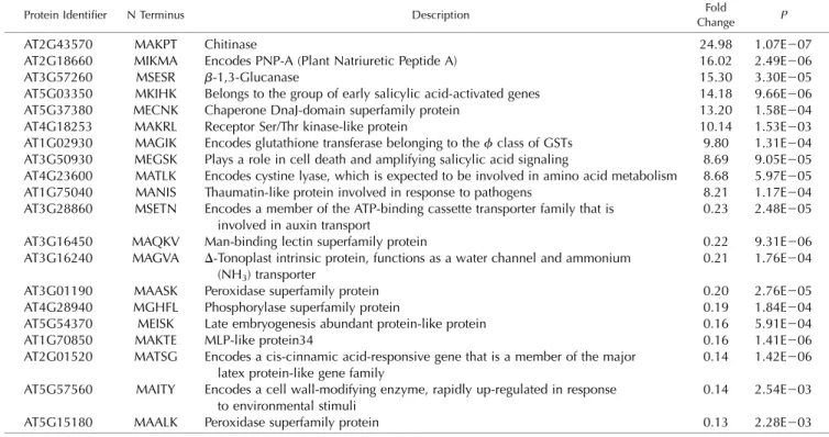
Table 2. List of the 20 most affected proteins by loss of NAA50
Proteins were extracted from 4-week-old naa50-2 mutants and wild-type plants grown on one-half-strength Murashige and Skoog (MS) plates under short-day conditions. This list only shows the 20 most prominently misregulated proteins in naa50-2. A full list of all quantified proteins can be found in Supplemental Table S2.
Protein Identifier N Terminus Description Fold
Change P
AT2G43570 MAKPT Chitinase 24.98 1.07E207
AT2G18660 MIKMA Encodes PNP-A (Plant Natriuretic Peptide A) 16.02 2.49E206 AT3G57260 MSESR b-1,3-Glucanase 15.30 3.30E205 AT5G03350 MKIHK Belongs to the group of early salicylic acid-activated genes 14.18 9.66E206 AT5G37380 MECNK Chaperone DnaJ-domain superfamily protein 13.20 1.58E204 AT4G18253 MAKRL Receptor Ser/Thr kinase-like protein 10.14 1.53E203 AT1G02930 MAGIK Encodes glutathione transferase belonging to thef class of GSTs 9.80 1.31E204 AT3G50930 MEGSK Plays a role in cell death and amplifying salicylic acid signaling 8.69 9.05E205 AT4G23600 MATLK Encodes cystine lyase, which is expected to be involved in amino acid metabolism 8.68 5.97E205 AT1G75040 MANIS Thaumatin-like protein involved in response to pathogens 8.21 1.17E204 AT3G28860 MSETN Encodes a member of the ATP-binding cassette transporter family that is
involved in auxin transport
0.23 2.48E205 AT3G16450 MAQKV Man-binding lectin superfamily protein 0.22 9.31E206 AT3G16240 MAGVA D-Tonoplast intrinsic protein, functions as a water channel and ammonium
(NH3) transporter
0.21 1.76E204 AT3G01190 MAASK Peroxidase superfamily protein 0.20 2.76E205 AT4G28940 MGHFL Phosphorylase superfamily protein 0.19 1.84E204 AT5G54370 MEISK Late embryogenesis abundant protein-like protein 0.16 5.91E204 AT1G70850 MAKTE MLP-like protein34 0.16 1.41E206 AT2G01520 MATSG Encodes a cis-cinnamic acid-responsive gene that is a member of the major
latex protein-like gene family
0.14 1.42E206 AT5G57560 MAITY Encodes a cell wall-modifying enzyme, rapidly up-regulated in response
to environmental stimuli
0.14 2.54E203 AT5G15180 MAALK Peroxidase superfamily protein 0.13 2.28E203
(i.e. plastid-imported proteins). Here, we show that in
the wild type, 66% (259 of 392) of the total quantified N
termini were fully acetylated (acetylation yield
.
95%), whereas 34% (133 of 392) were partially or not
acetylated. Similarly, in the naa50-2 mutant, 70% (215
of 309) of the total quantified N termini were fully
acetylated and 30% (94 of 309) were partially
acety-lated, revealing no significant changes in terms of
global NTA yield between the mutant and the wild
type (Fig. 5A). Based on the GAP assay (Fig. 1C),
AtNAA50 is expected to acetylate proteins retaining
their iMet, whereas the NatA complex acts on proteins
after removal of the iMet (Linster and Wirtz, 2018).
Hence, we analyzed both populations separately
(Fig. 5, B and C). When only N termini retaining their
iMet (Fig. 5B) are considered, 26 N termini (79%, 26 of
33) are fully acetylated in wild-type plants compared
with 13 N termini (65%, 13 of 20) retrieved in the
mutants, revealing no significant difference between
the two genotypes and therefore not providing any
straightforward candidates for in vivo substrates of
AtNAA50.
The same is true for N termini without iMet (Fig. 5C),
where 99 N termini (60%, 99 of 166) are fully acetylated
in wild-type plants compared with 74 N termini (59%,
74 of 126) in naa50-2, indicating that upon depletion of
NAA50, NatA-type N termini remain acetylated.
In-deed, previously identified NatA substrates, which are
fully (greater than 95%) acetylated in wild-type plants
and lose their acetylation marks upon knockdown of
NAA10, retain their N-terminal acetylation levels in the
naa50-2 background (Table 1).
Of 108 N termini that were quantified in both the
wild type and naa50-2, only two plastid proteins
(AT2G1488 and AT5G03370) were found to be
differ-entially acetylated (fold change
. 2, P , 0.05; Fig. 5D).
In both cases, the acetylation rates in the wild type
were already low (1.9% for AT2G1488 and 13.9% for
AT5G03370) and dropped to even lower levels in
naa50-2 (0.9% for AT2G1488 and 6.4% for AT5G03370).
Loss of NAA50 Causes the Accumulation of Stress
Response Proteins
Although we could not identify any in vivo NAA50
substrates with the SILProNAQ approach, the severe
phenotype of the naa50 mutants suggested a vital role of
AtNAA50 for the determination of proteome fate. A global
comparison of the naa50-2 mutant and the wild-type
pro-teome revealed differential expression (greater than
1.4-fold up- or down-regulated, P
, 0.05) of 732 protein
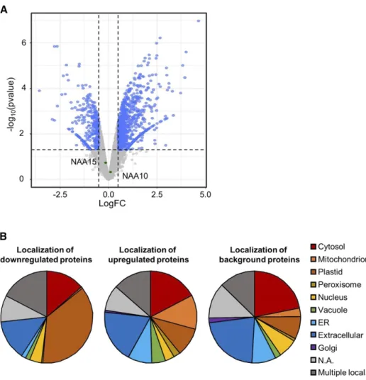
Figure 6. The expression of 732 pro-tein groups is deregulated in naa50-2 mutants compared with wild-type plants. A, Volcano plot depicting the significantly regulated (greater than 1.4-fold change, LIMMA P , 0.05, n 5 4) protein groups in blue. Proteins with unaltered abundance in naa50-2 are labeled in gray. NAA10 and NAA15 are indicated in green. Proteins were extracted from 4-week-old naa50-2 and wild-type seedlings grown under short-day conditions on one-half strength MS medium. B, The differentially regu-lated proteins (first pie chart, down-regulated in naa50-2 compared with the wild type; second pie chart, up-regulated in naa50-2 compared with the wild type) are localized in a variety of cellular compartments, according to the SUB-Acon localization of the SUBA4 Arabi-dopsis subcellular localization database (Hooper et al., 2017). For comparison, the distribution of localizations was calculated for all proteins detected in the MS approach (third pie chart, des-ignated background). The full list of quantified protein groups is available in Supplemental Table S2. N.A., Not available.
groups, which corresponds to 16% of the quantified
pro-tein groups (4,508 propro-tein groups; Supplemental Table S2).
The 20 most affected proteins are listed in Table 2. The
a-bundance of 541 protein groups increased significantly in
naa50-2, while 191 protein groups were found to be
down-regulated (Fig. 6A). The subcellular localization of these
up-regulated proteins was diverse (Fig. 6B). Remarkably, 19%
(compared with only 8% of all identified proteins) were
localized in the extracellular space. The down-regulated
proteins localize predominantly in the plastids (37%), the
cytosol (14%), and the extracellular space (14%). The
widespread subcellular distribution of the affected proteins
suggests that the loss of AtNAA50 causes the observed
alterations pleiotropically by disturbing relevant biological
processes. To identify biological processes associated with
NAA50, we performed a Gene Ontology enrichment
analysis of the differentially regulated proteins using the
DAVID Bioinformatics Resources tool v.6.8 (Table 3;
Supplemental Table S3). The pathway enrichment analysis
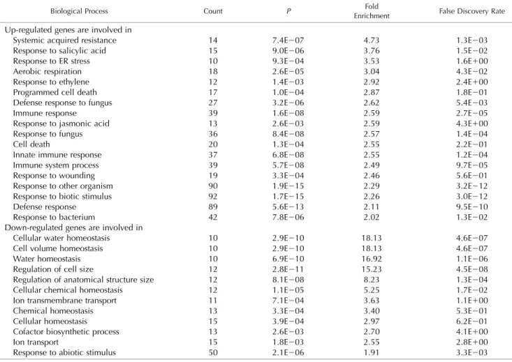
shows that proteins up-regulated in naa50-2 are involved in
a variety of stress responses, including immune system
processes (2.5-fold enriched, P
, 0.05) and the responses
to the stress-related phytohormones salicylic acid (4-fold
enriched, P
, 0.05), jasmonic acid (3-fold enriched, P ,
0.05), and ethylene (3-fold enriched, P
, 0.05). Among the
down-regulated proteins, proteins regulating cell size
(15-fold enriched, P
, 0.05), cell volume (18-fold enriched, P ,
0.05), and ion transport (;3-fold enriched, P , 0.05) were
overrepresented.
DISCUSSION
NAA50 Activity Is Functionally Conserved between
Humans and Plants
NTA is the most abundant cotranslationally
occur-ring protein modification in humans and Arabidopsis.
The essential NatA complex targets approximately 40%
of the proteome in both species, defining this complex
Table 3. NAA50 mutants accumulate proteins involved in stress responses
Proteins were extracted from 4-week-old naa50-2 and wild-type seedlings grown under short-day conditions on one-half strength MS medium. Differentially regulated proteins (greater than 1.4-fold up- or down-regulated compared with the wild type; P , 0.05) were subjected to a Gene Ontology enrichment analysis performed with the DAVID Bioinformatics Resources tool v.6.8 (http://david.abcc.ncifcrf.gov). Proteins involved in the depicted biological processes were significantly (greater than 2-fold, P , 0.05) enriched upon the up- and down-regulated proteins. Counts represent the number of regulated proteins. For clarity, only stress-related biological processes are listed here; a full list of all affected biological processes is available in Supplemental Table S3.
Biological Process Count P EnrichmentFold False Discovery Rate
Up-regulated genes are involved in
Systemic acquired resistance 14 7.4E207 4.73 1.3E203 Response to salicylic acid 15 9.0E206 3.76 1.5E202 Response to ER stress 10 9.3E204 3.53 1.6E100 Aerobic respiration 18 2.6E205 3.04 4.3E202 Response to ethylene 12 1.4E203 2.92 2.4E100 Programmed cell death 17 1.0E204 2.87 1.8E201 Defense response to fungus 27 3.2E206 2.62 5.4E203
Immune response 39 1.6E208 2.59 2.7E205
Response to jasmonic acid 13 2.6E203 2.59 4.3E100 Response to fungus 36 8.4E208 2.57 1.4E204
Cell death 20 1.3E204 2.55 2.2E201
Innate immune response 37 6.8E208 2.55 1.2E204 Immune system process 39 5.7E208 2.49 9.7E205 Response to wounding 19 3.3E204 2.46 5.6E201 Response to other organism 90 1.9E215 2.29 3.2E212 Response to biotic stimulus 92 1.7E215 2.26 3.0E212 Defense response 89 5.6E213 2.11 9.5E210 Response to bacterium 42 7.8E206 2.02 1.3E202 Down-regulated genes are involved in
Cellular water homeostasis 10 2.9E210 18.13 4.6E207 Cell volume homeostasis 10 2.9E210 18.13 4.6E207 Water homeostasis 10 6.9E210 16.92 1.1E206 Regulation of cell size 12 2.8E211 15.23 4.5E208 Regulation of anatomical structure size 12 8.1E208 8.23 1.3E204 Cellular chemical homeostasis 12 1.1E205 5.25 1.7E202 Ion transmembrane transport 11 7.1E204 3.63 1.1E100 Chemical homeostasis 13 3.3E204 3.40 5.3E201 Cellular homeostasis 15 3.9E204 2.97 6.2E201 Cofactor biosynthetic process 13 2.6E203 2.70 4.1E100
Ion transport 15 1.8E203 2.55 2.8E100
Response to abiotic stimulus 50 2.1E206 1.91 3.3E203
as one of the few globally acting protein modifiers in
eukaryotic cells. In all eukaryotes analyzed so far
(hu-man, fruit
fly, and yeast), NatA interacts with the
NAA50 protein to form NatE, which gives rise to an
independent NatE activity in humans and fruit
flies. In
contrast, ScNAA50 is enzymatically inactive (Deng
et al., 2019). While enzymatic activity and the
protein-protein interaction network of NAA50 have been
ex-tensively characterized in metazoa and fungi (Gautschi
et al., 2003; Williams et al., 2003; Arnesen et al., 2005;
Evjenth et al., 2009; Van Damme et al., 2011), the putative
ortholog in plants was not characterized so far. Our
work demonstrates that NAA50 is catalytically active in
the reference plant Arabidopsis. AtNAA50 has a broad
substrate specificity, which substantially overlaps with
the substrate specificity of HsNAA50 (Van Damme et al.,
2011; Reddi et al., 2016). Although we detect a preference
of AtNAA50 for iMet followed by Lys (K), Asn (N), Gln
(Q), and Thr (T) in the second position (Fig. 1C),
ex-pression of HsNAA50 is able to complement the loss of
AtNAA50 in naa50-2. Complementation with the
cata-lytically inactive ScNAA50 failed (Fig. 4C), suggesting
that NAA50 activity is required to rescue naa50-2.
Re-markably, catalytic residues are conserved in protein
sequences of putative NAA50s in algae and plants
(Supplemental Fig. S4), suggesting that NAA50 is
cata-lytically active in phototrophic eukaryotes.
NatA Activity Is Not Modulated by NAA50
Although we demonstrate that AtNAA50 is
catalyti-cally active, the N-acetylome profiling approach did not
allow us to identify any in vivo substrates that could
ex-plain the strong phenotype observed in the naa50-2 mutant
(Fig. 5D). This does not come as a surprise, since AtNAA50
is supposed to target only a few substrates (Ribeiro et al.,
2016). While the combined activities of NatA and NatB
target more than 75% of the extractable leaf proteome,
NAA30, NAA50, and NAA60 together are thought to
acetylate only 4% of the leaf proteome (Linster and Wirtz,
2018). For this reason, we cannot expect to identify a NatE
substrate by the N-acetylome profiling approach, although
it is state of the art. Despite the considerable overlap
be-tween the in vitro substrate specificities of AtNAA50 and
AtNAA60, the enzymes may target distinct substrate
pools due to their diverging subcellular localization
in vivo. This spatial separation of substrate pools
has also been suggested to occur in humans, where
NAA60 mainly acetylates transmembrane proteins
(Aksnes et al., 2015b; Van Damme et al., 2015).
Even though AtNAA50 displays catalytic activity on
its own, the enzyme might have additional functions in
association with the core NatA complex (Deng et al.,
2020). This core complex is conserved between
hu-mans, yeast, and plants and consists of the catalytic
subunit NAA10 and the auxiliary subunit NAA15
(Linster et al., 2015). In humans and yeast, the core
complex interacts with the accessory subunit NAA50
(Gautschi et al., 2003; Van Damme et al., 2015). Until
now, it was unclear whether NAA50 associates with the
core NatA complex in plants. However, the
colocali-zation of NAA10 and NAA50 was evidenced in the
cytoplasm (Neubauer and Innes, 2020). Although
ScNAA50 is inactive, it positively impacts NatA
activ-ity by modulating the enzymatic activactiv-ity of ScNAA10
(Deng et al., 2019) and by positioning the NatA complex
in close proximity to the ribosomal tunnel exit (Knorr
et al., 2019). The interaction of NAA50 with the NatA
complex is mediated via hydrophobic interactions
between the NAA50
b2-b3 and b4-a2 loops and the
NAA15
a22-a23 loop. A mutation in a highly
con-served Thr residue in NAA15 (T416 in yeast and T406 in
human) results in the dissociation of NAA15 from
NAA50 in both yeast and human (Deng et al., 2019).
This Thr residue (T412) is also conserved in Arabidopsis
NAA15, suggesting a common NAA50-NAA15
bind-ing mode. The N-terminome profilbind-ing approach,
how-ever, shows that the depletion of AtNAA50 does not
result in decreased acetylation of NatA-type N termini
(Table 1), revealing that, unlike ScNAA50, plant
NAA50 is not required for the proper function of the
NatA complex. Recently, the cryoelectron microscopy
structure of the quaternary human NatE/HypK
com-plex was reported and the authors show that HsNAA50
inhibits HsNatA activity in vitro (Deng et al., 2020). In
plants, however, the absence of NAA50 does not induce
any alteration of NTA on canonical NatA substrates
under nonstressed conditions. We cannot exclude the
possibility of stress-induced modifications of NatA/E
subunits, which might alter their enzymatic activity or
protein-protein interaction capability in plants,
result-ing in a more pronounced impact of NAA50 on NatA.
One such modification might be Lys acetylation of
NAA50, which has already been reported for the
resi-dues K37 and K155 (Hartl et al., 2017). The K37 residue
is an evolutionarily conserved Lys acetylation site in
humans and plants and is autoacetylated by HsNAA50
in vitro (Evjenth et al., 2009). Other modifications of the
plant NatA/E subunits are currently unknown.
Depletion of NAA50 Leads to a Severe Growth Retardation
A possible explanation for the inhibited growth of
naa50 mutants is the constitutive induction of
stress-related genes. We
find that among the proteins
accu-mulating in naa50-2 mutants, proteins involved in the
ER stress response are 3.4-fold overrepresented. The
accumulation of ER stress-associated proteins can be
explained by an enhanced transcription of these genes
due to increased splicing of the transcription factor
bZIP60 observed in naa50-1 seedlings (Neubauer and
Innes, 2020). Unspliced bZIP60 resides in the ER
membrane due to a C-terminal transmembrane
do-main. In response to ER stress, bZIP60 is spliced by
IRE1, which eliminates this C-terminal transmembrane
domain, resulting in the translocation of bZIP60 into the
nucleus, where it induces the expression of ER
stress-responsive genes (Nagashima et al., 2011). In line with
this
finding, proteins involved in the response to
sali-cylic acid accumulate in naa50-2 (3.7-fold). Salisali-cylic acid
is a primary regulator of plant growth particularly
under stress conditions. During ER stress, salicylic acid
diverts energy from plant growth to the induction of the
unfolded protein response, thereby protecting the plant
from misfolded proteins (Meng et al., 2017). Salicylic
acid is also an important positive regulator of the plant
immune response. In agreement with this
finding, an
increase in the expression of defense genes and salicylic
acid signaling is observed in naa50 mutants (Neubauer
and Innes, 2020). We
find this transcriptional increase
reflected at the protein level (2.1-fold up-regulation of
proteins involved in the defense response).
Since we show that AtNAA50-EYFP resides not only
at the ER but also in the nucleus (Fig. 4, A and B), we
cannot exclude a role of nucleus-localized NAA50 in
the constitutive induction of stress responses in naa50
mutants. The multiple localization of AtNAA50-EYFP
in the cytosol and the ER has been independently
con-firmed by Neubauer and Innes (2020). The authors
want to put a note of caution on the nuclear localization
of AtNAA50-EYFP, since EYFP on its own is known to
display nucleocytoplasmic localization (Chen et al.,
2020). Hence, we cannot exclude that EYFP
contrib-utes to the nucleocytoplasmic localization of
AtNAA50-EYFP. However, a nuclear localization of AtNAA50 is
in agreement with both the nucleocytoplasmic
locali-zation of HsNAA50 (Arnesen et al., 2006) and a
pre-dicted bipartite nuclear localization signal at position
125 of AtNAA50 (Kosugi et al., 2009). In other species,
acetyltransferases have already been shown to act as
transcriptional regulators. HsNAA10 for instance
reg-ulates the response to hypoxia by
«-Lys acetylation of
the transcription factor HIF-1a (Kang et al., 2018). Other
studies report on the import of NAA10 into the nuclei
of proliferating cells, where it is required for cell cycle
progression and cell proliferation (Park et al., 2014).
Such a scenario might also be conceivable for NAA50,
suggesting that NAA50 is not always present in the
nucleus. This would explain the apparent difference
in the subcellular localization shown in our study and
the results reported in Neubauer and Innes (2020).
CONCLUSION
In summary, we show that the catalytically active
N-acetyltransferase AtNAA50 is required for plant
growth and fertility. We demonstrate that its absence
causes a severe growth reduction and an increase
in diverse stress responses. Under optimal growth
conditions, the acetylation of canonical NatA
sub-strates was not affected by the loss of NAA50,
dem-onstrating that NAA50 is dispensable for NatA activity in
this condition. Our complementation studies point
to-ward a conserved function of enzymatically active
NAA50 in multicellular eukaryotes that is different
from the function of inactive NAA50 in the unicellular
budding yeast.
MATERIALS AND METHODS
Plant Material and Growth Conditions
This work refers to the Arabidopsis (Arabidopsis thaliana) ecotype Columbia-0 as the wild type. The T-DNA insertion lines naa50-1 (SAIL_1210_A02) and naa50-2 (SAIL_1186_A03) were obtained from the SAIL collection (Sessions et al., 2002). If not specified otherwise, experiments were conducted with plants grown on medium containing one-half soil and one-half substrate 2 (Klasmann-Deilmann) under short-day conditions (8.5 h of light, 100mE light photonflux density, 24°C/18°C day/night temperatures, and 50% humidity). For growth on plates, seeds were surface sterilized with 70% (v/v) ethanol (5 min) and 6% (v/v) NaClO (2 min) followed by three washing steps with sterile water. After 2 d of stratification at 4°C, seeds were germinated on AT medium [5 mMKNO3, 2.5 mMKH2PO4, pH 5.6, 2 mMMgSO4, 2 mMCa(NO3)2,
0.05 mMFe-EDTA, 0.01mMCoCl2, 0.02mMNa2MoO4, 0.5mMCuSO4, 1mM ZnSO4, 10mMNaCl, 14mMMnCl2, and 0.8% [w/v] micro agar, pH 5.8].
Identification of the Putative NAA50 Orthologs
The putative NAA50 homolog in Arabidopsis (AT5G11340) was identified as the result of a BLAST search (TAIR BLASTP2.2.8, www.arabidopsis.org) based on the Saccharomyces cerevisiae protein Naa50p (Yor253wp). AT5G11340 dis-played a homology higher than any of the other putative homologs (score 61, E-value 3E-10). Similarly, homologs of AtNAA50 were identified in other organisms via a BLAST search (https://blast.ncbi.nlm.nih.gov/Blast.cgi? PAGE5Proteins) in Homo sapiens (UniProt ID: Q9GZZ1; 54% identity), Oryza sativa (UniProt ID: A2WSI1; 76% identity), Glycine max (UniProt ID: C6SYN6; 79% identity), Chlamydomonas reinhardtii (UniProt ID A8I4R4; 48% identity), Vitis vinifera (UniProt ID: A5BJF0; 81% identity), and Populus trichocarpa (UniProt ID: B9H9R5; 79% identity).
Extraction of Genomic DNA
In order to genotype the naa50 mutants, genomic DNA was extracted from 50 to 100 mg of fresh Arabidopsis leaf material using Edwards buffer (200 mM Tris-HCl, 25 mMEDTA, 250 mMNaCl, and 0.5% [w/v] SDS). The tissue was ground in 400mL of Edwards buffer using a plastic pestle. After vigorous mixing and centrifugation (5 min at room temperature), the supernatant was transferred to a fresh reaction tube and mixed with an equal amount of iso-propanol. Subsequently, the samples were centrifuged at full speed for 10 min to allow nucleic acid precipitation. The supernatant was discarded and the pellet was washed with 70% (v/v) ethanol. After decanting the ethanol, the pellet was dried and resolved in 30mL of sterile water.
PCR
Homozygous naa50 mutants were identified by PCR-based genotyping using the FastGene Taq 2x Ready Mix (Nippon Genetics) and specific primer combinations for the wild-type (NAA50_fwd and NAA50_rev) and mutant (naa50-1, SAIL_LB and NAA50_rev; naa50-2, SAIL_LB and SAIL_RP) alleles.
For cloning, DNA was amplified with the high-fidelity DNA polymerase Phusion (New England Biolabs, M0530L). All enzymes were used according to the supplier’s instruction manual. The corresponding primer sequences are listed in Supplemental Table S4.
Constructs for Complementation and Subcellular
Localization in Plants
To analyze the conservation between putative NAA50 orthologs, the Ara-bidopsis naa50-2 line was transformed with the endogenous NAA50 sequence as well as the human and yeast NAA50 sequences. The genes of interest were amplified from human, yeast, and plant cDNA via PCR using Gateway-compatible primers (AtNAA15-N, AtNAA15-C, AtNAA50-N, AtNAA50_STOP-C, HsNAA50-N, HsNAA50-AtNAA50_STOP-C, ScNAA50-N, and ScNAA50-C). The protein sequences were then cloned into the binary vector pK2GW7.0, where they were expressed under the control of the CaMV 35S promoter. For localization, AtNAA50 was PCR amplified with the AtNAA50_N and AtNAA50_C primers and cloned via Gateway technology (Invitrogen) in the binary vector pB7YWG2.0, which contains the coding sequence for EYFP.
Stable Transformation of Arabidopsis
Stable transformation was conducted according to thefloral dip method for Agrobacterium tumefaciens-mediated transformation of Arabidopsis described by Clough and Bent (1998). Transformants were selected on solidified medium [5 mMKNO3, 2.5 mMKH2PO4, pH 5.6, 2 mMMgSO4, 2.5mMCa(NO3)2, 50mM Fe-EDTA, 70mMH3BO3, 14mMMgCl2, 10mMNaCl, 1mMZnSO4, 0.5mMCuSO4,
0.02mMNa2MoO4, 0.01mMCoCl2, and 0.8% micro agar, pH 5.8] supplemented
with 50mg mL21kanamycin.
Transient Transformation of
Nicotiana benthamiana and
Fluorescence Microscopy
The NAA50-EYFP fusion protein was expressed in N. benthamiana epidermal cells 24 to 48 h after A. tumefaciens-mediated transformation as described by Sparkes et al. (2006) with the pB7YWG2.0 construct. Leaves were either cotransfected with the ER marker VMA12-RFP (Viotti et al., 2013) or 2mg mL21 DAPI (Sigma-Aldrich). Thefluorescence was analyzed by confocal laser scan-ning microscopy using a Nikon automated Ti inverted microscope equipped with a Yokagawa CSU-X1 confocal scanning unit, a Hamamatsu C9100-02 EMCCD camera, and a Nikon S Fluor 403 numerical aperture 1.3 oil-immersion objective (Nikon). Images were taken in three channels (RFP, 561/ 615; DAPI, 405/445 nm; and EYFP, 488/527 nm). Additionally, a bright-field image was recorded. The resulting images were processed with the open-source image-analysis software Fiji.
Constructs for Expression in
Escherichia coli Cells
For the expression of AtNAA50 in E. coli, the NAA50 sequence was amplified from Arabidopsis cDNA with the primers NAA50_NcoI_fwd and NAA50_ BamHI_rev. Harnessing the newly established restriction sites, the resulting PCR fragment was cloned into the empty pETM20 or pETG10a vector (Dümmler et al., 2005).Expression and Purification of AtNAA50 in E. coli
E. coli Rosetta (DE3; Novagen) was transformed by heat shock with the pETM2-AtNAA50 or pETG10a-AtNAA50 plasmid encoding for the Trx-His6-AtNAA50 or His6-AtNAA50 fusion protein. The expression of these
recombi-nant proteins was induced with isopropyl-b-D-thiogalactoside (1 mM). After 5 h of incubation at 37°C, the cultures were harvested by centrifugation (10 min, 6,000g, 4°C). The pellet was dissolved in lysis buffer (250 mMNaCl, 20 mM imidazole, 0.5 mMphenylmethylsulfonylfluoride, and 50 mMTris, pH 8), and the cells were lysed by sonication. Trx-His6-AtNAA50 was purified from the
cell extracts by metal affinity chromatography on a HiTrap IMAC High-Performance column (GE Healthcare). For this purpose, the cell lysate was circulated on the column for 1 h. Afterward, the column was washed with wash buffer (250 mMNaCl, 80 mMimidazole, and 50 mMTris, pH 8) and the protein was eluted in elution buffer (250 mMNaCl, 400 mMimidazole, and 50 mMTris, pH 8). The quality of the purified protein was assessed by SDS-PAGE. The purified protein was used for in vitro acetyltransferase assays (Trx-His6
-AtNAA50; see below) and immunization of rabbits (His6-AtNAA50) to
gen-erate an NAA50-specific antibody.
Generation of an AtNAA50-Specific Antiserum in Rabbit
For the immunization of the rabbits (treatments at days 0 and 30), 300mL of purified His6-AtNAA60 (;1 mg) was sterile filtered and mixed with 300 mL ofFreund’s complete adjuvant (Sigma-Aldrich). For the first and second boosts, the antigen was mixed with Freund’s incomplete adjuvant (Sigma-Aldrich). The antibody titer was monitored 1 month after each injection via immuno-logical detection by comparing the preimmune sera with the antibody-containing sera of thefirst and second antigen treatments (Supplemental Fig. S5). The immunization of the rabbits was stopped after the second boost. The blood samples were taken at day 44 and were stored for 24 h at 4°C followed by 20 min of centrifugation at;30,000g and 4°C for 20 min. The resulting super-natant was supplemented with 0.2% (w/v) sodium azide and stored at280°C until usage.
In Vitro NAT Assay
To measure the NAT activity of AtNAA50, 4.5mg (150 pmol) of purified enzyme was mixed with 0.2 mMof a custom-made peptide (GeneCust), 0.2% (w/v) BSA in acetylation buffer (50 mMTris-HCl, pH 7.5, 8 mMEDTA, and 1 mM DTT), and 45mM[3H]acetyl-CoA (7.4 GBq mmol21; tritium labeled only at the acetyl group by Hartmann Analytics). The reaction mix was topped up to 0.1 mL with acetylation buffer and incubated at 37°C for 1 h. Subsequently, the samples were centrifuged at 1,500g for 4 min. To isolate the custom-made peptide, the supernatant was mixed with 0.1 mL of SP Sepharose (50% [w/v] in 0.5Macetic acid) and incubated for 5 min while shaking. After 4 min of centrifugation at 1,500g, the pellet was washed three times with 0.4 mL of 0.5M acetic acid and once with 0.4 mL of 100% methanol. The amount of incorporated
3H label was measured with a Tri-Carb 2810TR scintillation counter
(Perkin-Elmer). The custom-made peptides MLGP (MLGPEGGRWGRPVGRRRRPVR-VYP) and SESS (SESSSRSRWGRPVGRRRRPVR(MLGPEGGRWGRPVGRRRRPVR-VYP) share a C-terminal Arg-rich sequence resembling the human adrenocorticotropic hormone to facilitate peptide solubility and effective enrichment via Sepharose beads according to Evjenth et al. (2009).
In Vitro Kat Assay
To determine the Kat activity of AtNAA50, 11.5mg (385 pmol) of purified enzyme was mixed with 1.92mmol of acetyl-CoA in Kat buffer (50 mMTris, 10% [v/v] glycerol, and 1 mMEDTA, pH 8.5) and incubated at 37°C. After 0 to 60 min, the reaction was stopped by adding 5mL of 53 SDS loading buffer (0.1M Tris, pH 7, 5% [w/v] SDS, 3.63Mb-mercaptoethanol, 20% [v/v] glycerol, and 0.01% [w/v] bromophenol blue) and subsequent incubation at 95°C for 10 min. The Kat activity was determined via immunological detection of acetylated Lys residues with ana-acetylated Lys antibody (1:10,000 in Tris-buffered saline plus Tween 20 with 5% [w/v] BSA, incubation overnight at 4°C; Cell Signaling, 9441).
Protein Extraction from Arabidopsis Leaf Tissue
Total soluble protein extracts were isolated from 200 mg of frozen and ground leaf material with 400mL of precooled extraction buffer (50 mMHEPES, pH 7.4, 10 mMKCl, 1 mMEDTA, 11 mMEGTA, and 10% [v/v] glycerol) supplemented with 10 mMDTT and 0.5 mMphenylmethylsulfonylfluoride. Protein extracts were cleared by centrifugation (10 min, 20,200g, 4°C), and the protein concen-tration was quantified according to Bradford (1976).
SDS-PAGE and Immunological Detection
Protein extracts were analyzed via SDS-PAGE (Laemmli, 1970) and blotted onto polyvinylidene difluoride membranes using Mini-Protean II cells (Bio-Rad). The primary NAA50 antibody and the secondary horseradish peroxidase-linked anti-rabbit antibody (no. AS10 852, Agrisera) were diluted 1:5,000 and 1:25,000 in 13 Tris-buffered saline plus Tween 20 (50 mMTris, pH 7.6, 150 mM NaCl, and 0.05% [v/v] Tween 20) supplemented with 0.5% (w/v) BSA. Membranes were developed using the SuperSignal West Dura Extended Du-ration Substrate (Thermo Scientific) according to the manufacturer’s instruc-tions. The resulting signals were recorded using the ImageQuant LAS 4000 (GE Healthcare) and subsequently quantified with the ImageQuant TL software (GE Healthcare).
GAP and Data Analysis
GAP was performed as previously described by Dinh et al. (2015). For this purpose, 1 mg of E. coli lysates expressing AtNAA50-SUMO or SUMO-GFP as a negative control was subjected to chemical acetylation of free N termini with N-acetoxy-[2H3]-succinimide required for SILProNAQ quantification (Bienvenut et al., 2017a). The samples were digested with trypsin, and the N-terminal peptides were enriched by strong cation-exchange liquid chroma-tography before liquid chromachroma-tography-tandem mass spectrometry (LC-MS/ MS) analysis. Raw data were extracted and exported with Proteome Discoverer (Thermo Scientific, version 1.4). The MASCOT 2.4 software was used for the identification of proteins and their cotranslational and posttranslational mod-ifications. The E. coli K12 strain proteome extracted from UniProtKB (version 112) served as a reference proteome. The EnCOUNTer tool (Bienvenut et al., 2017b) was used to reprocess the quantified values determined by Mascot
Distiller and to supply afinal list of the different peptides associated with their NTA yield.
N-Terminomics of Arabidopsis Wild-Type Plants and
naa50-2 Knockout Mutants
Arabidopsis wild-type and naa50-2 mutant lines were grown for 4 weeks on one-half strength MS plates under short-day conditions. Whole plants were collected and pooled to obtain four replicates of 100 mg of plant material of wild-type and naa50-2 plants. The plant tissue was frozen in liquid nitrogen and ground to afine powder. Altogether, three biological replicates of naa50-2 were used to characterize and quantify NTA following the protocol described in the above GAP test (Dinh et al., 2015).
LC-MS/MS-Based Quantitative Proteome Analyses
For the quantitative proteome analyses, plants were grown for 4 weeks on one-half strength MS plates under short-day conditions. Whole plants were collected and pooled to obtain four replicates of 100 mg of plant material of wild-type and naa50-2 plants. The plant tissue was frozen in liquid nitrogen and ground to afine powder. Protein extraction, sample processing, and LC-MS/MS data acquisition were performed as described previously (Lassowskat et al., 2017). Briefly, proteins were extracted and digested using a modified filter-assisted sample preparation protocol. Peptides were dimethyl labeled, whereby two replicates of each genotype were light labeled (dimethyl mass shift,128.0313 D) and two replicates were medium labeled (dimethyl mass shift,132.0564 D). Equal amounts of light-labeled and medium-labeled peptides were pooled for each replicate, desalted, and frac-tionated using SDB-RPS Stagetips (Kulak et al., 2014) followed by LC-MS/MS analyses. LC-MS/MS analyses were performed by using an EASY-nLC 1200 (Thermo Fisher) coupled to a Q Exactive HF mass spectrometer (Thermo Fisher). Separation of peptides was performed on 17-cm frit-less silica emitters (New Ob-jective, 0.75mm i.d.) and packed in-house with reverse-phase ReproSil-Pur C18 AQ 1.9mm resin (Dr. Maisch). The column was constantly kept at 50°C. Peptides were eluted in 115 min applying a segmented linear gradient of 0% to 98% solvent B (solvent A, 0% acetonitrile and 0.1% [v/v] formamide; solvent B, 80% [v/v] ace-tonitrile and 0.1% [v/v] formamide) at aflow rate of 300 nL min21. Mass spectra were acquired in data-dependent acquisition mode according to the TOP15 method. MS spectra were collected by the Orbitrap analyzer with a mass range of 300 to 1,759 mass-to-charge ratio (m/z) at a resolution of 60,000 full width at half maximum, maximum injection time of 55 ms, and a target value of 33 106ions.Precursors were selected with an isolation window of 1.3 m/z, and higher-energy collisional dissociation fragmentation was performed at a normalized collision energy of 25. MS/MS spectra were acquired with a target value of 105 ions at a resolution of 15,000 full width at half maximum, maximum injection time of 55 ms, and afixed first mass of m/z 100. Peptides with a charge of 11, greater than 6, or with unassigned charge state were excluded from fragmentation for MS2, and the dynamic exclusion for 30 s prevented repeated selection of precursors.
Processing of raw data was performed using the MaxQuant software version 1.6.9.0 (Cox and Mann, 2008). MS/MS spectra were assigned to the Araport11 protein database. During the search, sequences of 248 common contaminant proteins as well as decoy sequences were automatically added. Trypsin specificity was required, and a maximum of two missed cleavages was allowed. Carbami-domethylation of Cys residues was set as afixed modification, and oxidation of Met, deamidation, and protein N-terminal acetylation were set as variable mod-ifications. Light and medium dimethylation of Lys residues and peptide N termini were set as labels. A false discovery rate of 1% for peptide spectrum matches and proteins was applied. Match between runs and requantify options were enabled. Further downstream analysis was performed using Perseus version 1.6.6.0 (Tyanova et al., 2016). Reverse hits and contaminants were removed, site ratios were log2transformed, andflip-label ratios were inverted. Normalized site ratios
were used for all further analyses. The resulting matrices were exported for the determination of significantly differential abundant protein groups using the LIMMA package (Ritchie et al., 2015) in R 3.3.1.
RT-qPCR
Nat transcript levels were analyzed by RT-qPCR as described previously (Huber et al., 2020). The corresponding primer sequences are listed in Supplemental Table S4.
Statistical Analyses
Statistical analysis was conducted using SigmaPlot 12.0. Means from dif-ferent sets of data were analyzed for statistically significant differences with the Holm-Sidak one-way ANOVA test or Student’s t test. Significant differences (P, 0.05) are indicated with different letters.
Accession Numbers
Sequence data from this article can be found in TAIR under Arabidopsis Ge-nome Initiative accession numbers AT5G11340 (AtNAA50), AT3G06610 (AtH-PYK) AT5G13780 (AtNAA10), and AT1G80410 (AtNAA15). The N-terminomics data are deposited at the PRIDE repository (https://www.ebi.ac.uk/pride/) with the data set identifier PXD017430. The full proteome data are deposited at the PRIDE repository (https://www.ebi.ac.uk/pride/) with the data set identifier PXD017663.
Supplemental Data
The following supplemental materials are available.
Supplemental Figure S1.NAA15-EYFP does not localize to the nucleus. Supplemental Figure S2.Genotyping of homozygous naa50 mutants. Supplemental Figure S3.RT-qPCR reveals that the full-length NAA50
transcript cannot be detected in naa50 mutants.
Supplemental Figure S4.Alignment of NAA50 from different phototro-phic organisms.
Supplemental Figure S5. Validation of the specific antiserum against AtNAA50.
Supplemental Table S1.AtNAA50 GAP test.
Supplemental Table S2.Misregulated (greater than 2-fold up- or down-regulated; P. 0.05) proteins in naa50-2 mutants compared with 4-week-old wild-type plants grown on one-half strength MS plates under short-day conditions.
Supplemental Table S3.Gene Ontology term enrichment analysis for dif-ferentially expressed proteins in NAA50-depleted plants.
Supplemental Table S4.Primers used for cloning, RT-qPCR, and genotyping.
ACKNOWLEDGMENTS
We thank Felix Alexander Weyer and Karine Lapouge (Heidelberg Univer-sity Biochemistry Center) for fruitful discussions and careful reading and comments on the article.
Received February 24, 2020; accepted May 15, 2020; published May 27, 2020.
LITERATURE CITED
Aksnes H, Drazic A, Marie M, Arnesen T(2016) First thingsfirst: Vital protein marks by N-terminal acetyltransferases. Trends Biochem Sci 41: 746–760
Aksnes H, Hole K, Arnesen T(2015a) Molecular, cellular, and physio-logical significance of N-terminal acetylation. Int Rev Cell Mol Biol 316: 267–305
Aksnes H, Marie M, Arnesen T(2015b) Holding it together: Naa60 at the Golgi. Oncotarget 6: 15726–15727
Aksnes H, Ree R, Arnesen T(2019) Co-translational, post-translational, and non-catalytic roles of N-terminal acetyltransferases. Mol Cell 73: 1097–1114
Arnesen T, Anderson D, Baldersheim C, Lanotte M, Varhaug JE, Lillehaug JR(2005) Identification and characterization of the human ARD1-NATH protein acetyltransferase complex. Biochem J 386: 433–443 Arnesen T, Anderson D, Torsvik J, Halseth HB, Varhaug JE, Lillehaug JR (2006) Cloning and characterization of hNAT5/hSAN: An evolution-arily conserved component of the NatA protein N-a-acetyltransferase
complex. Gene 371: 291–295


![Figure 5. N-terminal protein acetyla- acetyla-tion in NAA50-depleted mutants. A to C, Distribution of the NTA yields in naa50-2 (gray) and the wild type (black) for all quantified N termini (1/2N-terminal Met excision [NME];](https://thumb-eu.123doks.com/thumbv2/123doknet/13581336.422119/7.877.72.586.640.1093/figure-terminal-protein-depleted-distribution-quantified-terminal-excision.webp)