Wolbachia increases susceptibility to Plasmodium infection in a natural
system
Zélé F. 1,2*, Nicot A. 1,3, Berthomieu A. 2, Weill M. 2, Duron O. 1,2† and Rivero A.1†
1Maladies Infectieuses et Vecteurs: Ecologie, Génétique, Evolution et Contrôle, CNRS
(UMR CNRS-UM1-UM2 5290, IRD 224), Centre de Recherche IRD, 911 Avenue Agropolis, 34394 Montpellier, France
2Institut des Sciences de l'Evolution, CNRS (UMR 5554), Université de Montpellier II,
34095 Montpellier, France
3Centre d'Ecologie Fonctionnelle et Evolutive, CNRS (UMR 5175), 1919 Route de Mende,
34293 Montpellier, France † co-last authors *Corresponding author Email addresses: FZ: fezele@fc.ul.pt BA: arnaud.berthomieu@univ-montp2.fr NA: antoine.nicot@ird.fr WM: mylene.weill@univ-montp2.fr OD: olivier.duron@ird.fr AR: ana.rivero@cnrs.fr
Abstract
Current views about the impact of Wolbachia on Plasmodium infections are almost entirely based on data regarding artificially transfected mosquitoes. This work has shown that Wolbachia reduces the intensity of Plasmodium infections in mosquitos, raising the exciting possibility of using Wolbachia to control or limit the spread of malaria. Whether natural Wolbachia infections have the same parasite-inhibiting properties is not yet clear. Wolbachia-mosquito combinations with a long evolutionary history are, however, key for understanding what may happen with Wolbachia-transfected mosquitoes after several generations of coevolution. We investigate this issue using an entirely natural mosquito-Wolbachia-Plasmodium combination. In contrast to most previous studies, which have been centered on the quantification of the midgut stages of Plasmodium, we obtain a measurement of parasitaemia that relates directly to transmission by following infections to the salivary gland stages. We show that Wolbachia increases the susceptibility of Culex pipiens mosquitoes to Plasmodium relictum, significantly increasing the prevalence of salivary gland stage infections. This effect is independent of the density of Wolbachia in the mosquito. These results suggest that naturally Wolbachia-infected mosquitoes may, in fact, be better vectors of malaria than Wolbachia-free ones.
Key words: symbiont-mediated protection, vectorial competence, infection prevalence, infection intensity, oocysts, sporozoites.
Introduction
1Individual hosts are often simultaneously infected with more than one parasite species. Co-2
infections can impact both host fitness and parasite transmissibility, and can therefore have 3
important evolutionary and epidemiological consequences [1, 2]. Within a host, parasites may 4
interact in different ways. They may suppress each other because they are in competition for 5
a resource in limited supply, such as a particular nutrient or tissue, or because they stimulate 6
the same branch of the immune system [3]. In the most extreme cases, parasites can excrete 7
molecules that directly inhibit the growth of competitors [4]. Host sharing may also, however, 8
facilitate parasite development, most notably when one of the parasites immunosupresses the 9
host [2]. Co-infections have been intensely investigated in the biomedical literature, as 10
several important human infections are known to be complicated by the arrival of secondary 11
or opportunistic pathogens [3]. More recently, however, a great deal of attention has been 12
drawn to the impact of co-infections on vector-transmitted diseases with the realization that, 13
in the field, arthropod vectors are also often infected by multiple parasites [5-7]. 14
A few years ago, two seminal papers showed that Wolbachia, a maternally transmitted 15
bacterial endosymbiont of arthropods, protects Drosophila flies from several viral infections 16
[8, 9]. This stimulated a great deal of research into Wolbachia-mediated parasite interference 17
in other insect systems (see Supplementary Materials, Table S1), and raised the exciting 18
possibility of using Wolbachia to control or limit the spread of mosquito-transmitted diseases 19
such as dengue and malaria. Interestingly, although neither Aedes aegypti (vector of the 20
dengue virus) nor Anopheles gambiae or An. stephensi (vectors of Plasmodium falciparum) 21
are naturally infected by Wolbachia, they can be successfully transfected in the laboratory 22
using bacteria isolated from other insect species [10-12] although not always stably (in An 23
gambiae the infections are somatic and do not transmit vertically to the offspring [13, 14]). 24
As a consequence, in the last few years, a large number of studies have been conducted using 25
transfected mosquitoes. These studies have largely confirmed the results obtained in naturally 26
infected Drosophila: transfected Wolbachia exhibit considerable pathogen-intereference 27
properties against a wide range of parasite taxa (e.g. [12, 13, 15-17]; see also Table S1). In 28
contrast, studies of natural Wolbachia infections in mosquitoes have been much less 29
conclusive; some studies have shown no effect of Wolbachia on pathogen development [17-30
19] while others have shown that Wolbachia facilitates [20] or blocks [21] pathogen 31
replication (see Table S1 for a summary). This raises the question of whether the Wolbachia-32
mediated parasite protection observed in recently transfected mosquitoes can be maintained 33
across generations. Wolbachia-mosquito combinations with a long evolutionary history may 34
be key for understanding what will happen with Wolbachia transfected mosquitoes several 35
generations down the line if, as has been shown in other systems [22, 23], the novel 36
Wolbachia-mosquito interactions evolve rapidly. 37
Here we investigate whether a natural Wolbachia infection interferes or facilitates 38
Plasmodium development in mosquitoes. Previous work on the outcome of Plasmodium-39
Wolbachia coinfections has been carried out using transfected Wolbachia and/or mosquito-40
Plasmodium combinations that work well in the laboratory but do not exist in nature (Table 41
S1). The results obtained range from an increase [14, 19] to a decrease [12-15] in 42
Plasmodium parasitaemia in the presence of Wolbachia, depending on the particular 43
Wolbachia-Plasmodium combination used. Results from artificial mosquito-44
Plasmodium combinations are particularly difficult to interpret because there is growing 45
evidence that they do not behave in the same way as natural combinations [24, 25]. One 46
intriguing example from the Wolbachia literature is that of the human malaria vector, An. 47
gambiae, transfected with the wAlbB strain of Wolbachia. This strain of Wolbachia decreases 48
parasitaemia when mosquitoes are infected with a human (Plasmodium falciparum) malaria 49
parasite [13], but has the opposite effect when mosquitoes are infected with a rodent (P. 50
berghei) malaria parasite [14]. The reasons for these contrasting results are not yet known, 51
but one possibility is that the disparity may be immune-mediated, as the natural (P. 52
falciparum) and unnatural (P. berghei) parasites are controlled by different immune pathways 53
in An gambiae mosquitoes [25]. 54
We used an entirely natural system, consisting of the avian malaria parasite P. 55
relictum, its natural vector, the mosquito Cx pipiens, and its native (wPip) Wolbachia strain. 56
The aim was to establish whether the infection with Wolbachia decreases the prevalence 57
and/or intensity of Plasmodium infection. In contrast to most previous studies which have 58
been exclusively centered on the quantification of oocysts in the midgut of mosquitoes 7 days 59
after the infection (but see [12]), we aimed to obtain a measurement of parasitaemia that 60
would relate more directly to transmission by following the infections all the way to day 14, 61
when the sporozoites have infected the salivary glands of the female. Indeed the 62
epidemiological significance of having more or less oocysts in the gut remains to be 63
demonstrated: a single oocyst produces thousands of sporozoites, but as few as ten of these 64
sporozoites suffice to initiate a new infection in a host [26]. Thus, despite earlier studies 65
showing a difference in Plasmodium oocystaemia in Wolbachia-infected mosquitoes, the 66
question of whether natural Wolbachia infections can interfere with Plasmodium transmission 67
in mosquitoes has not been entirely resolved. 68
69 70
Material and methods
7172
Mosquito lines 73
We used two isogenic lines of Cx. pipiens quinquefasciatus that share the same nuclear 74
genome but differ in their Wolbachia infection. The first line (wSL) is naturally infected by 75
the Wolbachia wPip(Sl) strain. The second line (w(-)) was generated by antibiotic treatment of 76
wSL larvae to eliminate the Wolbachia infection (see [27] for details of the lines). The w(-) 77
was reared for ca. 30 generations before the experiment to eliminate side effects of the 78
tetracycline. Both lines, wSL and w(-) were reared throughout under identical conditions. 79
Newly hatched (L1) larvae from these two different lines were placed in plastic trays (34 x 23 80
x 7 cm) filled with 1L of water at a constant density of 300 larvae per tray (n = 10 trays per 81
line). The experiment took place under standard temperature (24 ± 2°C), humidity (65 ± 5%) 82
and photoperiod (12L:12D) conditions. Larvae were fed ad libitum on brewer's yeast on the 83
first day, and thereafter on ground Tetramin® fish flakes. On day 7 post hatching, each plastic
84
tray was individually placed inside an “emergence cage” (40 x 28 x 31 cm) and emerged 85
adults were allowed to feed ad libitum on a 10% glucose water solution. 86
87
Plasmodium strain and bird infections 88
We used a lineage of P. relictum known as SGS1. It is the most prevalent avian malaria 89
lineage in Europe, both in wild Passeriformes birds and in Cx. pipiens mosquitoes (MalAvi 90
database ; see [28]). The strain used in the experiment was isolated from wild sparrows and 91
has been since maintained in our animal house by carrying out regular passages between our 92
stock canaries every ca. 3 weeks [29]. Experimental canaries (n = 6) were haphazardly 93
allocated to one of two treatments: half of them were experimentally infected with our SGS1 94
Plasmodium lineage (“infected cages”), the other half were left as uninfected controls 95
(“control cages”). Experimental infections took place by intraperitoneal injection of ca. 50-96
100 µl of blood from our infected canary stock, and mosquito blood feeding took place 10 97
days after the infection, to coincide with the acute phase of the parasitaemia [29]. 98
Mosquito experimental infections and dissections 100
To estimate Plasmodium burden and Wolbachia density simultaneously, groups of 90 adult 101
Cx. pipiens females (8-10 days old) from each line (wSL and w(-)) were haphazardly chosen 102
from the different emergence cages and placed together to feed overnight inside an 103
experimental cage (n=3 infected cages, n=3 control cages). After the blood meal, the birds 104
were taken out and all the cages were supplied with ad libitum glucose water until the end of 105
the experiment. Mosquitoes that had not taken a blood meal (less than 8%) were removed 106
from the cages. To simplify the identification of the strains, three days before the blood meal 107
the mosquitoes were marked using a small amount (1µg/female) of either pink or blue 108
fluorescent powder (RadGlo® JST) applied as a dust storm. Preliminary trials have shown 109
that at this concentration the dust has no effect on mosquito survival or parasite burden [27]. 110
The two colours were used in rotation to mark the two strains so that the strain-colour code 111
was switched from cage to cage. 112
To count oocysts in the mosquito gut, 20 blood-fed females of each line were 113
haphazardly chosen from each cage 7-8 days post blood meal (dpbm), and dissected under a 114
binocular microscope in 100µl of 0.01M phosphate-buffered saline (PBS). One wing was also 115
extracted and measured along its longest axis as an estimate of female size. The dissected 116
midguts were stained with a 5% mercurochrome solution to assess infection rate (oocysts 117
present/absent) and oocyst burden (number of oocysts) under a phase contrast microscope. 118
The dissected abdomens (minus the midguts) were individually frozen at -20°C for the 119
subsequent Wolbachia quantification. A similar procedure was carried out at day 14 pbm, 120
when the sporozoites have migrated to the salivary glands. At this time, 40 blood-fed females 121
from each mosquito line were haphazardly sampled from each of the cages. Females were 122
first dissected to get rid of the midgut (at this stage, all oocysts in the midgut are expected to 123
have burst), and then the mosquito was severed to separate the thorax (containing the salivary 124
glands) and the abdomen, both of which were individually frozen at -20°C for the subsequent 125
quantification of Plasmodium and Wolbachia infections, respectively. 126
127
Wolbachia and Plasmodium sporozoite quantification 128
Real-time quantitative PCR was used to estimate the relative density of Wolbachia 129
(abdomen) and Plasmodium sporozoites (thorax) in each mosquito. We carried out two PCRs 130
on each of the body segments: one was specific for the Culex ace-2 locus [30], and the other 131
was either specific for the Wolbachia wsp locus [31] or for the mtDNA cytb gene of 132
Plasmodium. For the latter we used the primers CytSPO7F
AGTTTCATGGATATGTGGTGGA-3’) and CytSPO10R (5’-134
AAAGATTTGGATAGAAGGGTATTT-3’). For each of the genes under study, the 5µL 135
reaction mixture contained 1µl of template DNA (thorax at 5ng/µl and abdomens at 10ng/µl), 136
2.5µl of 2X LightCycler DNA Master SYBR Green I (Roche Applied Science), 0.25µl of 137
primers at 10µM, and 1µl of RNase-Free Water (QIAGEN). Amplification conditions were 138
as follows: 8 min at 95°C, followed by 45 cycles of 95°C for 10 s, 58°C for 20 s, 65°C for 20 139
s. Standard curves were plotted using dilutions of a pBluescriptKS vector containing one 140
copy of each of the ace-2, wsp and cytb gene fragments. Each abdomen (or thorax) DNA 141
template was analysed in triplicate for ace-2 and wsp (or cytb) quantification. Assuming that 142
each gene is present in a single copy per haploid genome, the ratio between the wsp (or cytb) 143
and ace-2 provides the number of Wolbachia (or Plasmodium) genomes relative to the Culex 144 genomes. 145 146 Statistical analysis 147
Analyses were carried out using the R statistical package (v2.12.0). The different statistical 148
models built to analyse the data are described in the electronic supplementary material (Table 149
S2). The general procedure for building the statistical models was as follows: mosquito lines 150
(wSL, and w(-)), dissection day (7-8 days pbm) and mosquito wing size were fitted as fixed 151
explanatory variables, whereas bird and qPCR plate were fitted as random explanatory 152
variables. Plasmodium infection prevalence (proportion of mosquitoes containing at least one 153
parasite; models 1 to 5, Table S2) was analyzed using generalized linear mixed models with a 154
binomial error distribution (lmer, lme4 package). Plasmodium infection intensity (oocyst and 155
sporozoite loads) was analyzed by including only individuals that became infected. As found 156
in other systems [32], oocyst count data were greatly overdispersed. One way of handling this 157
overdispersion is by using negative binomial pseudo distributions [32]. However, to our 158
knowledge, it is not currently possible to account for negative binomial distributions within a 159
mixed model lmer procedure. For this reason we used instead a glm model with a negative 160
binomial error distribution (glm.nb, MASS package; models 6 and 8, Table S2) and we fitted 161
bird and qPCR plate as fixed factors, next to our variables of interest (i.e. mosquito strain, 162
dissection day, mosquito wing size). Using fixed rather than mixed models results in some 163
loss of statistical power, but the results are likely to be conservative [33]. Sporozoite load 164
data were analysed using a glm model with a quasi error distribution and a log link with a 165
variance equal to µ² to correct for overdispersion (models 7 and 9). Wolbachia density was 166
Box-Cox transformed [34] (models 10 and 11) and subsequently analyzed using linear 167
mixed-effect models (lme, nlme package). Differences in wing size between the lines were 168
analysed using an ANOVA (aov). Maximal models, including all higher-order interactions, 169
were simplified by sequentially eliminating non-significant terms and interactions to establish 170
a minimal model [34]. The significance of the explanatory variables was established using a 171
likelihood ratio test (LRT), which is approximately distributed as a !" distribution [33]. The 172
significant !" values given in the text are for the minimal model [34]. Full dataset has been 173
deposited in the Dryad Digital Repository (doi.org/10.5061/dryad.m3752). 174
175
Ethical statement 176
Animal experiments were carried out in strict accordance with the “National Charter on the 177
Ethics of Animal Experimentation” of the French Government, and all efforts were made to 178
minimize suffering. Experiments were approved by the Ethical Committee for Animal 179
Experimentation established by the authors’ institution (CNRS) under the auspices of the 180
French Ministry of Education and Research (permit number CEEA- LR-1051). 181 182 183
Results
184 185During the blood meal, one infected canary died for an unknown reason, so this replicate was 186
eliminated from all subsequent analyses. The percentages of mosquitoes that did not blood 187
feed, or died before the dissections are detailed in the electronic supplementary material 188
(Table S3). In the end, a total of 77 wSL and 79 w(-) mosquitoes and 81 wSL and 83 w(-) 189
mosquitoes were dissected at the oocyst (day 7-8 pbm) and sporozoite (day 14 pbm) stages 190
respectively. Overall, w(-) females were smaller than wSL ones (mean ± se, w(-) 3.52 ± 0.01 191
mm, wSL 3.62 ± 0.01 mm, !#" = 8347, p < 0.0001). 192
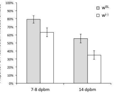
We first analysed whether Wolbachia influences Plasmodium prevalence. Our results 193
show that the probability of becoming infected with P. relictum is significantly higher when 194
Wolbachia is present (wSL). This effect is consistent across the oocyst (probability of
195
infection in wSL is on average 15.9 ± 7.1 % higher than in w(-), !#" = 5.42, p = 0.02, model 1,) 196
and the sporozoite (20.6 ± 7.7 % higher, χ#" = 10.74, p = 0.001, model 2) stages (Figure 1). 197
The combined analysis of the two measurement times revealed a mean (± standard error) 198
decrease of 26.2 (± 5.3) % in the Plasmodium prevalence between 7-8 and 14 dpbm 199
(Plasmodium stage effect: χ#" = 24.15, p < 0.0001, model 3), irrespective of the presence of 200
Wolbachia (Wolbachia x Plasmodium stage interaction: χ#" = 0.02, p = 0.88, model 3; Figure 201
1). In wSL females, the probability of becoming infected by Plasmodium when exposed to an 202
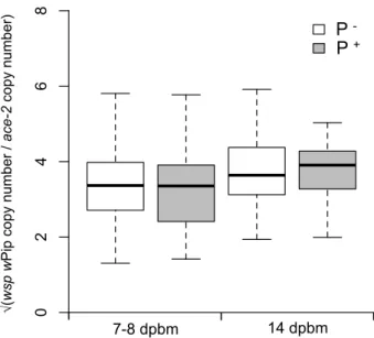
infected bird is independent of the density of Wolbachia (oocysts: χ#" = 0.21, p = 0.64, model 203
4; sporozoites: χ#" = 1.18, p = 0.28, model 5). Reciprocally, the Wolbachia density in female 204
abdomens was not different between mosquitoes fed on a Plasmodium-infected or uninfected 205
bird either at 7-8 dpbm (χ#" = 2.84, p = 0.09, model 10) or at 14 dpbm (χ #
" = 0.01, p = 0.91, 206
model 11; Figure 3). 207
We then analysed whether Wolbachia influences intensity of the Plasmodium 208
infection. The amount of oocysts that successfully developed in the mosquito midgut is 209
significantly higher in wSL than in w(-) females (χ#" =4.95, p = 0.03, model 6, Figure 2a). wSL
210
females have on average 3 more oocysts than w(-) ones (mean ± se, 8.4 ± 1.4 and 5.7 ± 0.8
211
oocysts, respectively). By contrast, the relative quantity of sporozoites present in infected 212
mosquito thoraxes is independent of the presence of Wolbachia (χ#" = 0.69, p = 0.55, model 7; 213
Figure 2b). As above, neither oocyst nor sporozoite load are correlated with Wolbachia 214
density (oocyst : χ#" = 2.64, p = 0.10, model 8; sporozoite : χ#" = 0.06, p = 0.84, model 9; 215 Figure 4). 216 217 218
Discussion
219Current views about the impact of Wolbachia on Plasmodium infections are almost entirely 220
based on data regarding artificially transfected mosquitoes. This work has shown that 221
Wolbachia reduces the number of Plasmodium oocysts in the midgut of mosquitoes. In
222
contrast, and probably because of the difficulty in finding natural Wolbachia infections in
223
epidemiologically significant malaria vectors, the role of natural Wolbachia infections on
224
Plasmodium development have either been ignored entirely or been given only cursory
225
attention. Wolbachia-mosquito combinations with a long evolutionary history may, however,
226
be key for understanding what will happen with Wolbachia-transfected mosquitoes several 227
generations down the line if, as has been shown in other systems [22, 23], the novel 228
Wolbachia-host interaction evolves rapidly. The number of generations needed for such 229
evolutionary change can be between 20 [22] and 200 [23, 35]. To our knowledge, the only 230
studies carried out using natural Wolbachia infections involve the mosquito Aedes fluviatilis 231
and the Asian avian malaria parasite P. gallinaceum. This work has shown that, far from 232
decreasing parasitaemia, Wolbachia either has no effect [17, 19] or increases [19] the number 233
of Plasmodium oocysts in the midgut of the mosquito. Ae. fluviatilis is, however, a South 234
American mosquito that serves a convenient laboratory host for P gallinaceum, but it is not 235
its natural vector. Previous work has indeed shown that Wolbachia can render contrasting 236
results on natural [23] and artificial [17, 19] Plasmodium combinations so the question, 237
which is relevant for the long term success of malaria control programs, of whether 238
Wolbachia can interfere with Plasmodium transmission in an entirely natural system is still 239
unresolved. 240
Here we used an entirely natural mosquito-Wolbachia-Plasmodium combination to 241
investigate whether Wolbachia increases or decreases the parasitaemia of mosquitoes. In 242
contrast to most previous studies, which have been centered on the quantification of oocysts 243
in the midgut of mosquitoes, we aimed to obtain a measurement of parasitaemia that would 244
relate more directly to transmission by following the infections all the way to the sporozoites 245
stage as recently done in An. stephensi [12]. We found that Wolbachia increases marginally, 246
albeit statistically significantly, the oocyst load of mosquitoes. However, the difference in 247
oocyst load found in the midguts on day 7 was not sufficiently marked to translate into a 248
difference in sporozoite load in the salivary glands seven days later. One potential 249
explanation for these results is that since a single oocyst can produce thousands sporozoites, 250
beyond a certain oocyst threshold the salivary glands of mosquitoes may have become 251
saturated by sporozoites [36]. Alternatively, the drastic loss of parasites that inevitably takes 252
place between the midgut and the salivary stages in any Plasmodium infection [32] may 253
upstage the marginal differences in oocystaemia that exist early on. Proof of the inefficient 254
migration from the midgut to the salivary glands is the significant (26%) decrease in 255
Plasmodium prevalence we observed between the oocyst and the sporozoite stages, which 256
was independent of the presence of Wolbachia. 257
Irrespective of the underlying mechanism, we believe that the epidemiological 258
significance of having more or less Plasmodium parasites in the gut or even in the salivary 259
glands remains to be demonstrated. As stated above, a single oocyst can produce between 260
2000 and 8000 sporozoites [37], and as few as ten sporozoites suffice to start a new infection 261
[26]. There is also no consistent evidence that the density of sporozoites in the salivary glands 262
correlates with the number of infecting sporozoites [38], or that this correlates with the 263
probability of a successful infection in the host (but see [39]). Mosquito infection intensity is, 264
indeed, conspicuously absent from current models of malaria transmission and epidemiology 265
[26, 40]. Infection intensity may, however, bear on epidemiology if it correlates negatively 266
with key life history traits of the vector, such as longevity, but the evidence for this is sparse 267
and comes from unrealistically high infections [41]. In contrast, infection prevalence, ie the 268
number of infectious mosquitoes in a population, is the keystone of epidemiological models 269
[26]. The proportion of infectious mosquitoes in a population, sometimes called the 270
sporozoite rate, is a key determinant of the rate at which hosts are bitten in a population [26, 271
40]. Here we show that the presence of Wolbachia increases sporozoite prevalence by as 272
much as 21%. Wolbachia does therefore play a major role in the transmission of Plasmodium 273
in the avian malaria system. 274
In several host species Wolbachia density can fluctuate both between individuals [31, 275
42] and within individuals over time [42, 43], and several Wolbachia-induced phenotypes, 276
such as cytoplasmic incompatibility [42] (but see [43]), longevity curtailment [44] or host 277
resistance to viruses [45], have been shown to depend on the density of infecting bacteria. 278
The correlation between Wolbachia density and parasite density can provide interesting 279
insights as to the mechanisms underlying the interaction. For example, a strong negative 280
correlation was found between Wolbachia density and dengue virus load in Ae. agypti and 281
Ae. albopictus cell lines [45], whereas in Ae. albopictus infected with the chikungunya virus, 282
the intensive phase of the viral replication is concomitant with a significant decrease in 283
Wolbachia load [20, 46, 47], leading the authors to suggest immune competition and resource 284
competition respectively, as the mechanisms driving the interaction between these two 285
players. Here, however, neither the probability nor the intensity of Plasmodium infection at 286
either the oocyst or sporozoite stages are explained by the density of Wolbachia. It would 287
therefore appear that it is the presence of Wolbachia, irrespective of its density, that 288
determines the increase in prevalence and intensity observed, as previously found in An. 289
gambiae with both P. falciparum and P. berghei [13, 14]. In addition, the density of bacteria 290
did not differ depending on whether the mosquitoes were infected by Plasmodium or not, 291
suggesting that the Wolbachia-Plasmodium interaction only works one way. 292
With this in mind, several different, but non-exclusive, mechanisms may be envisaged 293
to explain our results. First, we found that Wolbachia-infected mosquitoes were significantly 294
bigger than Wolbachia-free ones and may thus have simply taken larger blood meals, thereby 295
increasing their intake of Plasmodium gametocytes (the stage which is transmissible to 296
mosquitoes). We have previously shown that the number of P. relictum oocysts is 297
significantly correlated with the amount of blood ingested by the mosquitoes, albeit in a non-298
linear way [29]. Second, Wolbachia may facilitate the successful establishment of 299
Plasmodium within the mosquito tissues. One obvious way in which this could happen is 300
through a Wolbachia-induced down-regulation of the nonspecific arm of the mosquito 301
immune system, a form of self-protection that has been observed both in pill bugs (or 302
woodlice) [48] and parasitoids [49]. In this respect, these natural Wolbachia infections would 303
behave in a drastically different way to artificial infections, which are often found to up-304
regulate the immune system when introduced into a novel host [12, 13, 15, 17, 45]. 305
Third, the differences observed between our Wolbachia-infected and -free mosquito 306
lines could be mediated by differences in their midgut microbiota, which have been recently 307
shown to play a key role in mosquito resistance to Plasmodium infection [50, 51]. Using 308
tetracycline to eliminate Wolbachia is standard practice, the consensus being that mosquitoes 309
recover their microbial flora over a certain number of generations, a premise that, to our 310
knowledge has never been explicitly tested. Therefore, the possibility that the antibiotic 311
treatment may have irreversibly altered the midgut microbiota of mosquitoes, and therefore 312
the resistance to Plasmodium infection, cannot be totally eliminated. More interesting from a 313
biological point of view, but to our knowledge also hitherto unexplored, is the possibility that 314
Wolbachia itself may modify (through competition, or facilitation), the density and 315
composition of the microbial flora of their hosts. 316
Finally, w(-) was reared for ca. 30 generations before the experiment to eliminate side 317
effects of the tetracycline. Although the wSL and w(-) were kept throughout under identical 318
culturing conditions, we cannot entirely exclude the possibility that the two lines may have 319
diverged and that the results we obtain are due to different genetic backgrounds. Further work 320
should replicate these results with, if possible, several Wolbachia-infected and uninfected 321
lines. 322
Previous work in this system has shown that Plasmodium-infected females suffer 323
lower mortality rates if they are also infected with Wolbachia [27]. We had originally 324
advanced two potential explanations for these results: Wolbachia infected mosquitoes could 325
be either more resistant or more tolerant to a Plasmodium infection. Under the first 326
(resistance) scenario, Wolbachia would limit or inhibit parasite development, thereby 327
reducing overall parasitaemia. Dawes et al [41] have indeed shown that in rodent malaria the 328
number of oocysts in the mosquito midgut is correlated with mosquito longevity, but the 329
evidence comes from extremely high (100-2000) oocyst burdens. Under the second 330
(tolerance) scenario, Wolbachia would limit or compensate for the damage incurred by the 331
parasite, without necessarily altering the within-host growth rate of the parasite [52]. An 332
increase in tolerance to pathogens has been previously observed with native Wolbachia strain 333
of Drosophila flies when challenged with viruses [9, 53]. Elucidating which of these 334
mechanisms is at play is essential from a transmission perspective because parasite-resistant 335
vectors are expected to be worse vectors of diseases, while the opposite will be true for 336
parasite-tolerant ones (the "tragedy of tolerance" [54]). The results of the present experiments 337
show that Wolbachia-infected mosquitoes are in fact less resistant to Plasmodium, leaving a 338
higher Wolbachia-associated tolerance to Plasmodium as the only potential explanation for 339
the longevity results, the mechanisms underlying which remain to be explored. 340
In conclusion, we show that Wolbachia increases the susceptibility of Cx. pipiens 341
mosquitoes to P. relictum, significantly increasing the prevalence of salivary gland stage 342
infections. Previous work on this same system has shown that Wolbachia also protects 343
mosquitoes against a Plasmodium-induced mortality [27]. As both mosquito mortality and 344
infection prevalence are two key determinants of Plasmodium epidemiology, these results 345
suggest that naturally Wolbachia-infected mosquitoes may, in fact, be better vectors of 346
malaria than Wolbachia-free ones. 347
348
Acknowledgements
349We are grateful to S. Alizon and F. Vavre and the three anonymous referees for useful 350
discussions and comments on the manuscript. We also thank N. Barougier, P. Boutinaud, J. 351
Denoyelle, P. Perret and G. Sorci, for their help at different stages of the experiments. This 352
project is funded by the French ANR program (ANR “IRMAL”) to AR. AN was partly 353
funded by an ERC starting grant to Sylvain Gandon, FZ was funded by a PhD grant from the 354
CNRS and the Languedoc-Roussillon Region. This is contribution ISEM 2013-208 of the 355
Institut des Sciences de l'Evolution de Montpellier (UMR 5554 CNRS – Université 356
Montpellier 2). 357
Animal experiments were carried out in strict accordance with the ‘National Charter on the Ethics of Animal Experimentation’ of the French Government, and all efforts were made to minimize suffering. Experiments were approved by the Ethical Committee for Animal Experimentation established by the authors’ institution (CNRS) under the auspices of the French Ministry of Education and Research (permit number CEEA- LR-1051).
References
1. Alizon S., van Baalen M. 2008 Multiple infections, immune dynamics, and the evolution of virulence. Am Nat 172(4), E150-E168. (doi:10.1086/590958).
2. Pedersen A.B., Fenton A. 2007 Emphasizing the ecology in parasite community ecology. Trends Ecol Evol 22(3), 133-139. (doi:10.1016/j.tree.2006.11.005).
3. Graham A.L. 2008 Ecological rules governing helminth-microparasite coinfection. Proc Natl Acad Sci USA 105(2), 566-570. (doi:10.1073/pnas.0707221105).
4. Riley M.A., Wertz J.E. 2002 Bacteriocins: evolution, ecology, and application. Annual Review of Microbiology 56, 117-137. (doi:10.1146/annurev.micro.56.012302.161024). 5. Swanson S.J., Neitzel D., Reed K.D., Belongia E.A. 2006 Coinfections acquired from
Ixodes ticks. Clin Microbiol Rev 19(4), 708-727. (doi:10.1128/cmr.00011-06).
6. Hughes T., Irwin P., Hofmeister E., Paskewitz S.M. 2010 Occurrence of avian Plasmodium and West Nile virus in Culex species in Wisconsin. J Am Mosq Control Assoc 26(1), 24-31.
7. Vazeille M., Mousson L., Martin E., Failloux A.-B. 2010 Orally co-infected Aedes albopictus from La Reunion Island, Indian Ocean, can deliver both dengue and chikungunya infectious viral particles in their saliva. PLoS Negl Trop Dis 4(6). (doi:10.1371/journal.pntd.0000706).
8. Hedges L.M., Brownlie J.C., O'Neill S.L., Johnson K.N. 2008 Wolbachia and virus protection in insects. Science 322(5902), 702-702. (doi:10.1126/science.1162418). 9. Teixeira L., Ferreira A., Ashburner M. 2008 The bacterial symbiont Wolbachia induces
resistance to RNA viral infections in Drosophila melanogaster. PLoS Biol 6(12), 2753-2763. (doi:10.1371/journal.pbio.1000002).
10. Xi Z.Y., Khoo C.C.H., Dobson S.L. 2005 Wolbachia establishment and invasion in an Aedes aegypti laboratory population. Science 310(5746), 326-328. (doi:10.1126/science.1117607).
11. McMeniman C.J., Lane R.V., Cass B.N., Fong A.W.C., Sidhu M., Wang Y.F., O'Neill S.L. 2009 Stable introduction of a life-shortening Wolbachia infection into the mosquito Aedes aegypti. Science 323(5910), 141-144. (doi:10.1126/science.1165326). 12. Bian G., Joshi D., Dong Y., Lu P., Zhou G., Pan X., Xu Y., Dimopoulos G., Xi Z. 2013
Wolbachia invades Anopheles stephensi populations and induces refractoriness to Plasmodium infection. Science 340, 748-751. (doi:10.1126/science.1236192).
13. Hughes G.L., Koga R., Xue P., Fukatsu T., Rasgon J.L. 2011 Wolbachia infections are virulent and inhibit the human malaria parasite Plasmodium falciparum in Anopheles gambiae. PLoS Pathog 7(5), e1002043. (doi:10.1371/journal.ppat.1002043).
14. Hughes G.L., Vega-Rodriguez J., Xue P., Rasgon J.L. 2012 Wolbachia strain wAlbB enhances infection by the rodent malaria parasite Plasmodium berghei in Anopheles
gambiae mosquitoes. Appl Environ Microbiol 78(5), 1491-1495.
(doi:10.1128/aem.06751-11).
15. Kambris Z., Blagborough A.M., Pinto S.B., Blagrove M.S.C., Godfray H.C.J., Sinden R.E., Sinkins S.P. 2010 Wolbachia stimulates immune gene expression and inhibits Plasmodium development in Anopheles gambiae. PLoS Pathog 6(10), e1001143. (doi:10.1371/journal.ppat.1001143).
16. Kambris Z., Cook P.E., Phuc H.K., Sinkins S.P. 2009 Immune activation by life-shortening Wolbachia and reduced filarial competence in mosquitoes. Science 326(5949), 134-136. (doi:10.1126/science.1177531).
17. Moreira L.A., Iturbe-Ormaetxe I., Jeffery J.A., Lu G.J., Pyke A.T., Hedges L.M., Rocha B.C., Hall-Mendelin S., Day A., Riegler M., et al. 2009 A Wolbachia symbiont in Aedes aegypti limits infection with dengue, Chikungunya, and Plasmodium. Cell 139(7), 1268-1278. (doi:10.1016/j.cell.2009.11.042).
18. Blagrove M.S.C., Arias-Goeta C., Failloux A.-B., Sinkins S.P. 2012 Wolbachia strain wMel induces cytoplasmic incompatibility and blocks dengue transmission in Aedes albopictus. Proc Natl Acad Sci USA 109(1), 255-260. (doi:10.1073/pnas.1112021108). 19. Baton L.A., Pacidonio E.C., Goncalves D.d.S., Moreira L.A. 2013 wFlu:
Characterization and evaluation of a native Wolbachia from the mosquito Aedes fluviatilis as a potential vector control agent. PLoS One 8(3), e59619-e59619. (doi:10.1371/journal.pone.0059619).
20. Mousson L., Martin E., Zouache K., Madec Y., Mavingui P., Failloux A.B. 2010 Wolbachia modulates Chikungunya replication in Aedes albopictus. Mol Ecol 19(9), 1953-1964. (doi:10.1111/j.1365-294X.2010.04606.x).
21. Glaser R.L., Meola M.A. 2010 The native Wolbachia endosymbionts of Drosophila melanogaster and Culex quinquefasciatus increase host resistance to West Nile virus infection. PLoS One 5(8), e11977. (doi:10.1371/journal.pone.0011977).
22. McGraw E.A., Merritt D.J., Droller J.N., O'Neill S.L. 2002 Wolbachia density and virulence attenuation after transfer into a novel host. Proc Natl Acad Sci USA 99(5), 2918-2923. (doi:10.1073/pnas.052466499).
23. Weeks A.R., Turelli M., Harcombe W.R., Reynolds K.T., Hoffmann A.A. 2007 From parasite to mutualist: Rapid evolution of Wolbachia in natural populations of Drosophila. PLoS Biol 5(5), 997-1005. (doi:10.1371/journal.pbio.0050114).
24. Tripet F. 2009 Ecological immunology of mosquito-malaria interactions: Of non-natural versus non-natural model systems and their inferences. Parasitology 136(14), 1935-1942. (doi:10.1017/s0031182009006234).
25. Cohuet A., Osta M.A., Morlais I., Awono-Ambene P.H., Michel K., Simard F., Christophides G.K., Fontenille D., Kafatos F.C. 2006 Anopheles and Plasmodium: from laboratory models to natural systems in the field. EMBO Rep 7(12), 1285-1289. (doi:10.1038/sj.embor.7400831).
26. Smith D.L., McKenzie F.E. 2004 Statics and dynamics of malaria infection in Anopheles mosquitoes. Malaria J 3, 13. (doi:10.1186/1475-2875-3-13).
27. Zélé F., Nicot A., Duron O., Rivero A. 2012 Infection with Wolbachia protects mosquitoes against Plasmodium-induced mortality in a natural system. J Evol Biol 25(7), 1243-1252. (doi:10.1111/j.1420-9101.2012.02519.x).
28. Bensch S., Hellgren O., Perez-Tris J. 2009 MalAvi: a public database of malaria parasites and related haemosporidians in avian hosts based on mitochondrial cytochrome b lineages. Mol Ecol Res 9(5), 1353-1358. (doi:10.1111/j.1755-0998.2009.02692.x).
29. Vézilier J., Nicot A., Gandon S., Rivero A. 2010 Insecticide resistance and malaria transmission: infection rate and oocyst burden in Culex pipiens mosquitoes infected with Plasmodium relictum. Malaria J 9, 379. (doi:10.1186/1475-2875-9-379).
30. Weill M., Berticat C., Raymond N., Chevillon C. 2000 Quantitative polymerase chain reaction to estimate the number of amplified esterase genes in insecticide-resistant mosquitoes. Anal Biochem 285(2), 267-270. (doi:10.1006/abio.2000.4781).
31. Berticat C., Rousset F., Raymond M., Berthomieu A., Weill M. 2002 High Wolbachia density in insecticide-resistant mosquitoes. Proc R Soc Lond B 269(1498), 1413-1416. (doi:10.1098/rspb.2002.2022).
32. Vaughan J.A. 2007 Population dynamics of Plasmodium sporogony. Trends Parasitol 23(2), 63-70. (doi:10.1016/j.pt.2006.12.009).
33. Bolker B.M. 2008 Ecological models and data in R New Jersey, Princeton University Press.
34. Crawley M.J. 2007 The R Book Chichester, England, John Wiley & Sons, Ltd; 942 p. 35. Carrington L.B., Hoffmann A.A., Weeks A.R. 2010 Monitoring long-term evolutionary
changes following Wolbachia introduction into a novel host: the Wolbachia popcorn infection in Drosophila simulans. Proc R Soc B 277(1690), 2059-2068. (doi:10.1098/rspb.2010.0166).
36. Sinden R.E., Dawes E.J., Alavi Y., Waldock J., Finney O., Mendoza J., Butcher G.A., Andrews L., Hill A.V., Gilbert S.C., et al. 2007 Progression of Plasmodium berghei through Anopheles stephensi is density-dependent. PLoS Pathog 3(12), 2005-2016. (doi:10.1371/journal.ppat.0030195).
37. Wang Q., Fujioka H., Nussenzweig V. 2005 Exit of Plasmodium sporozoites from oocysts is an active process that involves the circumsporozoite protein. PLoS Pathog 1(1), e9. (doi:10.1371/journal.ppat.0010009).
38. Beier J.C. 1998 Malaria parasite development in mosquitoes. Ann Rev Entomol 43, 519-543. (doi:10.1146/annurev.ento.43.1.519).
39. Kebaier C., Voza T., Vanderberg J. 2009 Kinetics of Mosquito-Injected Plasmodium Sporozoites in Mice: Fewer Sporozoites Are Injected into Sporozoite-Immunized Mice. PLoS Pathog 5(4). (doi:10.1371/journal.ppat.1000399).
40. Smith D.L., Dushoff J., Snow R.W., Hay S.I. 2005 The entomological inoculation rate and Plasmodium falciparum infection in African children. Nature 438(7067), 492-495. (doi:10.1038/nature04024).
41. Dawes E.J., Churcher T.S., Zhuang S., Sinden R.E., Basanez M.G. 2009 Anopheles mortality is both age- and Plasmodium-density dependent: implications for malaria transmission. Malaria J 8, 228. (doi:10.1186/1475-2875-8-228).
42. Clark M.E., Veneti Z., Bourtzis K., Karr T.L. 2003 Wolbachia distribution and cytoplasmic incompatibility during sperm development: the cyst as the basic cellular unit of CI expression. Mech Dev 120(2), 185-198. (doi:10.1016/s0925-4773(02)00424-0).
43. Duron O., Fort P., Weill M. 2007 Influence of aging on cytoplasmic incompatibility, sperm modification and Wolbachia density in Culex pipiens mosquitoes. Heredity 98(6), 368-374. (doi:10.1038/sj.hdy.6800948).
44. Min K.T., Benzer S. 1997 Wolbachia, normally a symbiont of Drosophila, can be virulent, causing degeneration and early death. Proc Natl Acad Sci USA 94(20), 10792-10796. (doi:10.1073/pnas.94.20.10792).
45. Lu P., Bian G., Pan X., Xi Z. 2012 Wolbachia induces density-dependent inhibition to dengue virus in mosquito cells. PLoS Negl Trop Dis 6(7), e1754. (doi:10.1371/journal.pntd.0001754).
46. Tortosa P., Courtiol A., Moutailler S., Failloux A.B., Weill M. 2008 Chikungunya-Wolbachia interplay in Aedes albopictus. Insect Mol Biol 17(6), 677-684. (doi:10.1111/j.1365-2583.2008.00842.x).
47. Zouache K., Michelland R.J., Failloux A.-B., Grundmann G.L., Mavingui P. 2012 Chikungunya virus impacts the diversity of symbiotic bacteria in mosquito vector. Mol Ecol 21(9), 2297-2309. (doi:10.1111/j.1365-294X.2012.05526.x).
48. Sicard M., Chevalier F., De Vlechouver M., Bouchon D., Greve P., Braquart-Varnier C. 2010 Variations of immune parameters in terrestrial isopods: a matter of gender, aging and Wolbachia. Naturwissenschaften 97(9), 819-826. (doi:10.1007/s00114-010-0699-2).
49. Fytrou A., Schofield P.G., Kraaijeveld A.R., Hubbard S.F. 2006 Wolbachia infection suppresses both host defence and parasitoid counter-defence. Proc R Soc B 273(1588), 791-796. (doi:10.1098/rspb.2005.3383).
50. Dong Y.M., Manfredini F., Dimopoulos G. 2009 Implication of the mosquito midgut microbiota in the defense against malaria parasites. PLoS Pathog 5(5), e1000423. (doi:10.1371/journal.ppat.1000423).
51. Cirimotich C.M., Dong Y.M., Clayton A.M., Sandiford S.L., Souza-Neto J.A., Mulenga M., Dimopoulos G. 2011 Natural microbe-mediated refractoriness to Plasmodium infection in Anopheles gambiae. Science 332(6031), 855-858. (doi:10.1126/science.1201618).
52. Raberg L., Sim D., Read A.F. 2007 Disentangling genetic variation for resistance and tolerance to infectious diseases in animals. Science 318(5851), 812-814. (doi:10.1126/science.1148526).
53. Osborne S.E., Leong Y.S., O'Neill S.L., Johnson K.N. 2009 Variation in antiviral protection mediated by different Wolbachia strains in Drosophila simulans. PLoS Pathog 5(11), e1000656. (doi:10.1371/journal.ppat.1000656).
54. Vale P.F., Wilson A.J., Best A., Boots M., Little T.J. 2011 Epidemiological, evolutionary, and coevolutionary implications of context-dependent parasitism. Am Nat 177(4), 510-521. (doi:10.1086/659002).
Figure 1. Effect of Wolbachia on the prevalence of Plasmodium infection 7 days (oocyst stage) and 14 days post blood meal (sporozoite stage). Bars represent means (± SE) for Wolbachia-carrying females (grey bars) and Wolbachia-free ones (white bars).
Figure 2. Effect of the presence of Wolbachia on Plasmodium burden in mosquitoes. Distribution of the number of oocysts in the midgut of Plasmodium-infected females 7-8 days post blood meal (A), and of the relative quantity of sporozoites in the thorax of Plasmodium-infected females 14 days post blood meal (B), for Wolbachia-carrying females (grey circles) and Wolbachia-free ones (black circles). Horizontal lines represent medians.
0%# 10%# 20%# 30%# 40%# 50%# 60%# 70%# 80%# 90%# 100%# 7-8#dpbm# 14#dpbm# wSL# w(-)# wSL w(-) Pro po rt io n of Pl asmo di um in fe ct ed fe ma le s Mosquito lines N umb er of o ocyst s 0 10 20 30 40 50 60 W+ W-Mosquito lines cyt b Pl asmo di um co py nu mb er / Ace -2 co py nu mb er 0.00 0.05 0.10 0.15 0.20 0.25 W+ W-A! B! wSL w(-) wSL w(-) N umb er of oo cyst s cyt b Pl asmo di um co py nu mb er / ace -2 co py nu mb er
Figure 3. Boxplot of the Wolbachia density in wSL females according to the Plasmodium infection status at 7-8 days (oocysts) and 14 days (sporozoites) post blood meal. White boxes: Plasmodium uninfected mosquitoes (includes females fed on a control bird and females that did not become infected after feeding on a Plasmodium-infected bird) and grey boxes: Plasmodium infected mosquitoes. Wolbachia densities were Box-Cox transformed to linearize the data for the graphic representation.
Figure 4. Correlation between the density of Wolbachia and the intensity of Plasmodium infection at the oocyst (A) and sporozoite (B) stages (7-8 days and 14 days post blood meal respectively). Both Wolbachia and Plasmodium densities were Box-Cox transformed to linearize the data for the graphic representation.
Plasmodium infection at 7 and 14 days pbm
√(w sp w Pi p co py nu mb er / Ace -2 co py nu mb er) 0 2 4 6 8 P-P+ 7-8 dpbm 14 dpbm P - P + √( w sp w Pi p co py nu mb er / ace -2 co py nu mb er )
√(wsp wPip copy number / Ace-2 copy number)
lo g(cyt b Pl asmo di um co py nu mb er / Ace -2 co py nu mb er) 2.0 2.5 3.0 3.5 4.0 4.5 5.0 5.5 -7 -6 -5 -4 -3 -2 -1
√( wsp wPip copy number / Ace-2 copy number)
lo g(O ocyst s nu mb er) 1 2 3 4 5 6 0 1 2 3 4 A! B! log( cyt b Pl asmo di um co py nu mb er / ace -2 co py nu mb er ) log( oo cyst s nu mb er )
√(wsp wPip copy number / ace-2 copy number) √(wsp wPip copy number / ace-2 copy number)
Table S1. Summary of the different studies conducted to date on Wolbachia-mediated pathogen interference. a nature of the host-Wolbachia or host-parasite combination : “natural” when it occurs in nature, “artificial” when it was created in the lab. Lines that refer to complete natural combinations are highlighted in grey. bEffect of Wolbachia on parasite prevalence and intensity: (+) increase, (−) decrease, (×) no observed effects, (.) not studied. Abbreviations: IVV-6: Inluenza virus; LACV: La Crosse virus; FHV: Flock House virus; CHIKV: Chikungunya virus; WNV: West Nile virus; DENV-2: Dengue virus. Bacteria: 1 Pseudomonas aeruginosa, Serratia marcescens and Erwinia carotovora; and 2 Providencia rettgeri, Salmonella enterica and Listeria monocytogenes.
Host species Wolbachia strains Host-Wolbachia a Parasites Host-parasite a Parasite prevalence b Parasite intensity b References
Drosophila flies D. melanogaster
wMel natural DCV, Nora virus natural . − [1]
wMel natural FHV artificial . × [1]
wMel natural CHIKV, WNV artificial . − [2]
wMel natural IVV-6, LACV, Bacteria1,2 artificial . × [1-4]
D. simulans
wAu natural DCV natural . − [5]
wAu natural FHV artificial . − [5]
wHa / wNO natural DCV natural . × [5]
wHa / wNO natural FHV artificial . × [5]
? natural Leptopilina heterotoma natural × + [6]
wMel artificial DCV natural . − [5]
Mosquitoes
Aedes aegypti
wAlbB artificial DENV-2 natural . − [7-9]
wMel artificial DENV-2 natural . − [10]
wMelPop-CLA artificial CHIKV, DENV-2 natural − − [10, 11]
wMelPop-CLA artificial Brugia pahangi natural − − [12, 13]
wMelPop-CLA artificial P. gallinaceum artificial − − [11]
Ae. albopictus wAlbA&B natural CHIKV, DENV-2 natural × × [7, 14-17]
wMel artificial CHIKV, DENV-2 natural − − [16, 18]
Ae. fluvatilis wFlu natural P. gallinaceum artificial . × [11]
wFlu natural P. gallinaceum artificial . × + [19]
Ae. polynesiensis wPolA natural Brugia pahangi natural . × [20]
wAlbB artificial Brugia pahangi natural . − [20]
Culex pipiens quinquefasciatus wPip natural WNV natural × × − [2]
Anopheles gambiae
wAlbB artificial P. falciparum natural . − [21]
wAlbB artificial P. berghei artificial . + [22]
wMelPop-CLA artificial P. falciparum natural . − [21]
wMelPop-CLA artificial P. berghei artificial . − [12, 22]
An. stephensi wAlbB artificial P. falciparum natural − − [23]
Other insects
References
1. Teixeira L., Ferreira A., Ashburner M. 2008 The bacterial symbiont Wolbachia induces resistance to RNA viral infections in Drosophila melanogaster. PLoS Biol 6(12), 2753-2763. (doi:10.1371/journal.pbio.1000002).
2. Glaser R.L., Meola M.A. 2010 The native Wolbachia endosymbionts of Drosophila melanogaster and Culex quinquefasciatus increase host resistance to West Nile virus infection. PLoS One 5(8), e11977. (doi:10.1371/journal.pone.0011977).
3. Wong Z.S., Hedges L.M., Brownlie J.C., Johnson K.N. 2011 Wolbachia-mediated antibacterial protection and immune gene regulation in Drosophila. PLoS One 6(9), e25430. (doi:10.1371/journal.pone.0025430).
4. Rottschaefer S.M., Lazzaro B.P. 2012 No effect of Wolbachia on resistance to intracellular infection by pathogenic bacteria in Drosophila melanogaster. PLoS One 7(7), e40500.
5. Osborne S.E., Leong Y.S., O'Neill S.L., Johnson K.N. 2009 Variation in antiviral protection mediated by different Wolbachia strains in Drosophila simulans. PLoS Pathog 5(11), e1000656. (doi:10.1371/journal.ppat.1000656).
6. Fytrou A., Schofield P.G., Kraaijeveld A.R., Hubbard S.F. 2006 Wolbachia infection suppresses both host defence and parasitoid counter-defence. Proc R Soc B 273(1588), 791-796. (doi:10.1098/rspb.2005.3383).
7. Bian G.W., Xu Y., Lu P., Xie Y., Xi Z.Y. 2010 The endosymbiotic bacterium Wolbachia induces resistance to dengue virus in Aedes aegypti. PLoS Pathog 6(4), e1000833. (doi:10.1371/journal.ppat.1000833).
8. Lu P., Bian G., Pan X., Xi Z. 2012 Wolbachia induces density-dependent inhibition to dengue virus in mosquito cells. PLoS Negl Trop Dis 6(7), e1754.
9. Pan X., Zhou G., Wu J., Bian G., Lu P., Raikhel A.S., Xi Z. 2012 Wolbachia induces reactive oxygen species (ROS)-dependent activation of the Toll pathway to control dengue virus in the mosquito Aedes aegypti. Proc Natl Acad Sci USA 109(1), 13-14. (doi:10.1073/pnas.1116932108). 10. Walker T., Johnson P.H., Moreira L.A., Iturbe-Ormaetxe I., Frentiu F.D., McMeniman C.J., Leong
Y.S., Dong Y., Axford J., Kriesner P., et al. 2011 The wMel Wolbachia strain blocks dengue and invades caged Aedes aegypti populations. Nature 476(7361), 450-453. (doi:10.1038/nature10355). 11. Moreira L.A., Iturbe-Ormaetxe I., Jeffery J.A., Lu G.J., Pyke A.T., Hedges L.M., Rocha B.C.,
Hall-Mendelin S., Day A., Riegler M., et al. 2009 A Wolbachia symbiont in Aedes aegypti limits infection with dengue, Chikungunya, and Plasmodium. Cell 139(7), 1268-1278. (doi:10.1016/j.cell.2009.11.042).
12. Kambris Z., Blagborough A.M., Pinto S.B., Blagrove M.S.C., Godfray H.C.J., Sinden R.E., Sinkins S.P. 2010 Wolbachia stimulates immune gene expression and inhibits Plasmodium development in
13. Kambris Z., Cook P.E., Phuc H.K., Sinkins S.P. 2009 Immune activation by life-shortening
Wolbachia and reduced filarial competence in mosquitoes. Science 326(5949), 134-136.
(doi:10.1126/science.1177531).
14. Tortosa P., Courtiol A., Moutailler S., Failloux A.B., Weill M. 2008 Chikungunya-Wolbachia interplay in Aedes albopictus. Insect Mol Biol 17(6), 677-684. (doi:10.1111/j.1365-2583.2008.00842.x).
15. Mousson L., Martin E., Zouache K., Madec Y., Mavingui P., Failloux A.B. 2010 Wolbachia modulates Chikungunya replication in Aedes albopictus. Mol Ecol 19(9), 1953-1964. (doi:10.1111/j.1365-294X.2010.04606.x).
16. Blagrove M.S.C., Arias-Goeta C., Failloux A.-B., Sinkins S.P. 2012 Wolbachia strain wMel induces cytoplasmic incompatibility and blocks dengue transmission in Aedes albopictus. Proc Natl Acad
Sci USA 109(1), 255-260. (doi:10.1073/pnas.1112021108).
17. Zouache K., Michelland R.J., Failloux A.-B., Grundmann G.L., Mavingui P. 2012 Chikungunya virus impacts the diversity of symbiotic bacteria in mosquito vector. Mol Ecol 21(9), 2297-2309. (doi:10.1111/j.1365-294X.2012.05526.x).
18. Blagrove M.S.C., Arias-Goeta C., Di Genua C., Failloux A.-B., Sinkins S.P. 2013 A Wolbachia
wMel transinfection in Aedes albopictus is not detrimental to host fitness and inhibits Chikungunya
virus. PLoS Negl Trop Dis 7(3), E2152-E2152.
19. Baton L.A., Pacidonio E.C., Goncalves D.d.S., Moreira L.A. 2013 wFlu: Characterization and evaluation of a native Wolbachia from the mosquito Aedes fluviatilis as a potential vector control agent. PLoS One 8(3), e59619-e59619. (doi:10.1371/journal.pone.0059619).
20. Andrews E.S., Crain P.R., Fu Y., Howe D.K., Dobson S.L. 2012 Reactive oxygen species production and Brugia pahangi survivorship in Aedes polynesiensis with artificial Wolbachia infection types. PLoS Pathog 8(12), e1003075-e1003075. (doi:10.1371/journal.ppat.1003075). 21. Hughes G.L., Koga R., Xue P., Fukatsu T., Rasgon J.L. 2011 Wolbachia infections are virulent and
inhibit the human malaria parasite Plasmodium falciparum in Anopheles gambiae. PLoS Pathog
7(5), e1002043. (doi:10.1371/journal.ppat.1002043).
22. Hughes G.L., Vega-Rodriguez J., Xue P., Rasgon J.L. 2012 Wolbachia strain wAlbB enhances infection by the rodent malaria parasite Plasmodium berghei in Anopheles gambiae mosquitoes.
Appl Environ Microbiol 78(5), 1491-1495. (doi:10.1128/aem.06751-11).
23. Bian G., Joshi D., Dong Y., Lu P., Zhou G., Pan X., Xu Y., Dimopoulos G., Xi Z. 2013 Wolbachia invades Anopheles stephensi populations and induces refractoriness to Plasmodium infection.
Science 340, 748-751. (doi:10.1126/science.1236192).
!
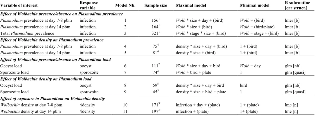
Table S2. Description of the statistical models used in the analyses. "Maximal model": model containing all explanatory variables and their interactions. "Minimal model"
: model containing only the significant variables and their interactions. Round brackets: variable fitted as a random factor. Square brackets: the error structure used (n: normal, b: binomial errors, nb: negative binomial, quasi: quasipoisson-like with link=log and variance = µ²).
Wolb: mosquito lines (wSL or w(-)), infection: exposed to a Plasmodium-infected bird vs. an uninfected bird, size: mosquito wing length, oocyst: number of oocysts in the
midgut, sporozoite: relative density of sporozoites in the thorax, density: Wolbachia density in the abdomen (wSL only) , stage: Plasmodium developmental stage (oocysts at
day 7-8 pbm or sporozoites at day 14 pbm).
Variable of interest Response
variable Model Nb. Sample size Maximal model Minimal model
R subroutine [err struct.]
Effect of Wolbachia presence/absence on Plasmodium prevalence
Plasmodium prevalence at day 7-8 pbm infection 1 1561 Wolb * size + day + (bird) Wolb + (bird) lmer [b] Plasmodium prevalence at day 14 pbm infection 2 1641 Wolb * size + (bird) Wolb + (bird/plate) lmer [b]
Total Plasmodium prevalence infection 3 3211 Wolb * stage * size + (bird) Wolb + stage + (bird) lmer [b]
Effect of Wolbachia density on Plasmodium prevalence
Plasmodium prevalence at day 7-8 pbm infection 4 754 density * size + day + (bird) 1 + (bird) lmer [b] Plasmodium prevalence at day 14 pbm infection 5 814 density * size + (bird) 1 + (bird) lmer [b]
Effect of Wolbachia presence/absence on Plasmodium load
Oocyst load oocyst 6 1112 Wolb * size + day + bird Wolb + day glm [nb]
Sporozoite load sporozoite 7 742 Wolb + bird + plate 1 glm [quasi]
Effect of Wolbachia density on Plasmodium load
Oocyst load oocyst 8 595 density * size + day + bird bird glm [nb]
Sporozoite load sporozoite 9 455 density * size + bird + plate 1 glm [quasi]
Effect of exposure to Plasmodium on Wolbachia density
Wolbachia density at day 7-8 pbm √density 10 1713 infection + day + (plate) 1 + (plate) lme [n] Wolbachia density at day 14 pbm √density 11 1973 infection + (plate) 1+ (plate) lme [n] 1 Includes only females exposed to a Plasmodium infected bird.
2 Includes only females with ≥ 1 oocysts. 3 Includes only wSL females.
4 Includes only wSL females exposed to a Plasmodium infected bird.
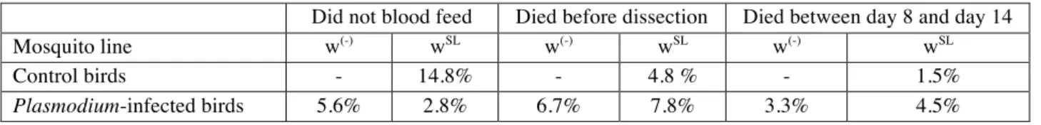
Table S3. Percentages of mosquitoes that did not blood fed or died before the dissections took place.
Did not blood feed Died before dissection Died between day 8 and day 14
Mosquito line w(-) wSL w(-) wSL w(-) wSL
Control birds - 14.8% - 4.8 % - 1.5%