HAL Id: hal-02550734
https://hal.umontpellier.fr/hal-02550734
Submitted on 22 Apr 2020
HAL is a multi-disciplinary open access
archive for the deposit and dissemination of
sci-entific research documents, whether they are
pub-lished or not. The documents may come from
teaching and research institutions in France or
abroad, or from public or private research centers.
L’archive ouverte pluridisciplinaire HAL, est
destinée au dépôt et à la diffusion de documents
scientifiques de niveau recherche, publiés ou non,
émanant des établissements d’enseignement et de
recherche français ou étrangers, des laboratoires
publics ou privés.
Pneumonia in Immunocompromised Patients: More
than Meets the Eye
Jeremy Signe, Boris Jung, Stephanie Nougaret, Fouad Belafia, Fabrizio
Panaro, Michael Bismuth, Samir Jaber
To cite this version:
Jeremy Signe, Boris Jung, Stephanie Nougaret, Fouad Belafia, Fabrizio Panaro, et al.. Pneumonia
in Immunocompromised Patients: More than Meets the Eye. American Journal of Respiratory and
Critical Care Medicine, American Thoracic Society, 2012, 186, �10.1164/rccm.201201-0161IM�.
�hal-02550734�
Images in Pulmonary, Critical Care, Sleep Medicine
and the Sciences
Pneumonia in Immunocompromised Patients
More than Meets the Eye
Jeremy Signe1, Boris Jung1, Stephanie Nougaret2, Fouad Belafia1, Fabrizio Panaro3,
Michael Bismuth4, and Samir Jaber1
1Intensive Care Unit, Saint Eloi Department of Critical Care and Anesthesiology,2Department of Abdominal Imaging,3Department of General and
Liver Transplant Surgery, and4Department of Hepato-Gastroenterology B, CHU de Montpellier, Hoˆpital Saint Eloi, Montpellier, France
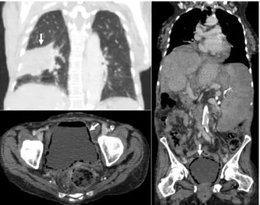
A 63-year-old patient was admitted to the ICU for severe sepsis and rapid onset of shortness of breath. She had had a liver transplant 6 years before and upon ICU admission presented with end-stage cirrhosis due to a sclerosing cholangitis relapse and type 1 diabetes mellitus. Examination led to the diagnosis of community-acquired pneumonia. Streptococcus pneumoniae urine antigen was found to be positive. In our department, a thoracoabdominal computed tomography (CT) scan facing sepsis in immunocompromised patients is always suggested to rule out an infection needing urgent source control. CT confirmed a right bilobar pneumonia, but incidentally showed a distended bladder with circumferential rosary-shaped pneumatosis within the wall and an intravesical hydroaeric level. The diagnosis of emphysematous cystitis was confirmed with urine culture showing 107colony-forming units/ml
amoxicillin-resistant Escherichia coli. Blood cultures found no bacteriemia. Emphysematous cystitis is a rare condition most often encountered in elderly diabetic women (1) and favored by immunodeficiency.
Late infections are a serious threat to patients who have undergone transplant, since they are implied in up to 9% of post–liver transplant deaths, with a global incidence of 0.4 per 1,000 transplant-days (2). Urinary tract infections account for nearly 37% of late infections in solid organ transplant recipients, whereas pneumonia accounts for 11%. Treatment combining urinary catheter-ization, broad spectrum systemic antibiotic therapy, norepinephrine for septic shock, and strict glycemic control was immediately started. The patient ultimately recovered without surgery. Subsequent CT scans showed normalized bladder images. Thus, making the right diagnosis when treating an infection is critical in immunocompromised patients.
Author disclosures are available with the text of this article at www.atsjournals.org. References
1. Thomas AA, Lane BR, Thomas AZ, Remer EM, Campbell SC, Shoskes DA. Emphysematous cystitis: a review of 135 cases. BJU Int 2007;100:17–20. 2. San Juan R, Aguado JM, Lumbreras C, Diaz-Pedroche C, Lopez-Medrano F, Lizasoain M, Gavalda J, Montejo M, Moreno A, Gurgui M, et al. Incidence,
clinical characteristics and risk factors of late infection in solid organ transplant recipients: data from the RESITRA study group. Am J Transplant 2007;7: 964–971.
Figure 1. (Top left panel) Coronal contrast-enhanced computed to-mography (CT) reveals a large alveolar condensation area in the right inferior lobe (arrow). (Bottom left panel) Axial and (right panel) coronal contrast-enhanced CT show “beaded necklace appearance” of the bladder mucosal surface in keeping with multiple diffuse cystic collec-tions of gas within the bladder wall (arrows). This finding reflects the irregular thickening produced by submucosal blebs as seen at direct cystoscopy.
Author Contributions: Drafting the manuscript for important intellectual content, J.S., B.J., F.B., S.J.; imaging, S.N.; primary care physicians, M.B., F.P.
Am J Respir Crit Care Med Vol 186, Iss. 11, p e18, Dec 1, 2012 Copyrightª 2012 by the American Thoracic Society DOI: 10.1164/rccm.201201-0161IM