Accessible and easy-to-use educational tools to teach molecular and
synthetic biology using freeze-dried, cell-free technology
By Ally Huang
B.S. Biomedical Engineering Johns Hopkins University, 2012
MASSACHUSETTS INSTITUTE
OF TECHNOLOGY
JUL 0
3
2019
LIBRARIES
ARCHIVES
Submitted to the Department of Biological Engineering in partial fulfillment of the requirements for the degree of
Doctor of Philosophy in Biological Engineering at the
MASSACHUSETTS INSTITUTE OF TECHNOLOGY June 2019
0 2019 Massachusetts Institute of Technology. All rights reserved.
Signature of author:....
Signature
redacted--Signature reda
Ally Huang Department of Biological Engineering
cted
Mayl,2019C ertified by: ... ...
/
James J. Collinsermeer Professor of Medical Engineering & Science Professor of Biological Engineering Thesis Supervisor
Signature redacted
A ccepted by: ... . ... Forest White Professor of Biological Engineering Chair of Graduate Program, Department of Biological Engineering
Thesis committee members
Angela Koehler, Ph.D. (Chair)
Assistant Professor of Biological Engineering Massachusetts Institute of Technology
James J. Collins, Ph.D. (Thesis Supervisor)
Termeer Professor of Medical Engineering & Science Professor of Biological Engineering
Massachusetts Institute of Technology Peter Dedon, M.D., Ph.D.
Underwood-Prescott Professor of Biological Engineering Massachusetts Institute of Technology
Accessible and easy-to-use educational tools to teach molecular and
synthetic biology using freeze-dried, cell-free technology
By Ally Huang
B.S. Biomedical Engineering Johns Hopkins University, 2012
Submitted to the Department of Biological Engineering on May 1, 2019 in partial fulfillment of the requirements for the degree of Doctor of Philosophy in Biological Engineering.
ABSTRACT
Hands-on demonstrations greatly enhance the teaching of STEM concepts and foster engagement and exploration in the sciences. While numerous chemistry and physics classroom demonstrations exist, few biology demonstrations are practical and accessible due to the challenges and concerns of growing living cells in classrooms. Here I introduce a platform to develop hands-on molecular and synthetic biology educational activities based on easy-to-use, shelf-stable, freeze-dried, cell-free (FD-CF) reactions, which are simply activated by water. By using fluorescent proteins as a visual output, I created a variety of engaging modules using this platform that can teach the central dogma of biology, how certain cellular functions work, and other basic molecular biology topics that are otherwise difficult to easily teach in a hands-on manner. By expanding the platform to other non-visual outputs (such as smell or touch), as well as further incorporating components, such as RNA switches, I also developed modules that can teach more advanced biology topics, such as biochemistry, biomaterials, and synthetic biology, as well as basic laboratory skills such as pipetting, experimental design, and the scientific method. Pilot testing of a prototype kit based on these elements were tested in classrooms across the country and initial results suggest that the activities are accessible, easy to use, educational, and engaging for high school students. Overall, the platform introduces low-cost, user-friendly, and hands-on activities that can be used in classrooms to improve the quality of biology education and open the door for student-driven, independent explorations in the life sciences.
Thesis Supervisor: James. J. Collins
Acknowledgements
I started my Ph.D. the fall of 2014 as a bright-eyed, optimistic young scientist who thought she wanted to do antibody engineering for cancer therapeutics and now am (hopefully) ending it as a more realistic (although still optimistic) experienced scientist creating low-cost education kits to teach molecular and synthetic biology. This journey has been filled with unexpected twists and turns but I would not change anything - every challenge and obstacle has been a valuable learning experience. And I certainly would not be here without the support of everyone in my life, both in lab and out.
My interest in biology first stated back in tenth grade, where my high school biology teacher, Kevin Engstrom opened my eyes to the wonderful and intricate world of molecular biology. My speech and debate coach, Todd Hering, taught me how to stand up and talk about anything without tripping over my words. When I got to college, my undergraduate lab advisor, Bill Matsui, took the time to mentor me about how to properly design experiments and interpret my results. Zeshaan Rasheed, the post-doc I worked under in the lab, (who somehow kept me on after I broke a serological pipette on my very first day in lab) taught me the basics of molecular biology and cell culture. At my post-graduate job at Genentech, all my supervisors - Aaron Miller, Kara Calhoun, Ambrose Williams, and Bob Kelley - convinced me it would be worth giving up my job to go back to graduate school (and they were right!).
Here at MIT, I was overwhelmed with the amount of support available. To Jim Collins - thank you for taking me on as a lost third-year graduate student and giving me the opportunity to carry out this unconventional thesis project. Thank you for pulling me back from the minute details of the work and helping me see the big picture, as well as providing invaluable advice and mentorship about the business and career side of things. To Doug Lauffenburger, Forest White, and Mark Bathe, for guiding me through the toughest point of my Ph.D. and helping me navigate the thorny subject of switching labs halfway through my graduate career. To the entire Collins lab for being a friendly and welcoming environment to work in. Peter Nguyen - your dedication and work ethic on your projects both inspire and terrify me and working on the education project with you has been such a rewarding experience. Also, I can only wish to match your Illustrator skills one day. Melissa Takahashi - thank you for teaching me everything you know about toeholds and cell-free, helping me grind out figures for education papers, and serving as a role model of how one should precisely organize experiments in lab. Aaron Dy - for being a friend, classmate, and labmate and willing to take on the more weird side of my project, where you sequenced strawberries and squished up bananas and kiwis in the name of science education. Nina Donghia - for teaching me everything about how to run freeze-dried reactions and proving that scientists can be rocker cool as well. Tom Ferrante - for being the amazing engineering wizard you are, designing all the equipment for the project, and teaching me how to solder, saw, and drill without losing any of my fingers. To Keith Pardee - even though we never met in person, your advice over email and the phone - not to mention starting this whole freeze-dried, cell-free branch in the lab and all the
extremely helpful papers that I constantly cite in this work - has been the reason that this project can even exist. To Chad Johnston, Nichole Daringer, Atti English, and Raph Gayet for all the great and amusing conversation we've had in the office. To my fellow Collins lab grad students, past and present - Dana Braff, Ning Mao, Sarah Bening, Bernie Cervantes, Ian Andrews, Nico Angenent-Mari, Luis Soenksen, Erika Zheng - for always having great advice in subgroup meetings and fun conversations at lunch. To my awesome team of undergraduate and high school research assistants, Sara Corchado, Audrey Ory, and Abby Mauermann, for quickly learning and carrying out experiments for me. To Michelle Morrison and Kostis Psimopoulos for being 100% responsible for keeping the lab at all functional and for keeping us all well-fed and happy. And to everyone else in the lab - at IMES, at the Broad, and at the Wyss - being always being friendly, helpful, and supportive and making the Collins lab an amazing place to work at.
To our collaborators at the Jewett lab at Northwestern - Jess Stark, it is a testament to this project that when we found out we were working on the same project, we decided to work together rather than race to scoop each other. Thank you for teaching me how to (properly) make cell-free extracts and for being such a dedicated and passionate member of this project (and for introducing me to the Bachelor, which may have been a mistake...). To Mike Jewett - for being so enthusiastic about science education and supportive of the project's efforts and for hosting me for a week at Northwestern to learn all your research secrets.
To my thesis committee, for not only agreeing to be on a committee with an unconventional project, but also being fully supportive of it and guiding me on the right path. Angela Koehler, you are my female role model for all things STEM -thanks for being a great thesis committee chair, as well as mentoring me both on things in and out of lab. Pete Dedon - fellow Minnesotan, thank you for sticking with me when I transitioned labs and remaining on my thesis committee, as well as understanding the importance of an education-applied project.
To all the resources that MIT provides - to Dalia Fares, Sue Jaskela, Aran Parillo, and Dan Darling for being the backbone of the Biological Engineering department. You all made sure that we all got paid, our posters got printed, we took the right classes, and could find jobs after graduation. To my funding sources - thanks to Claudia Urrea, Eric Klopfer, and Angela Belcher as part of MIT's Abdul Latif Jameel World Education Lab, Alison Hynd, Ariana Ricarte and the rest of the Priscilla King Gray Public Service Center at MIT, Ashley Richard and the Community Service Fund, Babi Mitra and Will Dickson and the Sandbox Innovation Fund Program, Penny Devoe at New England Biolabs, Beth Knudson at Eppendorf, and Lorna Bosworth at Beckman Coulter for making it even financially and logistically possible that I could develop curriculum and a prototype kit to send out to schools.
To each and every teacher and student that I worked with to develop the curriculum and test the prototype kit in their schools. Specifically, thanks to Helen Shao, Elizabeth Genovese, Crystal Shah, Jacqueline Theoharidis, and Faith Blake for taking time out of their busy lives teaching our 5
future bright minds and helping me design worksheets and instructional manuals for the prototype kits.
To Sebastian Kraves and Zeke Alvarez-Saavedra at miniPCR for having the same philosophy about accessible education as me, taking a chance on my ideas, and offering me my dream job - I can't wait to start developing novel educational kits with you both and the rest of the miniPCR team to teach molecular and synthetic biology to students
To the BE class of 2014 - from studying in the dungeon during first year to giant group dinners at Dumpling House to lounging on the beach in Puerto Rico to celebrate post-quals, every one of you have made grad school fun as we struggled and celebrated through it together. To Erika Handly for being a stellar roommate for our first three years, to Ross Jones and Brett Geiger for all those Catan games, to Alex Brown for binging TV and playing Minecraft with me, to Manu Kumar for our cooking/Game of Thrones/Westworld get-togethers. To all the other grad students in the department for making this truly the best bioengineering graduate program in the country.
To my roommates, George Sun and Jared Kehe, for all our late-night discussions at home, planning the interior decorating of our house, and of course, discovering the wonderful world of RuPaul's Drag Race together. If you can't love yourself, how the hell you gonna love somebody else? To all the amazing providers at MIT Medical for helping me keep my anxiety in check so I could survive grad school. To the MIT Club Golf team (especially Kim Dinh and Alex Triassi) for giving me an outlet when lab got tough. To the External Affairs Board at MIT and the MIT Museum, for giving me more opportunities to give back to the community. To my board gaming group for teaching me so many new board games - sorry I haven't been around lately, but will be backing to gaming once I finish this thesis!
And lastly, but certainly not least - to my amazing, wonderful family for all their unconditional love and support. For my parents, Frank and Alisa, for always encouraging me educationally, sacrificing and working so hard to provide the best opportunities for me, paying for college so I could be debt-free, and just being there for me, no matter what. I don't know if I've ever been have
- or be able to -properly express my appreciation and gratitude for all that you have done for me, so I'll just keep it short and sweet:
Th1R-J.
I4I41 llMT1:fi. Rf/"\Ir9.
To Katy - my baby sister, who has always been there for me, who understands me better than anyone else in this world, and proving that we're sisters by chance but friends my choice. And to our late beloved family Pomeranian, Doodle - sadly, you passed away three months shy of my Ph.D. defense, but you certainly did brighten our lives with your sassy personality while you were still with us.Table of Contents
CHAPTER ONE: BACKGROUND AND INTRODUCTION...10
1.1 Introduction to synthetic biology...10
1.2 Freeze-dried, cell-free (FD-CF) protein synthesis...11
1.2. 1 Overview of]FD-(F systems... ... ... ... ... ... ... ... ... ... 11
1.2.2 FD-CFfr di agnostic applications... ... ... ... ... ... ... ... ... ... ... ... ... ... ... 13
1.2.3 FD-CF.fr portable bio-manufzac/uring ... ... ... ... ... ... ... ... ... ... ... ... ... .. 19
1.3 Status quo of molecular biology education in secondary schools...20
1.4 D issertation overview ... 26
CHAPTER TWO: FUNDAMENTAL DEVELOPMENT OF THE EDUCATION KIT... .29
2 .1 Introduction ... . 29
2.2 Selection of basic set of fluorescent proteins plasmids...30
2.3 Selection of a cell-free system...34
2.4 Storage and stability of FD-CF...36
2.5 Development of accompanying hardware...43
2.6 Running FD-CF reactions in a "lab-free" environment...44
2.7 Examples of activities using the fluorescent proteins...46
CHAPTER THREE: MODULE TO ILLUSTRATE THE CENTRAL DOGMA OF BIOLOGY...50
3.1 Introduction ... 50
3.2.1 Screening aptamer-fluorescent protein pairs... ... ... ... ... ... ... ... ... 50
3.2.2 Optimization of the central dogma activity ... ... . ... ... ... ... ... ... ... .... 53
3.3 Testing the central dogma activity in actual classrooms...55
3.4 Addition of inquiry-based learning elements ... 58
3.4.1 Inhibition of transcription by targeting promoter sequences. ... 58
3.4.2 Inhibition of translation with antibiotics... ... ... ... ... ... ... ... ... ... 65
CHAPTER FOUR: MODULES TO ILLUSTRATE OTHER BASIC CONCEPTS...68
4 .1 Introduction ... 68
4.2 Protein structure/function relationship...69
4.3 Demonstrating antibiotic resistance...72
4.4 Fragrance-generating enzymes as olfactory outputs...74
4.5 Hydrogel-generating enzymes as tactile outputs...77
CHAPTER FIVE: MODULES TO ILLUSTRATE ADVANCES IN SYNTHETIC BIOLOGY...82
5.1 Introduction ... . 82
5.2 Performing ligation reactions in the FD-CF system... 82
5.2. 1 Development of a ligation activity in FD-CF... ... ... ... ... ... ... ... ... 82
5.2.2 Testing ligation activity in classroom settings... ... ... ... ... ... ... ... ... .84
5.2.3 Further optimization and improvement of ligation activity... ... 87
5.3 Development of a synthetic biology breadboard...88
5.3.1 Toehold switches in FD-CF ... ... ... ... ... ... ... ... ... ... ... ... ... 89
5.4 Developing diagnostics for educational use in FD-CF...95
5.5 Developing modules to teach CRISPR in FD-CF...100
5.5. 1 Development ofta module to introduce how C RISP R works...100
5.5.2 Development of/a module to demonstrate an application of/CRISPR...101
CHAPTER SIX: TESTING OF EDUCATION KIT MODULES IN CLASSROOMS...109
6 .1 Intro du ction ... 109
6.2 Prototype kit development...109
6.2.1 Designing set ofclassroom activities... ... ... .. ... ... 109
6.2.2 Optimization 0/ reaction conditions...112
6.3 Setup of pilot study ... 117
6.4 R esults of pilot study ... 119
6.5 Future D irections...172
6 .6 C onclusions...172
CHAPTER SEVEN: MATERIALS AND METHODS...174
REFERENCES...186
CHAPTER ONE: BACKGROUND AND INTRODUCTION
This chapter is in part adapted from:
Huang, A., Nguyen, P.Q., Stark, J.C., Takahashi, M.K., Donghia, N., Ferrante, T., Dy, A.J., Hsu, K.J., Dubner, R.S., Pardee, K., et al. (2018). BioBitsTM Explorer: A modular synthetic biology education kit. Sci. Adv. 4, eaat5105.
Stark, J.C., Huang, A., Nguyen, P.Q.. Dubner, R.S., Hsu, K.J., Ferrante, T.C., Anderson, M., Kanapskyte, A., Mucha, Q., Packett, J.S., et al. (2018). BioBitsTM Bright: A fluorescent synthetic biology education kit. Sci. Adv. 4, eaat5107.
With permission under the Creative Commons Attribution NonCommercial License 4.0 (CC BY NC).
1.1 Introduction to synthetic biology
Synthetic biology is a rapidly advancing field that utilizes engineering concepts to harness the
power and diversity of biology. As a highly interdisciplinary field, synthetic biology combines a
number of different disciplines beyond just bioengineering. including biophysics, chemical
engineering, electrical engineering and computer science/engineering, for a variety of different
applications. Through understanding and re-purposing naturally occurring biological processes,
synthetic biologists are able to create artificial biological systems, either through 1) engineering
living cells to take on new functions or 2) building novel in vitro biological systems comprised of abiotic biological parts for a specific application/function. At the base of these applications is the
ability to control gene expression in a predictable manner, which is often accomplished through
synthetic gene networks. To build these networks, synthetic biologists have developed modular
parts to control and tune the processes of transcription and translation (Medema et al. 2010;
for chemical and drug manufacturing (Smanski et al. 2016; Eisenstein 2016; Medema et al. 2010), clinical diagnostics (Gootenberg et al. 2017; Pardee et al. 2017), cell therapies (Fesnak et al. 2016; Roybal et al. 2017), as well as advanced fuels (Liu et al. 2016). In short, the field of synthetic biology has so far yielded many cutting-edge innovations, but has to potential to deliver even more.
1.2 Freeze-dried, cell-free (FD-CF) protein synthesis
1.2.1 Overview of FD-CF systems
For the synthetic biology advancements made in novel in vitro systems, cell-free protein synthesis systems are often utilized. Cell-free systems use essential cellular machinery, including polymerases, ribosomes, and transcription factors, in an in vitro setting to carry out the processes of transcription and translation, circumventing the need for specialized, sterile equipment and media to culture living cells. Moreover, the lack of living cells eliminates concerns of biocontainment. There are two general types of cell-free systems: crude extracts, where the required cellular components are harvested from whole-cell bacterial lysis (Kwon and Jewett 2015), and commercial purified recombinant systems, such as PURExpress@, (Shimizu et al. 2001), where each individual component is produced recombinantly, purified, and mixed together (Figure 1). In both system types, a separate buffer is included that contains essential components such as nucleotides and amino acids. Cell-free systems can be used to produce proteins and other biomolecules, as well as build and execute synthetic biology circuits (Gootenberg et al. 2017; Pardee et al. 2014; Pardee, Green, et al. 2016, Pardee et al. 2017; Carlson et al. 2012).
Due to the simplicity of the whole-cell lysate process, crude extracts are inexpensive to produce
-as low 1 0/ptL (see Chapter Seven: Materials and Methods, Table 11). Crude extracts can be derived from E. Coli, wheat germ, or mammalian cells, but the focus of this work here will be with E. Coli
derived crude extracts. Crude extracts derived from E. Coli also have the highest protein yield of all the cell-free systems (Villarreal and Tan 2017). The drawback to the crude system is that despite the high yield, the expressed proteins tend to be lower quality due to issues like aggregation (Gagoski et al. 2016). Furthermore, since the E. Coli crude extract is a prokaryotic system, expression of eukaryotic proteins can be challenging. For example, the prokaryotic crude extracts lack certain post-translation modifications that can cause improper protein folding. Finally, as the name implies, crude extracts are not purified or processed post-lysis, resulting in many cellular components, like nucleases and proteases, that will negatively interfere with protein expression processes (Villarreal and Tan 2017). Some innovations have been made to address these challenges, such as incorporating glycoprotein synthesis into crude cell-free systems to allow for expressed eukaryotic proteins to be glycosylated (Jaroentomeechai, Stark, et al 2018).
Commercial systems addresses the other drawbacks of the crude system. In this work, the focus will be on the PURExpress@ system (noted from this point as "PURE"). Since each component of the PURE system is individually expressed, purified and combined, it only contains the cellular machinery that is needed for transcription and translation, so even though the protein yield is not as high as in the crude extracts, the protein quality is higher (Hillebrecht and Chong 2008). The maj or limitation of the PURE system is the cost - the commercial cost of PURE is nearly $1 /pL, which is 100 times more costly than the crude system. Recent improvements to the efficiency of the PURE production system though suggest that its cost could be reduced to that of the crude system (Villarreal et al. 2017; Shepherd et al. 2017).
Importantly, cell-free systems can be freeze-dried along with genetic elements to form pellets that are stable at room temperature and can be easily transported (Pardee et al. 2014). The shelf-stable nature of the freeze-dried, cell-free (FD-CF) pellets eliminates the need for cold-chain transport or
dedicated freezers. Additionally, FD-CF reactions do not require specialized equipment, making them especially ideal for low-resource environments. Reactivation of the FD-CF components just requires the end user to rehydrate the reaction - or in even simpler terms, to "just add water."
Recombinant E. coli
CW Purification
HeLa Wheat Leishmania Escherichia I
cells germ tarentolae coli -A
Figure 1: Schematic of cell-free systems.
Schematic depicting the two main types of cell-free systems, lysates derived from whole cells (left) and individually purified and combined proteins (right). Figure taken from Villarreal and Tan 2017.
1.2.2 FD-CF for diagnostic applications
The standard for accurately detecting diseases, such as in the recent case of the Zika virus, is often
nucleic acid-based methods of amplifying DNA, such as PCR (de M Campos et al., 2016), but
these techniques require specialized equipment and expertise that many low-resource regions
cannot afford. Thus, the ease of use and low cost of FD-CF reactions make them an attractive
system for these kinds of diagnostic applications. Previous work in our lab group focused on
applying FD-CF to diagnostics by 1) developing programmable RNA sensors (toehold switches)
to allow detection and reporting of specific nucleic acid sequences (Green et al, 2014), and 2)
incorporating these sensors into the FD-CF platform in a way that they could be deployed and used
outside of a laboratory setting (Pardee et al., 2014; Pardee, Green, et al., 2016).
Lysate derived CFS ...--... PURE aystem
ribosomes: RNAP RNAP ribosome a~~ s+raMi Tram,40J mRNA e 4mRNA DNA . "0 0'mmm --- ~- .-.... - -a DNA
Toehold switch sensors are programmable synthetic riboregulators that allow protein expression only when a specific trigger RNA is present. A toehold switch sensor is an mRNA designed to include a hairpin structure that blocks gene translation in cis by sequestration of the ribosome binding site (RBS) and start codon. Upon hybridization to a complementary trigger RNA, sequestration of the RBS and start codon is relieved, allowing for ribosomal translation of an output gene (Figure 2). Toehold switch sensors provide a means for controlling the translation of a protein, usually a reporter protein (such as green fluorescent protein or a colorimetric b-galactosidase enzyme-mediated color change) that can be visualized as a diagnostic output. Unlike previous similar methods, toehold switches do not have any sequence constraints for the trigger, allowing a wide variety of applications (Green et al, 2014).
These toehold switches can be used in FD-CF systems, where the plasmid constructs encoding for the toehold switch sensors are freeze-dried along with the cell-free components and rehydrated with a patient sample containing the target virus or bacteria. The specific trigger sequence would then be a specific sequence from the virus or bacteria, so if the reporter protein is observed, then the patient sample contains the target pathogen. For further ease of use, the cell-free reactions were embedded on paper discs prior to freeze-drying to enable distribution and make handling these reactions much simpler (Figure 3; Pardee et al, 2014).
Toehold switch
Switch RNA
Trigger RNA
No sequence constraints
RBS
a*
b*
b
AUG
Linear-linear
a
=
Linker Repressed
interaction
Toehold
gene
a
b
Linker
Acie
a*
b*
Ribosome
Figure 2: Schematic of the toehold switch sensor.
Schematic depicting the toehold switch sensor. Hairpin structure in the switch RNA blocks gene translation by sequestration of the. ribosome binding site (RBS) and start codon. Hybridization to a complementary trigger RNA relieves the s RBS and start codon, allowing for ribosomal translation of an output gene. Figure taken from Green et al 2014.
Combing the toehold switches and the paper-based FD-CF platform, the lab group was able to show a rapidly prototyped proof of concept by developing sensors specific to the Ebola virus (Figure 4; Pardee et al. 2014). The group then further developed this proof of concept platform to incorporate sample handling to extract and amplify the specific RNA sequences needed to improve detection, this time using the Zika virus as the model disease. At less than $1/test, the "just add water/sample" protocol, and a result turnaround time of three hours, this platform is well-suited
for the limited medical infrastructure and/or expertise in developing areas (Pardee, Green, et al.
A
Syndthe gone network
Enzymes of dNTPs, tRNAs.
nranscron Trigger
& tranuleio, arnio acids.,
ril)oomes buers Synihetic gene '
-network Rehydrate Ml Ouu
Trhnuduosr slaments
Freeze-dry
Paper disc Paper-based synthedc gene network
ti
Figure 3: Schematic of the paper-based FD-CF system
Schematic depicting the paper-based FD-CF system. The cell-free system of choice (containing all necessary materials for transcription and translation) is freeze-dried on a paper disc with a synthetic gene network. Upon rehydration with water, the synthetic gene network will be activated, producing an output based on a given input. Figure taken from Pardee et al 2014.
A -U - tell rFree
> 1 year stability
Design &
Synthesis Screen Manufacture
4days 7 hr 1 day - 2T
Sensor Development Stage Distribution
(5+ days) & Storage
Visual Detection
Sample RNA RNA Toehold Collection Extraction Amplification Reaction
NI IN _N
Diagnostic Test (-3 hr)
Figure 4: Workflow to develop a novel diagnostic system
Workflow depicting how this system can be used to develop a novel diagnostic. The design and synthesis of the toehold sensors can be completed in less than a week. Once completed and applied to the paper-based FD-CF system, the new diagnostic test would only take about 3 hours to run and includes steps to process and amplify the RNA for toehold detection. Figure taken from Pardee, Green, et al 2016.
The paper-based FD-CF platform was also applied to detecting certain species, host biomarkers, and toxins within the gut microbiome as a more inexpensive and rapid technique to analyze patient microbiome samples (Figure 5; Takahashi et al 2018). This proof of concept further demonstrates the wide applicability in diagnostics of the FD-CF platform, as well as its potential as an easy-to-use research tool.
Target identification and sensor design
Paper-based detection platform
Sample collection
and RNA extraction
Fecal Total sample RNA Sensor development Toehold reaction Trigger RNA amplification
E
Microbiome composition[iL
Host biomarkers D U-Bacterial toxin identification 1 CDFigure 5: Application of paper-based FD-CF to the gut microbiome.
The paper-based FD-CF diagnostic workflow (Figure 4) can be applied tocreate a diagnostic and research tool to identify microbiome composition, host biomarkers, and bacterial toxins in the gut microbiome. Figure taken from Takahashi et al 2018.
The FD-CF platform can also be embedded on other materials besides paper as well. Current
ongoing work by the lab group includes running FD-CF reactions on fabrics and other materials
Gut microbiome - host
interactions
to create wearable diagnostics that could be deployed in the battlefield on solider uniforms to detect nerve agents and other biochemical threats or in hospitals on hospital gowns/bracelets to detect spread of specific diseases (Figure 6). The same fluorescent or colorimetric diagnostics outputs can be used here, but development of an electronic based sensor (also embedded in the clothing) to sense and display results is also ongoing.
Pathogens
Metabolites
Toxins
Weaving
Printing
Detection
Figure 6: Application of FD-CF to the wearable applications.
The FD-CF diagnostic workflow (Figure 4) can be applied to create a diagnostic tool embedded on fabric to detect various pathogens, metabolites, and toxins (i.e., on a soldier's uniform in the battlefield). Figure taken from Nguyen et al. 2019 (unpublished work).
1.2.3 FD-CF for portable bio-manufacturing
Besides diagnostics, FD-CF systems also have potential to be used in expressing proteins for therapeutic and medical applications (Figure 7). Besides cost and ease-of-use, FD-CF systems
also have the advantage that they will only express a specified protein, unlike living cultures that will express other proteins besides the one of interest. Since FD-CF systems will only express the proteins encoded by the added DNA constructs, this makes purifying and isolating the protein of interest easier. The lab group demonstrated the use of FD-CF to produce on-demand, small quantities of antimicrobial peptides to be used as defense molecules against infection and as an alternative to antibiotics, vaccines antigens to eliminate cold-chain vaccine distribution challenges to the developing world, antibody analogs for their research and therapeutic benefits, and biosynthesis of small molecules for therapeutic applications (Pardee, Slomovic, et al. 2016). The advantages of this FD-CF system allows expression of needed therapeutic proteins without need for a centralized lab with specialized equipment or expertise, meaning that proteins can be expressed on-demand in developing areas without having to worry about distribution, storage, or biosafety concerns.
Freeze-dried Freeze-dried reaction pellets DNA templates
Room temperature distribution and
storage
Mix and hydrate
A A
=rgs
Affinity (Va&3e AM
Figure 7: Application of FD-CF to the wearable applications.
The FD-CF diagnostic workflow (Figure 4) can be applied to create a diagnostic tool embedded on fabric to detect various pathogens, metabolites, and toxins (i.e., on a soldier's uniform in the battlefield). Figure taken from Pardee, Slomovic, et al. 2016.
1.3 Status quo of molecular biology education in secondary schools
Although these technologies were designed with clinical and medical applications in mind, they
also have great potential as educational tools to teach molecular and synthetic biology concepts.
The current status quo of STEM (Science, Technology, Engineering, and Math) education in the
United States seems to be less than ideal. An in-depth study conducted by the American
Association for the Advancement of Science (AAAS) in 2015 reveal that while both the general
public and scientists highly regard the country's scientific achievements, most consider the
Indeed, U.S. students' test scores in science rank quite low against those of other countries, with many countries surpassing the U.S. average (Figure 9; National Science Board 2012), which indicates that improvements could be made to our science education system.
Public and Scientists' Give High Marks
for U.S. Scientific Achievements, Are
Critical of K-12 STEM Education
% of U.S. adults andAAAS scientists rating scientific
achievements, medical treatment, and K-12 S7EM eduation in U.S. compared with other industrialized countries
0 Best in world/Above average m Average Below average U.& adits Scientific 9 achievements Medical 9_20 treatment K-12 STEM 29 AAAS scientists Scientific _2_ _ achievements Medical ____________ treatment K-12 STEM 46
Survey of U.S. adults August 15-25,2014. Q2a,gfl,e AAAS
scientists survey Sept 11-Oct. 13, 2014, Q3,4a,d. Those saving don't know or giving no answer are not shown.
PEW RESEARCH CENTER
Figure 8: Country's scientific achievements highly viewed, but K-12 STEM education are criticized. A survey of both US scientists and non-scientists adults to rate the scientific achievements and K- 12 STEM education on a scale of below average, average, or above average. Figure taken from Funk et al. 2015.
Average PISA scores of 15-year-old students on
science literacy scale, by OECD country: 2012 Higher than U.S. average
Not measurably different than U.S. average M Lower than U.S. average
Japan Finland Estonia Republic of Korea Poland Canada Germany Netherlanos Ireland Australia New Zea'and Sw itzerlan d Slovenia United Kingdom Czech Republic Austria Belgium OECD average France Denmark 'ited States Spa n Norway Hungary Italy Luxembourg Portugal Sweden Iceland Slovak Republic Israel Greece Turkey Chile Mex cc 0 100 200 300 400 500 60 Average score
Figure 9: Average PISA (Program for International Student Assessment) scores by country Average Program for International Student Assessment (PISA) scores of 15-year old students from Organisation for Economic Co-operation and Development (OECD) countries. Graph color-coded to indicate which countries scored above, the same, or below the U.S. Figure taken from National Science
Board 2012.
A lack of proficiency in basic science knowledge appears to trend after high school as well, with large gaps between scientists and non-scientists about their views on science-related issues. For example, 88% of the scientists surveyed for the study believe the genetically modified organisms (GMOs) are safe to eat (as they understand the basic science behind genetic modifications), while only 37% of non-scientist adults agree. Similar trends are found with other science issues, such as animal research, human evolution, and vaccines (Figure 10).
Opinion Differences Between Public and Scientists
" oT..dut n AAAS1 sent ,Ft _F g e -,c, ' t e +.o -g
Biomedical sciences A.S scientists
Safe to eat genetically V
modified foods 2 1 p IN F 6
Favor use of
animals in research 4 7
Safe toeat foods 68_________
grown with pesticides
Humans have evolved 65_9_
over time
Childhood vaccines such as MMR should
be required
Figure 10: Differences between the public and scientists concerning biomedical issues. A survey of both US scientists and non-scientists adults on if they agree with the list of biomedical science issues. Graph color-coded to highlight the % point difference between U.S. scientists and the general public. Figure taken from Funk et al. 2015.
Furthermore, the majority of scientists view the fact that the public has limited knowledge about science as a major problem and cite "not enough K-12 STEM" as a major reason why there is limited public science knowledge, with lack of interest in the public and media as a secondary reason (Figure 11; Funk et al. 2015). Since some of these issues, like GMO's and vaccines, have
real-world implications and impacts, it is critical that our public understands the fundamental science behind these topics, so they can make informed personal and policy decisions.
Scientists' Perspective: Limited Public Knowledge About Science Is a Major Problem
% fS2 s77?'t L2U2~ 1L 2 ?~1 N2Q YtL' C?7
genen l
0 Major problem e Minor problem
Public doesn't know much _AM
about science
soient5stssurvek Sept11 '-t, 2: 214 QCd Those s& ingth is t prbkrn or
9iving no nswer 9re nat sho n.
PEW RESEARCH CENTER
Scientists' Perspective: Too Little K-12 STEM Linked to Limited Public Science Knowledge
% of AAAS scientists saying each isa major "minor reasonfor the U.S. public hauing limited knowledge about science
* Major reason * Minor rea so n
Not enough K-12 STEM
Lack of public interest in science news
Lack of media interest in
-science Too few scientists who
com mu nicate find ing
5\ ieZ mnmtstssure\ Sep 11--Ot 1, 2'14 6bs-d Those 5ain1nots regson or givingrIo
5fn3sVr 5re not shicwn
PEW RESEARCH CENTER
Figure 11: Scientists' perspective on the impact of K-12 STEM on public science knowledge. A survey of US scientists on if they view limited public science knowledge as a minor or major problem, and what the major or minor reason for that limited knowledge is. Figure taken from Funk et al. 2015.
Many scientists can trace their initial fondness for the sciences to formative experiences with hands-on exploratory kits, such as chemistry sets, and research has supported that hands-on experiences enhances learning, especially in the science fields (Riskowski et al. 2009; Dhanapal and Shan 2014; Kontra et al 2015). In fact, it is estimated that classrooms that utilize frequent hands-on learning activities will outperform classrooms who do not by more than 40% of a grade level in science (National Center for Education Statistics 2011). This trend has expanded today to include a spectrum of educational kits that teach subjects such as physics, electronics, programming, or robotics (Benitti 2012; Resnick et al. 2009; Clark et al. 2016). However, there are few successful and engaging systems for teaching molecular or synthetic biology concepts in a hands-on manner (National Research Council 2013, Freeman et al. 2014). The efforts that do exist, such as the BioBuilder Education Foundation (Dixon and Kuldell 2011; Dixon and Kuldell 2012) and the International Genetically Engineered Machines (iGEM) competition (iGEM Team EPFL 2017; Kelwick et al. 2015), have had positive impact on its participants. These include self-reported academic gains, high level of student engagement, and increased self-identification as scientists or engineers (Campbell 2005; Mitchell and Kuldell 2015; Mitchell, Dori, and Kuldell 2011).
Unfortunately, these efforts involve traditional biology experimentation, which requires a cold chain to prevent the biological components from spoiling, sterile equipment and media to prevent contamination, specialized instruments such as shaking incubators, and concerns with the biocontainment of recombinant microorganisms. This limits these opportunities to schools who have the funding, time, and expertise to be able to implement these programs for their students, or the students who are fortunate enough to be near biotechnology-heavy cities (i.e. Boston, San Francisco). While there are some states who heavily invest in their students' education, other states
do not (Figure 12; National Science Board 2018). Students and schools in these more rural or low-resource areas will not be able to experience these hands-on molecular/synthetic biology activities as often, and thus, not gain the benefits associated with them.
15t Quartil
I~E2nd
Quartilee y3rd Quartile
U
~1' ~Nodt
AK HI
Figure 12: Public School Expenditures as a Percentage of Gross Domestic Product (GDP) Representation by state on what percentage of each state's GDP is spent on elementary and secondary public schools. Figure taken from National Science Board 2018
1.4 Dissertation overview
Here, I present the use of the FD-CF synthetic biology platform to circumvent these educational challenges, resulting in a shelf-stable and affordable educational kit for demonstrating advanced biological concepts with inquiry-based learning (Figure 13). The same advantages that make FD-CF a powerful tool for diagnostics and bio-manufacturing in low resource areas (i.e. low cost, ease of use) also make it a potentially beneficial addition into high school biology curriculum. I will cover the fundamental development using the FD-CF platform for educational applications, including utilizing fluorescent protein as engaging outputs for students, considering storage
logistics in the classroom, and providing some ideas of hands-on activities to teach molecular
biology concepts (Chapter 2). Then, I will delve deeper into one of these biological concepts, the
central dogma biology, present a specialized genetic circuit I developed to visually illustrate each
step of the central dogma, and suggest accompanying inquiry-based learning activities that can be
run with this genetic circuit (Chapter 3). I then describe a series of other modules that teach a
variety of other concepts, including some that utilize other sensory outputs (non-fluorescent), such
as smell and touch (Chapter 4). Moving beyond basic molecular biology, I then develop a series
of modules to create a "biological breadboard" to introduce students to basic synthetic biology
concepts and techniques and their real-world applications (Chapter 5). Finally, I conclude by
translating this work into actual classroom: first by developing a prototype kit with the help of
high school biology teachers, testing them with high school students in their classroom, and
gathering feedback from both the teachers and students to determine how effective the kits were
in teaching biological concepts, inspiring interest in biology/engineering, and addressing common
FD-CF Synthetic Biology Demonstrations
DNA Water
Mix DNA with FD-CF Incubate
powder and water at 25-37C
minutes -1-20hours
Traditional Biology Demonstrations
DNACY 'uving
DNA Cellsg
Transform Bacteria Select transform
with DNA on agar plates at
1-2 -18-24
hours hours
Observe
minutes
ants Grow colony,
3C shaking at 37C -18-24 hours Protein Purification -2-6 hours
Figure 13: Traditional vs. FD-CF Biology Demonstrations
FD-CF demonstrations require only the addition of water to the supplied reactions and incubation for 2-20 hours at 25- 37'C for observation and analysis by students. In contrast, traditional biology experiments require substantial time, resources, and specialized equipment. Figure taken from Huang, Stark, Nguyen
et al. 2018.
Observe
CHAPTER TWO: FUNDAMENTAL DEVELOPMENT OF THE EDUCATION KIT
This chapter is in part adapted from:
Huang, A., Nguyen, P.Q., Stark, J.C., Takahashi, M.K., Donghia, N., Ferrante, T., Dy, A.J., Hsu, K.J.,
Dubner, R.S., Pardee, K., et al. (2018). BioBitsTM Explorer: A modular synthetic biology education
kit. Sci. Adv. 4, eaat5105.
Stark, J.C., Huang, A., Nguyen, P.Q., Dubner, R.S., Hsu, K.J., Ferrante, T.C., Anderson, M., Kanapskyte, A., Mucha,
Q.,
Packett, J.S., et al. (2018). BioBitsTM Bright: A fluorescent synthetic biology education kit. Sci. Adv. 4, eaat5107.With permission under the Creative Commons Attribution NonCommercial License 4.0 (CC BY NC).
2.1 Introduction
The unique characteristics of FD-CF reactions that allow for efficient storage and usage also make them well suited for creating hands-on, educational activities for molecular biology concepts. The combination of FD-CF technology and a toolbox of synthetic biology parts provides the foundation for the creation of a hands-on biology educational kit that demonstrate a variety of outputs (Figure 14). Such a kit could be used to demonstrate both the breadth of synthetic biology activities that can be developed with FD-CF technology and how these platforms can increase student involvement, illustrate core concepts in molecular and synthetic biology, and provide opportunities for independent, student-directed research in the life sciences.
B
Inputs Freeze-Dried Plasmid DNAr Freeze-Dried Cell-Free Reaction Water_
_
Optional 0rC Substrate Molecules ? " 0 O He 40Components for Eductional Demonstrations Fluorescent proteins Colorimetnc proteins Enzyme-generated smells Enzyme-crosslinked hydrogels Programmable Synthetic Biosensors
Figure 14: Schematic for a freeze-dried, cell-free educational kit.
With the DNA template and any substrate molecules provided with the FD-CF reaction, the students just have to add water to run a number of bioscience activities and demonstrations. Figure taken from Huang, Stark, Nguyen et al. 2018.
2.2 Selection of basic set of fluorescent proteins plasmids
My first goal was to select a set of protein outputs that would be easily detected without any
specialized equipment and decided on visual outputs that could be easily seen by eye or with the
help of inexpensive equipment. Fluorescent proteins was selected as the visual output due to their
vibrant colors and efficient expression in FD-CF systems (determined from prior uses of FD-CF
fluorescent proteins).
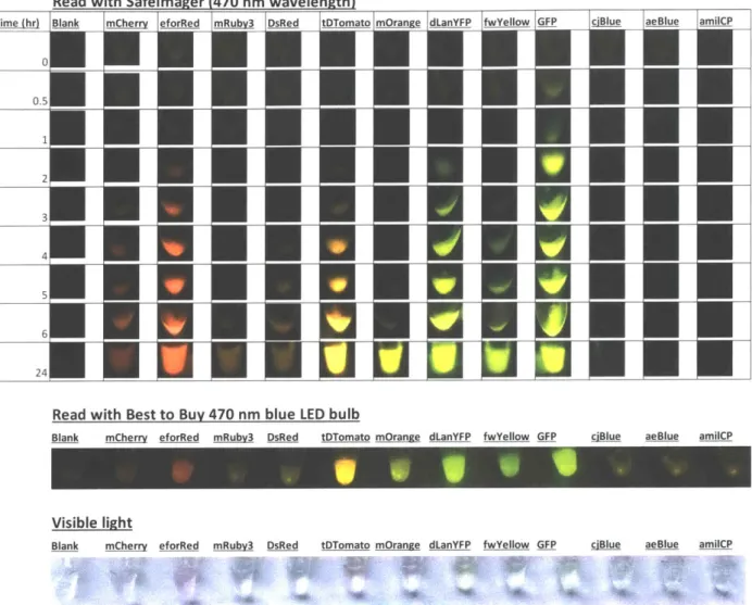
First, a panel of 12 different fluorescent proteins (readily available from previous projects) was
screened in fresh PURE reactions (Figure 15, top). The resulting protein outputs were visually
Read with Safelmager (470 nm wavelength) Time (hr) 0 0.5 1 2 3 4 5 6 24
Blank mCherry eforRed mRubv3 DsRed tDTomato mOrange dLanYFP fwYellow GFP ciBlue aeBlue amilCP
Visible light
Blank mCherry eforRed mRubv3 DsRed tDTomato mOrange dLanYFP fwYellow GFP ciBlue aeBlue amilCP
Figure 15: Screen of a fluorescent protein panel to determine set for kit
Plasmids containing fluorescent proteins were linearized, added to PURExpress reactions at 20 uL volume at saturating DNA conditions, and incubated at 30'C for 24 hours. Timecourse images were taken on a 470 nm Safelmager transilluminator. At 24 hours, an endpoint image was taken with the Safelmager (top), an inexpensive blue LED bulb (middle), and by eye on white paper under ambient light (bottom).
From this screen, I chose the brightest/fastest fluorescent protein from each color family to cover
a spectrum of colors comprised of a red (eforRed; Alieva et al. 2008; ten Buren et al. 2014), an
orange (tdTomato; Bianchi et al. 2015; Campbell et al. 2002), a yellow (mOrange; Bayle et al.
2008), and a green (GFP). Although the dLanYFP was much brighter and faster than mOrange, Read with Best to Buy 470 nm blue LED bulb
dLanYFP appeared green and was too similar to GFP. mOrange, while slower, still is bright after 24 hours and appears more yellow.
I also tested if the fluorescent proteins could be visualized with an inexpensive blue LED bulb, instead of the more expensive laboratory transilluminator (Figure 15, middle) and saw that the 24 hour endpoint image was visually comparable. Finally, the fluorescent proteins can also be visualized by eye under ambient light (Figure 15, bottom), although in the PURExpress system, the colors were very faint.
Notably, none of the blue fluorescent proteins could be visualized, as blue fluorescent proteins are difficult to visualize due to their shorter wavelength. After some experimentation with these and alternate blue fluorescent proteins, I selected Aqua (Aquamarine; Erard et al. 2013) (Figure 16). Although not as brightly expressed as the other proteins in the set, it is different enough from the GFP to be considered a blue-green, but still can be visualized under the same excitation wavelengths as the other proteins in the set. I also replaced the regular GFP with a superfolder
GFP (sfGFP; Heim and Tsien 1996) for a brighter and faster green output (Figure 16).
To validate that the fluorescent proteins could be expressed after the cell-free reaction was freeze-dried, FD-CF pellets including DNA encoding the five proteins were rehydrated and incubated overnight (~20 hours) at 30'C. The fluorescent proteins expressed robustly (Figure 16, top), and because this particular expression was done in crude extract, they were also easily visible by eye even without fluorescent excitation (Figure 16, bottom) (Huang, Stark, Nguyen et al. 2018).
II White U~wmInaSan
Figure 16: Fluorescent proteins as visual outputs.
A set of fluorescent proteins were expressed by FD-CF expression in crude extract and visualized with (i) a laboratory transilluminator (Safe Imager at 470 nm excitation) and a (ii) white light epi-illumination. Figure and caption taken from Huang, Stark, Nguyen et al. 2018.
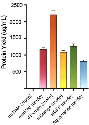
The resulting fluorescent proteins were also quantified by both protein yield (measured by
14C-Leucine incorporation; Figure 17) and by relative fluorescent units (RFU) (at optimal excitation
and emission spectra for each fluorescent protein; Figure 18). Both quantification methods showed
that the fluorescent proteins were highly expressed in the FD-CF system and confirms the visual
2500 E 2000 0D - 1500 *, 1000 CL 500
H
Figure 17: Quantification of fluorescent proteins expressed in crude FD-CF.
All of the FD-CF expressed fluorescent proteins used in the demonstration experiments had high soluble yields in crude extract (between 500 and >1000 ptg/mL), as measured by 14C-Leucine incorporation (done by Jessica Stark). Values represent averages and error bars represent standard deviations of n=3 biological replicates. Figure and caption taken from Huang, Stark, Nguyen et al. 2018.
2.3 Selection of cell-free system
As discussed in Chapter One, there are two general types of cell-free systems: crude extracts, where the required cellular components are harvested from bacterial lysis (Kwon and Jewett 2015), and the commercial PURE system (Shimizu et al. 2001), where each individual component is produced recombinantly and mixed together. In both system types, a separate buffer is included that contains essential components such as nucleotides and amino acids.
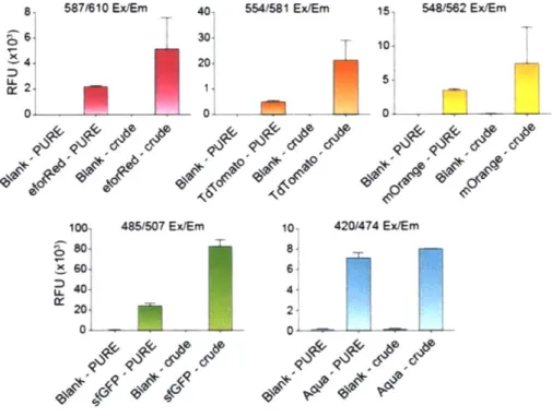
While screening and testing the fluorescent proteins, the PURE system was initially used because it had been validated in prior projects and was readily available for use. However, since the crude extracts are up to 100 times more inexpensive than PURE, economically, it would be the better system to use to develop a low-cost molecular biology kit. Furthermore, as noted above, the fluorescent proteins were also easily visible by eye even without fluorescent excitation in the crude system (Figure 16, bottom), but very faintly visible by eye in the PURE system (Figure 15, bottom). This was further quantifiably verified by measuring the relative fluorescent units (RFUs) of the fluorescent proteins in the PURE vs. crude system (Figure 18), where there was up to a 10-fold difference between the two systems (Huang, Stark, Nguyen et al. 2018).
8 587/610E X 4 4 0, 1 00 .80] ~6O1 .40 %ol2 01
ix/Em 40- 554/1 Ex/Em 15 548/562 Ex/Em 3010 20. 5.-0' 0 485/607 Ex/Em 10, 420/474 Ex/Em 8- 6-4
Figure 18: Fluorescent proteins expressed in the PURE and crude extract systems.
Endpoint fluorescent readouts of all expressed proteins in the commercial PURE or in-house crude system. Values represent averages and error bars represent average errors of n>2 biological replicates. Figure and caption taken from Huang, Stark, Nguyen et al. 2018.
Since the crude system is more inexpensive and has higher expression of the fluorescent proteins, I selected it as the primary cell-free system for developing my educational kits. Unless specifically noted, all experiments from this point on were done in the crude extract system. (For development of some of the other outputs besides constitutively expressed fluorescent proteins, PURE was used if the protein expression was low in the crude extracts).
2.4 Storage and stability of FD-CF
One of the advantages of FD-CF is that due to the freeze-dried conditions, the cell-free reactions do not need to be shipped or stored at -80'C, which makes it optimal for a classroom setting. Previous data show that PURE can be stored in a vacuum-sealed Mylar bag under nitrogen for up to a year with minimal loss of activity (Pardee et al. 2014). To help reduce kit production costs, I tested the stability of FD-CF reactions made with crude extract under conditions that were more inexpensive and realistic for a classroom setting.
For these stability studies, 5 uL FD-CF reaction pellets in PCR tubes were stored in vacuum-sealed bags and tested at each timepoint in triplicate with 1 ng/uL of sfGFP. After a 24 hour incubation at room temperature, the reactions were visualized (at 470 nm) and quantified on a plate reader for RFU (ex/em 485/520).
Any oxygen that remains in the vacuum bag and any water that remains bound in the reaction after freeze-drying can negatively affect the stability of the FD-CF pellets by degrading the reactions. To help mitigate these elements, I first tested the stability of the pellets at room temperature that had a cardboard desiccant card (Dri-Card, ~0.08g of Dri-Card per 5 uL pellet) and/or an oxygen scrubber packet (one packet per 3 x 5 uL pellets) sealed with the pellets in the vacuum-sealed bag.
Without any storage components, the pellets at room temperature last for less than a week (the one week timepoint shows a -85% loss in activity and a significant drop in visual signal). With the addition of Dri-Cards, the pellets at room temperature last up to three weeks before losing -95% activity by week 4. Although there was a ~50% loss of activity during weeks 1-3, this loss of activity does not correspond to a visual loss of signal. With the addition of both Dri-Cards and oxygen scrubber packets however, the pellets at room temperature show the same degradation rate as the pellets without any addition of storage components. This means that either the oxygen scrubber packet negatively affects the Dri-Card and prevents it from functioning properly or that the oxygen scrubber packet negatively affects the pellets themselves (Figure 19). From this study, I decided to include Dri-Cards, but not oxygen scrubber packets, with the pellets to extend their shelf life to three weeks at room temperature. As shipping typically will take less than three weeks, this will allow me to ship the kit at room temperature without any ice packs or dry ice, which will further reduce kit costs.
While a three-week shelf life at room temperature alleviates shipping concerns, many teachers would have to buy kits ahead of time and may not necessary use it within three weeks. To help extend the shelf life of the kit even further, I tested the stability of the pellets (with Dri-Cards added) at 4'C. At 4'C, the shelf life of the pellets is extended to 6 months. At 6 months, there is a ~50% loss of activity, but this loss of activity does not correspond to a visual loss of signal (Figure 20). (Note: this stability study is actually still ongoing, so the shelf life of the pellets at 4'C is potentially even longer than 6 months).
Since any water that remains bound in the reaction after freeze-drying can negatively affect the stability of the FD-CF pellets, I tested the stability of FD-CF pellets that had been freeze-drying for one week (instead of the overnight freeze-dry typically done in all other experiments) and
stored at room temperature with Dri-Cards. Although it does appear that a longer freeze-dry delays the loss in activity of the pellets (ex: 0% loss of activity vs. 50 % loss of activity at 2 weeks), it does not visually seem to make a significant visual difference (Figure 21). Combined with the fact that the pellets can be stored at 4'C to significantly extend shelf life, I decided that freeze-drying for a week was an unnecessary and time-consuming step.
With all of these stability studies, the sfGFP DNA plasmid used to test the pellets were stored at the same temperature that its corresponding pellets was stored at, in order to test overall kit stability (as the teachers will likely store the pellets and DNA together). However, I did test to see how long the DNA plasmid could be stored at room temperature (independent of the pellets) by testing the stored plasmid on freshly made FD-CF pellets. I found that the DNA plasmid has a similar shelf life to the FD-CF pellets, where at one month, there is a ~25% loss of activity and a significant visual drop in signal (Figure 22). However, as demonstrated by the other stability studies, this loss of activity can be mitigated by also storing the DNA plasmid at 4'C with the pellets.
From all these stability studies, I conclude that the FD-CF pellets (vacuum-sealed with Dri-Cards) and DNA plasmids that make up the education kit can be shipped at room temperature (with stability up to 3 weeks) and stored at 4'C (with stability up to 6 months). This should be a feasible storage condition as regular fridges have an equivalent temperature and are common in schools.
5000
42400C
In 5..3000U-2000
U-0
U.
0
1000
Weeks 0 2 3None Dri-Card Dri-Card/02 scrubber
4
Figure 19: Stability of FD-CF reaction pellets stored at room temperature with additional storage components.
5 uL FD-CF reaction pellets in PCR tubes were stored in vacuum-sealed bags with Dri-Cards, oxygen scrubbers, or no additional components at room temperature. The pellets were tested at each timepoint in triplicate with 1 ng/uL of sfGFP (also stored at room temperature). After a 24 hour incubation at room temperature, the reactions were visualized (470 nm) and quantified on a plate reader for RFU (ex/em 485/520). Note that the 3 week timepoint was done in duplicate (third pellet was lost).
Stability of pellets with storage components
0
* W/ no Storage Components
0
a W/ Dri-Card* W/ Dri-Card and Oxygen Scrubber
0 0 0 0 0
1
2
3
4
Time (week)
Stability Data
-50000, CN 1 4 0 0 00-1 LO30000
20000-L. W 20000-CL 210000-cc 0iLpellets stored at room temp vs 4C
0
i
.n
6Time (month)
Room Temp Storaze 4C Storage
Figure 20: Stability of FD-CF reaction pellets stored at 4*C.
5 uL FD-CF reaction pellets in PCR tubes were stored in vacuum-sealed bags with Dri-Cards at either
room temperature or 4'C. The pellets were tested at each timepoint in triplicate with 1 ng/uL of sfGFP (also stored at room temperature or 4'C). After a 24 hour incubation at room temperature, the reactions were visualized (470 nm) and quantified on a plate reader for RFU (ex/em 485/520). Note that the 0.75 month timepoint was done in duplicate (third pellet was lost).
Months 0 0.25 0.5 0.75 1 2 3 4.5 6 I * Room Iemp 0 4C
Stability of pellets freeze-dried for
1
week
80000
60000
0
FD 1 wk, store at RT E FD 1 day; store at RT f.40000
1
20000
I-.1
2
3
-200001
Time (week)
Weeks
Dried for
1
week
Dried forlday
0
4_
Figure 21: Stability of FD-CF reaction pellets freeze-dried for one week.
5 uL FD-CF reaction pellets in PCR tubes were freeze-dried for either one week or one day and stored in vacuum-sealed bags with Dri-Cards at room temperature. The pellets were tested at each timepoint in triplicate with 1 ng/uL of sfGFP (also stored at room temperature). After a 24 hour incubation at room temperature, the reactions were visualized (470 nm) and quantified on a plate reader for RFU (ex/em