HAL Id: hal-01764713
https://hal.archives-ouvertes.fr/hal-01764713
Submitted on 14 May 2018
HAL is a multi-disciplinary open access archive for the deposit and dissemination of sci-entific research documents, whether they are pub-lished or not. The documents may come from teaching and research institutions in France or abroad, or from public or private research centers.
L’archive ouverte pluridisciplinaire HAL, est destinée au dépôt et à la diffusion de documents scientifiques de niveau recherche, publiés ou non, émanant des établissements d’enseignement et de recherche français ou étrangers, des laboratoires publics ou privés.
Regeneration in White Sea sponge Leucosolenia
complicata (Porifera, Calcarea).
Alexander Ereskovsky, A Lavrov, F Bolshakov, D Tokina
To cite this version:
Alexander Ereskovsky, A Lavrov, F Bolshakov, D Tokina. Regeneration in White Sea sponge
Leucosolenia complicata (Porifera, Calcarea).. Invertebrate Zoology, 2017, 14 (2), pp.108-113. �10.15298/invertzool.14.2.02�. �hal-01764713�
Regeneration in White Sea sponge Leucosolenia
complicata (Porifera, Calcarea)
A.V. Ereskovsky
1,2, A.I. Lavrov
3,4, F.V. Bolshakov
3, D.B. Tokina
11 Institut Méditerranéen de Biodiversité et d’Ecologie marine et continentale (IMBE), Aix Marseille Université, CNRS, IRD, Marseille, France. E-mail: aereskovsky@mail.ru
2 Dept. Embryology, Faculty of Biology, Saint-Petersburg State University, Saint-Petersburg, Russia.
3 Pertsov White Sea Biological Station, Biological Faculty, Moscow State University M.V. Lomonos-ov, Moscow, Russia.
4 Koltzov Institute of Developmental Biology of Russian Academy of Sciences, Russia, Moscow.
ABSTRACT: Sponges (phylum Porifera) possess the remarkable regenerative abilities and great diversity of the regeneration mechanisms. The current study dealt with the regener-ation of calcareous sponge Leucosolenia complicata. Two types of experiments on L.
complicata regeneration were performed: 1) the regeneration of the body wall, and 2) the
regeneration of the amputated oscular tube. We have combined in vivo light microscopy and histological studies to reveal morphogenetic mechanisms and determine the cell types involved in the reparative regeneration in this sponge. The wound healing followed by complete restoration of lost body parts have been observed in both types of the experiments. The regeneration in Leucosolenia has a mode in which lost body parts are replaced by the remodeling of the remaining tissue. Epithelial morphogenesis, mainly spreading (flatten-ing) and fusion of exo- and endopinacoderm sheets accompanied by the transdifferentiation of the choanocytes to the endopinacocytes was found to be the key morphogenetic process during regeneration in this species. The epithelial nature of the regeneration in Leucosolenia makes it similar to the regeneration in homoscleromorphs sponges and Eumetazoans. How to cite this article: Ereskovsky A.V., Lavrov A.I., Bolshakov F.V., Tokina D. 2017. Regeneration in White Sea sponge Leucosolenia complicata (Porifera, Calcarea) // Invert. Zool. Vol.14. No.2. P.108–113. doi: 10.15298/invertzool.14.2.02
KEY WORDS: regeneration, epithelial morphogenesis, sponge, Leucosolenia.
Регенерация беломорской губки Leucosolenia
complicata (Porifera, Calcarea)
А.В. Ересковский
1,2, А.И. Лавров
3,4, Ф.В. Большаков
3, Д.Б. Токина
1 1 Средиземноморский институт биоразнообразия и экологии, НЦНИ, университет Экс-Марсель, ИРД, Экс-Марсель, Франция. E-mail: aereskovsky@mail.ru 2 Биологический факультет Санкт-Петербургского государственного университета, С-Петербург, Россия. 3 Беломорская биологическая станция им. Н.А. Перцова, Биологический факультет, МГУ им. М.В. Ломоносова, Москва, Россия. 4 Институт биологии развития им. Н.К. Кольцова РАН, Москва, Россия. РЕЗЮМЕ: Губки (тип Porifera) обладают значительными регенеративными способ-ностями и большим разнообразием механизмов регенерации. Настоящая работа109 Regeneration in White Sea sponge Leucosolenia complicata
Introduction
The morphological and cellular plasticity of sponges (phylum Porifera) allows them to adapt to the variations in the environmental condi-tions. Therefore, they often dominate the benth-ic communities in the diverse marine and freshwater ecosystems from tropical to polar regions. The ecological success of the sponges is partially a result of their rapid regeneration capacity enabling them to recover from damag-es (Ayling, 1983; Luter et al., 2012; Wulff, 2013). Sponges are known to possess remark-able regenerative and reconstitutive abilities ranging from the re-building of a functional body from dissociated cells to wound healing or body part regeneration (Korotkova, 1997).
Sponges show big diversity of the regenera-tion mechanisms (Alexander et al., 2015; Borisenko et al., 2015; Ereskovsky et al., 2015; Lavrov, Kosevich, 2016). As an ancient animal lineage, sponges are important models in stud-ies aimed at understanding of the evolution of animal regeneration mechanisms. For this pur-pose, it is necessary to expand the objects from different clades and with different body struc-ture.
Leucosolenia complicata (Montagu, 1818)
(class Calcarea) is a common species in littoral habitats along the North European coasts from the English Channel to the White Sea (Rapp, 2015), and is accessible throughout the year. This species has been successfully used for different experiments concerning the study of restoration morphogenesis (Jones, 1957; Ko-rotkova, 1961, 1969). It was showed their quick wound healing and high regeneration capacity after different surgical interventions indicating that Leucosolenia is a convenient model for sponge regeneration investigations.
The present study is aimed at revealing mor-phogenetic mechanisms and determining the cell types involved in the reparative regenera-tion in White Sea Leucosolenia complicata us-ing in vivo light microscopy and histological studies. Our study demonstrates that main mech-anism of reparative regeneration in this species is epithelial morphogenesis by spreading of cell layers that accompanied by choanocytes trans-differentiation.
Material and methods
Leucosolenia complicata is an asconoid
sponge with the body formed by the anastomos-посвящена исследованию регенерации известковой губки Leucosolenia complicata. Были проведены два типа экспериментов по регенерации L. complicata: 1) регенера-ция стенки тела и 2) регенерарегенера-ция отрезанных оскулярных трубок. Мы использовали прижизненные наблюдения и гистологические исследования, чтобы выявить морфо-генетические механизмы и определить клеточные источники, принимающие учас-тие репаративной регенерации этой губки. Заживление раны заканчивается полным восстановлением потерянных частей тела в обоих типах экспериментов. В регенера-ции L. complicata потерянные части тела восстанавливаются благодаря перераспре-делению оставшихся тканей. Основным механизмом регенерации Leucosolenia явля-ется эпителиальный морфогенез, в основном распространение (уплощение) и слия-ние экзо- и эндопинакодермы, которые сопровождаются трансдифференцировкой хоаноцитов в эндопинакоциты. Эпителиальный характер регенерации L. complicata сближает ее с регенерации губок из класса Homoscleromorpha и высших многокле-точных животных (Eumetazoa).
Как цитировать эту статью: Ereskovsky A.V., Lavrov A.I., Bolshakov F.V., Tokina D. 2017. Regeneration in White Sea sponge Leucosolenia complicata (Porifera, Calcarea) // Invert. Zool. Vol.14. No.2. P.108–113. doi: 10.15298/invertzool.14.2.02
КЛЮЧЕВЫЕ СЛОВА: регенерация, эпителиальные морфогенезы, губки,
ing hollow tubes with the internal cavities com-pletely lined with choanocytes, and external surface covered with the layer of the flat and T-shaped exopinacocytes. The narrow space in-between the exopinacoderm and choanoderm is occupied by the mesohyl – complex tissue com-prising abundant ECM, rare wandering amoe-boid cells, calcium carbonate spicules and sym-biotic bacteria.
The specimens of L. complicata were col-lected in the environs of Pertsov White Sea Biological Station of Lomonosov Moscow State University (Kandalakshsky Bay, White Sea) (66°34′N, 33°08′E) from the upper subtidal zone (0–2 m) at the low-tide. Prior the experi-ments the sponges were maintained in the 100 l aquarium with biological filters and natural sea water at temperature 6–10 °C no longer than 4 days.
Two types of experiments on L. complicata regeneration were performed: 1) the regenera-tion of the body wall, and 2) the regeneraregenera-tion of the amputated oscular tube. In the former, case the small square part (approximately 0.5x0.5 mm) of body wall were excised at the basis of the oscular tube. In the latter, case the oscular tubes were amputated from sponges and the regeneration of basal parts of such amputated oscular tube was observed. Several oscular tubes were amputated from a single sponge. All surgi-cal operations were done manually under a dissecting microscope using Castroviejo scis-sors. A total of 36 sponges were used in the experiments. Twenty-seven individuals were used in the body wall regeneration experiments. Twenty-seven oscular tubes obtained from nine individuals were used in the oscular tube regen-eration experiments.
The specimens were maintained in Petri dishes with 0.22 µm-filtered sea water (FSW) at temperature 10–11 °C after the surgical opera-tions. The half of FSW were changed every 12 h. At 3, 6, 12, 18, 24, 36, 48, 72 and 96 hours post operation (hpo) specimens were inspected and photographed using the stereomicroscope Leica M165FC (Leica, Germany) equipped with the digital camera Leica DFC 320 and Leica LAS Store and Recall v.3.6 software. The
fixa-tion of the specimens for the histological studies by 2.5% glutaraldehyde (Ted Pella, Inc., USA) on 0.2M Millonig’s phosphate buffer (pH 7.4) (Millonig, 1964) were performed at the same time points. After the fixation, the spicules were removed from specimens by treatment with 5% EDTA solution for 2 hours. Three individuals were fixed at every time point.
Results and Discussion
Regardless of the experiment type the wound healing can be subdivided into three stages according to the in vivo observations: 1) the alignment of the wound edges (0–6 hpo), 2) the regenerative membrane formation (6–24 hpo), and 3) the restoration of the body wall intact structure (4–5 days).
The complete wound healing occurs within 4–5 days post operation (dpo). The regenera-tion begins with the cleaning of the wound surface from the cell debris and broken spicules ending at 3 hpo. After that, the small irregular-ities of the wound edges which appeared due to the surgery gradually disappear and the wound edges become fairly smooth at 6 hpo (Fig. 1A, D). At this stage, the continuous epithelium appears on the wound edges. It is formed by joining of the intact exopinacocytes and endopinacocyte aris-ing from intact choanocytes near the wound edg-es through their transdifferentiation (Fig. 1G).
After the alignment of wound edges, the development of regenerative membrane begins. The regenerative membrane is a thin semitrans-parent two-layered cell membrane. It gradually covers the hole in the body wall or closes the opening in the basal part of the amputated oscu-lar tube. The regenerative membrane grows from the periphery of the wound to its center. The development of the membrane occurs quick-ly: the regenerative membrane closes a part of the wound at 12 hpo, and it completely closes wound at 24 hpo in the majority of the experi-ments (Fig. 1B, E).
The epithelial morphogenesis plays the main role during the development of the regenerative membrane. The regenerative membrane is formed due to the convergent spreading and
111 Regeneration in White Sea sponge Leucosolenia complicata
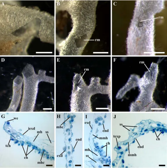
Fig. 1. The regeneration of the body wall and amputated oscular tubes in Leucosolenia complicata. A–C — the regeneration of the body wall, D–F — the regeneration of the amputated oscular tube, G–J — the histological structure of the different stages of the regeneration. A — the alignment of the wound edges, 6 hours post operation (hpo); B — regenerative membrane completely closing the wound, 24 hpo; C — the restoration of the body wall intact structure in the wound area, 72 hpo, black square indicates the newly formed spicules in the regenerative membrane; D — the alignment of the wound edges, 6 hpo; E — incomplete regenerative membrane closing a part of the wound, 24 hpo, white arrowhead indicates the hollow in the central part of the membrane; F — the regenerative membrane bearing numerous spicules, 72 hpo; G — the wound edge during its alignment, 6 hpo; H — the regeneration membrane, 24 hpo; I — the transition zone between intact tissue near the wound and regenerative membrane, 24 hpo; J — the restoration of the body wall intact structure, 96 hpo.
Abbreviations: ch — choanocytes; end — endopinacoderm of the regenerative membrane; exd — exopinacoderm of the regenerative membrane; iexd — intact exopinacoderm; mh — intact mesohyl; mhc — mesohyl cell; mmh — mesohyl of the regenerative membrane; rm — regenerative membrane; ss — space from removed spicule; tch — transdifferentiating choanocyte; texp — T-shaped exopinacocyte; we — wound edge. Scale bars: A–F — 500 µm; G–J — 10 µm.
Рис. 1. Регенерация стенки тела и отрезанных оскулярных трубок Leucosolenia complicata. A–C Ї регенерация стенки тела, D–F — регенерация отрезанной оскулярной трубки, G–J — гистологичес-кое строение различных стадий регенерации. A — выравнивание краев раны, 6 часов после операции
fusion of epithelial layers: the exopinacoderm on the external side of the membrane and the endopinacoderm — on the internal. The forma-tion of the endopinacoderm is most likely oc-curs due to the transdifferentiation of the cho-anocytes near the wound edges into endopina-cocytes through their flattening and losing of flagellum and microvilli collar (Fig. 1G). Thus, at this stage, the regenerative membrane con-sists of two epithelial layers (exo- and endopi-nacoderm) and the thin mesohyl between them (Fig. 1H, J). The mesohyl of the membrane differs from intact one — it contains fewer cells, less abundant ECM (as it does not acquire staining on the histological sections like the ECM in the intact mesohyl near the wound edges) and lacks spicules (Fig. 1H, J).
Such a rapid formation of the regenerative membrane, in our opinion, is associated with the necessity to restore the normal water flow through the aquiferous system of the sponge as soon as possible. Probably, the appearance of a large opening in the body wall during the exper-iments significantly changes the water flow in the aquiferous system of the asconoid sponge, since large volumes of the water may be inhaled and exhaled from the animal’s body through the wound opening rather than through the osculum and ostia. The formation of the regenerative membrane quickly returns the process of water pumping to the normal mode.
After the full development of the regenera-tive membrane, the restoration of the intact structure of the body wall proceeds. By 36–48 hpo, the wound size decreases, apparently, due to a decrease in the regenerative membrane
area. By 72 hpo, the wound surface further decreases in size and, in some cases, becomes poorly discernible. The mesohyl of the regener-ative membrane obtains intact structure – the cells become more abundant and spaces be-tween them occupy by ECM (Fig. 1J). In addi-tion, at this stage the new spicules appear in the regenerative membrane (Fig. 1C, F). These sp-icules can be formed either directly in the regen-erative membrane. The elucidation of the source of the new spicules requires further studies.
Leucosolenia complicata has been
success-fully used for in vivo and histological studies of regeneration by some authors (Jones, 1957; Korotkova, 1961, 1969). Our results confirm previous descriptions concerning the main stag-es of body wall regeneration. As in previous descriptions, we showed that the wound healing occurs by the formation of the regenerative membrane (Jones (1957) called this structure as “healing membrane”).
The epithelial morphogenesis, mainly spread-ing (flattenspread-ing) and fusion of epithelial sheets is involved in the reparative regeneration in L.
complicata. These processes are accompanied
by transdifferentiation of choanocytes to endop-inacocytes. Epithelial-mesenchymal transitions are absent during L. complicata regeneration. Moreover, we cannot reveal any morphological-ly distinct pluripotent cell types in this species. Therefore, regeneration in Leucosolenia has a mode in which lost body parts are replaced by the remodeling of the remaining tissue. Similar mechanisms of regeneration were described in other calcarean sponges Sycon lingua and S.
ciliatum (Korotkova, 1972; Laplant et al., 2014). (чпо); B — регенеративная мембрана, полностью закрывающая рану, 24 чпо; C — восстановление интактной структуры стенки тела в области раны, 72 чпо, черный квадрат отмечает новые спикулы в регенеративной мембране; D — выравнивание краев раны, 6 чпо; E — неполная регенеративная мембрана, 24 чпо, белая стрелка указывает на отверстие в центральной части регенеративной мембраны; F — регенеративная мембрана со множеством спикул, 72 чпо; G — край раны во время его выравнивания, 6 чпо; H — регенеративная мембрана, 24 чпо; I — переходная зона между интактными тканями вблизи раны и регенеративной мембраной, 24 чпо; J — восстановление исходной структуры стенки тела, 96 чпо. Обозначения: ch — хоаноциты; end — эндопинакодерма регенеративной мембраны; exd — экзопинакодерма регенеративной мембраны; iexd — интактная экзопинакодерма; mh — интактный мезохил; mhc — клетка мезохила; mmh — мезохил регенеративной мембраны; rm — регенеративная мембрана; ss — пространство от вытравленной спикулы; tch — трансдифференцирующийся хоаноцит; texp — T-образный экзопинакоцит; we — край раны. Масштаб: A–F — 500 µm; G–J — 10 µm.
113 Regeneration in White Sea sponge Leucosolenia complicata
The epithelial nature of the regeneration in
Leucosolenia makes it similar to the
regenera-tion in homoscleromorphs sponges, which was described in detail in Oscarella (Ereskovsky et al., 2015), and Eumetazoans, like Hydra, and to the last phase of the regeneration in the triplo-blastic animals (Salvenmoser et al., 2001; Gal-liot, Ghila 2010). In contrasts with Calcarea, Homoscleromorpha is notably the only sponge group to possess morphologically distinct base-ment membrane and specialized cell junctions and is therefore considered to possess true epi-thelia. The consequence of this peculiar organi-zation is the predominance of epithelial mor-phogenesis during ontogenesis regeneration in-clude, of these sponges (Ereskovsky et al., 2015). In order to understand whether there is a correlation between the dominance of the epi-thelial morphogenesis during Leucosolenia re-generation and the peculiarity of their epithelial layers (choanoderm and pinacoderm), further studies using electron microscopy and molecu-lar biology is necessary.
Knowledge on morphological basis of mor-phogenesis during Leucosolenia regeneration could have important implications for our un-derstanding of the diversity and evolution of the regeneration mechanisms in metazoans and pro-vides a strong basis for the future investigations with the genetic approaches.
Acknowledgements
Financial support by Russian Foundation for Basic Research #16-04-00084 and the Rus-sian Science Foundation #17-14-01089 (for the microscopy) is gratefully acknowledged.
References
Alexander B.E., Achlatis M., Osinga R., Van der Geest H.G., Cleutjens J.P.M., Schutte B., de Goeij J.M. 2015. Cell kinetics during regeneration in the sponge
Halisarca caerulea: how local is the response to tissue
damage? // PeerJ. Vol.3. e820.
Ayling A.L. 1983. Growth and regeneration rates in thinly encrusting demospongiae from temperate waters // Biol. Bull. Vol.165. P.343–352.
Borisenko I.E., Adamska M., Tokina D.B., Ereskovsky A.V. 2015. Transdifferentiation is a driving force of
regeneration in Halisarca dujardini (Demospongiae, Porifera) // PeerJ. Vol.3. e1211.
Ereskovsky A.V., Borisenko I.E., Lapebie P., Gazave E., Tokina D.B., Borchiellini C. 2015. Oscarella
lobu-laris (Homoscleromorpha, Porifera) regeneration:
Epithelial morphogenesis and metaplasia // PlosOne. Vol.10. e0134566.
Galliot B., Ghila L. 2010. Cell plasticity in homeostasis and regeneration // Mol. Reprod. Dev. Vol.77. P. 837– 55.
Jones W.C. 1957. The contractility and healing behavior of pieces of Leucosolenia complicata // Quart. J. Micr. Sci. Vol.98. P.203-217.
Korotkova G.P. 1961. Regeneration and somatic embryo-genesis in the calcareous sponge Leucosolenia
com-plicata Mont. // Acta Biol. Hung. Vol.2. P.315–334.
Korotkova G.P. 1969. [The peculiarities of morphogenesis during the development of the calcareous sponge
Leucosolenia complicata Montagu from the small
fragments of body wall] // Vest. Leningr. Univ. Vol.15. P.15–22 [in Russian, with English summary]. Korotkova G.P. 1972. [Comparative morphological
inves-tigations of development of sponges from dissociated cells] // Trans. Leningr. Soc. Natur. Vol.78. P.74–109 [in Russian, with English summary].
Korotkova G.P. 1997. [Regeneration in animals]. Saint-Petersburg: Saint-Petersburg University Press. 297p. [in Russian, with English summary].
Laplante M., Adamska M., Leininger S., Ereskovsky A. 2014. Sycon ciliatum (Calcarea, Calcaronea) regener-ation peculiarities [abstract] // 5th meeting of the European Society for Evolutionary Developmental Biology 22–25 July 2014, Program and Abstracts: Vienna, Austria, 2014. P.374–375.
Lavrov A.I., Kosevich I.A. 2016. Sponge cell reaggrega-tion: cellular structure and morphogenetic potencies of multicellular aggregates // J. Exp. Zool. A. Ecol. Genet. Physiol. Vol.325. P.158–177.
Luter H.M., Whalen S., Webster N.S. 2012. The marine sponge Ianthella basta can recover from stress-in-duced tissue regression // Hydrobiologia. Vol.687. P.227–235.
Millonig G. 1964. Study on the factors which influence preservation of fine structure // Sympos. on electron microscopy. Rome, Italy: Consiglio Nazionale delle Ricerche. P.347.
Rapp H.T. 2015. A monograph of the calcareous sponges (Porifera, Calcarea) of Greenland // JMBA. Vol.95. P.1395–1459.
Salvenmoser W., Riedl D., Ladurner P., Rieger R. 2001. Early steps in the regeneration of the musculature in
Macrostomum sp. (Macrostomorpha) // Belg. J. Zool.
Vol.131. P.105–109.
Wulff J. 2013. Recovery of sponges after extreme mortal-ity events: morphological and taxonomic patterns in regeneration versus recruitment // Integr. Comar. Biol. Vol.53. P.512–523.