Full Length Research Paper
Proliferation and rooting of wild cherry: The influence
of cytokinin and auxin types and their concentration
Akila Mansseri-Lamrioui
1, Ali Louerguioui
1, Jaqueline Bonaly
2*, Saliha Yakoub-Bougdal
3,
Nacer Allili
3and Salima Gana-Kebbouche
1 1Department of Biology, Faculty of Science, M’Hamed Bougara University, Boumerdes, Algeria. 2
Faculty of Pharmacy, University of Paris XI, France. 3
Department of Biology, Faculty of Biological and Agronomic Sciences, Mouloud Mammeri University, Tizi-ouzou, Algeria.
Accepted 17 June, 2011
Determination of the most optimal type and concentration of plant growth regulators as medium constituents is one of the most important aspects of successful micro propagation, among other in
vitro factors. With the aim of optimization of in vitro multiplication of wild cherry, the effect of the
following cytokinins was studied: 6-benzyladenine (BAP), 2-isopentenyl adenine (2iP) and kinetin (Kin)
at concentrations of 1, 2, 4 and 8 mg. l-1. Stem segments of seedlings from juvenile and adult materials
were disinfected and grown on a Quoirin and Lepoivre (1977) (QL) medium without growth regulators for 4 weeks. Each material responded differently to the tested cytokinins. The use of 6-benzyladenine resulted in the highest percentage of sprouting, the development of shoots and the ratios of multiplication for two materials of Prunus avium L. In the next experiment, seedlings from the juvenile and adult materials were grown on (MS2/5) medium in the presence of auxins indole-3-butyric acid (IBA), naphtaleneacetic acid (NAA), indole-3-acetic acid (IAA), when compared with concentrations of 0.5, 1, 2, and 4 mg. l-1. For the type of explants and its reactivity with the type and the concentration of auxin, significant differences among explants for root induction were observed. The adult material did not develop roots in any of the auxin and concentration used. In the case of the juvenile material, the IBA was distinguished from the other auxins tested and the highest induction of roots took place in 1
mg. l-1. The most significant induction of cal characterizes, especially, the mediums containing the NAA
followed by the IAA with concentrations of 2 and 4 mg. l-1, respectively, which block the emergence of
the roots partly and decreases the rate of rooting thereafter. The highest average number of roots and the highest average length of roots were obtained with the IBA with 1 mg. l-1.
Key words: Wild cherry tree, proliferation, cytokinins, rooting, auxins.
INTRODUCTION
Wild cherry (Prunus avium L.) is a fast-growing, high-value, squirrel-resistant and hardwood timber tree (Pryor, 1985; Hammatt and Grant, 1997), with potential for wider use in forestry and farm woodlands. It is also an excellent cultivar because of its edible fruit (Wünsch and Hormaza, 2003). In Algeria, studies related to this species are quite limited, although, there is an increasing interest in reforestation for both its economic and ecological value.
*Corresponding author. E-mail: mansseriakila@yahoo.fr .
Propagation of most Prunus spp. by conventional methods such as seedling and cutting, do not ensure healthy and disease-free plants and are time and labor consuming (Holtz et al., 1995). However, methods available for in vitro micro propagation have the potential to provide high multiplication rates of uniform genotypes, resulting in short-term gains (Durzan, 1988; Hammatt and Grant, 1993). Moreover, many studies have already been successfully applied to Prunus species such as sour and sweet cherries (Borkowska, 1983; Canli and Tian, 2008; Ruzic and Vujovic, 2008), cherry and peach rootstocks (Dalzotto and Docampo, 1997; Marino, 1997), Prunus
8614 Afr. J. Biotechnol.
armeniaca (Perez-Tornero and Burgos, 2007).
The optimal determination of growth regulator is impor-tant for a successful tissue culture. It is well known that cytokinins promote the growth of axillary buds, by reducing apical dominance in shoot cultures during the proliferation stage (Van Staden et al., 2008) and to achieve rooting, various auxins may be used, and the choice and their concentration can differ according to the specific culture conditions involved. In vitro technique encounters several problems generally related to the aseptic initiation of necroses, with the problems of oxida-tion of the culture medium, and the difficulty of rooting of the adult material. The use of a juvenile material pro-duced in greenhouse and resulting from in vitro germination can be very interesting for the rooting of the seedlings. In micro propagation, rooting of micro cuttings is often challenging and loses at this stage has vast economic consequences. Many woody species are difficult to root through cuttings after the seedling-derived stage. Wild cherry (P. avium L.) is particularly difficult to root both in vivo and in vitro. The response of cuttings to exogenous application of auxins is dependent on many internal and external factors. Both the concentration of the growth substance applied and, above all, the sensitivity of treated plant parts to this substance may be limiting factors (Dolezelova et al., 1996). Exogenously applied auxin is one of the external factors influencing the phytohormonal balance in the cuttings and thus adven-titious root formation. Moreover, the mineral concen-tration of the culture medium affects rooting charac-teristics and some researchers proposed its reduction to half normal strength for rooting improvement (Dimassi-Theriou, 1995).
We must optimize the tissue culture system for this species by using a simple efficient and reliable protocol. This can be achieved by optimizing the components in the tissue culture medium or by modifying the environ-mental conditions provided, or both. However, the age of explants plays an important role in the repeatability and reliability of any tissue culture protocol (Huang et al., 1992), and should be evaluated. It is well known that cytokinins promote cell division and cell expansion in plant tissue culture and many studies have reported suitable cytokinin types and their concentrations for each species. Hence, the first objective of this study was to develop a protocol for propagation of this wild cherry cultivar as well as to determine the most optimal condi-tions for multiplication phase with the use of 3 cytokinins: BAP, 2iP and Kin (from mature and juvenile material) of a local wild cherry cultivar.
To achieve rooting, various auxins may be used, in particular IBA, IAA or NAA. The choice of the auxin type and concentration can differ according to the specific culture conditions involved. The rooting of some woody species including Prunus can also be improved under darkness during the first week (Rugini et al., 1993; Caboni et al., 1997). The second objective of this study
was to find a reproducible method for the successful rooting of wild cherry. We tested the effects of the auxin type and concentration, on rooting of in vitro shoots of two types of explants.
MATERIALS AND METHODS
Establishment of sterile shoot cultures from seedlings
The vegetable material used (seedlings and seeds) was from the area of Kabylie (Larbaa-Nath-Inathen) located at the north of Algeria. The explants are from youthful material (seedlings resulting from in vitro germination of MJ1 and in vivo MJ2 of a one year old) and from adult material from forty-year old trees.
The in vivo germination is potted in the green-house in 20-cm plastic pots containing a 1:1:1 mixture of peat, sand and vermiculite. Plants were watered daily using drip irrigation systems and fertilized weekly with 250 ppm of a 20-20-20 NPK fertilizer solution.
The in vitro germination of the zygotic embryos, pre-treated at low temperature, took place in a medium of MS/4 selected previously. The taken stem segments containing 10 to 15 axillary buds of the adult material (young branches of the year in ongoing lignifications) and of juvenile material MJ2 were disinfected in mercury chloride (HgCl2) with the respective concentrations (2.5 g.
l-1 and 1 g. l-1) containing a few drops of Tween 20, and rinsed 3 times with sterilized distilled water (10 min each per rinse) in jars that were placed on an Orbital Shaker (Lab-Line Instruments) at 80 rpm. Two successive rinsing of 10 mn in the calcium chloride solution CaCl2 (2.5 g. l-1) were applied. The micro cuttings were
rinsed three times in sterile distilled water, for 10 min each.
Explants were cultured vertically in 25 x 15 mm test tubes containing 20 ml of solidified Quoirin and Lepoivre (QL) (1977) medium without growth regulations. After 1 month, axillary shoots of stem segments were used for subsequent experiments.
Experiments of axillary shoot development from stem explants
Shoot multiplication is the most crucial stage of micro propagation. The success of a micro propagation protocol, to a large extent, depends on the rate and mode of shoot multiplication. Stem segments from in vitro grown axillary buds shoots were cut into 1 to 2 cm sections with two to three axillary into jars containing 20 ml of the culture media. The basal medium contained QL salts, this mineral composition gave better results as compared to other formulations which we had compared beforehand (Mansseri-Lamrioui et al., 2009). We added the micronutrients of QL modified for the boron and manganese, Morel vitamins (1948) (Table 1), methionine (100 mg. l-1), sucrose (20 g. l-1) was supplemented with 1, 2, 4 and 8 mg. l-16-benzyladenine (BAP), 2 isopentenyl adenine (2iP) and kinetin (Kin). The medium was solidified with Difco Bacto-agar (7 g. l-1). Prior to autoclaving, the pH value of all the media was adjusted to 5.6 with 0.1 N KOH. The media were sterilized in an autoclave for 20 min at 120°C.
In the first experiment, the effect of three cytokinin treatments on percentage of sprouting, the development of shoots and the ratios of multiplication was investigated. A control treatment was also included in the experiment with no addition of cytokinin to the basal medium. For each treatment, 12 stem segments (each with two axillary buds) from each of the three explants MJ1, MJ2 and adult material were sub cultured vertically in a 25 x 15 mm test tubes. Eight replicate were used for each treatment. The explants placed in the test tubes were incubated in a culture room under a 16-h photoperiod at low-light intensity (15 to 20 µmol. m-2. s-1) provided
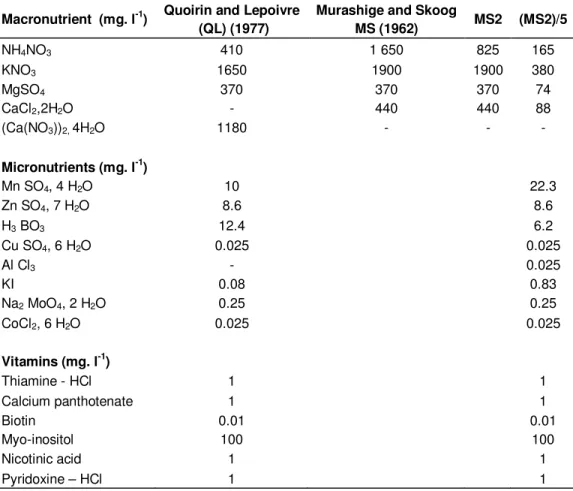
Table 1. Nutrient composition of proliferation and rooting media used of P. avium L.
Macronutrient (mg. l-1) Quoirin and Lepoivre (QL) (1977)
Murashige and Skoog
MS (1962) MS2 (MS2)/5 NH4NO3 410 1 650 825 165 KNO3 1650 1900 1900 380 MgSO4 370 370 370 74 CaCl2,2H2O - 440 440 88 (Ca(NO3))2, 4H2O 1180 - - - Micronutrients (mg. l-1) Mn SO4, 4 H2O 10 22.3 Zn SO4, 7 H2O 8.6 8.6 H3 BO3 12.4 6.2 Cu SO4, 6 H2O 0.025 0.025 Al Cl3 - 0.025 KI 0.08 0.83 Na2 MoO4, 2 H2O 0.25 0.25 CoCl2, 6 H2O 0.025 0.025 Vitamins (mg. l-1) Thiamine - HCl 1 1 Calcium panthotenate 1 1 Biotin 0.01 0.01 Myo-inositol 100 100 Nicotinic acid 1 1 Pyridoxine – HCl 1 1
by cool-white (40W) fluorescent tubes at 22 ± 1°C. Some specific issues, such as colour, leaf and callus size, leaf roll, incidence of chlorosis or necrosis were also monitored.
Experiment of the roots of seedlings
Shoots, 30 to 40 mm long with 2 to 4 leaves were isolated and used for the rooting experiments. The root induction phase consisted of culturing the shoots for 8 days in darkness on the macronutrients of (MS2/5), micronutrients of MS (1962), Morel vitamins (1948) (Table 1), L-proline (100 mg. l-1) and sucrose (30 g. l-1). The medium was solidified with Difco Bacto-agar (7 g. l-1). The pH of the medium was adjusted to 5.6 with 0.1 N KOH and 0.1 N HCl before autoclaving for 20 min at 120°C. After this period, the explants placed in test tubes were incubated in a culture room. Culture conditions were similar to those described earlier. For all experiments, data were collected after 4 weeks of in vitro culture. The influence of three auxins indole-3-butyric acid (IBA), naphtaleneacetic acid (NAA), indole-3-acetic acid (IAA), when compared with concentrations 0.5, 1, 2 and 4 mg. l-1 on the rooting rate, average number of roots and on the average length of roots was investigated. A control treatment was also included in the experiment with no auxin added to the basal medium. Twelve seedlings of explants MJ1, MJ2 and adult material were used. Eight replicates were used for each treatment.
Statistical analysis of the data was carried out by using analysis of variance (ANOVA) and differences among treatment means were compared by using least significance difference (LSD) test at 5% probability level.
RESULTS
The results gotten from this study represent a repro-ducible plant regeneration system through organogenesis from two types of explants of P. avium cultures. The efficiency of shoot regeneration depended on the type of explants, cytokinins and auxins, and their concentrations that are added to the basal medium.
Effect of the cytokinins on the percentage of sprouting
Aseptic culture and regeneration of shoots was obtained from axillary buds in 85% of explants, whereas 15% of explants were tarnished or infected for adult material. The sterilization protocol reduced the frequency of bacterial or fungal contamination of green-house material (MJ2) to a very low level. We estimated less than 5% visual culture contamination.
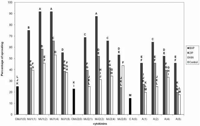
When the effect of different cytokinin treatments on the percentage of sprouting was investigated, there was statistically significant cytokinin x explants interaction. Among cytokinin treatment tested, BAP induced the highest percentage of sprouting with the MJ1 with
8616 Afr. J. Biotechnol.
Figure 1. Development of the explant in the proliferation phase of P. avium L. a, Transplantation of an explant from the establishment phase; b, Leaves tuft obtained after four weeks of culture on medium QL+ 2 mg/l of BAP (MJ1); c, Leaves tuft obtained after four weeks of culture on medium QL+ 1 mg/l of BAP (MJ2); d, Leaves tuft obtained after four weeks of culture on medium QL+ 1 mg/l of BAP (adult material). QL: Quoirin and Lepoivre (1977), BAP: 6-benzyladenine, MJ: juvenile material.
concentrations of 2 and 4 mg. l-1 (91.66%), followed by MJ2 with 87.49 and 65.62% to the respective concen-trations of 2 and 4 mg. l-1. Lower results were obtained with the adult material respectively, 64.58 and 52.08% (Figure 1). A high reactivity of the growths was also observed in the presence of 1 mg. l-1 of BAP with respectively, 75% for the MJ1; followed by MJ2 with 68.74% and only 45.83% for the adult material.
It should be noted that all the buds cultured in the presence of the BAP were sprouting, but the percentage of sprouting slightly decreased to 8 mg. l-1 respectively, 55.20% for the MJ1; 53.12% for MJ2 and only 45.83% for the adult material (Figure 2).
The cytokinins 2iP and kinetin had an average reactivity with the MJ1, of which the maxima rate of sprouting: 65.62 and 53.12% were obtained, respectively, with the 2ìP and the kinetin with a concentration of 4 mg. l-1, followed by MJ2: 52.08 and 43.74% with 2 mg. l-1 of 2ìP and 8 mg. l-1 of kinetin.
These rates are 45.83 and 33.33% for the adult mate-rial in the presence of 2 mg. l-1 of 2ìP and 4 mg. l-1 of kinetin. The sprouting was very weak with the absence of cytokinin in both juvenile and adult materials.
Effect of the cytokinins on the percentage of shoots The results of the analysis of the variance concerning the development of the new shoots, followed a tendency similar to that of sprouting, thus, the hormone and explants factors are highly significant with the threshold of confidence (P < 0.001). The best results were obtained with 2 mg. l-1 of BAP for juvenile material MJ1 91.66%; with 89.58% for the MJ2, and only 45.83% for the adult material (Figure 3), and the plants were big, with dark
green leaves.
The 2ìP and the kinetin had lower results in comparison with the BAP, with a bad aspect of explants, where we noted the appearance of vitrification. It should be noted that the adult explants induced callusing from the cut ends of the explants. The callus was friable and green in colour.
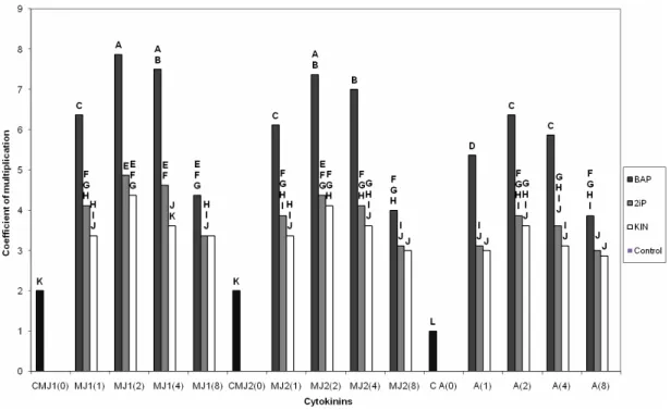
Effect of the cytokinins on the coefficient of multiplication
The statistical analysis reveals a significant difference between the coefficients of multiplication for all the cytokinins tested. Among the studied cytokinins, the BAP proved to be the most favorable for the multiplication, not only on the level of the coefficient of multiplication, but also on the level of the quality of the explants which remains better.
The axillary buds appear more numerous on average for concentrations of 2 and 4 mg. l-1 with 7.87 and 7.50 respectively, for the MJ1. Slightly lower results are recorded for the same concentrations for the MJ2 with 7.37 and 7. The coefficient of multiplication is decreased for the adult material with 6.37 and 5.87 for concen-trations 2 and 4 mg. l-1 of BAP (Figure 4). The use of both cytokinins: 2ìP and kinetin is accompanied by a reduction in the average coefficient of multiplication for the same concentrations respectively with, 4.87 and 4.62 for the 2ìP; 4.37 and 3.62 for the kinetin with regards to the MJ1. We noted that the lowest coefficient of multiplication (3.37) was recorded with the 2ìP and the kinetin with 8 mg. l-1. This is also done for the BAP where the coefficient of multiplication is 4.37 with the same 8 mg. l-1. Concerning the other types of explants, the
Figure 2. Percentage of sprouting according to the cytokinins for the juvenile and adult material.
Figure 3. Percentage of shoots according to the cytokinins for the juvenile and adult material.
results obtained highlight a slight variation of the coefficient of multiplication from one concentration to another. However, these fluctuations which remain for the majority of the concentrations, remain non significant.
Effect of the auxins on the rooting rate
Both the concentration and type of auxins used markedly influenced the percentage of root formation. The results
8618 Afr. J. Biotechnol.
Figure 4. Coefficient of multiplication according to the cytokinins for the juvenile and adult material.
of the analysis of the variance for the rooting rate, showed very high significant difference (P<0.001) for the factors explants and auxins. The adult material did not develop roots, in any of the auxin and concentration used.
Rooting was induced over the entire range of IBA concentrations tested; in the case of the juvenile materiel, the IBA was clearly distinguished from the other auxins tested and the highest induction of roots took place in the presence of 1 mg. l-1 (100%) for the MJ1, and increase in IBA concentration above 4 mg. l-1 resulted in a decline in rooting.
For MJ2, IBA produced rooting in maximum 80.53% shoots with 1 mg. l-1, whereas IAA produced rooting in maximum 77.77% shoots when used at concentration of 1 mg. l-1 for MJ1 and 56.94% for MJ2. NAA produced rooting in only 34.71% for MJ1 and 33.33% for MJ2 (Figures 5 and 6). The most significant induction of cal characterizes especially the mediums containing the NAA followed by the IAA with the concentrations of 2 and 4 mg. l-1, which blocks partly the emergence of the roots and thereafter decreases the rate of rooting.
Effect of the auxins on the average number of roots The rooted in vitro plants were maintained in the same medium in the presence of 3 auxins for the development of the roots under a normal photoperiod (16 h). A synergism was found between the rooting percentage and number of roots per rooted explant. The results show that IBA (1 mg. l-1) produced maximum number of roots
(2.83) per rooted explant for MJ1 against 2.5 for MJ2, while the maximum number of roots per explant produced by IAA was 2.33 at 1 mg. l-1 concentration for MJ1 and 1.66 for MJ2. For NAA, the maximum number of roots was only 1.16 for MJ1 and 1 for MJ2 for the same concentration (1 mg. l-1) (Figure 7).
Effect of the auxins on the average length of roots The results show that the trend followed by the previous two characters was maintained here. We noticed a slight advance of the IBA with 1 mg. l-1 for the MJ1 (1.40 cm) when compared with MJ2 (1.30 cm). The maximum root length produced by IAA was 1.1 for MJ1 and 1 for MJ2 with 1 mg. l-1. For NAA, the maximum roots length was only 0.82 for MJ1 and 0.62 for MJ2 (Figure 8). These results indicate a positive correlation between the rooting percentage, number of roots per rooted explants and root length. Like other developmental processes, cell elonga-tion involves sequential changes in levels and/or activity of enzymes (Cosgrove, 1999).
DISCUSSION
Contaminations were infrequent and mercuric chloride proved to be effective against bacterial and fungal micro flora in field collections of wild cherry. It was previously reported that mercuric chloride is a good disinfectant, but could be toxic to plant tissues of susceptible species and genotypes (Muna et al., 1999; Sedlák and Paprštein,
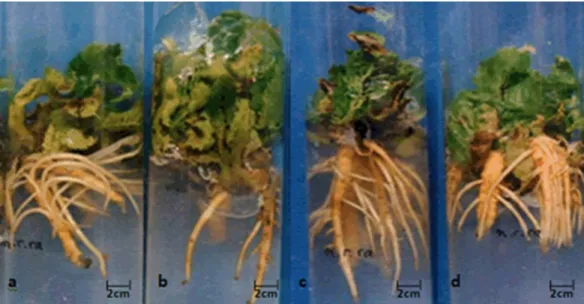
Figure 5. Different stages of explant in the rooting phase of P. avium L. a, Formation of many large and branched roots after 5 weeks of culture on MS2/5 + 1 mg/l of IBA (MJ1); b, Formation of many large and branched roots after 5 weeks of culture on MS2/5 + 2 mg/l of IBA (MJ1); c, Formation of many large and branched roots after 5 weeks of culture on MS2/5 + 1 mg/l of IBA (MJ2); d, Formation of many large and branched roots after 5 weeks of culture on MS2/5 + 2 mg/l of IBA (MJ2). MS2/5: MS (Murashige and Skoog, 1962) reduced five times with NH4NO3/10, IBA: indole-3-butyric acid, MJ:
juvenile material.
Figure 6. Percentage of rooting according to auxins.
2007).
On the basis of the obtained results, it could be noticed that there are differences in the uptake of cytokinins,
recognition by the cells or mechanisms of action of the cytokinin compounds. However, cytokinins in our experi-ment can be divided into two groups: very active
8620 Afr. J. Biotechnol.
Figure 7. Roots average number according to auxins.
Figure 8. Rooting average length according to auxins.
group (only BAP), which was more effective and more shoots of Prunus avium L. were formed, and the 2iP and kinetin which exhibited rather weak effects on multi-plication. The type and concentration of cytokinin influen-ced the percentage of sprouting and the percentage of shoots produced per explants. There was no sign of growth when explants were cultured in the media without cytokinin; this indicates that the contribution of cytokine has a positive and stimulating effect. Our results are in agreement with those of Ďurkovič (2006) and Mansseri-Lamrioui et al. (2009). They reported that a cytokinin was necessary for the development of P. avium L. and the
best result for shoot proliferation was obtained with 1 or 2 mg. l-1 of BAP.
These results agreed with those obtained by Kadota et al. (2001) and Kadota and Niimi (2003) with pear Pyrus pyrifolia N.; where they suggested that BAP displayed more noticeable effect than thidiazuron and kinetin, and also BAP is more suitable for shoot multiplication of pear than phenylurea derivatives. It is well known that high concentrations of cytokinins of adenine type are often necessary for growth and differentiation of tissue culture. It was previously reported that BAP could be used successfully to induce shoot multiplication in Prunus spp.
(Muna et al., 1999; Pruski et al., 2000). Saponari et al. (1999) successfully micro propagated Prunus mahaleb rootstocks on medium supplemented with 1 mg. l-1 BAP. Plant growth regulator 2iP proved to be unsuitable for the proliferation of sweet cherry cultivar Rivan (Sedlák and Paprštein, 2007).
Indeed, the caulogenesis depends on the concentration of the BAP. For Ruzic and Vujovic (2008), the best multiplication parameters with BAP cytokinin were obtained with the concentration of 5 µM with 0.5 variation of IBA for multiplication index and up to 2.5 µM for the shoot length of sweet cherry cv. Lapins (P. avium L.). They suggested that the choice of cytokinins for the phase of multiplication should be limited to BAP. Cytokinin 2iP and kinitin mainly influenced shoot growth of sweet cherry cv. Lapins, whereas it made little impact on multiplication. BAP concentrations of 8.87 to 12.82 µM gave optimal shoot proliferation in Chokecherry and 4.44 µM BAP in both cultivars of Pincherry (Pruski et al., 2000). For Vejsadová (2008), for most rhododendrons, the highest shoot multiplication was found on a medium with 8 to 10 mg/dm3 2iP in combination with 1 mg/dm3 IAA.
The hormonal balance: BAP (2.2 µM), IBA (0.49 µM) and GA3 (0.29 µM) with 1 mM phloroglucinol showed its positive influence in works of Hammatt and Grant (1997) of mature wild cherry. The 8.87 µM BAP level has been reported to be optimal for shoot proliferation of the Prunus cerasus x Prunus munsoniana rootstocks (Dalzoto and Docampo, 1997). Rodriguez et al. (1998) on the walnut tree Juglans spp. and Akbas et al. (2009) on Amygdalus communis L. noticed that the smallest percentages of sprouting and development of the shoots are due to the strong concentrations of BAP which inhibit them. The optima are reached with 1 mg. l-1 of the BAP. Concentrations of 5 and 10 mg. l-1 induced the formation of cal in Juglans spp.
Cytokinins 2iP and Kin are rarely used in micro propagation of fruit varieties (Hsia and Korban, 1997; Jaakola et al., 2001; Arinaitwe et al., 2004; Góralski et al., 2005). For Shekafandeh (2010), the best concentrations of BAP were 6.65 and 8.87 µM, which produced the highest rate of proliferation for Prunus dulcis Mill.
According to our results, all the regulators tested allowed sprouting and induced new shoots. Nevertheless, for the same amount, the results differ according to the regulator and the explants. Indeed, the specimen collec-tion of explants on juvenile structures improves the initial reaction and accelerates the multiplication. Moreover, the physiological state of explants is added (Vieitez and San-José, 1996).
Finally, we retained the BAP with 2 mg. l-1 since it pre-sents the highest coefficient of multiplication as well as a better quality of the in vitro plants for all the types of explants.
Recalcitrant nature of legumes with respect to rooting in culture conditions has limited the successful appli-
cation of any biotechnological approach for crop improve-ment. Either IAA or IBA at various concentrations are commonly used auxins to induce rooting from the explants both in culture and in vivo conditions (George and Sherrington, 1984). Indeed in vivo root induction by application of 2 mg. l-1 IBA has been reported for some Prunus spp. such as Prunus tomentosa, Prunus fructicosa, Prunus verginiaca and Prunus pensylvanica (Pruski et al., 2005).
Both the concentration and type of auxins used markedly influenced the percentage of root formation. Jay-Allemand (1993) and Bellamine et al. (1998) affirmed that auxin exerts primary role in root formation by its involvement in successive and interdependent phases. Sabatini et al. (1999) reported that differentiation of phloem ray parenchyma cells into root primordia depends upon the type and concentration of auxin. Further, it has been reported by Blakesley and Chaldecott (1997) that differentiating cells require the most appropriate auxin to become competent to respond to the organogenic signal.
Plant tissue culture methods have been used to study various aspects of rooting, and have improved the understanding of the uptake and metabolism of IBA (Reeves et al., 1985; Van Der Krieken et al., 1993; Baraldi et al., 1995), and the timing of root induction events (De Klerk et al., 1995). Indole-3-butyric acid (IBA) is commonly used to promote root initiation both in vitro and with cuttings (Pan and Zhao, 1994). Nissen and Sutter (1990) and Hausman (2003) have shown that in tissue culture media, IAA is photo-oxidized rapidly (50% in 24 h), while the IBA oxidized slowly (10%). Its slow movement and delayed degradation may be the primary reason for better performance of IBA as compared to IAA and NAA. IBA may also enhance rooting via increased internal free IBA or may synergistically modify the action of endogenous synthesis of IAA (Krieken et al., 1993). Thus, the poor performance with IAA may have been related to photochemical and/or enzymatic oxidation of IAA (Hammerschlag, 1982).
In wild cherry, rooting with NAA was poor. It might be explained by the fact that NAA is not destroyed by auxin-oxidase (Smulders et al., 1990), and its presence in the tissue in free form might block outgrowth of the root initials. NAA is very stable (De Klerk et al., 1999). Benelli et al. (2001), Tanimoto (2005) and Ansar et al. (2009) have proved that IBA is the most effective auxin in olive rhizogenesis as compared to NAA. As shown by Zhou et al. (2010), better rooting results were seen while using 4.92 µM IBA with 100%. In the literature, the concen-tration of 1 mg. l-1 of IBA is usually the one mostly used (Gorst et al., 1983; Drew et al., 1991, Drew et al., 1993; Kalinina and Brown, 2007). Fuernkranz et al. (1990) also reported that the concentration of 4.65 µM is most effective for the micro cuttings rooting resulting from Prunus serotina (84%). On the other hand, it is insufficient to stimulate the rooting explant of the apple tree (Malus domestica L.) (Marin et al., 1993).
8622 Afr. J. Biotechnol.
Moreover, Caboni et al. (1992) obtained a good rooting of a rootstock of an apple tree (M9 York) for a 16 h photoperiod in the presence of 2 mg. l-1 of IBA. As reported by Touqeer et al. (2004), the maximum number of roots was obtained with IBA (0.4 mg. l-1) for peach rootstock GF677. Oliveira and Browning (1993) and Ford et al. (2002) have further shown that GA3 treatment of P. avium L. enhanced adventitious rooting when treated shoots are taken as cuttings, both in terms of percentage of cuttings rooting and the number of roots produced per rooted cutting.
For the average number of roots, IBA (1 mg. l-1) produced the maximum number as compared with NAA. The inferior effect of NAA on the root number may be due to the fact that NAA is more persistent than IBA; remains present in the tissue and may block further development of root meristemoids (De Klerk et al., 1997; Nanda et al., 2004). Baker and Wetzstein (2004) have reported that higher concentrations of auxin induce the higher level of degradative metabolites in tissues, thus blocking the regeneration process. Moreover, Sugiyama (1999) and Baker and Wetzstein (2004) have reported that the effect of an auxin on rooting is promontory at low concen-trations and inhibitory at supra-optimal concenconcen-trations.
In this study, the use of the increasing auxinic con-centrations induced the cal formation in the mediums containing the NAA, followed by IAA with concentrations of 2 and 4 mg. l-1, which blocks partly, the emergence of the roots and consequently decreases the rooting rate.
In addition to that De Klerk et al. (1990) have reported that auxin showed a promotive effect during the early stages of rooting in Malus cuttings and an inhibitory effect during later stages. Wynne and McDonald (2002) have suggested that exogenous auxin is required only during the root induction stage in Betula pendula. Therefore, the difference in the response of cuttings to the auxin-like substances used was probably caused by the ability of the enzymatic system of cuttings to degrade these sub-stances. Moreover, the beneficial effect of the darkness was shown on various species of Prunus and Malus (Zimmerman and Fordham, 1985; Rugini et al., 1993; Caboni et al., 1997; Tsong-Ann et al., 2000; Karam and Al-Majathoub, 2000; Ning et al., 2004). Ripetti et al. (1994) also reported a beneficial effect of a dark pretreatment during root initiation in walnut.
The absence of rooting in the case of explants resulting from the adult material aged 40 years is common to other woody, fruit-bearing and forest species.
The renovation of the material by various means was suggested by several authors in order to improve their in vitro reactivity (Tereso et al., 2008).
Conclusion
The analysis of the effect of all 3 cytokinins on multi-plication phase of wild cherry infers that BAP gives the best results. Obtained results undoubtedly suggest that
the cytokinins type and concentration suitable for micro propagation of woody plants is probably genotype-dependent (depend on plant species). IBA proved to be better rooting hormone for wild cherry in terms of rooting percentage, number of roots per rooted explants and root length as compared to IAA and NAA.
Abbreviations
BAP, 6-Benzyladenine; 2iP, 2 isopentenyl adenine; Kin, kinetin; IBA, indole-3-butyric acid; NAA, naphtaleneacetic acid; IAA, indole-3-acetic acid; QL, macronutrients of Quoirin and Lepoivre (1977); MS, macronutrients Murashige and Skoog (1962); MS2, MS with NH4NO3 reduced by half; (MS2)/5, MS reduced five times with NH4NO3/10; MS/4, MS reduced four times; GA3,
gibberellic acid. REFERENCES
Akbas F, Isikalan C, Namli S, Erol Ak B (2009). Effect of plant growth regulators on in vitro shoot multiplication of Amygdalus communis L. cv. Yaltsinki. Afr. J. Biotechnol. 8(22): 6168-6174.
Ansar A, Touqeer A, Nadeem AA, Ishfaq AH (2009). Effect of different concentrations of auxins on in vitro rooting of olive cultivar ‘Moraiolo’. Pak. J. Bot. 41(3): 1223-1231.
Arinaitwe G, Rubaihayo PR, Magambo MJS (2004). Proliferation rate effects of cytokinins on banana (Musa spp.) cultivars. Sci. Hortic. 101(1-2): 121-126.
Baker CM, Wetzstein HY (2004). Influence of auxin type and concentration on peanut somatic embryogenesis. Plant Cell Tissue Organ Cult. 36(3): 361-368.
Baraldi R, Bertazza G, Bregoli AM, Fasola F, Rotondi A, Predieri S, Serafini-Fracassini D, Slovin JP, Cohen JD (1995). Auxins and polyamines in relation to differential in vitro root induction on microcuttings of two pear cultivars. J. Plant Growth Regul. 14: 49-59. Bellamine J, Penel C, Greppin H, Gaspar T (1998). Confirmation of the
role of auxin and calcium in the late phases of adventitious root formation. Plant Growth Reg. 26(3): 191-194.
Benelli C, Fabbri A, Grassi S, Lambardi M, Rugini E (2001). Histology of somatic embryogenesis in mature tissues of olive (Olea europaea L.). J. Plant Biotechnol. 76(1): 112-119.
Blakesley D, Chaldecott MA (1997). The role of endogenous auxin in root initiation. Plant Growth Regul. 13(1): 77-84.
Borkowska B (1983). Micropropagation of sour cherry cultivar-Schattenmorelle. Fruit Sci. Rep. 2:59-66.
Caboni E, Boumis G, Damiano C (1992). Effects of phénols, gibberellic acid and carbohydrates on the rooting of the Apple roots tock M9
York. Agronomie, 12(10): 789-794.
Caboni E, Tonelli MG, Lauri P, Kevers C, Damiano C, Gaspar T (1997). Biochemical aspects of almond microcuttings related to in vitro rooting ability. Biol. Plant, 39: 91-97.
Canli FA, Tian L (2008). In vitro shoot regeneration from stored mature cotyledons of sweet cherry (Prunus avium L.) cultivars. Sci. Hortic. 116(1): 34-40.
Cosgrove DJ (1999). Enzymes and other agents that enhance cell wall extensibility. Crit. Rev. Plant Physiol. Plant Mol. Biol. 50: 391-417. Dalzotto A, Docampo DM (1997). Micropropagation of rootstock from
the Marianna-2624 plum (Prunus cerasifera x Prunus munsoniana) and the pixy plum (P. insititia L.) under controlled conditions. Phyton. Int. J. Exp. Bot. 60: 127-135.
De Klerk GJ, Brugge JT, Smulders R, Benschop M (1990). Basic peroxidases and rooting in microcuttings of Malus. Acta Hortic. 280: 29-36.
De Klerk GJ, Keppel M, Brugge JT, Meekes H (1995). Timing of the phases in adventitious root formation in apple microcuttings. J. Exp.
Bot. 46: 965-972.
De Klerk GJ, Brugge JT, Marinova S (1997). Effectiveness of indoleacetic acid, indolebutyric acid and naphthaleneacetic acid during adventitious root formation in vitro in Malus ‘Jork 9’. Sci. Hortic. 31: 15-119.
De Klerk GJ, Krieken WVD, De Jong JC (1999). The formation of adventitious roots: new concepts, new possibilities. In Vitro Cell Dev. Biol., 35: 180-199.
Dimassi-Theriou K (1995). In vitro rooting of rootstock ‘GF677’ (Prunus
amygdalus × P. persica) as influenced by mineral concentration of the nutrient medium and type of culture-tube sealing material. J. Hort. Sci. 70: 105-108.
Dolezelova T, Psota V, Feiglová Z (1996). Endogenous indole-3-acetic acid during adventitious root formation in Populus x Canadensis Moench. Biol. Plant, 38(4): 617-619.
Drew RA, Simpson BW, Osborne WJ (1991). Degradation of exogenous indole-3-butyric acid and riboflavin and their influence on rooting response of papaya in vitro. Plant Cell Tissue Organ Cult. 26: 29-34. Drew RA, Mccomb JA, Considine JA (1993). Rhizogenesis and root
growth of Carica papaya L. in vitro in relation to auxin sensitive phases and use of riboflavin. Plant Cell Tissue Organ Cult. 33: 1-7. Ďurkovič J (2006). Rapid micropropagation of mature Wild cherry. Biol.
Plant. 50(4): 733-736.
Durzan DJ (1988). Applications of cell and tissue culture in tree improvement. In: Bock G, Marsh J (eds). Applications of plant cell and tissue culture (Ciba Foundation Symposium 137). Chichester: Wiley, pp 36-59.
Ford, YY, Taylor JM, Blake PS, Marks TR (2002). Gibberellin A3
stimulates adventitious rooting of cutting from cherry (Prunus avium). Plant Growth Regul. 37: 127-133.
Fuernkranz HA, Nowak CA, Maynard CA (1990). Light effects on in vitro adventitious root formation in axillary shoots of mature Prunus
serotina. Physiol. Plant, 80: 337-341.
George EF, Sherrington PD (1984). Plant propagation by tissue culture (Exegetics Ltd., Basingstoke, England).
Góralski G, Popielarska M, Siwinska D, Batycka M (2005). Organogenesis in endosperm of Actinidia deliciosa cv Hayward cultured in vitro. Acta Biol. Cracoviensia, 47: 121-128.
Gorst JR, Slaytor M, De Fossard RA (1983). The effect of Indole-3-Butyricacid and riboflavin on the morphogenesis of adventitious roots of Eucalyptus ficifolia F. Muell. Grown in vitro. J. Exp. Bot. 34(11): 1503-1515.
Hammatt N, Grant NJ (1993). Apparent rejuvenation of mature Wild cherry (Prunus avium L.) during micropropagation. J. Plant Physiol. 141(3): 341-346.
Hammatt N, Grant NJ (1997). Micropropagation of mature British wild cherry. Plant Cell Tissue Organ Cult. 47:103-110.
Hammerschlag F (1982). Factors influencing in vitro multiplication and rooting of the plum rootstock Myrobolan (Prunus cerasifera Ehrh). J. Am. Soc. Hort. Sci. 107(1): 44-47.
Hausman JF (2003). Changes in peroxidase activity, auxin level and ethylene production during root formation by poplar shoots raised in
vitro. Plant Growth Regul. 13(3): 263-268.
Holtz B, Ferguson L, Allen GE (1995). Rootstocck Production and Budding. In: Ferguson L (eds). University of California Pistachio Production, pp. 54-56.
Hsia CH, Korban SS (1997). The influence of cytokinins and ionic strength of Anderson’s medium on shoot establishment and proliferation of evergreen azalea. Euphytica, 93: 11-17.
Huang LC, Lius S, Huang BL, Murashige T, Mahdi EFM, Van Gundy R (1992). Rejuvenation of Sequoia sempervirens by repeated grafting of shoot tips onto juvenile rootstocks in vitro. Plant Physiol. 98: 166-173.
Jaakola L, Tolvanen A, Laine K, Hohtola A (2001). Effect of N6-isopentenyladenine concentration on growth initiation in vitro and rooting of bilberry and lingonberry microshoots. Plant Cell Tissue Organ Cult. 66: 73-77.
Jay-Allemand C (1993). Micropropagation of hybrid walnut trees some factors involved in rooting. Acta Hortic. 311: 117-123.
Kadota M, Hirano T, Imizu K (2001). Double-phase in vitro culture using sorbitol increases shoot proliferation and reduces, hyperhydricity in Japanese pear. Sci. Hortic. 89: 207-215.
Kadota M, Niimi Y (2003). Effect of cytokinin types and their concentration on shoot proliferation and hyperhydricity in in vitro pear cultivar shoots. Plant Cell Tissue Organ Cult. 72: 261-265.
Kalinina A, Brown DCW (2007). Micropropagation of ornemental Prunus spp and GF305 peach, a Prunus viral andicator. Plant Cell Rep. 26(7): 927-935.
Karam N, Al-Majathoub M (2000). In vitro shoot regeneration from mature tissue of wild Cyclamen persicum Mill. Sci. Hortic. 86(1): 13-21.
Krieken WVD, Breteler H, Visser MHM, Mavridou D (1993). The role of the conversion of IBA into IAA on root regeneration in apple. Introduction of test system. Plant Cell Rep. 12: 203-206
Mansseri-Lamrioui A, Louerguioui A, Abousalim A (2009). Effect of the medium culture on the microcutting of material resulting from adult cuttings of Wild cherry trees (Prunus avium L.) and of in vitro germination. Eur. J. Sci. Res. 25(2): 345-352.
Marino G (1997). The influence of ethylene on in vitro rooting of GF-677 (Prunus persica x Prunus amygdalus) hybrid peach rootstock. In Vitro Cell Dev. Biol. Plant, 33: 26-29.
Marin JA, Jones OP, Hadlow WC (1993). Micropropagation of columnar apple trees. J. Hort. Sci. 68: 289-297.
Morel G (1948). Research on the associated cultures of obligatory parasites and plant tissues. Ann. Epiphyt. 14: 123-134.
Muna AS, Ahmad AK, Mahmoud K, Abdul-Rahman K (1999). In vitro propagation of a semi-dwarfing cherry rootstock. Plant Cell Tissue Organ Cult. 59:203-208.
Murashige T, Skoog F (1962). A revised medium for rapid growth and bioassay with tobacco tissue culture Physiol. Plant, 15: 473-497. Nanda RM, Das P, Rout GR (2004). In vitro clonal propagation of
Acacia mangium and its evaluation of genetic stability through RAPD marker. Ann. For. Sci. 61: 381-386.
Ning Y, Sheng L, Xiuchun W, Ziyi C (2004). Rapid propagation of almond’s rootstock. Acta Bot. Boreali-Occidentalia Sinica, 24: 324-328.
Nissen SJ, Sutter EG (1990). Stability of IAA and IBA in nutrient medium of several tissue culture procedures. Hort. Sci. 25: 800-802. Oliveira CM, Browning G (1993). Gibberellin structure-activity effects on
flower initiation in mature trees and on shoot growth in juvenile and mature Prunus avium. Plant Growth Regul. 13: 55-63.
Pan R, Zhao Z (1994). Synergistic effects of plant growth retardants and IBA on the formation of adventitious roots in hypocotyl cuttings of mung bean. Plant Growth Regul. 14: 15-19.
Perez-Tornero O, Burgos L (2007). Apricot micropropagation. In: Protocols for micropropagation of woody trees and fruits. Ed Springer USA, pp. 267-278.
Pruski KW, Lewis T, Astatkie T, Nowak J (2000). Micropropagation of Chokecherry and Pincherry cultivars. Plant Cell Tissue Organ Cult. 63: 93-100.
Pruski KW, Astatkie T, Nowak J (2005). Tissue culture propagation of Mongolian cherry (Prunus fruticosa) and Nanking cherry (Prunus
tomentosa). Plant Cell Tissue Organ Cult. 82: 207-211.
Pryor SN (1985). The silviculture of wild cherry or gean (Prunus avium L.) Q. J. For. 79: 95-109.
Quoirin M, Lepoivre Ph (1977). Study of medium adapted to the in vitro cultures of Prunus. Acta Hort. 78: 437-442.
Reeves DW, Couvillon A, Horton BD (1985). Effect of gibberellic acid (GA3) on elongation and rooting of “St. Julien” rootstock in vitro. Sci.
Hort. 26: 253-259.
Ripetti V, Kevers C, Gaspar T (1994). Two successive media for rooting of walnut shoots in vitro. Changes in peroxidase activity and in ethylene production. Adv. Hort. Sci. 8: 29-32.
Rodriguez R, Revilla A, Albuerne M, Perez C (1998). Walnut (Juglans spp). In: Bejaj YPS (eds). Springer-verlag Berlin Heidelgerg. Biotechnologie in Agriculture and Foresty, 5: 13-127.
Rugini E, Jacoboni A, Luppino M (1993). Role of basal shoot darkening and exogenous putrescine treatments on in vitro rooting and on endogenous polyamine changes in difficultto-root woody species. Sci. Hortic. 53: 63-72.
Ruzic DjV, Vujovic TI (2008). The effects of cytokinin types and their concentration on in vitro multiplication of sweet cherry cv. Lapins (Prunus avium L.). Hort. Sci. 35 (1): 12-21.
8624 Afr. J. Biotechnol.
organizer of pattern and polarity in the Arabidopsis root. Sci. Hortic. 99: 463-472.
Saponari M, Bottalico G, Savino G, (1999). In vitro propagation of
Prunus mahaleb and its sanitation from Prune dwarf virus. Adv. Hort. Sci. 13: 56-60.Sedlák J, Paprštein F (2007). In vitro propagation of blue honeysuckle. Hort. Sci. 34: 129-131.
Shekafandeh A (2010). The effects of Ph Levels and Plant Growth Regulators on in vitro regeneration of Almond (Prunus dulcis Mill.). W. Appl. Sci. J. 8 (11): 1322-1326.
Smulders MJM, Van Devan ETWM, Croes AF, Wullems GJ (1990). Metabolism of 1-naphthaleneancetic acid in explants of tobacco: evidence for release of free hormone from conjugates. J. Plant Growth Regul. 9: 27-34.
Sugiyama M (1999). Organogenesis in vitro. Sci. Hort. 116(2): 61-64. Tanimoto E (2005) Regulation of root growth by plant hormones: Roles
for auxin and Gibberellin. Crit. Rev. Plant Sci. 24(4): 249-265. Tereso S, Miguel CM, Mascarenhas M, Roque A, Trindade H, Maroco
J, Oliveira MM (2008). Improved in vitro rooting of Prunus dulcis Mill. Cultivars. Biol. Plant, 52(3): 437-444.
Touqeer A, Haffez-ur- R, Laghari MH (2004). Effect of different auxins on in vitro rooting of peach rootstock GF677. Sarhad J. Agric. 20(3): 373-375.
Tsong-Ann Y, Shyi-Dong Y, Ying-Hucy C, Jiu-Sherng Y (2000). Efficient rooting for establishment of Papaya plantlets by micropropagation. Plant Cell Tissue Organ Cult. 61: 29-35.
Van Der Krieken WM, Breteler H, Visser MHM, Mavridou D (1993). The role of the conversion of IBA into IAA on root regeneration in apple: introduction of a test system. Plant Cell Rep., 12: 203-206.
Van Staden J, Zazimalova E, George EF (2008). Plant Growth Regulators II: Cytokinins, their Analogues and Antagonists. In: Plant propagation by tissue culture, volume one: The Background. Ed Springer, Netherlands, pp. 205-226.
Vejsadová H (2008). Growth regulator effect on in vitro regeneration of rhododendron cultivars. Hort. Sci. 35(2): 90-94.
Vieitez AM, San-José MC (1996). Adventitious shoot regeneration from
Fagus sylvatica leaf explants in vitro. In Vitro Cell Dev. Biol. Plant, 32: 140-147.
Wünsch A, Hormaza JI (2003). Identificación y secuenciación de nuevos alelos de incompatibilidad en cerezo (Prunus avium L.). Acta Hort. 39: 180-182.
Wynne J, Mcdonald MS (2002). Adventitious root formation in woody plant tissue: the influence of light and indole-3-butyric acid (IBA) on adventitious root induction in Betula pendula. In Vitro Cell Dev. Biol. Plant 38: 210-212.
Zimmerman RH, Fordham I (1985). Simplified method for rooting apple cultivars in vitro. J. Am. Soc. Hort. Sci. 110: 34-38.
Zhou H, Li M, Zhao X, Fan X, Guo A (2010). Plant regenerati on from in
vitro leaves of the peach rootstock ‘Nemaguard’ (Prunus persica x P.