HAL Id: tel-01374859
https://tel.archives-ouvertes.fr/tel-01374859
Submitted on 2 Oct 2016HAL is a multi-disciplinary open access archive for the deposit and dissemination of sci-entific research documents, whether they are pub-lished or not. The documents may come from teaching and research institutions in France or abroad, or from public or private research centers.
L’archive ouverte pluridisciplinaire HAL, est destinée au dépôt et à la diffusion de documents scientifiques de niveau recherche, publiés ou non, émanant des établissements d’enseignement et de recherche français ou étrangers, des laboratoires publics ou privés.
Biochemical characterization of the plastid terminal
oxidase and its implication in photosynthesis
Kathleen Feilke
To cite this version:
Kathleen Feilke. Biochemical characterization of the plastid terminal oxidase and its implica-tion in photosynthesis. Biophysics. Université Paris Saclay (COmUE), 2015. English. �NNT : 2015SACLS051�. �tel-01374859�
NNT : 2015SACLS051
THESE
DE
DOCTORAT
DE
L’UNIVERSITE
PARIS-SACLAY
PREPAREE
A
I2BC, CEA Saclay, Institut de Biologie et de Technologie de Saclay,
Centre National de la Recherche Scientifique, 91191 Gif-sur-Yvette, France
ÉCOLE DOCTORALE N°567
Sciences du végétal : du gène à l'écosystème
Spécialité Biologie
Par
Mme Kathleen FEILKE
Biochemical characterization of the plastid terminal oxidase and its implication in
photosynthesis
Thèse présentée et soutenue à Gif-sur-Yvette, le 23 octobre 2015 devant le jury composé de:
M. Fabrice RAPPAPORT Directeur de Recherche CNRS (IBPC Paris)
Rapporteur
M. Peter NIXON Professeur
Imperial College (Londres)
Rapporteur
M. Graham NOCTOR Professeur
Université Paris-Sud (Orsay)
Président
M. Marcel KUNTZ Directeur de Recherche
CNRS (CEA Grenoble)
Examinateur Mme. Anja KRIEGER-LISZKAY Directeur de Recherche
CNRS (CEA Saclay)
Directeur de thèse ACKNOWLEDGEMENTS ... 5 ABBREVIATIONS ... 7
2
1 INTRODUCTION AND AIMS ... 9
1.1 Chloroplast and photosynthesis ... 11
Organisation of the thylakoid membrane structure ... 11
The primary processes of photosynthesis ... 12
Light harvesting antenna ... 13
Photosystem II ... 14 Plastoquinone ... 15 Cytochrome b6f complex ... 15 Plastocyanin ... 16 Photosystem I ... 16 ATP synthase ... 17
The secondary processes of photosynthesis ... 17
The Calvin-Benson-Bassham cycle ... 17
Photorespiration ... 18
Malate valve ... 19
Alternative electron routes in photosynthesis ... 20
Cyclic electron flow around PSI ... 20
Mehler reaction ... 22
Chlororespiration ... 22
Regulation of photosynthetic electron transport ... 23
Reactive oxygen species and antioxidant defenses ... 24
Photosynthetic control at the cytochrome b6f complex ... 26
Non-photochemical quenching of chlorophyll a fluorescenece ... 26
Acclimatisation of photosynthesis to environmental changes ... 28
1.2 Non-heme diiron proteins ... 30
3
Ribonucleotide reductase ... 33
Alternative oxidase ... 33
Plastid terminal oxidase ... 34
PTOX and carotenoid biosynthesis ... 35
PTOX and chlororespiration ... 36
PTOX and abiotic stress ... 37
1.3 Aims ... 38
2 In vitro characterization of PTOX ... 39
2.1 Functional and molecular characterization of plastid terminal oxidase from rice.40 Introduction ... 41
Paper 1 ... 42
PTOX oligomerization and its catalytically active state ... 77
2.2 In vitro characterization of recombinant PTOX in photosynthetic electron transport ... 78
Introduction ... 79
Effect of Zn2+ on PTOX activity... 113
3 In vivo characterization of PTOX ... 114
3.1 Effect of constitutive expression of bacterial phytoene desaturase CRTI on photosynthetic electron transport in Arabidopsis thaliana ... 116
Introduction ... 116
Paper 3 ... 117
Crossing immutans with CRTI: Effect of PTOX mutation on photosynthetic electron transport without impact on phytoene desaturation ... 156
3.2 Chlamydomonas plastid terminal oxidase expressed in tobacco competes with photosynthetic electron transport and its activity is regulated by the proton-gradient across the thylakoid membrane ... 158
4
Paper 4 ... 159
4. The dual role of the plastid terminal oxidase PTOX: between a protective and a pro-oxidant function (Paper 5) ... 207
5. Expression of PTOX in Synechocystis ... 203
Abstract ... 204
Introduction ... 204
Material and Methods ... 205
Results and discussion ... 208
6. DISCUSSION ... 213 7. CONCLUSION ... 225 8. SUMMARY ... 229 9. RESUMEE ... 233 10. ZUSAMMENFASSUNG ... 237 11. REFERENCES ... 241
5
ACKNOWLEDGEMENTS
Though only my name appears on the cover of this manuscript, a great number of people have contributed to its production. I owe my gratitude to all those people who have made this possible. Thanks for everybody who helped me get to this day.
I am very grateful to my supervisor Anja Krieger-Liszkay for the excellent guidance, her encouragement, her patience and help during the past few years. I have been amazingly fortunate to have a supervisor who gave me the freedom to explore on my own and at the same time the guidance to recover when I failed.
Then I would like to thank Peter Nixon for providing me with seeds overexpressing PTOX and for accepting to review my manuscript. At the same time I would like to thank Fabrice Rappaport for reviewing my manuscript and for useful discussion time. Moreover, I would like to thank Marcel Kuntz for his time to discuss about PTOX and supply with very useful PTOX antibody. I would also like to thank him and Graham Noctor for accepting to examine my manuscript and being part of my jury at the oral defense.
Moreover, I would like to thank Peter Beyer and Abby Yu for providing me the plasmid expressing PTOX and the purification protocol and their helpful replies to every question I asked. Without them, the project would not have been possible like that. I would like to thank Belen Naranjo, who provided me her power point so I could fast finish the introduction figures.
I would like to thank all my colleagues in CEA for their support: Ghada Ajlani who devoted her time to introduce me into moleular biology and started investigation of PTOX in cyanobacteria. Diana Kirilovsky for helpful discussion and introduction to measurements of fluorescence with cyanobacteria. Pierre Sétif especially for setting up everything to measure electrochromic shift and my questions concerning mathematical problems. Sun Un, who critically reviewed oral and written reports. Ghada Ajlani, Vincent Ching and Antoine for proof reading parts of this manuscript and Thomas Barbot and Pierre Sétif for correcting the french part.
Of course I want to thank all the others, who have been very helpful in getting my mind clear concerning an unsolved problem or just had a mind-freeing break with me.
In addition I would like to thank Peter Streb and Gabriel Cornic for measuring gas exchange with me. Peter Streb and Pierre Carrol need also a great thanks for being part of my thesis committee, which helped me focusing my ideas and organizing my time. I would like to acknowledge University Paris-Sud and the French Ministry for Research for their financial support. Moreover, I would like to thank the Doctoral School ED145 for their quick answers towards any questions.
Last but not least I would like to appreciate the support of my family and my friends, who always gave a little booster, when I was tired. Their support helped me to overcome difficult situations and finish this manuscript.
A special thanks goes to Ant(oo)ine: your confidence in me has enhanced my ability to get to the end.
7
ABBREVIATIONS
ABA: abscisic acid AOX: alternative oxidase APX: ascorbate peroxidase ATP: adenosine triphosphate
3
Car*: triplet carotenoid
CBB cycle: Calvin-Benson-Bassham cycle CEF: cyclic electron flow
Chl: chlorophyll 1 Chl*: singlet chlorophyll 3 Chl*: triplet chlorophyll Cl: chlorin
cox: cytochrome c oxidase complex
Cr-PTOX1: PTOX1 from Chlamydomonas reinhardtii crr2: chlororespiratory reduction mutant
cyd: cytochrome bd-quinol oxidase complex cyt: cytochrome
Cyt b6f: cytochrome b6f coomplex
e-: electron
EPR: electron-paramagnetic resonance Fd: ferredoxin
Flv: flavodiiron proteins
FNR: Ferredoxin-NADPH oxidoreductase G3P: glyceraldehyde 3-phosphate
HPLC: high performance liquid chromatography LHCI/II: light-harvesting complex I/II
LEF: linear electron flow
MMO: methane monooxygenase MOT: malate-oxalacetate valve
NAD(P)H: nicotinamide adenine dinucleotide (phosphate) reduced NDA2: NAD(P)H like dehydrogenase in green algae (non-electrogenic) NDH: NAD(P)H dehydrogenase
8 NPQ: non-photochemical quenching
OG: octyl gallate PC: plastocyanin
PDS: phytoene desaturase PG: propyl gallate
PGA: 3-phosphoglycerate PGRL1: pgr5-like 1
PGR5: proton gradient regulation 5 Phe: pheophytin
PIFT: post-illumination fluorescent transient PQ: plastoquinone (oxidized)
PQ-•: semi-plastoquinone PQH2: plastoquinol (reduced)
PsbA: D1 protein of PSII PsbC: CP43 protein of PSII PS I, II: photosystem I, II PTOX: plastid terminal oxidase
qE: high energy quenching component of NPQ qI: photoinhibition component of NPQ
qT: state transition component of NPQ
RNR R2: R2 subunit of ribonucleotide reductase class I ROS: reactive oxygen species
RubisCO: 1,5 bisphosphate carboxylase/oxygenase RuBP: 1,5 ribulose bisphosphate
SOD: superoxide dismutase
TCA cycle: tricarboxylic acid cycle wt: wildtype
9
11
Chloroplast and photosynthesis
Chloroplasts belong to the family of plant organelles called plastids and are responsible for photosynthesis and carbon dioxide (CO2)fixation. Plastids are bounded by a double
membrane and contain their own genome, but can differ in structure and function. They are thought to have originated from cyanobacteria, which were taken up by an eukaryotic cell in a process called endosymbiosis (around 1.5 billion years ago) and enabled eukaryotes to carry out oxygenic photosynthesis (e.g. McFadden and van Dooren, 2004). Aside from chloroplasts other types of plastids are also known, such as chromoplasts and leucoplasts. Chromoplasts develop from chloroplasts and contain carotenoids. These are responsible for the yellow, orange and red colours of some flowers and fruits. Leucoplasts are non-pigmented plastids, which store a variety of energy sources in non-photosynthetic tissues. Chloroplasts and mitochondria function to generate metabolic energy. However, chloroplasts are more complex than mitochondria. Chloroplasts have a larger genome and they perform several critical tasks in addition to the generation of ATP and NAD(P)H. Chloroplasts are organelles capable of performing photosynthetic conversion of CO2 to carbohydrates. In the primary reaction,
the absorption of light energy leads to the electron transfer through protein complexes of the thylakoid membrane. This special function occurs at a third internal membrane system, in addition to the outer and inner envelope membranes, the thylakoid membrane and leads to the reduction of NADP+ and generates a proton (H+) gradient which drives ATP synthesis. Together ATP and NADPH serve as substrate for the secondary reaction, the fixation of CO2 in the Calvin-Benson-Bassham (CBB) cycle in the stroma.
Organisation of the thylakoid membrane structure
The thylakoid membrane consists of lipids, proteins and protein bound pigments. The vast majority of thylakoid membrane proteins is organized into five integral membrane spanning complexes: light-harvesting complex II (LHCII), photosystem II (PSII), cytochrome b6f complex (Cyt b6f), photosystem I (PSI) and the ATP synthase complex
(ATP synthase). These protein complexes together with a number of extrinsic proteins are responsible for the primary processes during photosynthesis. Light-driven charge separation events occur at the level of PSII and PSI. The two light reactions operate in series in a linear electron transfer chain (Z-scheme) where electrons extracted from
12
water (H2O) by PSII are transferred through the plastoquinone (PQ) pool, the Cyt b6f
and plastocyanin (PC) to PSI and finally to ferredoxin (Fd) and NADP+ to produce NADPH. This electron transfer is coupled with an establishment of a pH gradient, which drives ATP synthesis. Both fuel the CBB cycle for CO2 fixation and other
assimilatory processes.
The thylakoid membrane of higher plants can be defined into 3 different regions, the appressed (grana lamellae), the non-appressed (stroma lamellae) and the marginal regions. This structural organization is important for the processes taking place and changes under varying environmental conditions (Kirchhoff, 2014). The grana lamellae are thylakoid membrane stacks and contain mainly PSII and LHCII. The space which is enclosed by the thylakoid membrane is called lumen. The stroma lamellae are unstacked and link the grana lamellae. They contain mainly PSI and ATP synthase; Cyt b6f is
equally distributed between grana and stroma lamellae (Anderson, 1989). The space inside the envelope, but outside the thylakoid is called stroma, the location of CO2
fixation.
The primary processes of photosynthesis
Light-driven electron transport from water (H2O) to NADP+ involves the
supramolecular integral membrane complexes PSII, Cyt b6f and PSI. The oxidation of
water leads to proton release into the lumen. Between PSII and Cyt b6f the electrons are
transported by the mobile electron transporter PQ and between Cyt b6f and PSI by PC
(Figure 1). At the same time, the Q-cycle in Cyt b6f complex leads to the translocation
13
Figure 1: Linear electron transport and photophosphorylation in plants. Photosystem II (PSII) oxidizes H2O and reduces plastoquinone (PQ). Cytochrome b6f complex (Cyt b6f)
oxidizes plastoquinol (PQH2) at the Qo site. The electrons are used to reduce PQ at the Qi site or transferred to photosystem I (PSI) via plastocyanin (PC) and finally to NADP+ producing NADPH. At the same time the lumen gets acidified, which drives generation of ATP via the ATP synthase. Light energy is absorbed by pigments in the light harvesting complexes (LHC) associated to PSI and PSII.
Light harvesting antenna
In the first steps of photosynthesis sunlight is collected by the light harvesting antenna, that transfer the excitation energy to the reaction centres in the core complex of photosystem I and II. Plant antenna proteins are members of the light-harvesting complex (Lhc) multigenic family(Jansson, 1999), which bind various pigment molecules and associate with PSI and/or PSII to form supercomplexes (Lee et al., 2015). The most abundant complex of green plants is the light harvesting complex II (LHCII). The structure has been solved to high resolution (2.72 Å), which has helped our understanding of its function as a light harvesting complex (Liu et al., 2004; Standfuss et al., 2005). LHCII polypeptides have three transmembrane α-helices and coordinate chlorophylls (a and b) and different carotenoids, which absorb the light. LHCII is organized as heterotrimers. In addition to LHCII three other antenna complexes called Lhcb4 (CP29), Lhcb5 (CP26) and Lhcb6 (CP24) exist, and they are monomeric. Besides their major function of capturing light energy, they also play a role
14
in photoprotection. When the absorbed energy exceeds the photosynthetic capacity, the antenna can dissipate the excess energy as heat (Goss and Lepetit, 2015). This process is called high energy quenching (qE) and is one component of non-photochemical quenching (NPQ, e.g. Müller et al., 2001) of chlorophyll fluorescence. Another mechanism which adapts to different light quantity and quality is the reversible phosphorylation of LHCII, known as state transitions: Thereby a portion of LHCII detaches reversibly from PSII whereby the PSII/PSI energy distribution changes (Grieco et al., 2012).
In cyanobacteria phycobilisomes serve as light-harvesting antenna, large soluble protein complexes anchored to the thylakoid membrane (Watanabe and Ikeuchi, 2013) containing phycobiliproteins and pigments for light aborption. Here also state transitions control the relative extent of energy transfer from phycobilisomes to PS I or PS II by movement of phycobilisomes on the membrane surface (Mullineaux, 2008).
Photosystem II
The PSII complex catalyses the light-driven oxidation of water, evolving molecular oxygen (O2) and four protons per two molecules of H2O oxidized. Up till now, no
atomic-resolution structure of eukaryotic PSII has been reported, but there are structures from cyanobacteria (1.9 Å; Umena et al., 2011). PSII consists of more than 20 subunits, about 36 chlorophylls, 11 β-carotenes and protein bound PQ, as well as Mn, Ca, Cl and Fe which act as cofactors. The reaction centre (RC) is made up of a heterodimer of the D1 and D2 protein. D1 and D2 bind the cofactors involved in light-driven electron transport. The RC contains a core of four Chl a (PD1 and PD2, and ChlD1 and ChlD2) and
two pheophytin a molecules (PheoD1, PheoD2) that are arranged in two symmetric
branches formed by the D1 and D2 proteins. The peripheral accessory chlorophylls are coordinated by a pair of symmetry-related histidine residues (D1-H118 and D2-H117; also called ChlzD1 and ChlzD2) and participate in energy transfer from the proximal
antennae complexes (CP43 and CP47) to the RC core chromophores.
After excitation at room temperature the excited state P680* is formed at the four
pigments PD1, PD2, ChlD1 and ChlD2 with the highest probability of being localized at
ChlD1. From the excited state the charge separation between P680 and pheophytin (Pheo),
15
The chlorophyll cation is mainly localized at PD1 (Renger and Schlodder, 2011). The
charge separation is stabilized by rapid electron transfer processes both at the donor and at the acceptor sides of PSII. On the acceptor side the electron is transported from reduced Pheo first to plastoquinone A (QA) and then to plastoquinone B (QB). On the
donor side, P680+• is reduced by tyrosine Z, a redox active tyrosine residue of D1 that
itself extracts an electron from the water-oxidizing complex. Water oxidation is catalyzed by the Mn4CaO5 cluster that is bound in a pocket mainly from D1 on the
lumenal side of the thylakoid membrane. The ability to oxidize water is unique to PSII and still not fully understood. The water oxidation takes place on a Mn cluster, which cycles through five oxidation states S0 to S4. O2 is released during the S3 S4 S0
transition, where S4 is an intermediate with a very short life-time (Shen, 2015). The
oxidation of two H2O molecules gives four electrons(for reduction of P680+ via tyrosine
Z), O2 and four protons. The protons are released into the thylakoid lumen.
Plastoquinone
Plastoquinones form the PQ pool in the thylakoid membrane and transfer the electrons from PSII to Cyt b6f. QB is a not tightly-bound substrate for the electrons of PSII, and it
takes in addition to the two electrons from PSII two protons from the stroma. The reduced plastoquinol (PQH2) then exchanges with an oxidized PQ molecule from the
PQ pool in the thylakoid membrane.
Cytochrome b6f complex
The Cyt b6f complex functions as plastoquinol-plastocyanin oxidoreductase linking
electron transport from PSII to PSI and releasing protons into the lumen (Baniulis et al., 2008). The complex contains three different redox active polypeptides: the Fe2S2 Rieske
protein, cyt f (c-type heme) and cyt b6 (b-type hemes bL and bH; c-type heme ci). PQH2
can bind to the quinone oxidation site Qo of cyt b6, where itis oxidized to semiquinone
and then to quinone and two protons are released into the lumen. The electrons from the Qo site can be delivered in two different pathways called c- and b-chain (Hasan et al.,
2013). In the c-chain, the electron is transferred via F2S2 Rieske to cyt f and then via PC
to PSI. In the b-chain the electron goes via heme bL and heme bH to the catalytic
16
process is repeated and the semi-plastquinone (PQ-•) at the Qi site is reduced to
plastoquinol. Thereby the Q-cycle translocates protons from stroma to lumen and helps to generate a pH gradient (Deniau and Rappaport, 2000; Sacksteder et al., 2000). Heme bL and bH are called low- and high-potential heme due to their different redox potentials
allowing them to operate in series in the Q-cycle.
Plastocyanin
PC is a soluble copper protein found in the lumen. As a one-electron transporter it transfers the electrons from Cyt b6f to PSI. In cyanobacteria cytochrome c (cyt c) and
PC can operate as electron donor for PSI. In green algae and some cyanobacteria PC can be replaced by cyt c under copper depleted conditions (Howe et al., 2006).
Photosystem I
PSI functions as a plastocyanin-ferredoxin oxidoreductase and is a multi-protein complex that consists of a heterodimeric core. It harbours the intrinsic pigments and cofactors for excitation energy and electron transfer. This core antenna is extended by the light-harvesting complex I (LHCI), which serve as antenna system to collect light energy. The most recent crystal structure refinement of the eukaryotic PSI–LHCI complex at 2.8 Å identified a total of 18 protein subunits, 173 Chls, 15 β-carotenes, 3 (Fe4–S4) clusters, and 2 phylloquinones (Qin et al., 2015). The subunits PsaH, PsaL,
PsaO, PsaP and PsaI have been shown to be important for interaction with LHCII in the ‘state 2’ of the so-called state transitions. On the lumenal side PsaF is important for interaction with the soluble electron carrier PC (Farah et al., 1995; Sommer et al., 2006). The reaction centre of PSI is organized into two almost identical branches in which each branch is bound by either PsaA (A-branch) or PsaB (B-branch). From the central Chl a/a' pair (P700) each branch contains two Chl a (accessory Chl and the A0)
and a phylloquinone (A1). The two branches join again at the Fe4–S4-cluster Fx. Then
the electron reduces the Fe4–S4-clusters FA and FB bound to PsaC and finally ferredoxin,
a soluble iron-sulfur protein in the stroma containing a Fe2S2 centre. Ferredoxin-NADP
oxidoreductase (FNR), a flavoprotein, transfers the electrons to the final electron acceptor NADP+, which is reduced to NADPH. Traditionally the special Chl a pair P700
17
showed that the initial charge separation occurs independently in each branch within the accessory Chl a pair, rather than at P700. The radical pair would then, in a second step,
be reduced by P700. The resulting P700+ is then reduced by an electron from PC (Busch
and Hippler, 2011).
ATP synthase
The proton gradient established during photosynthesis serves as driving force to generate ATP via ATP synthase. ATP synthase is made up of different subunits with an overall mass of about 500 kDa. The ATP-synthesizing component CF1, projecting into
the stroma, consists of five subunits with a stoichiometry of 3311 and 1. The
membrane spinning proton-translocating component CF0 is formed by 3 different
subunits with stoichiometry of a1b2c10–15. The subunit rotates against the α3β3 ring
during catalysis. The rotation is driven by the proton translocation from the lumen to the stroma. The activity of the ATP synthase is redox-regulated. A disulfide bond located on the -subunit is reduced by reducing equivalents supplied by thioredoxin, which is, in turn, reduced by the photosynthetic electron transfer reaction and activates the ATP synthase (Hisabori et al., 2013). The ATP synthase complex can also hydrolyse ATP (ATPase).
The secondary processes of photosynthesis
In the Calvin-cycle ATP is used as an energy source and NADPH is used as a reductant to fix CO2 in the form of sugar. For the formation of 1 molecule glucose, meaning the
fixation of 6 CO2, 12 molecules NADPH and 18 ATP are consumed, giving a ratio of
ATP/NADPH of 1.5 (Bassham et al., 1954).
The Calvin-Benson-Bassham cycle
The Calvin-Benson-Bassham (CBB) cycle can be divided into 3 phases: Phase 1 (Carboxylation)
The enzyme ribulose 1,5 bisphosphate carboxylase/oxygenase (RubisCO) incorporates CO2 into the five-carbon sugar 1,5 ribulose bisphosphate (RuBP). RubisCO is the most
18
of the reaction is a six-carbon intermediate which immediately splits to form two molecules of 3-phosphoglycerate (C3).
Phase 2 (Reduction)
ATP and NADPH are used to convert 3-phosphoglycerate (3-PGA) to glyceraldehyde 3-phosphate (G3P). This reaction is catalyzed by the PGA kinase and the G3P dehydrogenase. G3P can be used for glucose and starch synthesis in the chloroplast, for sucrose synthesis in the cytoplasm or be recycled to RuBP.
Phase 3 (Regeneration)
Some of the G3P is used to regenerate RuBP, the CO2 acceptor, at the expense of one
more ATP per molecule RuBP produced.
Photorespiration
Aside from its carboxylase function, the RubisCO also has an oxygenase function. If RubisCO is binding O2 instead of CO2, it is active as oxygenase and the
photorespiration cycle is activated instead of the CBB cycle. Photorespiration is primarily an energy-dissipating, not an energy conserving mechanism. The oxygenation of RuBP leads to the formation of one molecule 3-PGA and one molecule phosphoglycolate. PGA is used in the Calvin-cycle. To remove phosphoglycolate it is dephosphorylated and the glycolate is transported into the peroxisome, where it is oxidized to glyoxylate. This reaction produces hydrogen peroxide (H2O2), which is
detoxified by catalase. Glyoxylate is converted to Glycine, which is transported to the mitochondria, where two molecules of glycine are converted to one molecule of serine, one molecule of ammonium and one molecule of CO2. Ammonium is toxic for the cell
and therefore recycled in the chloroplast or in the cytoplasm by the glutamine synthase/glutamate synthase system. The serine is converted to glycerate in the peroxisome and transformed to 3-PGA and then to RuBP in the chloroplast.
The affinity of RubisCO towards O2 is much lower than its affinity towards CO2,
but the concentration of O2 (21 %) in the atmosphere is much higher than that of CO2
(0.038 %). Therefore RubisCO acts with a probability of 25-50 % as oxygenase and not as carboxylase. Photorespiration is ATP- and NADPH-consuming, which is important under stress conditions, such as high light. It helps dissipating excess energy and thereby decreasing harmful ROS formation in the chloroplast (Voss et al., 2013).
19
Malate valve
It is generally accepted that the mitochondrial respiration is inhibited in the light (Atkin et al., 2000). Redox equilibrium between chloroplast and mitochondrium can be established due to exchange of metabolites. Reducing conditions in the chloroplast (low NADP+/NADPH ratio) lead to the activation of NADP-malate dehydrogenase by thioredoxin, which transfers the electrons from NADPH to oxalacetate forming malate and regenerating NADP+ (Fridlyand et al., 1998). This also contributes to the reequilibration of the ATP/NADPH ratio in the chloroplast. The malate can be shuttled into the cytoplasm via the malate-oxalacetate valve (MOT) where the NAD-malate dehydrogenase transfers the electrons to NAD to form NADH which then can be used for other processes as nitrogen assimilation, respiration and photorespiration.
Alternative electron routes in photosynthesis
The fixation of one CO2 in the Calvin-cycle requires three ATP and two NADPH,
giving a ATP/NADPH ratio of 1.5 (Bassham et al., 1954). Estimating the ATP/NADPH ratio from linear electron flow gives about 1.28 and this is not sufficient to drive CO2
fixation (Joliot and Joliot, 2002). Therefore additionally to linear electron transport cyclic electron flow (CEF) around PSI serves to pump protons from the stroma into the lumen and generates more ATP. Moreover, when linear electron transport to NADP+ is saturated, other alternative electron routes exist to relieve pressure from the photosynthetic electron chain (Rutherford et al., 2012).
Cyclic electron flow around PSI
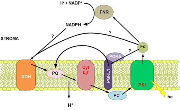
CEF around PSI was discovered by Arnon (Arnon et al., 1954) and contributes, in addition to linear electron flow (LEF), to the formation of a pH gradient. The acidification of the lumen is essential for protection of PSI and PSII against stress (Joliot and Johnson, 2011). Electrons are recycled from ferredoxin to plastoquinone, therefore no NADPH is accumulated. Genetic approaches demonstrated that 2 different pathways for CEF exist in Arabidopsis (Figure 2): NAD(P)H dehydrogenase- (NDH) and PGR5/PGRL1 (proton gradient regulation 5/pgr5-like 1)-dependent pathway (Munekage et al., 2004). NDH mediates CEF in cyanobacteria (Battchikova et al., 2011) and since plastids also contain a NDH complex it seems to be a good candidate for CEF
20
in higher plants, too. Interestingly, for plant NDH the subunit for NAD(P)H oxidation has not yet been identified and it seems that ferredoxin can also be the electron donor (Yamamoto et al., 2011). In Arabidopsis NDH forms a supercomplex with PSI (Peng et al., 2009). A clear phenotype of ndh deficient Arabidopsis was observed in pgr5 mutant background (Munekage et al., 2004). The PGR5/PGRL1 pathway is antimycin A-sensitive (LEF is not antimycin A-A-sensitive) and it is supposed to mediate electron transfer between ferredoxin and PQ. The PGR5/PGRL1 dependent pathway is needed to increase lumenal acidification and induces non-photochemical quenching (NPQ; Munekage et al., 2002). The pgr5 mutant from Arabidopsis has reduced NPQ (Jahns and Holzwarth, 2012). In Chlamydomonas a supercomplex including PSI, Cyt b6f, FNR
and PGRL1 (but not PGR5) performing CEF was discovered (Iwai et al., 2010).
Figure 2: Cyclic electron flow around photosystem I in plants. Electrons from photosystem I (PSI) are recycled through ferredoxin (Fd) and reduce the plastoquinone (PQ) pool without net NADPH production. Two different pathways of cyclic electron flow exist around PSI, one involves the NAD(P)H dehydrogenase (NDH) complex and the other one PGR5 and PGRL1. The proton gradient keeps forming through the Q-cycle in cytochrome b6f complex (Cyt b6f).
21
Mehler reaction
In the Mehler reaction (also known as pseudocyclic electron transfer) molecular oxygen is reduced by PSI to produce superoxide (O2•-), which is converted to hydrogen
peroxide (H2O2) by superoxide dismutase (SOD) and then to H2O and O2 by
peroxidases (water-water cycle; Asada, 2000). This results in an increase of the pH gradient and in ATP synthesis, but not in net NADPH production. Thereby over-reduction of the electron transport chain is minimized.
Chlororespiration
Chlororespiration was discovered by Bennoun in Chlamydomonas, when in the dark in the absence of photosynthetic electron transport the redox state of the PQ pool changed (Bennoun, 1982). The term chlororespiration was designated for a chloroplastic respiration analogue to the one in mitochondria driving the non-photochemical reduction of the PQ pool and its non-photochemical oxidation using O2 as final electron
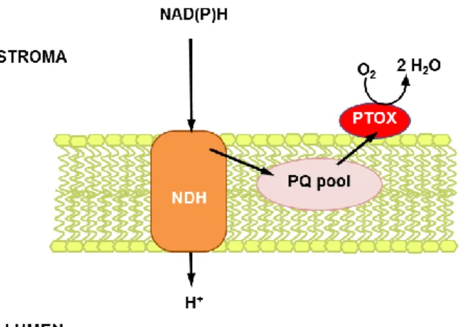
acceptor. The corresponding oxidase, the plastid terminal oxidase (PTOX), was discovered in Chlamydomonas lacking PSI and Cyt b6f, where the PQ pool was still oxidized under O2 consumption (Cournac et al., 2000). Chlamydomonas contains 2
PTOX isoforms. In plants, the discovery of immutans in Arabidopsis favoured the suggestion that PTOX is the oxidase oxidizing PQH2 which is reduced by the NDH
complex (Carol et al., 1999; Nixon, 2000, Figure 3). Thereby, in plants the NDH complex transfers protons from the stroma to the lumen, generating a proton gradient for ATP synthesis in the dark. In green algae chlororespiration is not electrogenic, they contain NDA2 instead of NDH which does not pump protons across the thylakoid membrane.
22
Figure 3: Chlororespiration in plants. The NAD(P)H dehydrogenase (NDH) complex reduces the PQ pool at the expense of NAD(P)H and transfers protons into the lumen. The plastid terminal oxidase (PTOX) oxidizes the PQ pool and reduces oxygen to water.
1.1.1 Regulation of photosynthetic electron transport
Photosynthetic organisms need to acclimate to a variety of environmental changes and stress conditions, especially plants, since they are sessile. The photosynthetic apparatus does not only provide energy for the cellular metabolism, but also acts as a sensor for stress and helps the plant to acclimate to the changes. The ability of oxygenic photosynthetic organisms to oxidize H2O and reduce plastoquinone is connected to the
formation of reactive oxygen species (ROS) and increases strongly when the absorption of light by chlorophylls exceeds the capacity of the photosynthetic apparatus to use this energy in photosynthesis.
1.1.1.1 Reactive oxygen species and antioxidant defenses
ROS participate in signalling (Noctor et al., 2007; Suzuki et al., 2012), but especially hydroxyl radical (•OH) and singlet oxygen (1O2)are also very reactive towards cellular
compounds and can be toxic. Most ROS are generated as by-products during electron transport chains in chloroplasts and mitochondria: O2•-is formed when O2 receives one
electron (Figure 4, reaction a). O2•-gets spontaneously dismutated to H2O2 and O2 at
neutral or alkaline pH values (pK=4.8). SOD catalyses this reaction making it more efficient (Figure 4, reaction b). •OH can be formed from H2O2 in the presence of
reduced transition metals such as Fe2+ or Cu+, known as Fenton reaction (Figure 4, reaction c). In the Fenton reaction the transition metal is oxidized. Re-reduction can be achieved by O2
•-
23
Figure 4: Formation of different reactive oxygen species. (a) Superoxide O2•-; (b) hydrogen peroxide H2O2; (c) hydroxyl anion -OH and hydroxyl radical •OH.
Because the hydroxyl radical (•OH) is so destructive, an effective antioxidant defence system against ROS is required. •OH formation is avoided by limiting the amount of its precursors through the action of antioxidants (tocopherol, ascorbate and gluthathione) and antioxidative enzymes (SOD, catalase and ascorbate peroxidase (APX)). SOD is a soluble enzyme. APX exists in a soluble form and a form attached to the stromal side of the thylakoid membrane (Shigeoka et al., 2002). For the regeneration of antioxidants NADPH is used, linking this mechanism to the photosynthetic electron transport. During photosynthesis ROS are formed easily when the photosynthetic electron chain is highly reduced (Rutherford et al., 2012). O2•- is mainly formed at the acceptor site of
PSI (Mehler reaction) and, to a lower extent, at the electron acceptor site of PSII (Pospíšil, 2012), and at the Cyt b6f complex (Baniulis et al., 2008).
Singlet oxygen (1O2)is generated by triplet-triplet excitation transfer from triplet
chlorophyll (3Chl*) to the triplet ground state of molecular oxygen (3O2). The 3Chl* is
formed either by intersystem crossing from the singlet excited state of chlorophyll (1Chl*) in the PSII antenna complex or by charge recombination of the primal radical pair P680+•Phe-• in the PSII reaction centre. This is likely, when the forward electron
transfer is blocked and is considered to be the main reaction pathway for 1O2 generation
(Krieger-Liszkay, 2005). Carotenoids can avoid 1O2 generation by quenching 3Chl*,
since the triplet energy level of carotenoids (3Car*) is lower than the triplet energy level of chlorophyll. Carotenoids can also quench 1O2 by singlet-triplet energy transfer. This
is of special importance in the PSII reaction centre, where carotenoids and chlorophylls are too far from each other for 3Chl* quenching (Telfer, 2005). The energy of 3Car* is converted into heat and the carotenoid returns to its ground state. Due to charge recombination, PSI can also generate 3Chl*, but 1O2 generation in PSI is unlikely mainly
due to shielding of P700 from O2 (Setif et al., 1981). Other quenchers of 1O2 are
24
1.1.1.2 Photosynthetic control at the cytochrome b6f complex
The oxidation of PQH2, after its binding to Cyt b6f, is the rate limiting step in
photosynthesis (Haehnel, 1976). This reaction is pH sensitive and depends on lumen acidification, which slows down PQH2 oxidation at the Qo siteof Cyt b6f and therefore
also electron transport to PSI, protecting PSI from over-reduction. As an initial step of PQH2 oxidation and proton release into the lumen, the proton of the hydroxyl group of
PQH2 forms a hydrogen bond with the nitrogen of a specific histidine of Fe2S2 Rieske
protein. The protons accumulated in the lumen decrease the probability of proton dissociation from histidine of F2S2 Rieske protein and therefore Cyt b6f turnover. The
pH optima for reduction of cyt f are in the range of 6,5-7,25 (Finazzi, 2002; Hope et al., 1994; Tikhonov, 2013).
1.1.1.3 Non-photochemical quenching of chlorophyll a fluorescenece
Light absorption leads to an excited state of Chl a. This excitation energy can drive photosynthesis (photochemical quenching), be dissipated as heat (non-photochemical quenching, NPQ) or reemitted as fluorescence. At least three different NPQ components have been defined: high energy quenching (qE); photoinhibition (qI) and state transitions (qT). The relative contribution of the different processes to the overall inducible NPQ varies on light-intensity and light exposure time.
High energy quenching qE
This energy quenching mechanism is based on the dissipation of excess energy as heat in the light harvesting antenna proteins and involves the xanthophyll violaxanthin and sometimes also lutein (Niyogi et al., 2001). There is currently no consensus on the precise physical mechanism of quenching, nor on the structural changes which take place (Bianchi et al., 2008; Passarini et al., 2009; Ruban et al., 2007). High energy quenching qE is triggered by acidification of the thylakoid lumen, which becomes lower than pH 6 under excessive light conditions. Firstly, this leads to the attachment of violaxanthin de-epoxidase to the membrane, where it de-epoxidises violaxanthin to zeaxanthin via antheraxanthin using ascorbate (Hager, 1969; Müller-Moulé et al., 2002). Zeaxanthin can deactivate excited singlet chlorophyll (1Chl*) in the PSII antenna and dissipate the energy as heat. Non-protein bound zeaxanthin has also a function as 1O2
25
quencher (Jahns and Holzwarth, 2012). Secondly, the acidification of the lumen leads also to the protonation of the PsbS protein that seems to act as a sensor of the lumen pH (Li et al., 2000). The protonation of PsbS induces a conformational change in the PSII antenna leading to the detachment of parts (LHCII, CP29 and CP24) from the PSII-LHCII supercomplex and a switch of the PSII antenna from a light-harvesting to an energy-dissipating state (Holzwarth et al., 2009). These changes are reversed, when the pH in the lumen increases.
Photoinhibition qI
Photoinhibition is defined as very slow-relaxing (>30 min) quenching component including the downregulation, inactivation or damage of PSII. Photoinhibition is not directly reversible. It can be caused by acceptor side inhibition of PSII, donor side inhibition of PSII or direct destruction of the Mn cluster by UV-B light (Vass, 2012). Photoinhibition is often associated with the inactivation of the D1 protein which has to be degraded and replaced by newly synthesized D1 (Edelman and Mattoo, 2008). Photoinhibition becomes visible when the rate of PSII inhibition exceeds the rate of de novo D1 protein synthesis. The contribution of the different mechanisms of photodamage leading to loss of PSII activity is still under discussion.
State transitions qT
State transitions refer to a redox-controlled LHCII phosphorylation/ dephosphorylation process by which excitation energy is redistributed among PSI and PSII when they become exposed to light conditions that preferably excite either PSI or PSII (Grieco et al., 2012; Minagawa, 2011). When PSII is preferentially excited, the PQ pool becomes more reduced, which favors PQH2 binding to the Qo site of the Cyt b6f (Rochaix, 2011)
and leads to a conformational change, which in turn activates kinase STN7 bound to Cyt b6f. The kinase is then released from Cyt b6f, and migrates to the margins between
stroma and grana membranes, where it phosphorylates LHCII. Phosphorylated LHCII has a decreased affinity for the grana stacks because of the negatively charged phosphate groups and thus a lateral movement of phosphorylated LHCII from grana to stroma occurs. It seems that the phosphorylated LHCII partly associates with PSI and increases PSI antenna size (Galka et al., 2012; Kouril et al., 2005). The movement from
26
PSII towards PSI is the state transition from ‘state 1’ to ‘state 2’ in plants. The PSII-LHCII supercomplexes decrease and leave PSII with a smaller absorption cross-section. In plants most of the LHCII is not accessible for the kinase, probably because a tight stacking of grana membranes restricts the LHCIIs that can be phosphorylated to those near the margins. The process is reversible: phosphatase PPH1 can dephosphorylate LHCII (Shapiguzov et al., 2010; Tikkanen et al., 2010).
Regulation of gene expression is crucial to acclimatisation to environmental changes and involves among others the redox state of the PQ pool. Both photosystems differ in their light absorption characteristics with maxima at 680 nm for PSII and 700 nm for PSI, respectively. PSII specific light induces genes encoding PSI proteins and vice versa (Pfannschmidt et al., 1999). Dependent on the light quality either PSI of PSII is excited more strongly, which leads to a more oxidized or reduced PQ pool, respectively. The redox state of the PQ pool modulates redox signalling pathways; by posttranslational modification of proteins (STN7), but also by changing gene transcription (Escoubas et al., 1995). Besides phosphorylation of LHCII STN7 also activates a signalling cascade regulating gene transcription (Allen et al., 2011). Other plastid signals can be ROS (Foyer and Noctor, 2009; Gadjev et al., 2006) and different pigment biosynthesis intermediates or metabolites (Bräutigam et al., 2007). Different signalling pathways have been reported to be induced by 1O2 (Kim and Apel, 2013) and
by H2O2 (Mullineaux et al., 2006).
1.1.1.4 Acclimatisation of photosynthesis to environmental changes
The photosynthetic electron chain needs to be protected against photodamage and electron flow has to be in balance with carbon fixation capacity. The regulation of photosynthesis is important for photosynthetic organisms to acclimate to changing environmental conditions. In addition to diurnal light changes, photosynthetic organisms also have to cope with light fluctuations during the day. Cyanobacteria, green algae and higher plants developed different mechanisms to acclimate to changing light conditions (Allahverdiyeva et al., 2015; Kono et al., 2014).
Cyanobacteria contain respiratory terminal oxidases: Synechocystis contains mainly two in the thylakoid membrane: Cyd (cytochrome bd-quinol oxidase complex) and Cox (cytochrome c oxidase complex). A cox/cyd double mutant showed to be lethal under
27
fluctuating light, but grew normally under continuous light (Lea-Smith et al., 2013). Regarding the photosynthetic apparatus, damage was located in PSII. Moreover, in cyanobacteria, four special flavodiiron proteins (Flv) are involved in protection, Flv2 and Flv4 protect PSII (Bersanini et al., 2014) and Flv1 and Flv3 protect PSI (Allahverdiyeva et al., 2013; Helman et al., 2005). Flv1 and Flv3 are necessary for acclimatisation to fluctuating light, flv1/flv3 cells showed strong acceptor side limitation of PSI and longer incubation under these conditions lead to cell death. Flv1 and Flv3 function as NADPH-oxygen oxidoreductases (Vicente et al., 2008) and reduce O2 to H2O (Mehler-like reaction) and help to minimize PSI acceptor site limitation.
Green algae and lower land plants contain two genes, flvA and flvB, homologous to cyanobacterial flv1 and flv3, but also pgr5 and pgrl1 (Peltier et al., 2010; Zhang et al., 2009). In contrast, higher plants only contain pgr5 and pgrl1, which are involved in CEF around PSI. The pgr5 mutant showed strong acceptor site limitation of PSI and was lethal under fluctuating light, but grew normally under constant light (Tikkanen et al., 2010). The wildtype showed some PSI acceptor-side limitation and accumulated ROS-scavenging enzymes. On the other hand, the double mutant pgr5 crr2 (chlororespiratory reduction, NDH mutant) had a severe phenotype already under constant light and strongly reduced linear photosynthetic electron flow. This demonstrated the importance of CEF for photosynthesis by protection against over-reduction of the stroma (Munekage et al., 2004). CEF also protects PSI from ROS production and due to its participation in lumen acidification, cyclic electron transport is necessary for triggering qE of NPQ to protect PSII from photodamage. The increase of the pH gradient serves as a stress signal and leads to the slow-down of the Cyt b6f
turnover. Moreover, a highly reduced PQ pool induces the detachment of LHCII from PSI (state transitions). As pgr5, the stn7 mutant (affected in state transitions) showed a very strong phenotype when grown under fluctuating light, probably due to overexcitation of PSII, which is especially harmful when NPQ relaxes (Tikkanen et al., 2010).
These different regulation mechanisms on different levels help adapting photosynthetic electron flow to a fluctuating environment and balance ATP and NADPH supply with their demand in assimilation.
28
Non-heme diiron proteins
Non-heme diiron carboxylate proteins are a ubiquitous family of metal containing enzymes. From their primary sequence motif, they share structural properties: a four-helix bundle surrounds the two iron atoms and highly conserved carboxylate and histidine ligands coordinate the diiron centre (Figure 5, Nordlund and Eklund, 1995). Despite the highly conserved active site residues, their overall sequence similarity is quite low. Also the reactions they catalyse are quite diverse and some examples include: hydroxylation of aliphatic or aromatic residues (soluble methane monooxygenase, MMO), generation of stable tyrosyl radical (R2 subunit of ribonucleotide reductase class I, R2 RNR), desaturation of fatty acids (9-desaturase, 9D), peroxidation by ruberythrin, NO/O2 reduction (Flavo-diirion proteins) and quinol oxidation (alternative
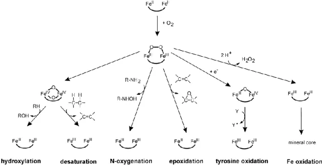
oxidase, AOX; plastid terminal oxidase, PTOX). The catalytic reaction is initiated by the reaction of the diiron centre with O2 (Krebs et al., 2011). The reaction with O2 leads
to oxidation of the irons and a peroxo-Fe2III intermediate P is formed, but the
intermediate species have different possible structures. This is probably one of the reasons for the diversity of the reactions catalyzed by this family (Figure 6, Krebs et al., 2011). The reaction intermediates are likely to be sensitive to oxidation. This may lead to the generation of ROS. The structural similarity of the active site makes it difficult to explain the variety of reactions catalyzed by these enzymes from crystallographic data alone. The catalytic mechanism of some of these enzymes has been studied in great detail using a number of different methods to obtain a detailed idea of the geometric and electronic structure of their iron active sites.
29
Figure 5: (A) The consensus sequence of the iron binding motif of the non-heme diiron carboxylate family. (B) Diiron active sites of methane monooxygenase MMO, ribonucleotide reductase RNR and 9-desaturase 9-D in the reduced state (Solomon et al., 2000).
Figure 6: Different possibilities for the reaction following the formation of the peroxo-Fe2III intermediate P after binding of O2 (Krebs et al., 2011).
1.1.2 Methane monooxygenase
Methane monooxygenases (MMO) from methanotrophic bacteria catalyse the conversion of methane to methanol. The soluble MMO complex is a multienzyme system consisting of a diiron active site for hydroxylase (MMOH), an oxidoreductase subunit containing FAD and Fe2S2-cluster which provides the electrons for O2
activation from NADPH (MMOR) and a regulatory subunit required for efficient catalysis (MMOB). MMOH is a mixed-function oxidase; one atom of O2 is transferred
to substrate and the other forms water. It converts methane to methanol in a process coupled to the oxidation of NADH (Tinberg and Lippard, 2011). The first
30
spectroscopically observed intermediate after the reductive activation of O2 at the diiron
centre of MMOH is the peroxodiiron intermediate P. The oxygen atoms abstract one electron from each iron and form (FeIIIFeIII / O--O-). The transfer of a proton leads to the 2nd peroxodiiron intermediate Hperoxo (Liu et al., 1994). The exact structures of the
MMOH peroxodiiron intermediates remain to be elucidated. The Hperoxo leads to
cleavage of the O-O bond and generates the intermediate Q. Q contains two high-valent FeIV ions and has been only detected in MMOH so far (Shu et al., 1997). Q hydroxylates methane, but the detailed mechanism of how the strong C-H bond of methane is activated by Q is not yet fully understood. To avoid harmful side reactions, the diiron centre is only reduced in the presence of the substrate.
1.1.3 Ribonucleotide reductase
In contrast to MMO, where the diiron centre directly participates in substrate oxidation, the role of the diiron centre in ribonucleotide reductase (RNR) is to generate a stable tyrosyl radical. RNRs catalyze the NTP/dNTP conversion and are divided into three classes. They are activated by ATP binding. Class I RNR consists of two dimeric not identic subunits, R1 and R2. Only R2 of class I RNR contains the non-heme diiron centre (Nordlund and Reichard, 2006). O2 oxidizes the RNR R2 diiron site and leads to
the peroxodiiron centre intermediate P. This gets reduced and forms the intermediate X (mixed-valent oxo-bridged FeIII-FeIV centre). The FeIII-FeIV generates a stable tyrosyl radical Y• (Figure 7, Solomon et al., 2000) and restores the FeIII-FeIII state, which can be reduced to FeII-FeII. The stable tyrosine radical activates R2 and induces proton-coupled electron transfer between the two subunits to the catalytic centre of R1, where the nucleotide is reduced by two cysteine residues (Stubbe, 2003). Electrons from a ferredoxin-like Fe2S2 protein regenerate the diiron centre (Wu et al., 2007).
Figure 7: Catalytic cycle of O2 activation and tyrosyl radical formation in ribonucleotide reductase RNR R2 (Friedle et al., 2010).
31
1.1.4 Alternative oxidase
Another example of a diiron carboxylate protein where a tyrosyl radical is also likely to be involved in the reaction mechanism is the alternative oxidase (AOX). This special tyrosine residue is conserved across all known AOX sequences and essential for enzymatic activity (Albury et al., 2002). AOX is found in plants, some fungi and protozoa and some -proteobacteria. AOX is attached to the membrane and involved in mitochondrial respiration, where it catalyses the oxidation of ubiquinol coupled with the reduction of O2 to H2O. AOX was discovered because its catalytic centre is not
inhibited by cyanide (Albury et al., 1996). The structure of AOX from Trypanosoma brucei was solved to atomic resolution (2.85 Å; Shiba et al., 2013) and showed the four -helix bundle harbouring the diiron centre common to all non-heme diiron carboxylate proteins (Figure 8A). Additionally AOX contains two amphipathic -helices which are believed to be involved in attachment to the mitochondrial membrane (Figure 8B). It is an antiparallel dimer with 6 universally conserved residues and 8 highly conserved residues which are believed to be involved in dimerization.
A catalytic cycle has been proposed based on different spectroscopic studies (Figure 8C, Moore et al., 2013). The cycle starts with the binding of O2 to the diferrous
state. Then two short-lived intermediates are formed, first a superoxo- and then a hydroperoxo-intermediate. A transfer of a proton and electron from bound ubiquinol forms the peroxodiiron species and water is released. Cleavage of the O-O bridge follows extraction of a proton and electron from the tyrosine, generating the tyrosyl radical. This is rereduced by the semiquinone. The second ubiquinol serves to regenerate the diferrous state and the second water is released (Moore et al., 2013).
32
Figure 8: Trypanosoma alternative oxidase overall structure and catalytic model (Moore et al., 2013). (A) Hydrogen bond network in the diiron centre. (B) Alternative oxidase dimer modeled into the membrane; 1 and 4 have a hydrophobic region which inserts into the membrane. (C) Scheme of a possible catalytic redox cycle of alternative oxidase AOX. The binding of O2 to the reduced diiron centre (1) leads, via 2 short-lived intermediates and the oxidation of ubiquinol to ubisemiquinone (2), to the formation of the peroxodiiron intermediate (3). Cleavage of the O-O bond and oxidation of the tyrosine radical results in a oxodiiron state (4). The tyrosine radical is rereduced by semiquinone. The oxodiiron species is rereduced by a second ubiquinol.
33
In plants AOX seems to serve as an alternative electron sink under conditions where the respiratory chain is overreduced. O2 is converted to H2O and harmful ROS formation is
minimized (Maxwell et al., 1999; Yip and Vanlerberghe, 2001). AOX activity is redox-regulated and reduction of an intermolecular disulfide bond between two cysteines increases the enzyme activity (Umbach et al., 2006). Activity of the reduced AOX can then additionally be increased by -keto acids such as pyruvate (Carré et al., 2011). This is in line with its role as a safety valve. Pyruvate is provided by glycolysis and is metabolized in the mitochondrial tricarboxylic acid cycle (TCA) cycle. The TCA cycle is inhibited at a high ATP/ADP ratio. The respiratory chain generates a proton gradient, which serves for ATP synthesis. AOX does not pump protons and reduces ATP synthesis since electrons flowing to AOX bypass proton pumping complex III and IV. Gene expression studies of AOX support its proposed physiological role under stress conditions. Plant AOXs are encoded by a small gene family, consisting of two distinct subfamilies termed AOX1 and AOX2. Expression of the AOX1a genes is highly responsive to abiotic and biotic stress, as well as dysfunctions in respiratory metabolism (Vanlerberghe, 2013). Exogenous treatment with ROS did also induce AOX expression (Ho et al., 2008), indicating a direct role of ROS in AOX signalling. On the other hand, overexpression of mitochondrial SOD attenuated AOX induction by stress (Li et al., 2013). AOX1a expression is regulated by the transcription factor ABSCISIC ACID INSENSITIVE 4 (ABI4, (Giraud et al., 2009), which is an ABA-responsive transcription factor and also involved in chloroplast retrograde signalling (León et al., 2012).
1.1.5 Plastid terminal oxidase
Analog to AOX in the mitochondria PTOX catalyses the oxidation of PQH2 coupled
with the reduction of O2 to H2O in the chloroplast (Cournac et al., 2000; Joët et al.,
2002). PTOX is only found in organisms capable of performing oxygenic photosynthesis (McDonald et al., 2011). It is of prokaryotic origin and most organisms encode one PTOX gene. In some green algae, as Chlamydomonas reinhardtii, two genes encode for PTOX (Ahmad et al., 2012). The primary sequence contains the consensus motif for iron binding conserved in non-heme diiron carboxylate proteins. Moreover, it contains a highly conserved tyrosine residue important for protein function
34
(Fu et al., 2005), suggesting a role in catalysis similar to the specific tyrosine in AOX and ribonucleotide oxidase R2. Spectroscopic studies on PTOX investigating intermediates of the catalytic cycle (a semiquinone or a tyrosine radical) or different redox states of the diiron centre are missing.
PTOX is bound interfacially to the thylakoid membrane, mainly to the stroma lamellae (Joët et al., 2002; Lennon et al., 2003). Expression of PTOX in E.coli showed that it oxidizes PQH2 and that its activity is cyanide-resistant, but sensible to propyl-
and octyl-gallate and that it has a strong preference for PQH2 (Josse et al., 2003).
Crystallographic and other spectroscopic studies of PTOX do not exist up to now. AOX and PTOX primary sequence show limited similarity (Nawrocki et al., 2015). The cysteine residues responsible for regulation of AOX activity are not conserved in PTOX and how its activity is regulated is not yet understood.
1.1.5.1 PTOX and carotenoid biosynthesis
PTOX was discovered as immutans in Arabidopsis showing a light-dependent variegated phenotype (Wetzel et al., 1994; Carol et al., 1999). When immutans plants were grown under low light intensity all leaves were green. Variegated leaves contained white and green sectors. Chloroplasts of the green sectors appeared wildtype-like, but chloroplasts from white sectors contained no pigments or any membrane structures and accumulated phytoene, a colourless intermediate of carotenoid synthesis. This indicated a problem in desaturation of phytoene to ζ-carotene, which is catalyzed by the phytoene desaturase (PDS), but PDS itself was functional in immutans (Wetzel et al., 1994). Later the Arabidopsis mutants pds1 and pds2, affected in PQ synthesis, also accumulated phytoene, implicating a role for plastoquinone in phytoene desaturation (Norris et al., 1998). It was postulated that phytoene needs to transfer its electrons to PQ to form ζ-carotene. PTOX oxidizes the PQ pool in the absence of light-driven electron transport, and provides thereby the substrate, oxidized plastoquinone, for desaturation of phytoene to ζ-carotene by PDS (Carol et al., 1999). The synthesized coloured carotenoids are essential for protecting the chloroplast against photooxidation and serve as light-harvesting pigments in the photosynthetic antenna. In addition, β-carotene is a component of the reaction centers. Moreover, carotenoids are the precursors for the plant hormones abscisic acid (ABA, (Nambara and Marion-Poll, 2005)) and
35
strigolactone (Alder et al., 2012). ABA is involved in acclimatisation to abiotic stress and in processes as stomatal closure. It was shown that ABA induced PTOX expression in rice under salt stress (Kong et al., 2003).
The question remains why some sectors in immutans leaves are green and develop functional chloroplasts. Studies led to the hypothesis that phytoene desaturase activity must reach a certain threshold to guarantee normal plastid development (Foudree et al., 2012). The PTOX mutant of tomato, ghost, also shows a light-dependent variegated phenotype. It was seen that the expression pattern of genes involved in carotenoid biosynthesis differed between wildtype and ghost (Barr et al., 2004). Interestingly a NDH-mutant showed a similar phenotype to ghost (Nashilevitz et al., 2010). NDH and PTOX are involved in chlororespiration. Chlororespiration builds up a pH gradient for ATP generation and might supply the energy for carotenoid synthesis, which takes place in the plastid and is an energy consuming pathway (Nawrocki et al., 2015). An immutans/pgr5 or immutans/crr2-2 double mutant suppressed the immutans phenotype (Wetzel et al., 1994; Carol et al., 1999), probably due to less electron flow back into the PQ pool, since PGR5 and CRR2-2 (NDH-mutant) are involved in cyclic electron flow (Munekage et al., 2004). All these enzymes have an impact on the redox state of the PQ pool, therefore the PQ pool redox state seems to play a key role in carotenoid synthesis. The PQ pool redox state is important for retrograde signaling (Jung and Mockler, 2014) and many plastid enzymes, among these PTOX and enzymes for carotenoid biosynthesis, are nuclear encoded and posttranslationally imported into the plastid. A detailed study of immutans revealed, that not only the white, but also the green sectors have a different morphology. Additionally, the green sectors had higher photosynthetic activity than wildtype leaves and it was suggested that retrograde signaling is impaired in immutans (Aluru et al., 2001). It may be that certain carotenoid intermediates play a role as signalling compound in retrograde signalling (see Chapter 4, Galzerano et al., 2014).
1.1.5.2 PTOX and chlororespiration
PTOX is involved in chlororespiration and might be important in plants to generate a pH gradient with NDH. Moreover, it might be important to keep the redox poise of the PQ pool in the dark, especially in algae, where chlororespiration is non-electrogenic
36
(Nawrocki et al., 2015). Even after long dark incubation the redox state of the PQ pool remains 30% reduced in Chlamydomonas (Houille-Vernes et al., 2011). On the contrary, in most plants the redox state is almost completely oxidized (Nawrocki et al., 2015). However, there is some disagreement. In tomato the PQ pool was about 20% reduced (Trouillard et al., 2012). Both enzymes are often upregulated under stress such as nitrogen starvation in Chlamydomonas (Peltier and Schmidt, 1991) or abiotic stress in some plants.
1.1.5.3 PTOX and abiotic stress
It is generally accepted that PTOX has a low activity compared to photosynthetic electron flow. Depending on the organism different electron transport activities were measured for PTOX: The maximum rate of PTOX was reported to be 4.5 e- s-1 PSII-1 for PTOX2 in Chlamydomonas (Houille-Vernes et al., 2011) and 1 e- s-1 PSII-1 in tomato (Trouillard et al., 2012) while the maximal rate of photosynthesis is approximately 150 e- s-1 PSII-1 (Nawrocki et al., 2015). Under normal growth conditions PTOX abundance is quite low (about 1% PTOX protein per 100 PSII; Lennon et al., 2003), but PTOX level were shown to be upregulated in plants exposed to stress such as cold, drought, high temperatures and high light (Díaz et al., 2007a; Ivanov et al., 2012; Laureau et al., 2013; Quiles, 2006; Stepien and Johnson, 2009). The alpine plant Ranunculus glacialis increased its PTOX level about 3-times when exposed to high light (Laureau et al., 2013) and the tolerant Thellungiella halophila even 4-times when exposed to salt-stress (Stepien and Johnson, 2009). The increased PTOX content correlated with increased electron flow capacity and higher PQ pool oxidation. Therefore PTOX is regarded as a safety valve, serving as an alternative electron sink and preventing the PQ pool and the photosynthetic electron chain from overreduction during stress, thereby reducing ROS formation and PSI/PSII photodamage. Regarding its low activity compared to PSII, PTOX might not provide photoprotection alone but together with other regulatory processes. Evidence in PTOX serving as a safety valve also comes from mutant analyses. The tobacco mutant rbcl (lacking RubisCO) showed also a much higher PTOX level than the wild-type (Allahverdiyeva et al., 2005). Additionally, by preventing an over-reduction of the PQ pool, PTOX has been proposed to reduce the formation of ROS, analog to AOX in the mitochondria (McDonald et al., 2011). A
37
mutant lacking catalase and peroxidase, showed stimulated PTOX expression (Rizhsky et al., 2002). Moreover, plants with increased PTOX level during stress showed less ROS formation (Díaz et al., 2007a; Ibáñez et al., 2010) and inhibition of PTOX activity increased photooxidative damage during high light in the green algae Haematococcus pluvialis (Wang et al., 2009).
However, plant transformants with increased PTOX levels have been shown to exhibit either no phenotype under photoinhibitory conditions (Rosso et al., 2006) or signs of chronic photoinhibition (Ahmad et al., 2012; Heyno et al., 2009; Joët et al., 2002) and increased ROS production under high light (Heyno et al., 2009). This implies that a high level of PTOX itself is not sufficient to acclimate to abiotic stress and that upregulation of PTOX is linked to other processes (Sun and Wen, 2011).
38
Aims
Studies on mutant, overexpressor and stressed plants and algae have provided a lot of information concerning the plastid terminal oxidase, but neither the biochemistry nor the physiological role of PTOX during photosynthesis are fully understood. Moreover, it is still under debate, if PTOX is working in an antioxidant or in a prooxidant manner. The aim of my thesis was to contribute to the understanding of the function of PTOX.
My thesis focused on:
1. Biochemical characterization of recombinant purified PTOX from rice, a construct with the maltose binding protein-tag (MBP-OsPTOX, Yu et al., 2014). Therefore, recombinant PTOX was first studied with an enzymatic assay using decylPQH2 as substrate. In a second approach PTOX was added to PSII enriched membrane fragments and its attachment and functionality were studied. PTOX activity was measured as oxygen consumption or by chlorophyll fluorescence.
2. PTOX activity in planta and its effect on photosynthetic electron transport. Two different transformants were used: Arabidopsis thaliana expressing the cyanobacterial desaturase CRTI in addition to the endogenous desaturase (Schaub et al., 2012) which contain a higher PTOX level and Nicotiana tabacum plastid transformants expressing PTOX1 from Chlamydomonas (Ahmad et al., 2012). A combination of spectroscopic, biochemical and physiological techniques allowing in vivo and in vitro analysis were used.
3. MBP-OsPTOX expression in Synechocystis sp. PCC6803. MBP-OsPTOX was cloned into Synechocystis sp. PCC6803 and its expression and attachment to the membrane investigated. Further, PTOX activity was studied by chlorophyll fluorescence.
39


![Table 1 PTOX (µg) Diaph. (µg) NADH (µM) DPQ (µM) DPQH 2 (µM) Act. [nmol O 2 min -1 ml -1 ] O 2 reduced (µM) NADH/O 2PQH2/O2 10 10 25 100 - 39.2±2.6 13.0±0.5 1.92 10 10 50 100 - 78.2±5.3 24.0±1.2 2.08 10 10 100 100 - 112.6±6](https://thumb-eu.123doks.com/thumbv2/123doknet/12888102.370465/60.892.120.774.558.1127/table-ptox-diaph-nadh-dpq-dpqh-reduced-nadh.webp)